Article

Investigating the Neurotoxic Impacts of Arsenic and the Neuroprotective Effects of Dictyophora Polysaccharide Using SWATH-MS-Based Proteomics

1
The Key Laboratory of Environmental Pollution Monitoring and Disease Control, School of Public Health, Ministry of Education, Guizhou Medical University, Guiyang 550025, China
2
College of Life Science and Oceanography, Shenzhen University, Shenzhen 518060, China
3
Department of Psychology, Emory University, Atlanta, GA 30322, USA
*
Authors to whom correspondence should be addressed.
These authors contributed equally to this paper.
Molecules 2022, 27(5), 1495; https://doi.org/10.3390/molecules27051495
Submission received: 6 February 2022 / Revised: 18 February 2022 / Accepted: 18 February 2022 / Published: 23 February 2022

Abstract

:
Arsenic (As) is one of the most important toxic elements in the natural environment. Currently, although the assessment of the potential health risks of chronic arsenic poisoning has received great attention, the research on the effects of arsenic on the brain is still limited. It has been reported that dictyophora polysaccharide (DIP), a common bioactive natural compound found in dietary plants, could reduce arsenic toxicity. Following behavioral research, comparative proteomics was performed to explore the molecular mechanism of arsenic toxicity to the hippocampi of SD (Sprague Dawley) rats and the protective effect of DIP. The results showed that exposure to arsenic impaired the spatial learning and memory ability of SD rats, while DIP treatment improved both the arsenic-exposed rats. Proteomic analysis showed that arsenic exposure dysregulated the expression of energy metabolism, apoptosis, synapse, neuron, and mitochondria related proteins in the hippocampi of arsenic-exposed rats. However, DIP treatment reversed or restored the expression levels of these proteins, thereby improving the spatial learning and memory ability of arsenic-exposed rats. This study is the first to use high-throughput proteomics to reveal the mechanism of arsenic neurotoxicity in rats as well as the protective mechanism of DIP against arsenic neurotoxicity.

1. Introduction

Arsenic (As) is a common environmental toxicant and occupational contaminant that affects over 200 million people worldwide, primarily in developing countries, and arsenic poisoning is still a world public health issue [1]. The accumulation of arsenic in the human body can cause organ damage and tissue carcinogenesis. While it enhances the production of reactive oxygen species (ROS) such as superoxide and hydrogen peroxide, it increases protein oxidation, disrupts enzymes and DNA [2], causes lipid peroxidation, impairs mitosis, and promotes apoptosis.
Arsenic can easily cross the blood–brain barrier and accumulate in various parts of the brain including the striatum and hippocampus, amplifying arsenic toxicity and tissue injury [3]. Long-standing evidence has shown the role of hippocampal neurons in the formation of spatial learning and memory. By impairing hippocampal neurons, arsenic toxicity leads to decreased learning and memory capacity [4]. Arsenic, when present in high concentrations in the brain, disrupts neurocellular signaling pathways and functions, resulting in induced synaptic transmission and neurological dysfunction [5]. However, there is still no consensus on the exact mechanism of arsenic-induced neurotoxicity.
Chronic arsenic poisoning is caused by long-term consumption of contaminated food and water, and there is currently no effective treatment for symptoms. Recent research has found that bioactive natural compounds found in dietary plants can improve arsenic-induced toxicity [6], and their medicinal value is gaining in popularity. Among them, dictyophora polysaccharide (DIP) was found to have anti-inflammatory, anti-tumor, and immunomodulatory activities [7,8,9]. Recent studies have noted that it has a neuroprotective effect [10,11]. However, the effect of DIP on the neurotoxicity caused by sodium arsenite is still unknown.
In this study, the learning and memory capability of arsenic exposed-SD (Sprague Dawley) rats was shown to be impaired, but DIP alleviated the impairment. To investigate the molecular mechanism of arsenic-induced hippocampus toxicity and the protective effect of DIP on arsenic-induced neurotoxicity, a comparative proteomics analysis based on SWATH (sequential windowed acquisition of all theoretical fragment ion mass spectra) was performed. SWATH-MS (mass spectrum) analysis was chosen because of its high reproducibility and quantitative accuracy.

2. Results

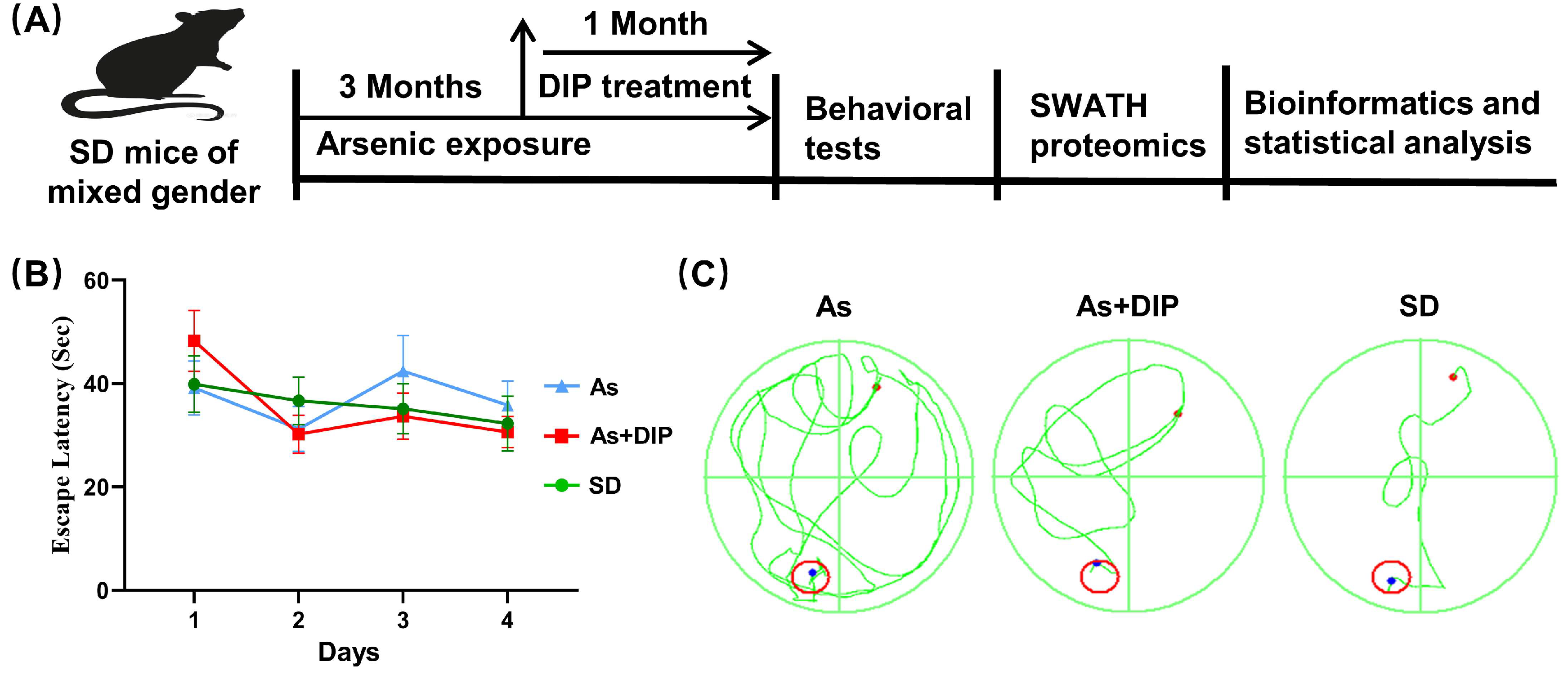
2.1. DIP Improved Spatial Learning and Memory of Arsenic Treated Rats

In the Morris Water Maze (MWM) test, compared to the control group, the swimming track lines of the arsenic-treated group were more irregular with much higher escape latency (Figure 1B,C). The latency on the fifth day was also higher in the arsenic-treated group (Figure 1D). Following that, a probe trial was conducted to assess the rats’ memory 24 h after the 4-day successive training. The DIP + As group rats crossed the platform’s hidden location much more frequently than the arsenic-treated group rats (Figure 1E). In addition, the DIP + As group rats spent significantly more time in the target quadrants than the arsenic-treated rats (Figure 1F).

2.2. Overview of Protein Expression Characteristics of Different Groups

SWATH-MS analysis identified a total of 3833 quantitative proteins with 2674 quantitative proteins meeting the quality control requirements after QC correction. We then compared and analyzed the protein expression profiles between different groups including the arsenic-treated group versus the control group (As/Ctrl group); DIP treatment + NaAsO2-treated group compared with the NaAsO2-treated group (DIP + As/As group); and DIP treatment + NaAsO2-treated group compared with control group (DIP + As/Ctrl group). The cluster analysis revealed that the protein expression pattern of the As/Ctrl group differed significantly from that of the other two groups, whereas the DIP + As/As group and the DIP + As/Ctrl group exhibited more similar patterns (Figure 2A).
Among them, 172, 75, and 82 proteins were identified as DEPs in the As/Ctrl group, DIP + As/As group, and DIP + As/Ctrl groups, respectively (Figure 2B–D). Of these, 44 DEPs were found to be common between the As/Ctrl group and DIP + As/As group, while 43 DEPs were discovered to have the opposite expression trend in these two groups (named as reversed proteins; Table 1, Figure 2E). When compared to the As/Ctrl group, the expression of most DEPs in the As/Ctrl group was restored or reversed in the As + DIP/As group (Table 1, Figure 2F).

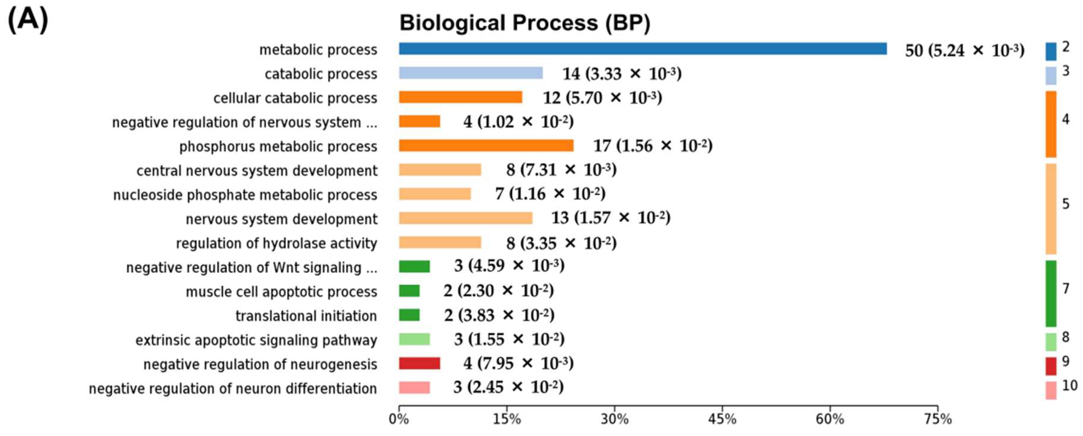
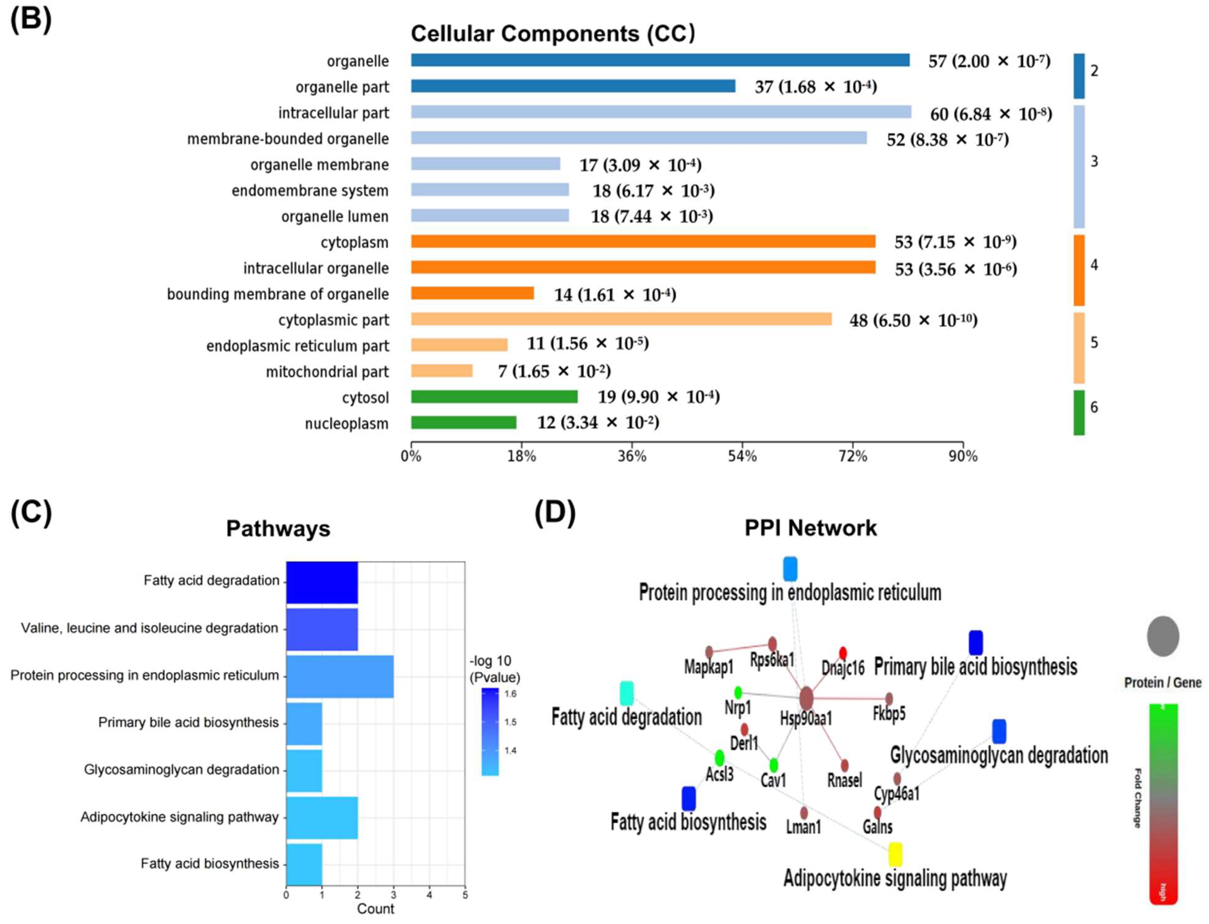
2.3. Differentially Expressed Proteins between NaAsO2-Treated Group and Control Group (As/Ctrl Group)

According to the bioinformatics analysis, the biological process (BP) associated with DEPs in the As/Ctrl group was mainly involved in the cellular catabolic process and regulation of apoptotic process, regulation of translational, protein and enzyme signal regulation, and organophosphate catabolic process. Several signal transduction pathways were involved including Ras protein signal transduction, Wnt signaling pathway, small GTPase mediated signal transduction, and MAP kinase activity (Figure 3A, Table S1). Among the DEPs, 12 were related to mitochondrion organization including Ddhd2, Pmpca, Bcs1l, Ndufaf7, Slc25a46, Hars2, Elmod1, Tfam, U2af2, Bag3, Fam162a, and Msto1; 10 DEPs (i.e., Lrp1, Krt18, Grm7, Itgb1, Eif2ak2, Bag3, Rps6ka1, Map4k4, Anp32b, and Cav1) were related to negative regulation of apoptotic process; four DEPs (i.e., Unc13a, Shank1, Tbc1d24, and Clstn1) were associated with the regulation of synaptic transmission; and two DEPs (including Kcna1 and Fgf12) were involved in the neuronal action potential process (Table S1). By cellular components (CC) analysis, the DEPs were mainly distributed in the cytoplasm, cytoplasmic parts, mitochondrion, and mitochondrial matrix. Noticeably, multiple DEPs were distributed in synapse and neuron part: 16 DEPs belonged to synaptic proteins and 10 of them were downregulated; 19 DEPs belonged to the neuronal part and 14 of them were downregulated (Figure 3B, Table S2). These DEPs were found to be involved in metabolic pathways and glutamatergic synapse. The metabolic pathways included pyruvate metabolism, propanoate metabolism, ether lipid metabolism, ascorbate, aldarate metabolism, and drug metabolism-other enzymes (Figure 3C). Ten DEPs were found to be involved in these metabolic pathways with six of them being downregulated (Table S3). Figure 3D depicts the protein−protein interaction (PPI) network related to DEPs. The findings agreed with the Kyoto Encyclopedia of Genes and Genomes (KEGG) pathway analysis.
We further analyzed the DEPs enriched into the key pathway of Gene Ontology (GO) analysis. The PPI of DEPs involved in mitochondrion organization, negative regulation of apoptotic process, and synapse and neuron parts are shown in Figure 4A–D, respectively.

2.4. Differentially Expressed Proteins between DIP + As-Treated Group and As-Treated Group (DIP + As/As Group)

The DIP + As/As group contained 75 DEPs. The BP associated with these DEPs was mainly involved in the metabolic process, catabolic process, regulation of hydrolase activity, translational initiation, extrinsic apoptotic, Wnt signaling pathway, nervous system development, and regulation of neuron differentiation. These were widely distributed in cells. Among them, 50 were involved in the metabolic process (Figure 5A, Table S4), seven were mitochondrial part proteins, and 11 were endoplasmic reticulum part proteins (Figure 5B, Table S5). According to the KEGG and PPI network, they were linked to glycosaminoglycan degradation pathways, protein processing in the endoplasmic reticulum, and fatty acid biosynthesis and degradation (Figure 5C,D, Table S6). The PPI of DEPs involved in catabolic process, the extrinsic apoptotic signaling pathway, and the endoplasmic reticulum and mitochondrion parts are shown in Figure 6A–D, respectively.

2.5. Differentially Expressed Proteins between DIP + As-Treated Group and Control Group (DIP + As/Ctrl Group)

We also compared the protein expression profiles of the DIP + As-treated group and the control group. Eighty-two DEPs were identified. Compared with the results from the As/Ctrl comparison, about half of the DEPs suggested that DIP treatment changed the protein expression profile in the hippocampi of rats exposed to arsenic, turning it closer to normal expression.

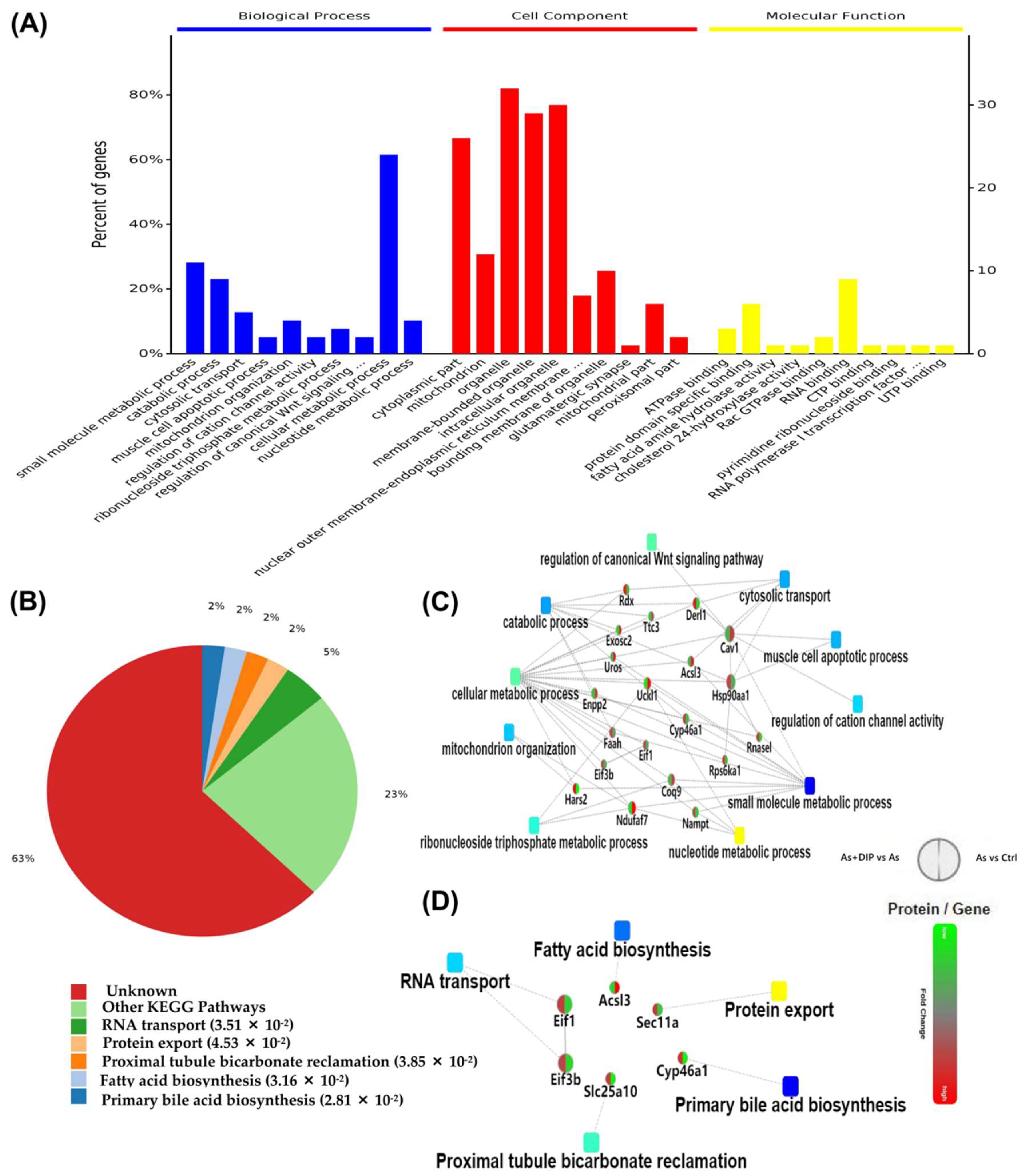
2.6. The Reversed Proteins between the As/Ctrl Group and DIP + As/As Group

Subsequently, we analyzed the 43 reversed proteins between the As/Ctrl group and DIP + As/As group. Among these, 22 proteins were upregulated and 21 proteins were downregulated in the As/Ctrl group, while their expression trends were reversed in the DIP + As/As group. GO analysis showed that these proteins were mainly implicated in the catabolic process and apoptotic process, and mitochondrion organization (Figure 7A, Table S7), and were widely distributed in cells. They were involved in the regulation of biosynthesis pathways and other biological processes such as RNA transport and protein export (Figure 7B, and Table S8). The BP-related PPI network revealed that these reversed proteins were associated with the catabolic process, mitochondrion organization, apoptotic process, and other signaling pathways. According to the PPI network and KEGG analysis, these proteins were correlated with protein export, acid biosynthesis, and RNA transport pathways (Figure 7C,D).

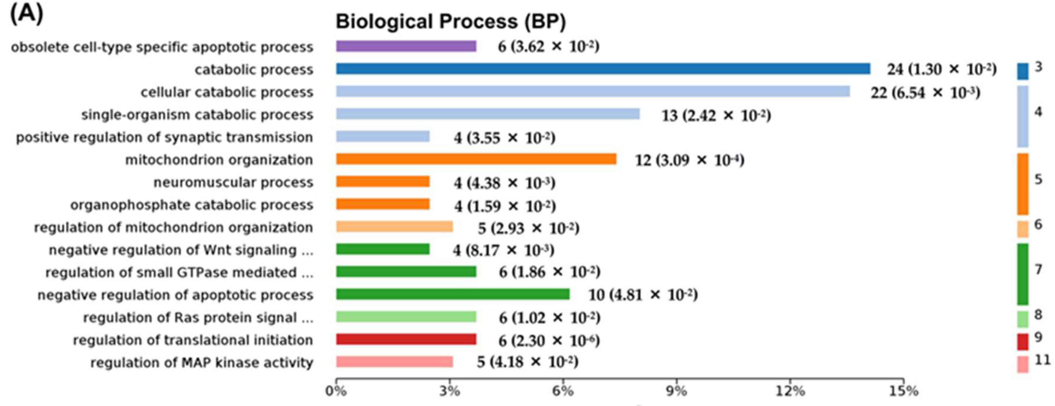
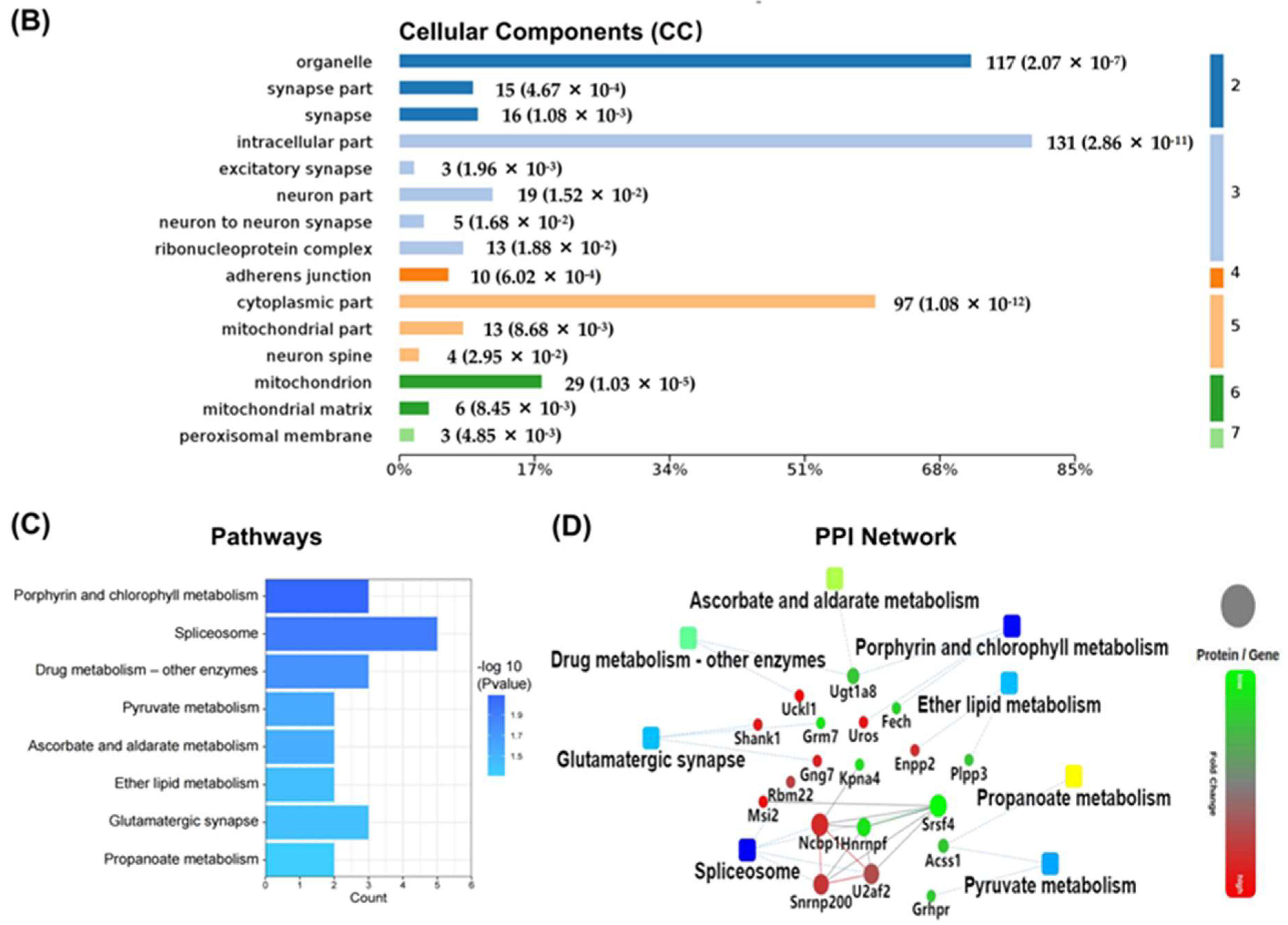
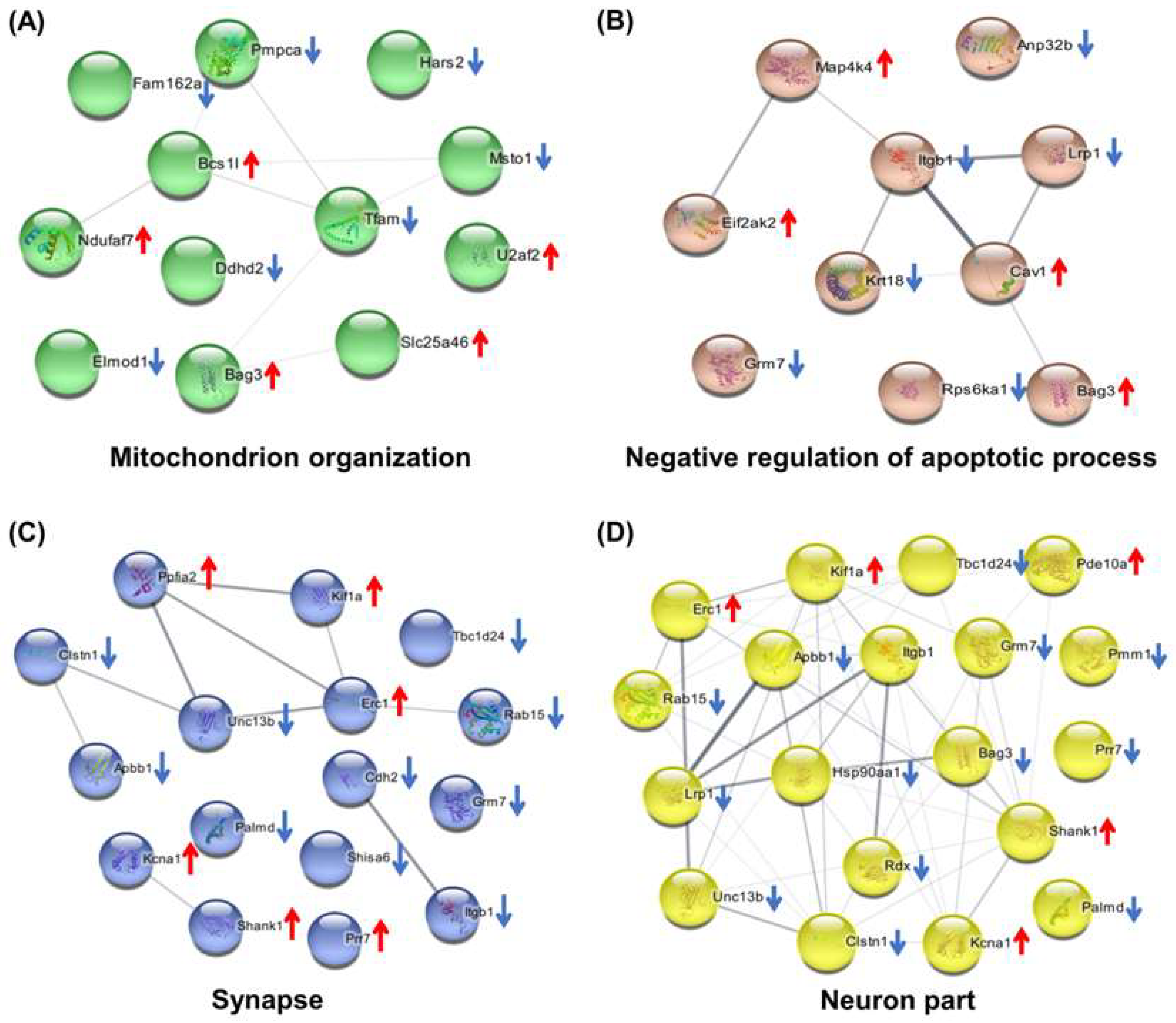
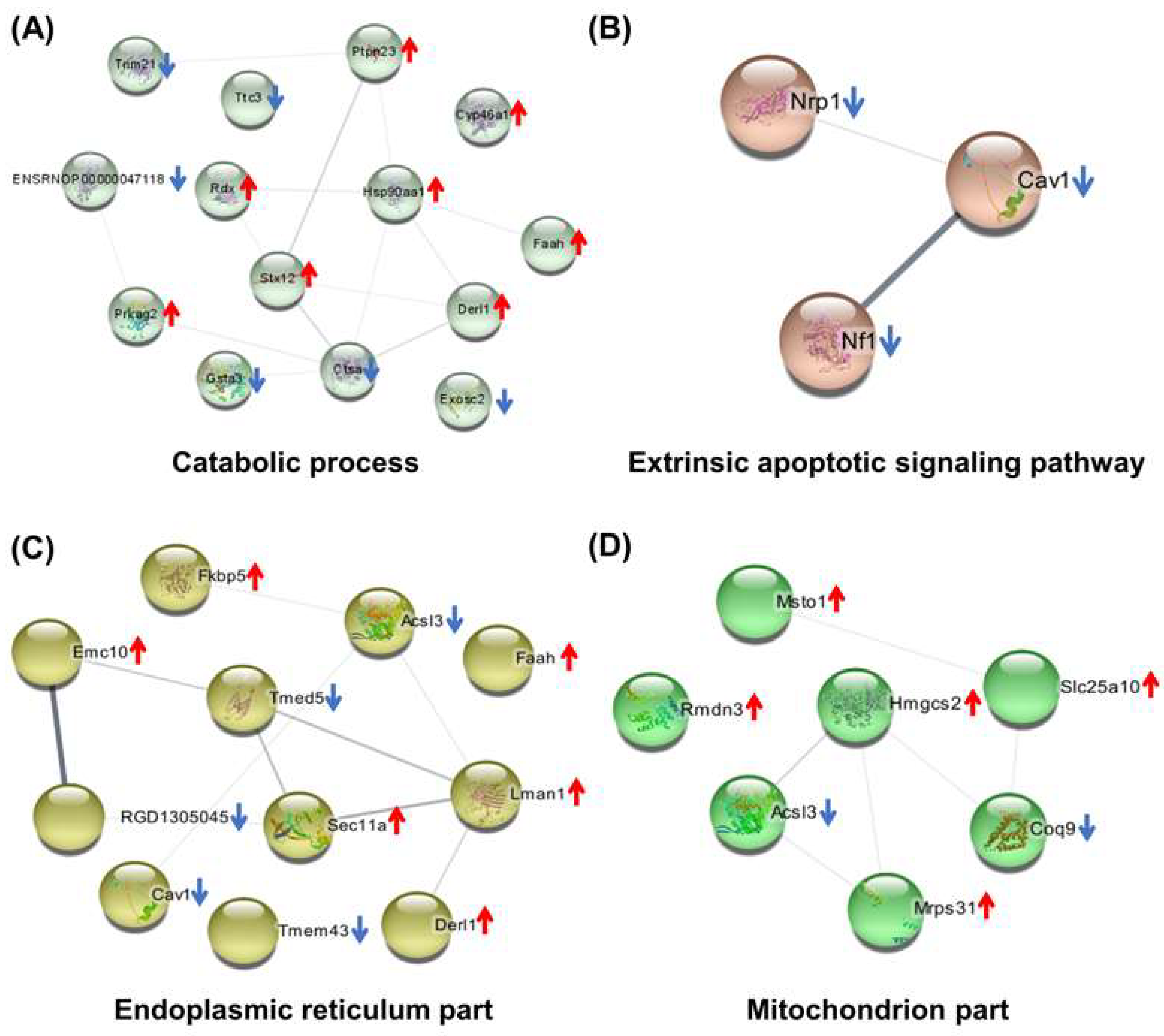
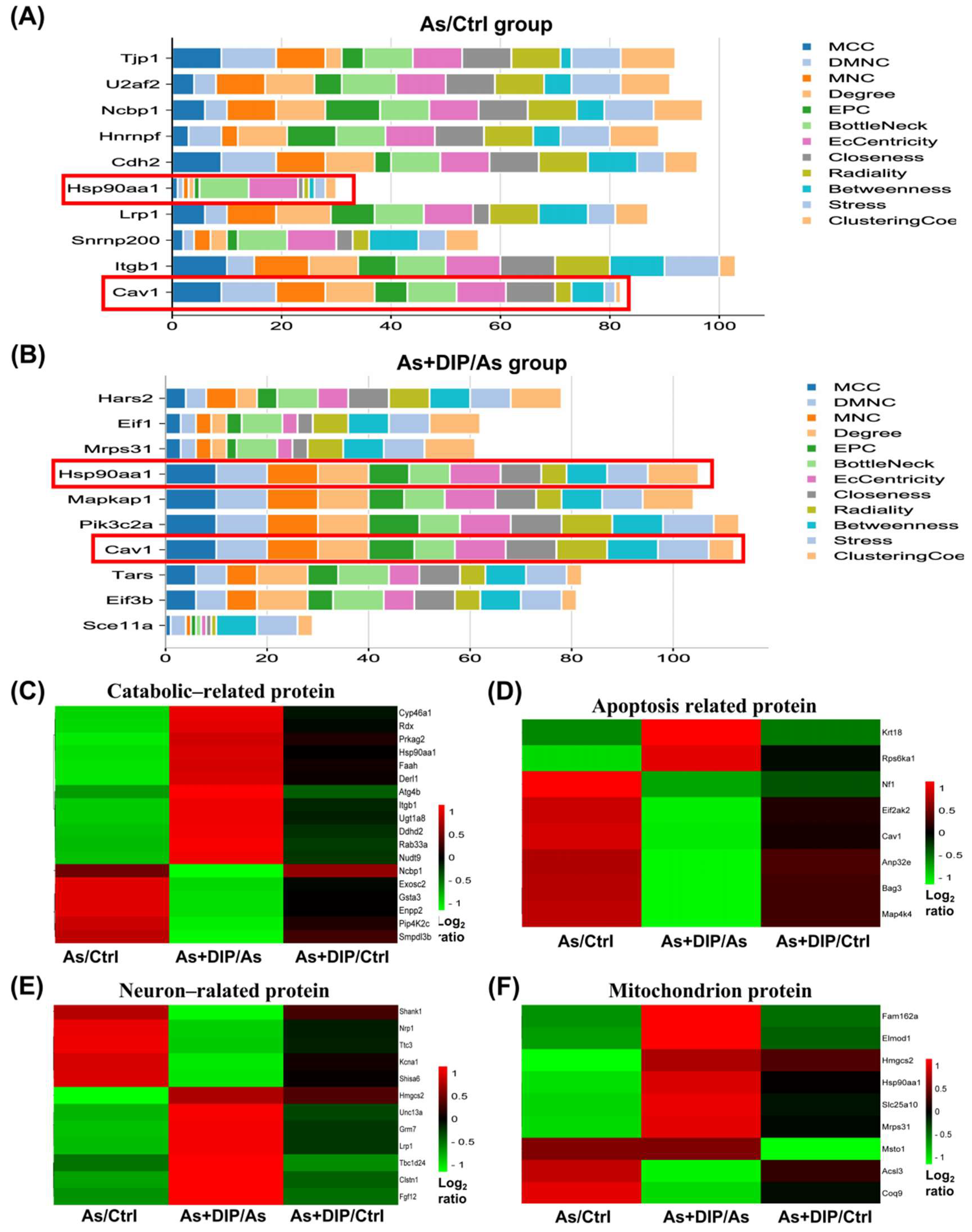
2.7. Hub Genes Analysis and Cluster Analysis of Differentially Expressed Proteins in Key Pathways

Cytohubba software was used to analyze the hub genes in the As/Ctrl and DIP + As/As groups. The top ten hub genes in the As/Ctrl group were identified in Figure 8A. Among them, Cav1 and Hsp90aa1 belonged to reversed proteins. The top ten hub genes in the DIP + As/As group were also identified in Figure 8B. Among these, Cav1, Eif1, Eif3b, Hars2, Hsp90aa1, and Mrps31 belonged to reversed proteins. Evidently, Cav1 and Hsp90aa1 were common hub genes in these two groups. These hub genes were associated with catabolic, apoptosis, mitochondrion, and neuron.
According to UniProt annotation and GO analysis results, we further clustered and analyzed the expressions of DEPs in the key pathways (catabolic, neuron, and apoptosis-related protein, mitochondrial protein) in different groups. The results showed that their expression was reversed or restored after treatment with DIP (Figure 8C–F).

3. Discussion

Arsenite exposure impaired the spatial learning and memory ability of rodents have been reported in several previous studies [12,13]. Consistently, here, our results showed that arsenic exposure could damage the spatial learning and memory ability of SD rats. However, DIP treatment could improve the learning and memory ability of arsenic-exposed rats.
Through proteomics analysis, the results showed that the DEPs in the As/Ctrl groups were mainly associated with the synapse, neuron part, apoptosis, mitochondrial and energy metabolism, and catabolic process. Among them, 16 DEPs belonged to synaptic proteins, 19 DEPs belonged to the neuron part, and four DEPs were related to synaptic transmission. These results suggest that arsenic treatment dysregulated the expression of synapses and neuron-related proteins in the hippocampus of SD rats, thus contributing to the impairment of learning and memory in rats exposed to arsenic. This is similar to previous research where arsenic exposure disrupted synapse-related protein expression and signals [14].
Glutamate is the major excitatory neurotransmitter in the mammalian central nervous system and plays an important role in spatial learning and memory function [12]. We found that the expression of several proteins associated with it in the As/Ctrl groups had changed. For example, the expression of Unc13a and GRM7 were downregulated in the hippocampus of arsenic-treated SD rats. Unc13a is essential for synaptic vesicle maturation in most excitatory/glutamatergic that enhances neurotransmitter release [15]. Grm7, as the G-protein coupled receptor activated by glutamate, is related to neurodevelopmental disorders [16]. Therefore, dysregulation of glutamate related proteins might inhibit the growth of glutamatergic and reduce the release of neurotransmitters, which may be responsible for the learning and memory impairment of arsenic-exposed rats. The expression of these synaptic and neuron-related proteins was reversed or restored in the DIP + As/Ctrl group, indicating that DIP improved spatial memory and exploring ability in arsenic-exposed SD rats. Furthermore, in the DIP + As/As group, 13 DEPs were associated with nervous system development, implying that the protective effect of DIP was related to synaptic and neuron-related proteins.
In addition, in the As/Ctrl group, 10 DEPs were found to be associated with regulation of the apoptotic process, suggesting that apoptosis might be one of the important mechanisms of arsenic toxicity in rat hippocampus. Among these DEPs, Krt18 was a regulator of neuronal apoptosis. Eif2ak2 plays a role in apoptosis, cell proliferation, and differentiation. It regulates various signaling pathways (p38 MAP kinase, NF-kappa-B, and insulin signaling pathways). Bag3 is involved in the regulation of the extrinsic apoptotic signaling pathway. Cav1 is a scaffold protein in the cell membrane, which is closely related to cell apoptosis. The expression of these proteins was increased in the As/Ctrl group, which may be related to the apoptosis of hippocampal cells induced by NaAsO2. On the other hand, Krt18 plays a role in the negative regulation of apoptotic process. Rps6ka1 functions as a differentiation factor by modulating mTOR signaling and suppressing the pro-apoptotic functions of BAD and DAPK1. Their downregulation in the As/Ctrl group may also contribute to NaAsO2-induced apoptosis. Interestingly, Krt18 [17], Cav1 [18], and Bag [19] have been reported to be associated with arsenic-induced apoptosis. In the current study, Cav1 was also identified as a common Hub protein in the As/Ctrl group and the As DIP + As/As group.
Previous studies have shown that arsenic could induce neuronal cell death through oxidative stress [20]. Arsenic toxicity can increase the level of ROS in organs, cause damage to the mitochondrial respiratory chain, disrupt glucose homeostasis in tissues, and induce oxidative stress [21] as well as lead to ROS-induced apoptosis [22]. As expected, in this study, the expression of 29 mitochondrial-associated proteins in the hippocampus of SD rats altered after arsenic treatment. However, their expression was reversed in the DIP + As/As group. The results suggest that they may be related to the neurotoxicity of arsenic and responsible for DIP attenuating arsenic-induced mitochondrial function damage. Indeed, in our previous proteomics study on hepatocytes treated with arsenic, the proteins related to apoptosis and mitochondria were the most dysregulated proteins, while pretreatment with DIP reversed or restored the expression of those proteins [23].
One of the main functions of mitochondria is related to energy metabolism. In this study, catabolic pathways were enriched including the ribonucleotide catabolic process, ascorbate and aldarate metabolism, pyruvate metabolism, and glycosaminoglycan degradation. These pathways have been observed in proteomics studies of arsenic-exposed rats [24] or yeast [25]. We found that most of the proteins in these pathways were downregulated in the As/Ctrl group. This indicated that NaAsO2 treatment resulted in abnormal catabolic pathways in the hippocampus of SD rats. However, the expression of these DEPs was reversed in the As + DIP/As group, suggesting that DIP reduced the effect of arsenic. In particular, pyruvate is closely related to adenosine triphosphate (ATP). When pyruvate metabolism is inhibited, it may affect the production of ATP by hindering the aerobic decomposition of pyruvate, increasing the consumption of ATP, and eventually reducing the energy levels [26]. Of note, seven DEPs were identified as ATP binding related proteins including Hsp90aa1, Rdx, Pip4K2c, Rab33a, Nudt9, Derl1, and Prkag2. Among them, the expression levels of six DEPs showed a downward trend, while Hsp90aa1 may play a key role. HSP90AA1 has a variety of functions including involvement in ATPase activation, transcriptional regulation, mitochondrial import, cell growth, development, and apoptosis. In this study, it was found to be a common Hub protein in the As/Ctrl group and the As DIP + As/As group.
Therefore, our results agree with previous studies that arsenic inhibits ATP production, leading to abnormal energy metabolism. As the main manufactory of cellular ATP, energy-deficiency, mainly resulting from mitochondrial damage, can lead to the dysfunction of synaptic neurotransmission, and further cause nerve injury. DIP has a neuroprotective effect, which can reduce the neurotoxicity of arsenic and improve the nerve injury caused by arsenic. In addition, improved mitochondrial biosynthesis and redox status can alleviate mitochondrial dysfunction. DIP may serve as a potential scavenger for products of oxidative stress products [27], thereby reducing ROS levels to ameliorate mitochondrial dysfunction caused by arsenic. We also found 50 DEPs related to metabolic processes in the DIP + As/Ctrl group, indicating that DIP improved hippocampal metabolism in arsenic-exposed SD rats by being associated with metabolism-related proteins and pathways. Indeed, the relationship between energy-deficiency and nerve injury associated with arsenic has been well reported [28]. However, most of the studies on the effect of arsenic on mitochondrial function have been conducted on cardiac, liver, kidney, and cells. To the best of our knowledge, this is the first study to show that arsenic causes a decrease in the expression of ATP binding proteins and mitochondrial proteins in the hippocampus, resulting in impaired learning and memory function in arsenic-treated SD rats, whereas DIP improved the arsenic-treated SD rats’ learning and memory by reversing or restoring their expression. In addition, we performed subgroup analysis on the sex of rats, and there was no significant difference between the results of each group and the original results. Taken together, the toxic mechanisms of arsenic on energy metabolism, apoptosis, synapse, neuron, and mitochondrion in rat hippocampus and the intervention effect of DIP are summarized in Figure 9.
Moreover, we finally focused on 43 reversed expressions and hub genes, with expression patterns supporting the above discussion. Likewise, we found that some DEPs in the As/Ctrl group were related to the Ras protein signal transduction, small GTPase mediated signal transduction, MAP kinase activity pathway, and Wnt signaling pathway. Most of these DEPs were downregulated in the As/Ctrl group and reversed in the As + DIP/As group. Several studies have shown that arsenic can inhibit the Wnt signaling pathway and affect cell proliferation, differentiation, and apoptosis [29]. Inhibition of the GTPase signaling pathway can induce neuronal apoptosis, which is mediated by a variety of mitogen-activated protein kinase signal cascades [30]. Arsenic has been shown to induce apoptosis in rat neurons by activating p38 MAP kinases [31]. Our results suggest that arsenic-induced neurotoxicity and the improvement in DIP on arsenic induced learning and memory impairment in rats may be related to these signal pathways.

4. Materials and Methods

4.1. Chemicals and Dictyophora Polysaccharide Preparation

Food-grade Dictyophora was obtained from Zhijin Sifang Hongye Co., Ltd., (Zhijin City, China). Sodium arsenite (NaAsO2) was purchased from Sigma Chemical Corp (St. Louis, MO, USA). DIP was prepared in accordance with our previous study [11,23,32]. Briefly, the Dictyophora fruiting body was ground into a powder, extracted, filtered, and concentrated. The crude polysaccharides of Dictyophora were obtained by freeze-drying after overnight precipitation with four volumes of 100% ethanol.

4.2. Animals and Experimental Design

The whole work scheme is shown in Figure 1A. Sixty 6-week-old SD rats (30 male and 30 female) were obtained from Huafukang Biotechnology Co., Ltd., (Beijing, China). Rats were fed a standard diet and allowed to drink freely for one week while being kept at a temperature of 20 ± 2 °C and a relative humidity of 55 ± 5%.
After one week of adaptive feeding, they were randomly divided into three groups with 20 rats in each group (half male and half female). The As and DIP concentrations used in the groups were determined according to the previous studies [10,32,33]. The details were as follows: for the control group, common feedstuff and distilled water were given for four months, with physiological saline (0.9 percent NaCl) gavage once a day in the fourth month. For the As group, processed feedstuff with 50 mg/kg NaAsSO2 and distilled water were given for four months, with physiological saline (0.9 percent NaCl) gavage once a day in the fourth month. For the As + DIP group, processed feedstuff with 50 mg/kg NaAsSO2 and distilled water were given for four months. Gavage bamboo fungus polysaccharide beverage was then administered once a day in the fourth month.

4.3. Morris Water Maze (MWM) Test

The MWM test was used to check the spatial learning and memory capacities of rats [34]. Briefly, each rat was trained four times over the course of four days. On the fifth day of the space exploration experiment (SPaceex Plorationex Periment), the platform was removed and the 120 s exploration training began. The following information was recorded as the rats entered: the number of times the rats passed through the target (the original platform’s position) and the swimming time they took to cross the target quadrant (the quadrant where the platform was originally placed).

4.4. Sample Preparation for Proteomic Analysis

After four months of treatment, rats were euthanized by cervical dislocation with their brains rapidly excised on an ice-cold plate for hippocampal dissection. Ten rats (five male and five female) were chosen randomly for proteomic analysis in each group. Hippocampal tissue was homogenized by using the Freezing grinder (Jingxin, Shanghai, China) and lysed in RIPA lysis buffer (KeyGEN BioTECH, Nanjing, Jiangsu, China), and centrifuged at 12,000 rpm, 4 °C for 30 min. The supernatants were collected with protein concentrations quantified using the BCA assay (KeyGEN BioTECH) and stored at −80 °C until use. For trypsin digestion, 200 μg protein of each sample was reduced with 10 mM DTT (dithiothreitol) at 37 °C for 2 h before being alkylated with 55 mM iodoacetamide (IAM) at room temperature for 30 min in darkness. After this, it was digested with trypsin at 37 °C overnight. The peptides were dried up in a vacuum centrifuge and stored at 4 °C for LC-MS/MS analysis.

4.5. Generation of the Reference Spectral Library by Independent Data Analysis (IDA) Acquisition

In order to perform SWATH-MS analysis, a reference spectral library needs to be established. A mixed representative sample of all samples was obtained by mixing 15 μg of protein from each sample and used for quality control (QC). The liquid chromatography (LC) was carried out by Agilent 1100 Serise liquid phase system (Agilent Technologies, Santa Clara, CA, USA). The conditions for chromatographic separation of protein samples were as follows: flow rate—0.30 mL/min; injection volume—100 μL; mobile phase: phase A, water (containing 0.1% formic acid); phase B, acetonitrile:water (95:5, v/v); the gradient of liquid phase elution was 70 min, and phase B increased from 5% to 80%: 5% phase B was the initial concentration (0−3 min), 5−38% phase B (3−60 min), 38−80% phase B (60−65 min), and 5% phase B equilibrium to 70 min. The mass spectrometer was a Triple TOF 6600 (SCIEX, Framingham, MA, USA). In the IDA mode, a time-of-flight mass spectrometry (TOF-MS) survey scan was acquired (350–1500 m/z, 0.25 s) with the 40 most intense multiply charged ions (2+ to 5+; exceeding 100 counts per s). MS/MS spectra were accumulated for 50 ms in the mass range of 100–1500 m/z with rolling collision energy. To create the library, the MS raw files and databases searches were combined and performed using ProteinPilot software v.5.0.1 (SCIEX).

4.6. Quantification of Proteins by SWATH Acquisition

The SWATH-MS acquisition was performed in DIA mode, with the peptides from each sample collected separately after enzymatic hydrolysis. The QC sample was collected first, and subsequent quality control analyses were performed by collecting a QC sample every five samples. Data were acquired with a 2.4 kV ion spray voltage, 35 psi curtain gas, 12 psi nebulizer gas, and an interface heater temperature of 150 °C. The scanning resolution of the primary mass spectrometry was 70,000, the scanning range was 100–1500 m/z, and the maximum injection time was 40 ms. Each cycle contained 34 distinct windows. The variable width between windows was 80 w, the window mass range was 12.5–158 Da, and the maximum ion implantation time of secondary mass spectrometry was 50 ms. The collision chamber energy (high energy collision induced dissociation, HCD) was set to 28 ev. Accurate quantitation was conducted using MultiQuant™ software. The cutoff values were 1.5-fold for upregulated and 0.67-fold for downregulated proteins.

4.7. Statistical Analysis

The MWM test data from the three groups were subjected to one-way ANOVA analysis. Previously, normal distribution and variance homogeneity were investigated (Shapiro–Wilk). Statistical analysis was carried out using the SPSS package (SPSS 23.0, Chicago, IL, USA).

4.8. Bioinformatics Analysis

The data were corrected using the R language software statTarget tool based on QC samples. Heatmap was plotted on an online platform for data analysis and visualization (http://www.bioinformatics.com.cn, accessed on 7 October 2021). Bioinformatics analysis was performed using OMICSBEAN online tools (http://www.omicsbean.cn, accessed on 1 October 2021) and the String database (http://string-db.org, version 9.1, accessed on 15 September 2021). DEPs were analyzed by GO (gene ontology) annotation, KEGG (Kyoto Encyclopedia of Genes and Genomes) pathways, and protein−protein interaction (PPI) networks. GO annotations included the biological process (BP) and cellular component (CC) [35]. Hub genes were screened by cytohubba software.

5. Conclusions

In this study, we showed that NaAsO2 induced learning and memory impairments in SD rats. Nevertheless, DIP treatment restored their learning and memory. Changes in energy metabolism, apoptosis, synapse, neuron, and mitochondrial related proteins were observed in the hippocampus of arsenic induced rats. Their changes led to mitochondrial dysfunction, abnormal energy metabolism, apoptosis, and abnormal synaptic structure and neurotransmitter, resulting in learning and memory impairment of As induced rats. Several pathways including the Ras protein signal transduction, small GTPase mediated signal transduction, MAP kinase activity pathway, and Wnt signaling pathway may also play an important role. Importantly, this is the first study to observe that arsenic induces downregulation of ATP binding and mitochondrial-related protein expression in the hippocampus of rats. The expression of these proteins was reversed by DIP treatment. This is also the first study to link DIP to arsenic-induced apoptosis and neurotoxicity. These findings may provide a direction for arsenic neurotoxicity research using DIP treatment. Future studies are needed to validate the molecular mechanisms of arsenic-induced hippocampal toxicity and the implications presented by the findings in the current study.

Supplementary Materials

The following supporting information can be downloaded at, Table S1: Fifteen important biological process in GO analysis in the As/Ctrl group, Table S2: Fifteen important cellular component in GO analysis in the As/Ctrl group, Table S3: KEGG pathways associated with the differentially expressed proteins in the As/Ctrl group, Table S4: Fifteen important biological process in GO analysis in the As + DIP/As group, Table S5: Fifteen important cellular component in GO analysis in the As + DIP/As group, Table S6: KEGG pathways associated with the differentially expressed proteins in the As + DIP/As group, Table S7: Ten important biological process associated with the reversed proteins in GO analysis, Table S8: The pathways associated with the reversed proteins in KEGG analysis.

Author Contributions

Conceptualization, P.L. and L.S.; Study design, J.Z. and T.H.; Performed the experiments and analyzed the data, J.Z., Y.W., X.Z., H.Z., J.L., X.T. and X.L.; Writing—original draft preparation, J.Z. and T.H.; Writing—review and editing, J.Z., T.H., M.C., N.U.K., P.L. and L.S.; Funding acquisition, P.L. and L.S. All authors have read and agreed to the published version of the manuscript.

Funding

This work was supported by the National Natural Science Foundation of China (P.L. 81860560, U1812403-6-2-4), the Shenzhen–Hong Kong Institute of Brain Science-Shenzhen Fundamental Research Institutions (L.S. 2021SHIBS0003), the Guizhou Science Combined Support (P.L.[2021]134), and the Science and Technology Fund Project of Guizhou Provincial Health Commission (P.L. gzwjkj2020-1-188).

Institutional Review Board Statement

All animal experiments were conducted according to the Declaration of Helsinki, approved by the Ethics Committee of Guizhou University (syxk 201803).

Informed Consent Statement

Not applicable.

Data Availability Statement

The data presented in this study are available on request.

Acknowledgments

We are grateful to the instrument analysis center of Shenzhen University.

Conflicts of Interest

The authors declare that they have no known competing financial interests or personal relationships that could have appeared to influence the work reported in this paper.

References

  1. Duan, T.; Hu, T.; Wu, C.; Yeh, Y.T.; Lu, J.; Zhang, Q.; Li, X.; Jian, W.; Luo, P. PINK1/Parkin-mediated mitophagy is involved in NaAsO2-induced apoptosis of human hepatic cells through activation of ERK signaling. Toxicol. Vitr. 2020, 66, 104857. [Google Scholar] [CrossRef] [PubMed]
  2. Kitchin, K.T. Recent advances in arsenic carcinogenesis: Modes of action, animal model systems, and methylated arsenic metabolites. Toxicol. Appl. Pharm. 2001, 172, 249–261. [Google Scholar] [CrossRef] [PubMed] [Green Version]
  3. Itoh, T.; Zhang, Y.F.; Murai, S.; Saito, H.; Nagahama, H.; Miyate, H.; Saito, Y.; Abe, E. The effect of arsenic trioxide on brain monoamine metabolism and locomotor activity of mice. Toxicol. Lett. 1990, 54, 345–353. [Google Scholar] [PubMed]
  4. Pandey, R.; Rai, V.; Mishra, J.; Mandrah, K.; Kumar, R.S.; Bandyopadhyay, S. From the Cover: Arsenic Induces Hippocampal Neuronal Apoptosis and Cognitive Impairments via an Up-Regulated BMP2/Smad-Dependent Reduced BDNF/TrkB Signaling in Rats. Toxicol. Sci. 2017, 159, 137–158. [Google Scholar] [CrossRef] [Green Version]
  5. Lu, C.; Zhao, F.; Sun, D.; Zhong, Y.; Yu, X.; Li, G.; Lv, X.; Sun, G.; Jin, Y. Comparison of speciated arsenic levels in the liver and brain of mice between arsenate and arsenite exposure at the early life. Environ. Toxicol. 2014, 29, 797–803. [Google Scholar] [CrossRef]
  6. Kuzu, M.; Kandemir, F.M.; Yıldırım, S.; Çağlayan, C.; Küçükler, S. Attenuation of sodium arsenite-induced cardiotoxicity and neurotoxicity with the antioxidant, anti-inflammatory, and antiapoptotic effects of hesperidin. Environ. Sci. Pollut. Res. 2021, 28, 10818–10831. [Google Scholar] [CrossRef]
  7. Hua, Y.; Gao, Q.; Wen, L.; Yang, B.; Tang, J.; You, L.; Zhao, M. Structural characterisation of acid-and alkali-soluble polysaccharides in the fruiting body of Dictyophora indusiata and their immunomodulatory activities. Food Chem. 2021, 132, 739–743. [Google Scholar] [CrossRef]
  8. Han, S.; Ma, C.; Hu, M.; Wang, Y.; Ma, F.; Tao, N.; Qin, Z. A polysaccharide from Dictyophora indusiata inhibits the immunosuppressive function of cancer-associated fibroblasts. Cell Biochem. Funct. 2017, 35, 414–419. [Google Scholar] [CrossRef]
  9. Ruksiriwanich, W.; Khantham, C.; Linsaenkart, P.; Chaitep, T.; Rachtanapun, P.; Jantanasakulwong, K.; Sringarm, K. Anti-inflammation of bioactive compounds from ethanolic extracts of edible bamboo mushroom (Dictyophora indusiata) as functional health promoting food ingredients. Int. J. Food. Sci. Technol. 2021, 57, 110–122. [Google Scholar] [CrossRef]
  10. Yu, W.X.; Lin, C.Q.; Zhao, Q.; Lin, X.J.; Dong, X.L. Neuroprotection against hydrogen peroxide-induced toxicity by Dictyophora echinovolvata polysaccharide via inhibiting the mitochondria-dependent apoptotic pathway. Biomed. Pharmacother. 2017, 88, 569–573. [Google Scholar] [CrossRef]
  11. Zhang, J.; Shi, R.; Li, H.; Xiang, Y.; Xiao, L.; Hu, M.; Ma, F.; Ma, C.W.; Huang, Z. Antioxidant and neuroprotective effects of Dictyophora indusiata polysaccharide in Caenorhabditis elegans. J. Ethnopharmacol. 2016, 192, 413–422. [Google Scholar] [CrossRef] [PubMed]
  12. Jing, J.; Zheng, G.; Liu, M.; Shen, X.; Zhao, F.; Wang, J.; Zhang, J.; Huang, G.; Dai, P.; Chen, Y.; et al. Changes in the synaptic structure of hippocampal neurons and impairment of spatial memory in a rat model caused by chronic arsenite exposure. Neurotoxicology 2012, 33, 1230–1238. [Google Scholar] [CrossRef] [PubMed]
  13. Luo, J.H.; Qiu, Z.Q.; Shu, W.Q.; Zhang, Y.Y.; Zhang, L.; Chen, J.A. Effects of arsenic exposure from drinking water on spatial memory, ultra-structures and NMDAR gene expression of hippocampus in rats. Toxicol. Lett. 2009, 184, 121–125. [Google Scholar] [CrossRef] [PubMed]
  14. Luo, J.H.; Qiu, Z.Q.; Zhang, L.; Shu, W.Q. Arsenite exposure altered the expression of NMDA receptor and postsynaptic signaling proteins in rat hippocampus. Toxicol. Lett. 2012, 211, 39–44. [Google Scholar] [CrossRef]
  15. Betz, A.; Ashery, U.; Rickmann, M.; Augustin, I.; Neher, E.; Südhof, T.C.; Rettig, J.; Brose, N. Munc13-1 is a presynaptic phorbol ester receptor that enhances neurotransmitter release. Neuron 1998, 21, 123–136. [Google Scholar] [CrossRef] [Green Version]
  16. Song, J.M.; Kang, M.; Park, D.H.; Park, S.; Lee, S.; Suh, Y.H. Pathogenic GRM7 mutations associated with neurodevelopmental disorders impair axon outgrowth and presynaptic terminal development. J. Neurosci. 2021, 41, 2344–2359. [Google Scholar] [CrossRef]
  17. Fortier, A.M.; Asselin, E.; Cadrin, M. Keratin 8 and 18 loss in epithelial cancer cells increases collective cell migration and cisplatin sensitivity through claudin1 up-regulation. J. Biol. Chem. 2013, 288, 11555–11571. [Google Scholar] [CrossRef] [Green Version]
  18. Luanpitpong, S.; Wang, L.; Stueckle, T.A.; Tse, W.; Chen, Y.C.; Rojanasakul, Y. Caveolin-1 regulates lung cancer stem-like cell induction and p53 inactivation in carbon nanotube-driven tumorigenesis. Oncotarget 2014, 5, 3541. [Google Scholar] [CrossRef]
  19. Roy, S.; Narzary, B.; Ray, A.; Bordoloi, M. Arsenic-induced instrumental genes of apoptotic signal amplification in death-survival interplay. Cell Death Discov. 2016, 2, 1–6. [Google Scholar] [CrossRef] [Green Version]
  20. TLu, T.H.; Tseng, T.J.; Su, C.C.; Tang, F.C.; Yen, C.C.; Liu, Y.Y.; Yang, C.Y.; Wu, C.C.; Chen, K.L.; Hung, D.Z.; et al. Arsenic induces reactive oxygen species-caused neuronal cell apoptosis through JNK/ERKmediated mitochondria-dependent and GRP 78/CHOPregulated pathways. Toxicol. Lett. 2014, 224, 130–140. [Google Scholar]
  21. Rezaei, M.; Keshtzar, E.; Khodayar, M.J.; Javadipour, M. SirT3 regulates diabetogenic effects caused by arsenic: An implication for mitochondrial complex II modification. Toxicol. Lett. 2019, 301, 24–33. [Google Scholar] [CrossRef]
  22. Chirumbolo, S.; Bjørklund, G. PERM hypothesis: The fundamental machinery able to elucidate the role of xenobiotics and hormesis in cell survival and homeostasis. Int. J. Mol. Sci. 2017, 18, 165. [Google Scholar] [CrossRef]
  23. Hu, T.; Shen, L.; Huang, Q.; Wu, C.; Zhang, H.; Zeng, Q.; Wang, G.; Wei, S.; Zhang, S.; Zhang, J.; et al. Protective Effect of Dictyophora Polysaccharides on Sodium arsenite-induced Hepatotoxicity: A Proteomics Study. Front. Pharmacol. 2021, 12, 749035. [Google Scholar] [CrossRef] [PubMed]
  24. Wu, S.; Li, J.; Jin, X. iTRAQ-based quantitative proteomic analysis reveals important metabolic pathways for arsenic-induced liver fibrosis in rats. Sci. Rep. UK 2018, 8, 1–9. [Google Scholar] [CrossRef] [PubMed]
  25. Guerra-Moreno, A.; Isasa, M.; Bhanu, M.K.; Waterman, D.P.; Eapen, V.V.; Gygi, S.P.; Hanna, J. Proteomic analysis identifies ribosome reduction as an effective proteotoxic stress response. J. Biol. Chem. 2015, 290, 29695–29706. [Google Scholar] [CrossRef] [PubMed] [Green Version]
  26. Saunier, E.; Antonio, S.; Regazzetti, A.; Auzeil, N.; Laprévote, O.; Shay, J.W.; Coumoul, X.; Barouki, R.; Benelli, C.; Huc, L.; et al. Resveratrol reverses the Warburg effect by targeting the pyruvate dehydrogenase complex in colon cancer cells. Sci. Rep. UK 2017, 7, 1–16. [Google Scholar] [CrossRef] [PubMed] [Green Version]
  27. Deng, C.; Hu, Z.; Fu, H.; Hu, M.; Xu, X.; Chen, J. Chemical analysis and antioxidant activity in vitro of a beta-D-glucan isolated from Dictyophora indusiata. Int. J. Biol. Macromol. 2012, 51, 70–75. [Google Scholar] [CrossRef]
  28. Panchal, K.; Tiwari, A.K. Mitochondrial dynamics, a key executioner in neurodegenerative diseases. Mitochondrion 2019, 47, 151–173. [Google Scholar] [CrossRef]
  29. Li, S.; Ren, Q. Effects of arsenic on wnt/β-catenin signaling pathway: A systematic review and meta-analysis. Chem. Res. Toxicol. 2020, 33, 1458–1467. [Google Scholar] [CrossRef]
  30. Stankiewicz, T.R.; Ramaswami, S.A.; Bouchard, R.J.; Aktories, K.; Linseman, D.A. Neuronal apoptosis induced by selective inhibition of Rac GTPase versus global suppression of Rho family GTPases is mediated by alterations in distinct mitogen-activated protein kinase signaling cascades. J. Biol. Chem. 2015, 290, 9363–9376. [Google Scholar] [CrossRef] [Green Version]
  31. Namgung, U.K.; Xia, Z. Arsenic induces apoptosis in rat cerebellar neurons via activation of JNK3 and p38 MAP kinases. Toxicol. Appl. Pharm. 2001, 174, 130–138. [Google Scholar] [CrossRef] [PubMed] [Green Version]
  32. Hu, T.; Zhang, K.; Pan, D.; Pan, X.; Yang, H.; Xiao, J.; Shen, X.; Luo, P. Inhibition Effect of Dictyophora Polysaccharides on Human Hepatocellular Carcinoma Cell Line HCC-LM3. Med. Sci. Monitor. 2020, 26, e918870. [Google Scholar] [CrossRef] [PubMed]
  33. Sun, H.; Yang, Y.; Shao, H.; Sun, W.; Gu, M.; Wang, H.; Jiang, L.; Qu, L.; Sun, D.; Gao, Y. Sodium arsenite-induced learning and memory impairment is associated with endoplasmic reticulum stress-mediated apoptosis in rat hippocampus. Front. Mol. Neurosci. 2017, 10, 286. [Google Scholar] [CrossRef] [Green Version]
  34. He, Z.; Han, S.; Wu, C.; Liu, L.; Zhu, H.; Liu, A.; Lu, Q.; Huang, J.; Du, X.; Li, N.; et al. Bis (ethylmaltolato) oxidovanadium (iv) inhibited the pathogenesis of Alzheimer’ s disease in triple transgenic model mice. Metallomics 2020, 12, 474–490. [Google Scholar] [CrossRef] [PubMed]
  35. Du, X.; Shi, Q.; Zhao, Y.; Xie, Y.; Li, X.; Liu, Q.; Iqbal, J.; Zhang, H.; Liu, X.; Shen, L. Se-Methylselenocysteine (SMC) Improves Cognitive Deficits by Attenuating Synaptic and Metabolic Abnormalities in Alzheimer’s Mice Model: A Proteomic Study. ACS Chem. Neurosci. 2021, 12, 1112–1132. [Google Scholar] [CrossRef] [PubMed]
Figure 1. Schematics for study design and behavioral tests. (A) Schematic diagram of the whole experiment. (B) The escaping latencies of rats in the 4-day trials. (C) The representative swimming track lines of rats in the 4-day trials. (D) The escaping latencies of rats. (E) The times of rats crossing over the hidden location of the platform. (F) The time of mice spent in the target quadrants. * p < 0.05, n = 20. (DF) In the probe trail at 24 h after the 4-day successive training.
Molecules 27 01495 g001aMolecules 27 01495 g001b
Figure 2. Differentially expressed proteins identified in different groups. (A) Cluster map comparing the proteins identified in the As/Ctrl, DIP + As/As, and DIP + As/Ctrl groups. Red color indicates higher expression (upregulation), blue indicates lower expression (downregulation), and white indicates similar expression levels. (B) Volcano plots depicted the distribution of proteins in the As/Ctrl group. (C) Volcano plots depicted the distribution of proteins in the As + DIP/As group. (D) Volcano plots depicted the distribution of proteins in the As + DIP/Ctrl group. For parts (BD), the log2 fold change (FC) is plotted versus the −log10 of the p-value. Red dots = hits with p < 0.05 and mean log2 FC > 0.58; blue dots = hits with p < 0.05 and mean |log2 FC| < 0.58. (E) Venn diagrams of DEPs among the As/Ctrl group, As + DIP/As group, and As + DIP/Ctrl. (F) The cluster analysis results of different groups of DEPs.
Molecules 27 01495 g002
Figure 3. Bioinformatics analysis of the differentially expressed proteins in the As/Ctrl group. (A) The top 15 categories of enriched BP associated with DEPs. (B) The top 15 categories of enriched CC associated with DEPs. (C) The significantly enriched KEGG pathways linked to DEPs. (D) Pathway PPI network linked to the differentially expressed proteins in the As/Ctrl group. (AC) The number of proteins and p values associated with each category is shown to the right of each term bar.
Molecules 27 01495 g003aMolecules 27 01495 g003b
Figure 4. Bioinformatics analysis of the differentially expressed proteins in the As/Ctrl group. (AD) PPI of DEPs involved in mitochondrion organization, negative regulation of apoptotic process, synapse, and neuron part, respectively. ↑, upregulated; ↓, downregulated.
Molecules 27 01495 g004
Figure 5. Bioinformatics analysis of the differentially expressed proteins in the As + DIP/As group. (A) The top 15 categories of enriched BP associated with DEPs. (B) The top 15 categories of enriched CC associated with DEPs. (C) The significantly enriched KEGG pathways linked to DEPs. (D) Pathway PPI network linked to the differentially expressed proteins in the As + DIP/As group. (AC) The number of proteins and p-values is shown to the right of each term bar.
Molecules 27 01495 g005aMolecules 27 01495 g005b
Figure 6. Bioinformatics analysis of the differentially expressed proteins in the As + DIP/As group. (AD) PPI of DEPs involved in the catabolic process, extrinsic apoptotic signaling pathway, and endoplasmic reticulum and mitochondrion parts, respectively. ↑, upregulated; ↓, downregulated.
Molecules 27 01495 g006
Figure 7. Bioinformatics analysis of the reversed proteins between the As/Ctrl, and DIP + As/As group. (A) The GO annotations of the reversed proteins. (B) The KEGG pathways associated with the reversed proteins. (C) Biological process PPI network linked to the reversed proteins in this group. (D) Pathway PPI network linked to the reversed proteins in this group.
Molecules 27 01495 g007
Figure 8. Hub gene and cluster analysis of the differentially expressed proteins in the key pathways in different groups. (A) The horizontal stack bar charts the top ten hub genes associated with the As/Ctrl group based on 12 hybrid algorithms. (B) The horizontal stack bar charts the top ten hub genes associated with the DIP + As/As group based on 12 hybrid algorithms. (C) Cluster analysis of catabolic-related proteins. (D) Cluster analysis of apoptosis-related proteins. (E) Cluster analysis of neuron-related proteins. (F) Cluster analysis of mitochondrial proteins.
Molecules 27 01495 g008
Figure 9. The underlying mechanisms of sodium arsenite exposure impaired learning and memory in SD rats and DIP attenuated this impairment. Arsenic exposure can cause dysregulation of energy metabolism, apoptosis, synapses, neurons, and mitochondria-related proteins in the hippocampus of SD rats. The mechanisms by which these proteins are associated may be interactive. Arsenic induces ROS production, leads to mitochondrial dysfunction, inhibits ATP production, and further causes oxidative stress, ultimately leading to cell apoptosis and synaptic dysfunction. In contrast, DIP reversed or restored the expression of these proteins, helping to improve NaAsO2-induced learning and memory impairments in rats. This may be related to the antioxidant effect of DIP. ↑: upregulation. ↓: downregulation. Red is related to As3+, blue is related to DIP.
Molecules 27 01495 g009
Table 1. Reversed proteins Identified in the present study.
No.Protein NameGene NameUniprot AssentionAs/CtrlAs + DIP/AsAs + DIP/Ctrl
1Prefoldin 5 (Predicted), isoform CRA_aPfdn5B5DFN40.47a0.57a0.27a
2Protein PRRC1Prrc1G3V8340.21a2.87a0.61
3Histidine--tRNA ligaseHars2F1M9C90.26a3.36a0.89
4Family with sequence similarity 219, member AFam219aD4AAI70.33a1.82a0.60
5Ribonuclease LRnaselG3V9150.43a2.16a0.93
6Ribosomal protein S6 kinaseRps6ka1F1LXV00.45a1.97a0.88
7Nicotinamide phosphoribosyltransferaseNamptA0A0G2K0I30.45a1.71a0.77
8DerlinDerl1F7FNS30.46a2.55a1.17
9Protein misato homolog 1Msto1D3ZMW30.46a1.64a0.76
1028S ribosomal protein S31, mitochondrialMrps31B0BN560.47a1.92a0.91
11Cyp46a1 proteinCyp46a1F7EN520.50a1.71a0.85
12RadixinRdxE9PT650.52a2.69a1.39
13Mitochondrial dicarboxylate carrierSlc25a10O890350.53a1.62a0.86
14PiggyBac transposable element derived 5 (Predicted)Pgbd5D3ZSZ40.53a1.67a0.89
15Sorbin and SH3 domain-containing protein 2Sorbs2F1LPM30.55a1.80a1.00
16BolA family member 2Bola2D4A9P70.60a1.56a0.93
17Protein Dr1Dr1Q5XI680.61a1.57a0.95
18Heat shock protein HSP 90-alphaHsp90aa1P829950.62a1.68a1.03
19Eukaryotic translation initiation factor 3 subunit BEif3bQ4G0610.62a1.54a0.96
20Eukaryotic translation initiation factor 1Eif1B0K0080.64a1.59a1.02
21Fatty-acid amide hydrolase 1FaahP976120.65a1.72a1.11
22Regulator of microtubule dynamics protein 3Rmdn3Q66H150.65a1.60a1.04
23Signal peptidase complex catalytic subunit SEC11Sec11aQ6P9X20.66a1.61a1.07
24E3 ubiquitin-protein ligase TTC3Ttc3D3ZSP71.57a0.50a1.16
25Shisa family member 6Shisa6D4A4M01.60a0.66a1.06
26BCS1-like proteinBcs1lQ5XIM01.65a0.65a1.07
27LIM and calponin homology domains 1Limch1F1M3921.69a0.52a0.88
284-trimethylaminobutyraldehyde dehydrogenaseAldh9a1A0A0G2JSI11.71a0.62a1.07
29Ubiquinone biosynthesis protein COQ9, mitochondrialCoq9Q68FT11.72a0.53a0.91
30MAP7 domain-containing 2Map7d2D4A4L41.72a0.54a0.92
31MTSS I-BAR domain-containing 2Mtss1lD4A3S61.80a0.54a0.97
32Proline-rich transmembrane protein 3Prrt3D3ZWQ01.82a0.61a1.10
33PTPRF-interacting protein alpha 2Ppfia2F1M8A41.82a0.46a0.84
34Pleckstrin and Sec7 domain-containing 3Psd3D4A2Q31.83a0.66a1.21
35CaveolinCav1Q2IBC61.88a0.63a1.18
36ER membrane protein complex subunit 10Emc10Q6AYH61.89a0.66a1.24
37Ectonucleotide pyrophosphatase/phosphodiesterase family member 2Enpp2Q646101.97a0.51a1.00
38Glutathione S-transferase alpha-3Gsta3P049042.07a0.49a1.02
39Ribosomal RNA-processing protein 4Exosc2D3ZBP32.11a0.44a0.94
40Potassium voltage-gated channel subfamily A member 1Kcna1P104992.30a0.53a1.21
41Hydroxymethylbilane hydrolyase [cyclizing]UrosQ5XIF22.41a0.62a1.51
42Long-chain-fatty-acid--CoA ligase 3Acsl3Q631512.50a0.63a1.57
43Uridine-cytidine kinaseUckl1D3ZYQ82.95a0.33a0.96
44Protein arginine methyltransferase NDUFAF7, mitochondrialNdufaf7Q5XI793.71a0.37a1.38
45Uncharacterized proteinDock2F7F7H40.15a2.560.38a
46RAB33A, member RAS oncogene familyRab33aD3ZCU80.26a1.720.45a
47Ubiquitin carboxyl-terminal hydrolaseFam63bD3ZWA10.27a1.150.31a
48Mitogen-activated protein kinase kinase kinase 2Map4k2D3ZXB10.32a0.920.30a
49Metallo-beta-lactamase domain-containing 2Mblac2D4A2490.33a1.330.44a
50Hypothetical protein MGC:15854RGD1302996G3V6280.34a1.360.46a
51Alpha-MPPPmpcaQ68FX80.37a1.340.50a
52Microtubule associated protein 11RGD1305455A0A0G2KAX20.40a0.850.34a
53Keratin, type I cytoskeletal 18Krt18Q5BJY90.43a1.050.45a
54CalminClmnD4A6260.44a1.020.45a
55Tight junction protein ZO-1Tjp1A0A0G2K2P50.45a1.100.49a
58Solute carrier family 7, member 14Slc7a14A0A0G2K1G80.47a1.280.61a
59CD151 antigenCd151Q9QZA60.47a0.870.41a
60Cysteine proteaseAtg4bA0A0G2QC330.48a1.140.54a
61HECT-type E3 ubiquitin transferaseUbe3cD3ZHB70.48a0.870.42a
62Proline-rich protein 7Prr7P0C6T30.48a0.700.34a
63DystrophinDmdQ7TPH40.50a1.040.52a
64Inosine-5’-monophosphate dehydrogenase 2Impdh2E9PU280.52a1.200.62a
65Amyloid-beta A4 precursor protein-binding family B member 1Apbb1P469330.56a1.090.61a
66TBC1 domain family member 24Tbc1d24D4A3Z30.56a0.970.54a
67Transmembrane protein 151ATmem151aM0RAG00.56a1.080.61a
68Kelch-like protein 22Klhl22A0A0G2KA060.57a0.750.43a
69Protein FAM162AFam162aQ4QQV30.57a1.060.61a
70SCY1-like pseudokinase 2Scyl2D4A1Y00.58a1.050.61a
71Ras GTPase-activating protein 2Rasa2Q637130.61a0.690.42a
72Chromatin-modifying protein 4B-like 1Chmp4bl1D4A9Z80.62a0.810.50a
73Prostamide/prostaglandin F synthaseFam213bD3ZVR70.64a0.940.61a
74Isochorismatase domain-containing protein 1LOC103694869F2Z3T70.65a1.020.66a
75RCG43947Txndc5D3ZZC10.66a0.790.52a
76Calpain-5Capn5G3V7U60.66a0.990.65a
77Phosphatidic acid phosphatase type 2BPlpp3Q6IMX40.66a0.900.60a
78Similar to RIKEN cDNA 1110063G11 (Predicted)Tmcc2D3ZE260.66a0.870.57a
79Capping protein regulator and myosin 1 linker 2Carmil2D3ZC151.68a0.931.57a
80Proton myo-inositol cotransporterSlc2a13Q921A21.69a1.242.10a
81Retinoid-inducible serine carboxypeptidaseScpep1Q920A61.72a0.901.56a
82ELKS/Rab6-interacting/CAST family member 1Erc1A0A0G2JYT11.74a0.961.68a
83ASPSCR1 tether for SLC2A4, UBX domain-containingAspscr1F1LR711.78a1.132.00a
84Hemoglobin subunit beta-1HbbP020911.89a0.811.53a
85Hemoglobin subunit beta-2ENSRNOG00000031230P115171.92a0.841.61a
86Nuclear cap-binding protein subunit 1Ncbp1Q56A272.06a1.062.17a
87Acid sphingomyelinase-like phosphodiesteraseSmpdl3bQ4V7D92.23a0.731.62a
88SH3 and multiple ankyrin repeat domains protein 1Shank1Q9WV482.33a0.741.73a
89Proteasome inhibitor PI31 subunitPsmf1F1M7S22.45a0.892.17a
90YTH N(6)-methyladenosine RNA-binding protein 1Ythdf1Q4V8J62.50a1.142.87a
91Spermine synthaseSmsQ3MIE93.36a0.923.07a
92Serine/threonine-protein kinase PRP4 homologPrpf4bQ5RKH14.79a0.643.07a
93Fam81aFam81aD4A7T80.830.60a0.50a
9440S ribosomal protein S15Rps15P628451.030.60a0.58a
95Ras suppressor protein 1Rsu1D4A8F20.841.82a1.52a
96Target of rapamycin complex 2 subunit MAPKAP1Mapkap1Q6AYF11.081.56a1.68a
97CaM kinase-like vesicle-associated proteinCamkvA0A0G2K1R50.932.81a2.66a
a Log2 Fold change, in bolded text, p < 0.05.
Publisher’s Note: MDPI stays neutral with regard to jurisdictional claims in published maps and institutional affiliations.

