
Antiviral Activities of Algal-Based Sulfated Polysaccharides

 , , , and
Abstract
:1. Introduction
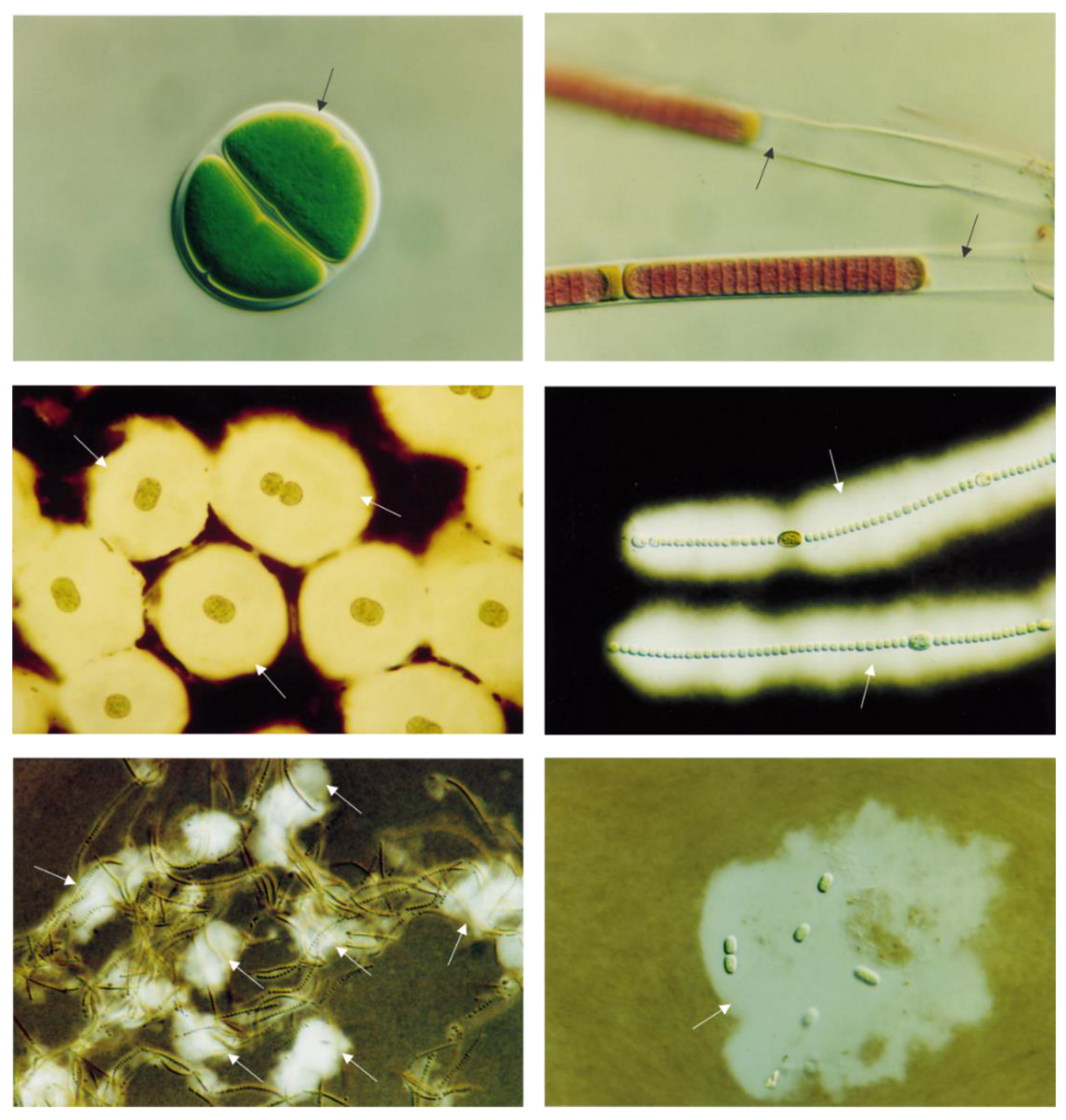
2. Macroalgae and Microalgae: An Overview
2.1. Macroalgae
2.2. Microalgae
3. Algal-Based Sulfated Polysaccharides
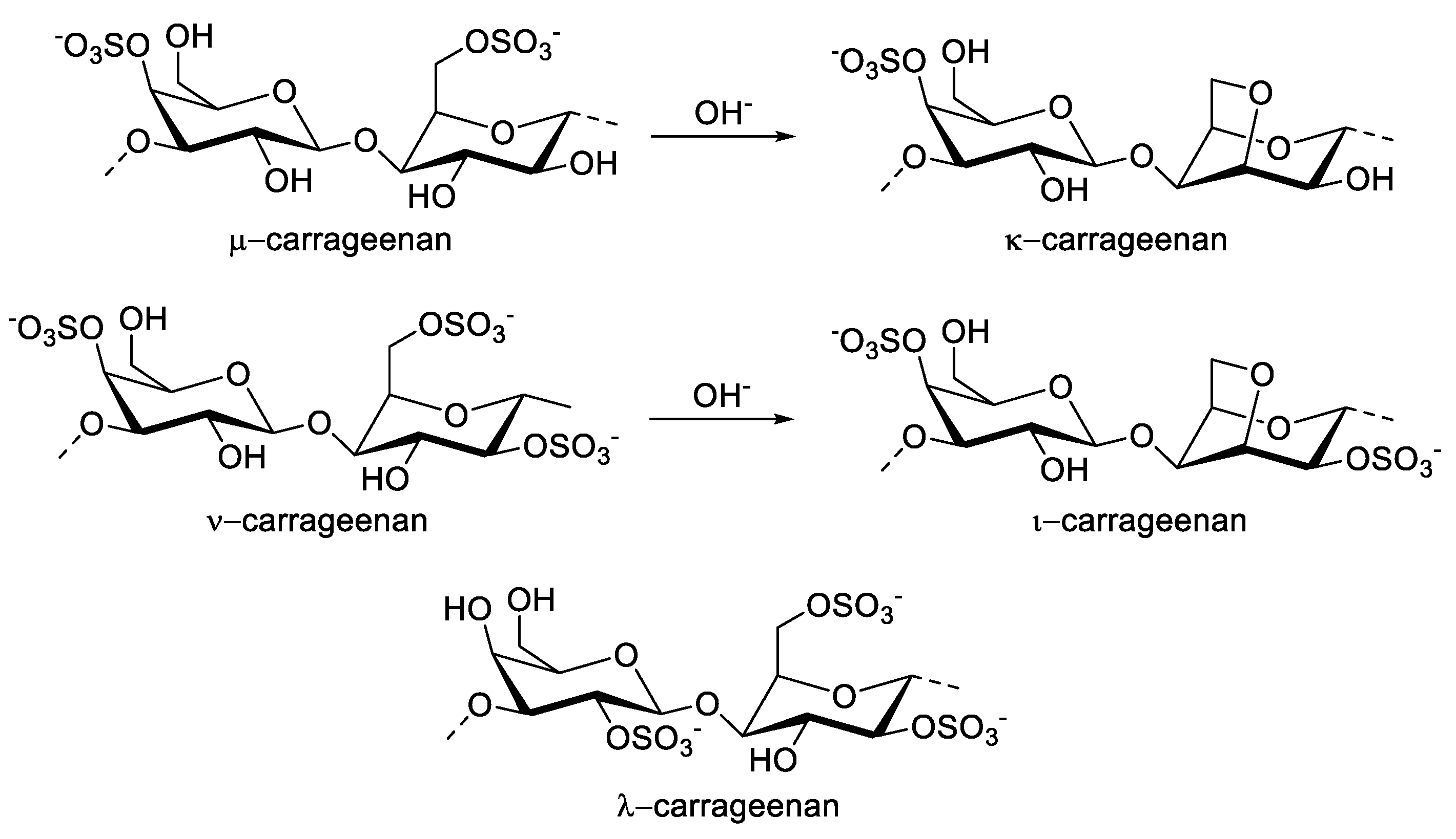
3.1. Carrageenan
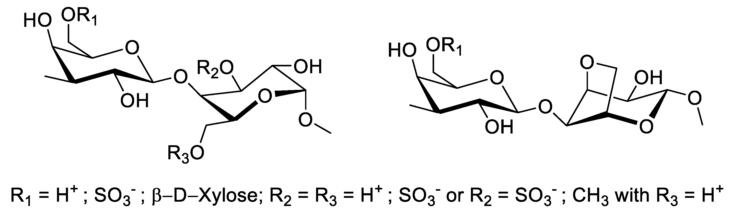
3.2. Agaran
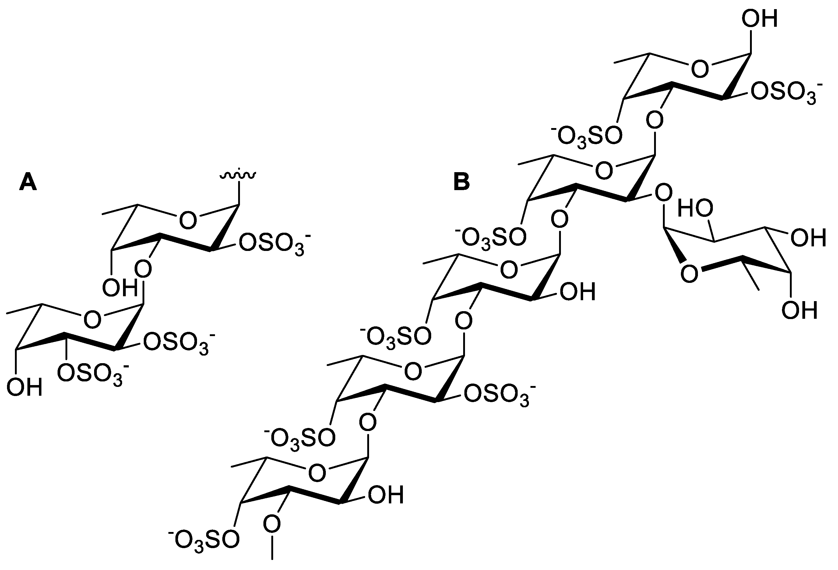
3.3. Fucoidan
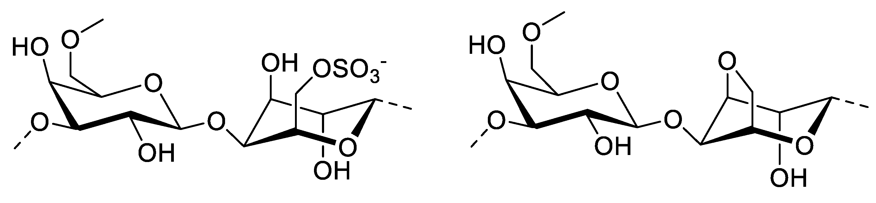
3.4. Porphyran
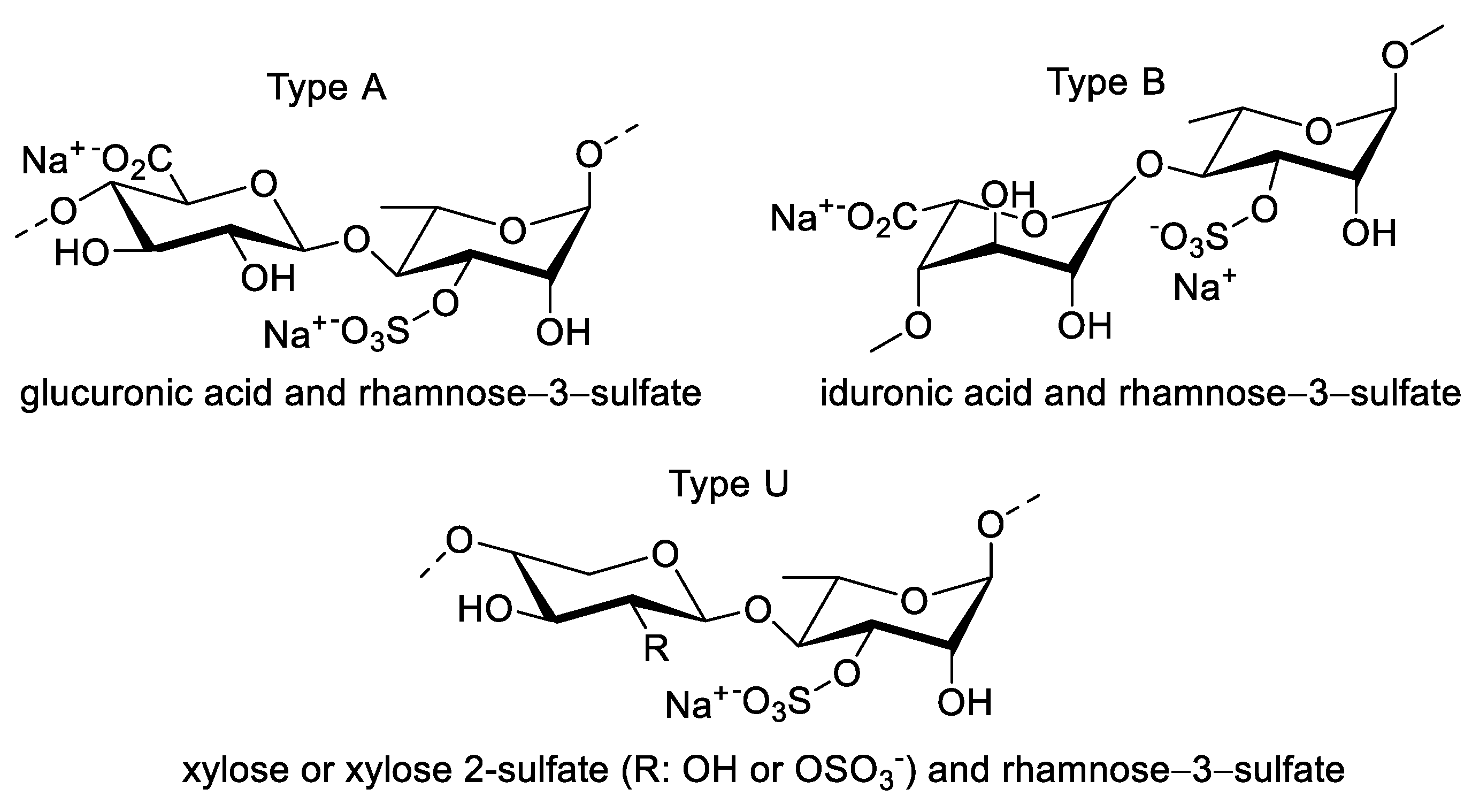
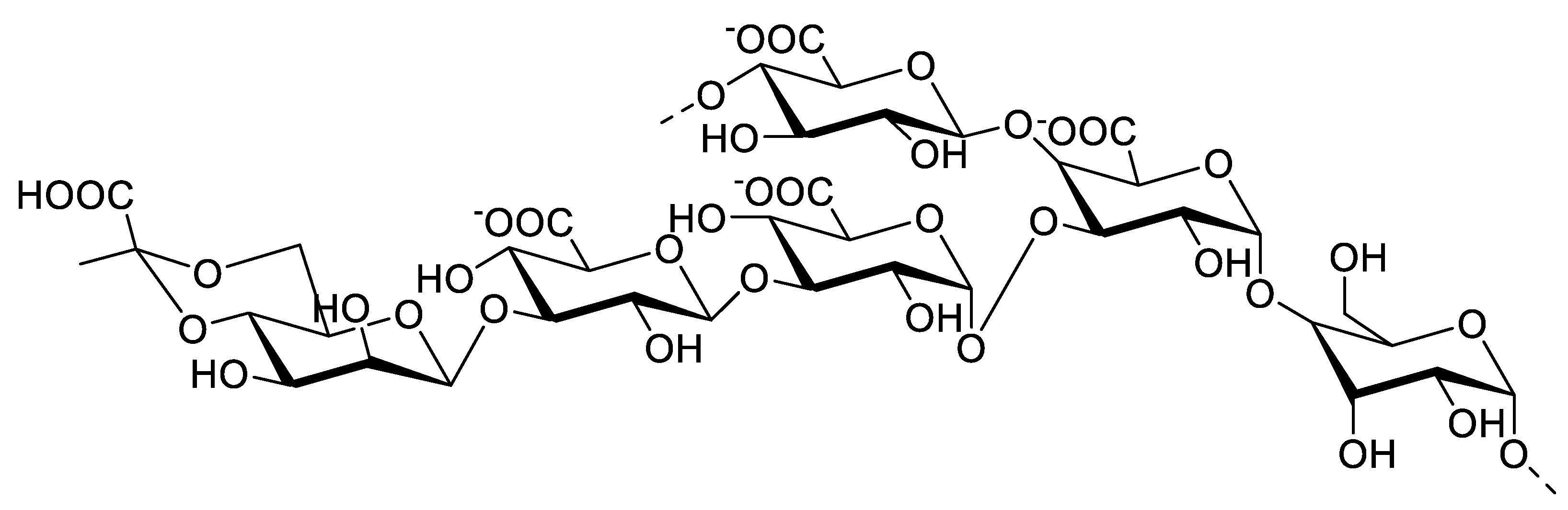
3.5. Ulvan
3.6. Exopolysaccharides
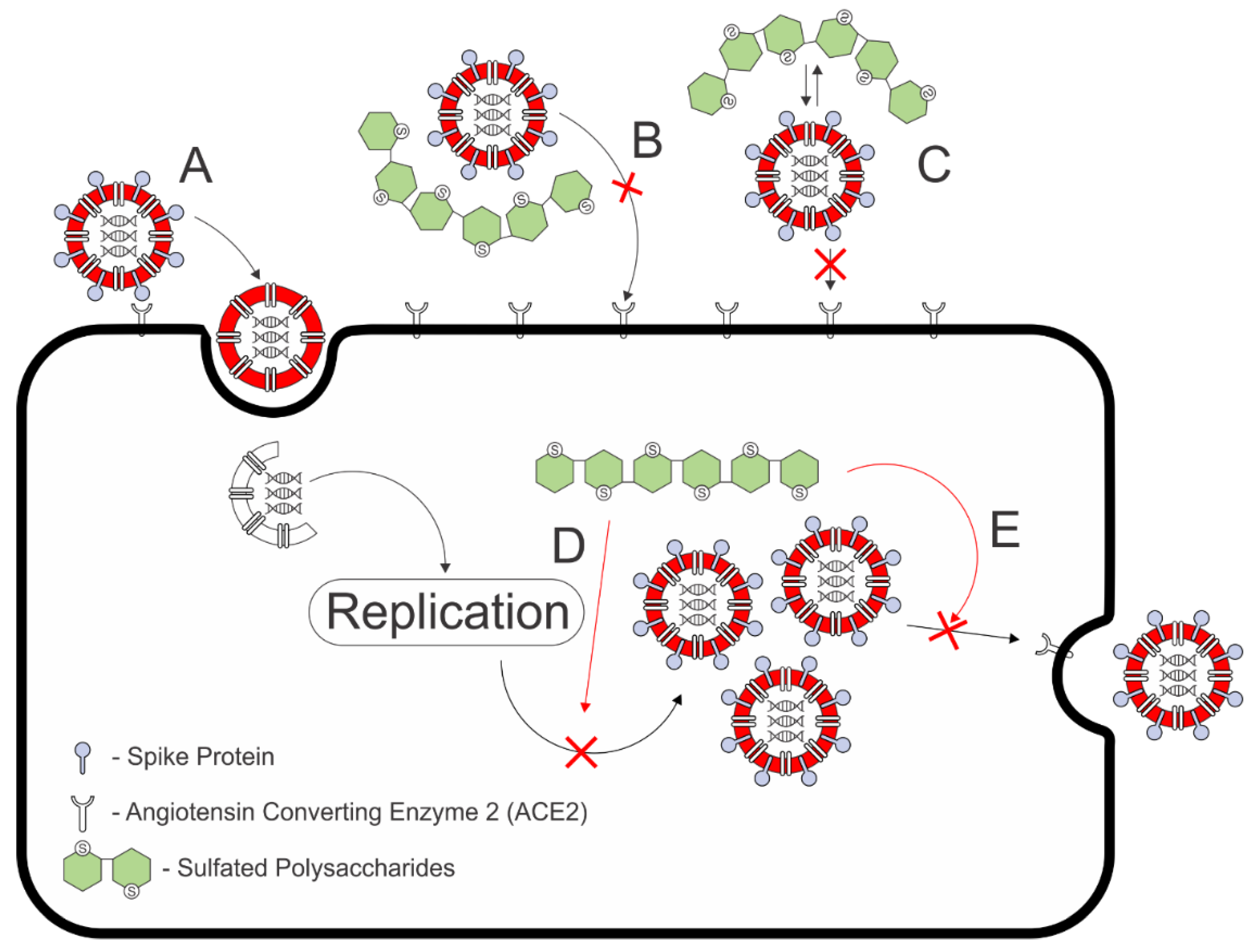
4. Antiviral Activities of Algal-Based Sulfated Polysaccharides
4.1. Carrageenan

| Sulfated Polysaccharide | Virus Strain | Antiviral Activities | Proposed Mechanism of Action | Toxicity (Cell) | Remarks on Molecular Weight and Sulfate Content | Refs. |
|---|---|---|---|---|---|---|
| CP (%) | ||||||
| ι-Carrageenan | HRV2 | 100% 2 | ι-Carrageenan inhibits HRV2 entry to infect HeLa cell line | >1000 µg/mL (HeLa) | [11] | |
| κ-Carrageenan | 62% 2 | |||||
| λ-Carrageenan | 55% 2 | |||||
| log TCID50 1 | ||||||
| ι-Carrageenan | HRV2 | <2 2 | ||||
| κ-Carrageenan | ~6 2 | |||||
| λ-Carrageenan | ~6 2 | |||||
| Viral Replication Inh. at 5 µg/mL | ||||||
| ι-Carrageenan | HRV1A | >99% | Reduces production of HRV particles on HeLa cell line | >500 µg/mL (HNep) | [11] | |
| HRV14 | >99% | |||||
| HRV16 | >99% | |||||
| HRV83 | >99% | |||||
| HRV84 | >99% | |||||
| Neutralization Activity | ||||||
| ιCarrageenan | SARS-CoV-2 Spike pseudotyped lentivirus | 79% 3 | Inhibits cell entry of the SARS-CoV-2 spike pseudotyped lentivirus | >100 µg/mL (Vero B4) | 1. High molecular weight Fucoidan from U. pinnatifida and F. vesiculosus shows less than 50% reduction in infection 2. Polymer without sulfate found to be inactive | [88] |
| κ-Carrageenan | ~80% 4 | |||||
| λ-Carrageenan | ~80% 4 | |||||
| EC50 (µg/mL) | ||||||
| λ-Carrageenan | SARS-CoV-2 | 0.9 | 1. Neutralizing viral glycoprotein HA 2. Blocking the VP harboring viral ribonucleoprotein complexes | >300 µg/mL (MDCK) | λ-carrageenan (1025 kDa) shows better solubility in cold water than other carrageenans because of its higher sulfate content | [90] |
| Influenza A (H1N1) | 0.3 | |||||
| Influenza A (H3N2) | 0.3 | |||||
| Influenza B | 1.4 | |||||
4.2. Fucoidan
4.3. Agaran
4.4. Porphyran
4.5. Ulvan
4.6. Exopolysaccharides
5. Outlook and Future Prospects
- The encapsulation amount, which correlates with the efficacy concentration;
- Consideration of the drug preparation (e.g., injections, solids, or transdermal application);
- Their preservation capabilities, alongside stabilities both before and after uptake;
- The impact of the bioavailability of the released drug on systemic circulation;
- The possibility of controlled released tuning in antiviral delivering agents [127].
Author Contributions
Funding
Institutional Review Board Statement
Informed Consent Statement
Data Availability Statement
Acknowledgments
Conflicts of Interest
References
- World Health Organization. WHO Coronavirus Disease 2019 Situation Report 51—11th March 2020. WHO Bull. 2020, 2019, 2633. [Google Scholar]
- Zhu, N.; Zhang, D.; Wang, W.; Li, X.; Yang, B.; Song, J.; Zhao, X.; Huang, B.; Shi, W.; Lu, R.; et al. A Novel Coronavirus from Patients with Pneumonia in China, 2019. N. Engl. J. Med. 2020, 382, 727–733. [Google Scholar] [CrossRef] [PubMed]
- Cohen, J.; Kupferschmidt, K. Countries test tactics in “war” against COVID-19. Science 2020, 367, 1287–1288. [Google Scholar] [CrossRef] [PubMed] [Green Version]
- Leung, K.; Shum, M.H.H.; Leung, G.M.; Lam, T.T.Y.; Wu, J.T. Early transmissibility assessment of the N501Y mutant strains of SARS-CoV-2 in the United Kingdom, October to November 2020. Eurosurveillance 2020, 26, 2002106. [Google Scholar] [CrossRef] [PubMed]
- Sanches, P.R.S.; Charlie-Silva, I.; Braz, H.L.B.; Bittar, C.; Freitas Calmon, M.; Rahal, P.; Cilli, E.M. Recent advances in SARS-CoV-2 Spike protein and RBD mutations comparison between new variants Alpha (B.1.1.7, United Kingdom), Beta (B.1.351, South Africa), Gamma (P.1, Brazil) and Delta (B.1.617.2, India). J. Virus Erad. 2021, 7, 100054. [Google Scholar] [CrossRef]
- Burki, T. COVID-19 and diabetes in Africa: A lethal combination. Lancet Diabetes Endocrinol. 2021, 10, 23. [Google Scholar] [CrossRef]
- Tregoning, J.S.; Flight, K.E.; Higham, S.L.; Wang, Z.; Pierce, B.F. Progress of the COVID-19 vaccine effort: Viruses, vaccines and variants versus efficacy, effectiveness and escape. Nat. Rev. Immunol. 2021, 21, 626–636. [Google Scholar] [CrossRef]
- Bandyopadhyay, S.S.; Navid, M.H.; Ghosh, T.; Schnitzler, P.; Ray, B. Structural features and in vitro antiviral activities of sulfated polysaccharides from Sphacelaria indica. Phytochemistry 2011, 72, 276–283. [Google Scholar] [CrossRef]
- Karmakar, P.; Alberto, C.; Beatriz, E.; Ghosh, T.; Ray, B. Polysaccharides from Padina tetrastromatica: Structural features, chemical modification and antiviral activity. Carbohydr. Polym. 2010, 80, 513–520. [Google Scholar] [CrossRef]
- Pujol, C.A.; Ray, S.; Ray, B.; Damonte, E.B. Antiviral activity against dengue virus of diverse classes of algal sulfated polysaccharides. Int. J. Biol. Macromol. 2012, 51, 412–416. [Google Scholar] [CrossRef]
- Grassauer, A.; Weinmuellner, R.; Meier, C.; Pretsch, A.; Prieschl-Grassauer, E.; Unger, H. Iota-Carrageenan is a potent inhibitor of rhinovirus infection. Virol. J. 2008, 5, 5–7. [Google Scholar] [CrossRef] [PubMed] [Green Version]
- Wang, W.; Wu, J.; Zhang, X.; Hao, C.; Zhao, X.; Jiao, G.; Shan, X.; Tai, W.; Yu, G. Inhibition of influenza A virus infection by fucoidan targeting viral neuraminidase and cellular EGFR pathway. Sci. Rep. 2017, 7, 1–14. [Google Scholar] [CrossRef] [PubMed]
- Muto, S.; Niimura, K.; Oohara, M.; Oguchi, Y.; Matsunaga, K.; Hirose, K.; Kakuchi, J.; Sugita, N.; Furusho, T.; Yoshikumi, C.; et al. Polysaccharides from Marine Algae and Antiviral Drugs Containing the Same as Active Ingredient. U.S. Patent 5,089,481 A, 18 February 1992. [Google Scholar]
- Santos, S.; Ferreira, T.; Almeida, J.; Pires, M.J.; Colaço, A.; Lemos, S.; Da Costa, R.M.G.; Medeiros, R.; Bastos, M.M.S.M.; Neuparth, M.J.; et al. Dietary supplementation with the red seaweed porphyra umbilicalis protects against DNA damage and pre-malignant dysplastic skin lesions in HPV-transgenic mice. Mar. Drugs 2019, 17, 615. [Google Scholar] [CrossRef] [PubMed] [Green Version]
- Hasui, M.; Matsuda, M.; Okutani, K.; Shigeta, S. In vitro antiviral activities of sulfated polysaccharides from a marine microalga (Cochlodinium polykrikoides) against human immunodeficiency virus and other enveloped viruses. Int. J. Biol. Macromol. 1995, 17, 293–297. [Google Scholar] [CrossRef]
- Fabregas, J.; García, D.; Fernandez-Alonso, M.; Rocha, A.I.; Gómez-Puertas, P.; Escribano, J.M.; Otero, A.; Coll, J.M. In vitro inhibition of the replication of haemorrhagic septicaemia virus (VHSV) and African swine fever virus (ASFV) by extracts from marine microalgae. Antivir. Res. 1999, 44, 67–73. [Google Scholar] [CrossRef]
- Huleihel, M.; Ishanu, V.; Tal, J.; Arad, S. Antiviral effect of red microalgal polysaccharides on Herpes simplex and Varicella zoster viruses. J. Appl. Phycol. 2001, 13, 127–134. [Google Scholar] [CrossRef]
- Yim, S.K.; Kim, K.; Kim, I.; Chun, S.H.; Oh, T.H.; Kim, J.U.; Kim, J.; Jung, W.; Moon, H.; Ku, B.; et al. Inhibition of sars-cov-2 virus entry by the crude polysaccharides of seaweeds and abalone viscera in vitro. Mar. Drugs 2021, 19, 219. [Google Scholar] [CrossRef]
- Figueroa, J.M.; Lombardo, M.E.; Dogliotti, A.; Flynn, L.P.; Giugliano, R.; Simonelli, G.; Valentini, R.; Ramos, A.; Romano, P.; Marcote, M.; et al. Efficacy of a Nasal Spray Containing Iota-Carrageenan in the Postexposure Prophylaxis of COVID-19 in Hospital Personnel Dedicated to Patients Care with COVID-19 Disease. Int. J. Gen. Med. 2021, 14, 6277–6286. [Google Scholar] [CrossRef]
- Lauritano, C.; Andersen, J.H.; Hansen, E.; Albrigtsen, M.; Escalera, L.; Esposito, F.; Helland, K.; Hanssen, K.; Romano, G.; Ianora, A. Bioactivity screening of microalgae for antioxidant, anti-inflammatory, anticancer, anti-diabetes, and antibacterial activities. Front. Mar. Sci. 2016, 3, 1–2. [Google Scholar] [CrossRef] [Green Version]
- Pangestuti, R.; Kim, S.K. Biological Activities of Carrageenan, 1st ed.; Elsevier Inc.: Amsterdam, The Netherlands, 2014; Volume 72, ISBN 9780128002698. [Google Scholar]
- Ciancia, M.; Fernández, P.V.; Leliaert, F. Diversity of Sulfated Polysaccharides From Cell Walls of Coenocytic Green Algae and Their Structural Relationships in View of Green Algal Evolution. Front. Plant Sci. 2020, 11, 1–15. [Google Scholar] [CrossRef]
- Muthukumar, J.; Chidambaram, R.; Sukumaran, S. Sulfated polysaccharides and its commercial applications in food industries—A review. J. Food Sci. Technol. 2021, 58, 2453–2466. [Google Scholar] [CrossRef] [PubMed]
- El Gamal, A.A. Biological Importance of Marine Algae. In Handbook of Marine Macroalgae: Biotechnology and Applied Phycology; John Wiley & Sons: Chichester, UK, 2011; pp. 1–35. [Google Scholar] [CrossRef] [Green Version]
- Kerrison, P.D. Algae as Crops Seaweed, 2nd ed.; Elsevier: Amsterdam, The Netherlands, 2016; Volume 3, ISBN 9780123948083. [Google Scholar]
- Wang, J.; Wang, D.; Zhang, Y.; Dong, J. Synthesis and Biopharmaceutical Applications of Sugar-Based Polymers: New Advances and Future Prospects. ACS Biomater. Sci. Eng. 2021, 7, 963–982. [Google Scholar] [CrossRef] [PubMed]
- Domínguez, H. Algae as a source of biologically active ingredients for the formulation of functional foods and nutraceuticals. In Functional Ingredients from Algae Foods Nutraceuticals; Woodhead Publishing: Sawston, UK, 2013; pp. 1–19. [Google Scholar] [CrossRef]
- Dominguez, H.; Loret, E.P. Ulva lactuca, A Source of Troubles and Potential Riches. Mar. Drugs 2019, 17, 357. [Google Scholar] [CrossRef] [PubMed] [Green Version]
- Cunha, L.; Grenha, A. Sulfated seaweed polysaccharides as multifunctional materials in drug delivery applications. Mar. Drugs 2016, 14, 42. [Google Scholar] [CrossRef]
- Ahmed, A.B.A.; Adel, M.; Karimi, P.; Peidayesh, M. Pharmaceutical, Cosmeceutical, and Traditional Applications of Marine Carbohydrates, 1st ed.; Elsevier Inc.: Amsterdam, The Netherlands, 2014; Volume 73, ISBN 9780128002681. [Google Scholar]
- Thakur, V.; Lu, J.; Roscilli, G.; Aurisicchio, L.; Cappelletti, M.; Pavoni, E.; White, W.L.; Bedogni, B. The natural compound fucoidan from New Zealand Undaria pinnatifida synergizes with the ERBB inhibitor lapatinib enhancing melanoma growth inhibition. Oncotarget 2017, 8, 17887–17896. [Google Scholar] [CrossRef] [PubMed]
- Hong, S.W.; Jung, K.H.; Lee, H.S.; Zheng, H.M.; Choi, M.J.; Lee, C.; Hong, S.S. Suppression by fucoidan of liver fibrogenesis via the TGF-β/smad pathway in protecting against oxidative stress. Biosci. Biotechnol. Biochem. 2011, 75, 833–840. [Google Scholar] [CrossRef]
- Berri, M.; Olivier, M.; Holbert, S.; Dupont, J.; Demais, H.; Le Goff, M.; Collen, P.N. Ulvan from Ulva armoricana (Chlorophyta) activates the PI3K/Akt signalling pathway via TLR4 to induce intestinal cytokine production. Algal Res. 2017, 28, 39–47. [Google Scholar] [CrossRef]
- Li, W.; Jiang, N.; Li, B.; Wan, M.; Chang, X.; Liu, H.; Zhang, L.; Yin, S.; Qi, H.; Liu, S. Antioxidant activity of purified ulvan in hyperlipidemic mice. Int. J. Biol. Macromol. 2018, 113, 971–975. [Google Scholar] [CrossRef]
- Albuquerque, J.C.S.; Araújo, M.L.H.; Rocha, M.V.P.; de Souza, B.W.S.; de Castro, G.M.C.; Cordeiro, E.M.S.; de Suosa Silva, J.; Benevides, N.M.B. Acid hydrolysis conditions for the production of fine chemicals from Gracilaria birdiae alga biomass. Algal Res. 2021, 53, 102139. [Google Scholar] [CrossRef]
- Fayaz, M.; Namitha, K.K.; Murthy, K.N.C.; Swamy, M.M.; Sarada, R.; Khanam, S.; Subbarao, P.V.; Ravishankar, G.A. Chemical composition, iron bioavailability, and antioxidant activity of Kappaphycus alvarezzi (Doty). J. Agric. Food Chem. 2005, 53, 792–797. [Google Scholar] [CrossRef]
- Yang, Y.; Zhang, M.; Alalawy, A.I.; Almutairi, F.M.; Al-Duais, M.A.; Wang, J.; Salama, E.-S. Identification and characterization of marine seaweeds for biocompounds production. Environ. Technol. Innov. 2021, 24, 101848. [Google Scholar] [CrossRef]
- Yu, G.; Zhang, Q.; Wang, Y.; Yang, Q.; Yu, H.; Li, H.; Chen, J.; Fu, L. Sulfated polysaccharides from red seaweed Gelidium amansii: Structural characteristics, antioxidant and anti-glycation properties, and development of bioactive films. Food Hydrocoll. 2021, 119, 106820. [Google Scholar] [CrossRef]
- Caballero, E.; Flores, A.; Olivares, A. Sustainable exploitation of macroalgae species from Chilean coast: Characterization and food applications. Algal Res. 2021, 57, 102349. [Google Scholar] [CrossRef]
- Lorenzo, J.M.; Agregán, R.; Munekata, P.E.S.; Franco, D.; Carballo, J.; Şahin, S.; Lacomba, R.; Barba, F.J. Proximate composition and nutritional value of three macroalgae: Ascophyllum nodosum, Fucus vesiculosus and Bifurcaria bifurcata. Mar. Drugs 2017, 15, 360. [Google Scholar] [CrossRef] [PubMed] [Green Version]
- Kumar, M.; Gupta, V.; Kumari, P.; Reddy, C.R.K.; Jha, B. Assessment of nutrient composition and antioxidant potential of Caulerpaceae seaweeds. J. Food Compos. Anal. 2011, 24, 270–278. [Google Scholar] [CrossRef]
- Bahari, A.; Moelants, K.; Wallecan, J.; Mangiante, G.; Mazoyer, J.; Hendrickx, M.; Grauwet, T. Understanding the effect of time, temperature and salts on carrageenan extraction from Chondrus crispus. Algal Res. 2021, 58, 102371. [Google Scholar] [CrossRef]
- Mansilla, A.; Ávila, M. Using Macrocystis pyrifera (L.) C. Agardh from southern Chile as a source of applied biological compounds. Rev. Bras. Farmacogn. 2011, 21, 262–267. [Google Scholar] [CrossRef] [Green Version]
- Garcia-Vaquero, M.; Rajauria, G.; Miranda, M.; Sweeney, T.; Lopez-Alonso, M.; O’Doherty, J. Seasonal Variation of the Proximate Composition, Mineral Content, Fatty Acid Profiles and Other Phytochemical Constituents of Selected Brown Macroalgae. Mar. Drugs 2021, 19, 204. [Google Scholar] [CrossRef]
- Ethier, S.; Woisard, K.; Vaughan, D.; Wen, Z. Continuous culture of the microalgae Schizochytrium limacinum on biodiesel-derived crude glycerol for producing docosahexaenoic acid. Bioresour. Technol. 2011, 102, 88–93. [Google Scholar] [CrossRef]
- Bongiorno, T.; Foglio, L.; Proietti, L.; Vasconi, M.; Lopez, A.; Pizzera, A.; Carminati, D.; Tava, A.; Vizcaíno, A.J.; Alarcón, F.J.; et al. Microalgae from Biorefinery as Potential Protein Source for Siberian Sturgeon (A. baerii) Aquafeed. Sustainability 2020, 12, 8779. [Google Scholar] [CrossRef]
- Grossmann, L.; Hinrichs, J.; Weiss, J. Cultivation and downstream processing of microalgae and cyanobacteria to generate protein-based technofunctional food ingredients. Crit. Rev. Food Sci. Nutr. 2020, 60, 2961–2989. [Google Scholar] [CrossRef] [PubMed]
- Khatoon, H.; Abdu Rahman, N.; Banerjee, S.; Harun, N.; Suleiman, S.S.; Zakaria, N.H.; Lananan, F.; Abdul Hamid, S.H.; Endut, A.; Abdu, N.; et al. Effects of different salinities and pH on the growth and proximate composition of Nannochloropsis sp. and Tetraselmis sp. isolated from South China Sea cultured under control and natural condition. Int. Biodeterior. Biodegrad. 2014, 95, 11–18. [Google Scholar] [CrossRef]
- Menegol, T.; Diprat, A.B.; Rodrigues, E.; Rech, R. Effect of temperature and nitrogen concentration on biomass composition of Heterochlorella luteoviridis. Food Sci. Technol. 2017, 37, 28–37. [Google Scholar] [CrossRef] [Green Version]
- Kumaran, J.; Poulose, S.; Joseph, V.; Bright Singh, I.S. Enhanced biomass production and proximate composition of marine microalga Nannochloropsis oceanica by optimization of medium composition and culture conditions using response surface methodology. Anim. Feed Sci. Technol. 2021, 271, 114761. [Google Scholar] [CrossRef]
- Abu-Ghosh, S.; Dubinsky, Z.; Verdelho, V.; Iluz, D. Unconventional high-value products from microalgae: A review. Bioresour. Technol. 2021, 329, 124895. [Google Scholar] [CrossRef]
- Di Lena, G.; Casini, I.; Lucarini, M.; Sanchez del Pulgar, J.; Aguzzi, A.; Caproni, R.; Gabrielli, P.; Lombardi-Boccia, G. Chemical characterization and nutritional evaluation of microalgal biomass from large-scale production: A comparative study of five species. Eur. Food Res. Technol. 2020, 246, 323–332. [Google Scholar] [CrossRef]
- Assunção, M.F.G.; Varejão, J.M.T.B.; Santos, L.M.A. Nutritional characterization of the microalga Ruttnera lamellosa compared to Porphyridium purpureum. Algal Res. 2017, 26, 8–14. [Google Scholar] [CrossRef]
- Alagawany, M.; Taha, A.E.; Noreldin, A.; El-Tarabily, K.A.; Abd El-Hack, M.E. Nutritional applications of species of Spirulina and Chlorella in farmed fish: A review. Aquaculture 2021, 542, 736841. [Google Scholar] [CrossRef]
- Grossmann, L.; Ebert, S.; Hinrichs, J.; Weiss, J. Effect of precipitation, lyophilization, and organic solvent extraction on preparation of protein-rich powders from the microalgae Chlorella protothecoides. Algal Res. 2018, 29, 266–276. [Google Scholar] [CrossRef]
- Estevez, J.M.; Ciancia, M.; Cerezo, A.S. The system of sulfated galactans from the red seaweed Gymnogongrus torulosus (Phyllophoraceae, Rhodophyta): Location and structural analysis. Carbohydr. Polym. 2008, 73, 594–605. [Google Scholar] [CrossRef]
- Perez Recalde, M.; Canelón, D.J.; Compagnone, R.S.; Matulewicz, M.C.; Cerezo, A.S.; Ciancia, M. Carrageenan and agaran structures from the red seaweed Gymnogongrus tenuis. Carbohydr. Polym. 2016, 136, 1370–1378. [Google Scholar] [CrossRef] [PubMed]
- Estevez, J.M.; Ciancia, M.; Cerezo, A.S. The system of galactans of the red seaweed, Kappaphycus alvarezii, with emphasis on its minor constituents. Carbohydr. Res. 2004, 339, 2575–2592. [Google Scholar] [CrossRef] [PubMed]
- Qureshi, D.; Nayak, S.K.; Maji, S.; Kim, D.; Banerjee, I.; Pal, K. Carrageenan: A Wonder Polymer from Marine Algae for Potential Drug Delivery Applications. Curr. Pharm. Des. 2019, 25, 1172–1186. [Google Scholar] [CrossRef] [PubMed]
- Li, L.; Ni, R.; Shao, Y.; Mao, S. Carrageenan and its applications in drug delivery. Carbohydr. Polym. 2014, 103, 1–11. [Google Scholar] [CrossRef]
- Kariduraganavar, M.Y.; Kittur, A.A.; Kamble, R.R. Polymer Synthesis and Processing, 1st ed.; Elsevier Inc.: Amsterdam, The Netherlands, 2014; ISBN 9780123969835. [Google Scholar]
- Campo, V.L.; Kawano, D.F.; da Silva, D.B.; Carvalho, I. Carrageenans: Biological properties, chemical modifications and structural analysis—A review. Carbohydr. Polym. 2009, 77, 167–180. [Google Scholar] [CrossRef]
- Pereira, L. Seaweeds as Source of Bioactive Substances and Skin Care Therapy—Cosmeceuticals, Algotheraphy, and Thalassotherapy. Cosmetics 2018, 5, 68. [Google Scholar] [CrossRef] [Green Version]
- Alba, K.; Kontogiorgos, V. Seaweed Polysaccharides (Agar, Alginate Carrageenan); Elsevier: Amsterdam, The Netherlands, 2018; ISBN 9780128140451. [Google Scholar]
- Imeson, A.P. Carrageenan and furcellaran. In Handbook of Hydrocolloids; Elsevier: Amsterdam, The Netherlands, 2009; pp. 164–185. ISBN 9781845695873. [Google Scholar]
- García-Poza, S.; Leandro, A.; Cotas, C.; Cotas, J.; Marques, J.C.; Pereira, L.; Gonçalves, A.M.M. The Evolution Road of Seaweed Aquaculture: Cultivation Technologies and the Industry 4.0. Int. J. Environ. Res. Public Health 2020, 17, 6528. [Google Scholar] [CrossRef]
- Zúñiga, E.A.; Matsuhiro, B.; Mejías, E. Preparation of a low-molecular weight fraction by free radical depolymerization of the sulfated galactan from Schizymenia binderi (Gigartinales, Rhodophyta) and its anticoagulant activity. Carbohydr. Polym. 2006, 66, 208–215. [Google Scholar] [CrossRef]
- Stanley, N.F. Agars. In Food Polysaccharides and Their Applications; CRC Press: Boca Raton, FL, USA, 2016; pp. 231–252. [Google Scholar]
- Ale, M.T.; Meyer, A.S. Fucoidans from brown seaweeds: An update on structures, extraction techniques and use of enzymes as tools for structural elucidation. RSC Adv. 2013, 3, 8131–8141. [Google Scholar] [CrossRef] [Green Version]
- Oliveira, C.; Neves, N.M.; Reis, R.L.; Martins, A.; Silva, T.H. A review on fucoidan antitumor strategies: From a biological active agent to a structural component of fucoidan-based systems. Carbohydr. Polym. 2020, 239, 116131. [Google Scholar] [CrossRef]
- Isnansetyo, A.; Lutfia, F.N.L.; Nursid, M.; Trijoko, T.; Susidarti, R.A. Cytotoxicity of fucoidan from three tropical brown algae against breast and colon cancer cell lines. Pharmacogn. J. 2017, 9, 14–20. [Google Scholar] [CrossRef] [Green Version]
- Zayed, A.; Dienemann, C.; Giese, C.; Krämer, R.; Ulber, R. An immobilized perylene diimide derivative for fucoidan purification from a crude brown algae extract. Process Biochem. 2018, 65, 233–238. [Google Scholar] [CrossRef]
- Zhang, Q.; Qi, H.; Zhao, T.; Deslandes, E.; Ismaeli, N.M.; Molloy, F.; Critchley, A.T. Chemical characteristics of a polysaccharide from Porphyra capensis (Rhodophyta). Carbohydr. Res. 2005, 340, 2447–2450. [Google Scholar] [CrossRef]
- Qiu, Y.; Jiang, H.; Fu, L.; Ci, F.; Mao, X. Porphyran and oligo-porphyran originating from red algae Porphyra: Preparation, biological activities, and potential applications. Food Chem. 2021, 349, 129209. [Google Scholar] [CrossRef] [PubMed]
- Isaka, S.; Cho, K.; Nakazono, S.; Abu, R.; Ueno, M.; Kim, D.; Oda, T. Antioxidant and anti-inflammatory activities of porphyran isolated from discolored nori (Porphyra yezoensis). Int. J. Biol. Macromol. 2015, 74, 68–75. [Google Scholar] [CrossRef] [PubMed]
- Chen, Y.-y.; Xue, Y.-t. Optimization of microwave assisted extraction, chemical characterization and antitumor activities of polysaccharides from porphyra haitanensis. Carbohydr. Polym. 2019, 206, 179–186. [Google Scholar] [CrossRef] [PubMed]
- Lahaye, M.; Robic, A. Structure and function properties of Ulvan, a polysaccharide from green seaweeds. Biomacromolecules 2007, 8, 1765–1774. [Google Scholar] [CrossRef] [PubMed]
- Kidgell, J.T.; Magnusson, M.; de Nys, R.; Glasson, C.R.K. Ulvan: A systematic review of extraction, composition and function. Algal Res. 2019, 39, 101422. [Google Scholar] [CrossRef]
- Glasson, C.R.K.; Sims, I.M.; Carnachan, S.M.; de Nys, R.; Magnusson, M. A cascading biorefinery process targeting sulfated polysaccharides (ulvan) from Ulva ohnoi. Algal Res. 2017, 27, 383–391. [Google Scholar] [CrossRef]
- Pereira, S.; Zille, A.; Micheletti, E.; Moradas-Ferreira, P.; De Philippis, R.; Tamagnini, P. Complexity of cyanobacterial exopolysaccharides: Composition, structures, inducing factors and putative genes involved in their biosynthesis and assembly. FEMS Microbiol. Rev. 2009, 33, 917–941. [Google Scholar] [CrossRef]
- Mancuso Nichols, C.A.; Guezennec, J.; Bowman, J.P. Bacterial exopolysaccharides from extreme marine environments with special consideration of the Southern Ocean, sea ice, and deep-sea hydrothermal vents: A review. Mar. Biotechnol. 2005, 7, 253–271. [Google Scholar] [CrossRef] [PubMed] [Green Version]
- Gaignard, C.; Laroche, C.; Pierre, G.; Dubessay, P.; Delattre, C.; Gardarin, C.; Gourvil, P.; Probert, I.; Dubuffet, A.; Michaud, P. Screening of marine microalgae: Investigation of new exopolysaccharide producers. Algal Res. 2019, 44, 101711. [Google Scholar] [CrossRef]
- Kim, K.J.; Lee, O.H.; Lee, B.Y. Fucoidan, a sulfated polysaccharide, inhibits adipogenesis through the mitogen-activated protein kinase pathway in 3T3-L1 preadipocytes. Life Sci. 2010, 86, 791–797. [Google Scholar] [CrossRef] [PubMed]
- Andri Frediansyah, S.S. The antiviral activity of iota-, kappa-, and lambda-carrageenan against COVID-19: A critical review. Clin. Epidemiol. Glob. Health 2021, 12, 100826. [Google Scholar] [CrossRef]
- Jabeen, M.; Dutot, M.; Fagon, R.; Verrier, B.; Monge, C. Seaweed sulfated polysaccharides against respiratory viral infections. Pharmaceutics 2021, 13, 733. [Google Scholar] [CrossRef]
- Reynolds, D.; Huesemann, M.; Edmundson, S.; Sims, A.; Hurst, B.; Cady, S.; Beirne, N.; Freeman, J.; Berger, A.; Gao, S. Viral inhibitors derived from macroalgae, microalgae, and cyanobacteria: A review of antiviral potential throughout pathogenesis. Algal Res. 2021, 57, 102331. [Google Scholar] [CrossRef]
- Adalja, A.; Inglesby, T. Broad-Spectrum Antiviral Agents: A Crucial Pandemic Tool. Expert Rev. Anti-infect. Ther. 2019, 17, 467–470. [Google Scholar] [CrossRef] [Green Version]
- Morokutti-Kurz, M.; Fröba, M.; Graf, P.; Große, M.; Grassauer, A.; Auth, J.; Schubert, U.; Prieschl-Grassauer, E. Iota-carrageenan neutralizes SARS-CoV-2 and inhibits viral replication in vitro. PLoS ONE 2021, 16, e0237480. [Google Scholar] [CrossRef]
- Song, S.; Peng, H.; Wang, Q.; Liu, Z.; Dong, X.; Wen, C.; Ai, C.; Zhang, Y.; Wang, Z.; Zhu, B. Inhibitory activities of marine sulfated polysaccharides against SARS-CoV-2. Food Funct. 2020, 11, 7415–7420. [Google Scholar] [CrossRef]
- Jang, Y.; Shin, H.; Lee, M.K.; Kwon, O.S.; Shin, J.S.; Kim, Y.-i.; Kim, C.W.; Lee, H.R.; Kim, M. Antiviral activity of lambda-carrageenan against influenza viruses and severe acute respiratory syndrome coronavirus 2. Sci. Rep. 2021, 11, 1–12. [Google Scholar] [CrossRef]
- Hans, N.; Malik, A.; Naik, S. Antiviral activity of sulfated polysaccharides from marine algae and its application in combating COVID-19: Mini review. Bioresour. Technol. Rep. 2021, 13, 100623. [Google Scholar] [CrossRef] [PubMed]
- Aguilar-Briseño, J.A.; Cruz-Suarez, L.E.; Sassi, J.F.; Ricque-Marie, D.; Zapata-Benavides, P.; Mendoza-Gamboa, E.; Rodríguez-Padilla, C.; Trejo-Avila, L.M. Sulphated polysaccharides from Ulva clathrata and Cladosiphon okamuranus seaweeds both inhibit viral attachment/entry and cell-cell fusion, in NDV infection. Mar. Drugs 2015, 13, 697–712. [Google Scholar] [CrossRef] [PubMed]
- Jiao, G.; Yu, G.; Zhang, J.; Ewart, H.S. Chemical structures and bioactivities of sulfated polysaccharides from marine algae. Mar. Drugs 2011, 9, 196–233. [Google Scholar] [CrossRef] [Green Version]
- Ponce, N.M.A.; Pujol, C.A.; Damonte, E.B.; Flores, M.L.; Stortz, C.A. Fucoidans from the brown seaweed Adenocystis utricularis: Extraction methods, antiviral activity and structural studies. Carbohydr. Res. 2003, 338, 153–165. [Google Scholar] [CrossRef]
- Lee, J.-B.; Hayashi, K.; Hashimoto, M.; Nakano, T.; Hayashi, T. Novel Antiviral Fucoidan from Sporophyll of Undaria pinnatifida (Mekabu). Chem. Pharm. Bull. 2004, 52, 1091–1094. [Google Scholar] [CrossRef] [PubMed] [Green Version]
- Marks, R.M.; Lu, H.; Sundaresan, R.; Toida, T.; Suzuki, A.; Imanari, T.; Hernáiz, M.J.; Linhardt, R.J. Probing the interaction of dengue virus envelope protein with heparin: Assessment of glycosaminoglycan-derived inhibitors. J. Med. Chem. 2001, 44, 2178–2187. [Google Scholar] [CrossRef]
- Lu, W.; Yang, Z.; Chen, J.; Wang, D.; Zhang, Y. Recent advances in antiviral activities and potential mechanisms of sulfated polysaccharides. Carbohydr. Polym. 2021, 272, 118526. [Google Scholar] [CrossRef]
- Zhang, H.; Wang, J.Q.; Nie, S.P.; Wang, Y.X.; Cui, S.W.; Xie, M.Y. Sulfated modification, characterization and property of a water-insoluble polysaccharide from Ganoderma atrum. Int. J. Biol. Macromol. 2015, 79, 248–255. [Google Scholar] [CrossRef]
- Bao, H.; Choi, W.S.; You, S. Effect of sulfated modification on the molecular characteristics and biological activities of polysaccharides from Hypsizigus marmoreus. Biosci. Biotechnol. Biochem. 2010, 74, 1408–1414. [Google Scholar] [CrossRef]
- Helen Fitton, J.; Park, A.Y.; Karpiniec, S.S.; Stringer, D.N. Fucoidan and lung function: Value in viral infection. Mar. Drugs 2021, 19, 4. [Google Scholar] [CrossRef]
- Nadeeka Madushani Herath, K.H.I.; Kim, H.J.; Kim, A.; Sook, C.E.; Lee, B.Y.; Jee, Y. The role of fucoidans isolated from the sporophylls of undaria pinnatifida against particulate-matter-induced allergic airway inflammation: Evidence of the attenuation of oxidative stress and inflammatory responses. Molecules 2020, 25, 2869. [Google Scholar] [CrossRef] [PubMed]
- Dutot, M.; Grassin-Delyle, S.; Salvator, H.; Brollo, M.; Rat, P.; Fagon, R.; Naline, E.; Devillier, P. A marine-sourced fucoidan solution inhibits Toll-like-receptor-3-induced cytokine release by human bronchial epithelial cells. Int. J. Biol. Macromol. 2019, 130, 429–436. [Google Scholar] [CrossRef] [PubMed]
- Duarte, M.E.R.; Noseda, D.G.; Noseda, M.D.; Tulio, S.; Pujol, C.A.; Damonte, E.B. Inhibitory effect of sulfated galactans from the marine alga Bostrychia montagnei on herpes simplex virus replication in vitro. Phytomedicine 2001, 8, 53–58. [Google Scholar] [CrossRef]
- Duarte, M.E.R.; Cauduro, J.P.; Noseda, D.G.; Noseda, M.D.; Gonçalves, A.G.; Pujol, C.A.; Damonte, E.B.; Cerezo, A.S. The structure of the agaran sulfate from Acanthophora spicifera (Rhodomelaceae, Ceramiales) and its antiviral activity. Relation between structure and antiviral activity in agarans. Carbohydr. Res. 2004, 339, 335–347. [Google Scholar] [CrossRef] [PubMed]
- Hardouin, K.; Bedoux, G.; Burlot, A.S.; Donnay-Moreno, C.; Bergé, J.P.; Nyvall-Collén, P.; Bourgougnon, N. Enzyme-assisted extraction (EAE) for the production of antiviral and antioxidant extracts from the green seaweed Ulva armoricana (Ulvales, Ulvophyceae). Algal Res. 2016, 16, 233–239. [Google Scholar] [CrossRef] [Green Version]
- Chiu, Y.H.; Chan, Y.L.; Li, T.L.; Wu, C.J. Inhibition of Japanese Encephalitis Virus Infection by the Sulfated Polysaccharide Extracts from Ulva lactuca. Mar. Biotechnol. 2012, 14, 468–478. [Google Scholar] [CrossRef] [PubMed]
- Zhao, T.; Zhang, Q.; Qi, H.; Zhang, H.; Niu, X.; Xu, Z.; Li, Z. Degradation of porphyran from Porphyra haitanensis and the antioxidant activities of the degraded porphyrans with different molecular weight. Int. J. Biol. Macromol. 2006, 38, 45–50. [Google Scholar] [CrossRef]
- Li, Y.T.; Huo, Y.F.; Wang, F.; Wang, C.; Zhu, Q.; Wang, Y.B.; Fu, L.L.; Zhou, T. Improved antioxidant and immunomodulatory activities of enzymatically degraded Porphyra haitanensis polysaccharides. J. Food Biochem. 2020, 44, 1–13. [Google Scholar] [CrossRef]
- López-Vázquez, C.; Bandín, I.; Panzarin, V.; Toffan, A.; Cuenca, A.; Olesen, N.J.; Dopazo, C.P. Steps of the replication cycle of the viral haemorrhagic septicaemia virus (VHSV) affecting its virulence on fish. Animals 2020, 10, 2264. [Google Scholar] [CrossRef]
- Gaudreault, N.N.; Madden, D.W.; Wilson, W.C.; Trujillo, J.D.; Richt, J.A. African Swine Fever Virus: An Emerging DNA Arbovirus. Front. Vet. Sci. 2020, 7, 1–17. [Google Scholar] [CrossRef]
- Kleandrova, V.V.; Speck-Planche, A. The urgent need for pan-antiviral agents: From multitarget discovery to multiscale design. Future Med. Chem. 2021, 13, 5–8. [Google Scholar] [CrossRef] [PubMed]
- Raposo, M.F.D.J.; De Morais, A.M.M.B.; De Morais, R.M.S.C. Influence of sulphate on the composition and antibacterial and antiviral properties of the exopolysaccharide from Porphyridium cruentum. Life Sci. 2014, 101, 56–63. [Google Scholar] [CrossRef] [PubMed]
- Usov, A.I.; Zelinsky, N.D. Chemical Structures of Algal Polysaccharides; Woodhead Publishing Limited: Sawston, UK, 2013; ISBN 9780857095121. [Google Scholar]
- Aquino, R.S.; Grativol, C.; Mourão, P.A.S. Rising from the sea: Correlations between sulfated polysaccharides and salinity in plants. PLoS ONE 2011, 6, e18862. [Google Scholar] [CrossRef] [PubMed] [Green Version]
- Popper, Z.A.; Michel, G.; Hervé, C.; Domozych, D.S.; Willats, W.G.T.; Tuohy, M.G.; Kloareg, B.; Stengel, D.B. Evolution and diversity of plant cell walls: From algae to flowering plants. Annu. Rev. Plant Biol. 2011, 62, 567–590. [Google Scholar] [CrossRef] [PubMed] [Green Version]
- Michel, G.; Tonon, T.; Scornet, D.; Cock, J.M.; Kloareg, B. The cell wall polysaccharide metabolism of the brown alga Ectocarpus siliculosus. Insights into the evolution of extracellular matrix polysaccharides in Eukaryotes. New Phytol. 2010, 188, 82–97. [Google Scholar] [CrossRef]
- Merchant, S.S.; Prochnik, S.E.; Vallon, O.; Harris, E.H.; Karpowicz, S.J.; Witman, G.B.; Terry, A.; Salamov, A.; Fritz-Laylin, L.K.; Maréchal-Drouard, L.; et al. The Chlamydomonas Genome Reveals the Evolution of Key Animal and Plant Functions. Science 2007, 318, 245–250. [Google Scholar] [CrossRef] [Green Version]
- Hebar, A.; Koller, C.; Seifert, J.M.; Chabicovsky, M.; Bodenteich, A.; Bernkop-Schnürch, A.; Grassauer, A.; Prieschl-Grassauer, E. Non-clinical safety evaluation of intranasal iota-carrageenan. PLoS ONE 2015, 10, e0122911. [Google Scholar] [CrossRef]
- Wang, W.; Zhang, P.; Hao, C.; Zhang, X.E.; Cui, Z.Q.; Guan, H.S. In vitro inhibitory effect of carrageenan oligosaccharide on influenza A H1N1 virus. Antivir. Res. 2011, 92, 237–246. [Google Scholar] [CrossRef]
- Kwon, P.S.; Oh, H.; Kwon, S.J.; Jin, W.; Zhang, F.; Fraser, K.; Hong, J.J.; Linhardt, R.J.; Dordick, J.S. Sulfated polysaccharides effectively inhibit SARS-CoV-2 in vitro. Cell Discov. 2020, 6, 4–7. [Google Scholar] [CrossRef]
- Copeland, R.; Balasubramaniam, A.; Tiwari, V.; Zhang, F.; Bridges, A.; Linhardt, R.J.; Shukla, D.; Liu, J. Using a 3-O-sulfated heparin octasaccharide to inhibit the entry of herpes simplex virus type 1. Biochemistry 2008, 47, 5774–5783. [Google Scholar] [CrossRef] [Green Version]
- Venkatpurwar, V.; Shiras, A.; Pokharkar, V. Porphyran capped gold nanoparticles as a novel carrier for delivery of anticancer drug: In vitro cytotoxicity study. Int. J. Pharm. 2011, 409, 314–320. [Google Scholar] [CrossRef] [PubMed]
- Manivasagan, P.; Bharathiraja, S.; Bui, N.Q.; Jang, B.; Oh, Y.O.; Lim, I.G.; Oh, J. Doxorubicin-loaded fucoidan capped gold nanoparticles for drug delivery and photoacoustic imaging. Int. J. Biol. Macromol. 2016, 91, 578–588. [Google Scholar] [CrossRef] [PubMed]
- Rajeshkumar, S.; Malarkodi, C.; Al Farraj, D.A.; Soliman Elshikh, M.; Mohana Roopan, S. Employing sulphated polysaccharide (fucoidan) as medium for gold nanoparticles preparation and its anticancer study against HepG2 cell lines. Mater. Today Commun. 2021, 26, 101975. [Google Scholar] [CrossRef]
- Xian, H.; Wang, P.; Jing, H.; Chen, G.-q.; Cheng, D.-f.; Ji, F.; Song, S.; Zhang, L. Comparative study of components and antioxidative effects between sulfated polysaccharide and its iron complex. Int. J. Biol. Macromol. 2018, 118, 1303–1309. [Google Scholar] [CrossRef]
- Netanel Liberman, G.; Ochbaum, G.; Arad, S.; Bitton, R. The sulfated polysaccharide from a marine red microalga as a platform for the incorporation of zinc ions. Carbohydr. Polym. 2016, 152, 658–664. [Google Scholar] [CrossRef] [PubMed]
- Delshadi, R.; Bahrami, A.; McClements, D.J.; Moore, M.D.; Williams, L. Development of nanoparticle-delivery systems for antiviral agents: A review. J. Control. Release 2021, 331, 30–44. [Google Scholar] [CrossRef] [PubMed]
| Algal Strain | Carbohydrate (%) | Protein (%) | Lipid (%) | Ash (%) | Refs. |
|---|---|---|---|---|---|
| Red Algae | |||||
| Gracilaria birdiae | 73.01 | 8.03 | 0.46 | 6.05 | [35] |
| Kappaphycus alvarezzi | 27.4 | 16.24 | 0.74 | 19.7 | [36] |
| Mastocarpus stellatus | 35.08 | 9.14 | 4.63 | [37] | |
| Porphyra tenera | 46.0 | n.d. 1 | [18] | ||
| Gelidium amansii 2 | 79.24–86.78 | 2.22–3.46 | [38] | ||
| Brown Algae | |||||
| Durvillaea antarctica | 54.57 | 10.79 | 0.43 | 26.06 | [39] |
| Macrocystis pyrifera | 52.71 | 9.81 | 0.21 | 30.47 | |
| Lessonia nigrescens | 48.36 | 9.88 | 0.23 | 33.03 | |
| Sargassum thunbergii | 37.00 | 7.14 | 7.88 | [37] | |
| Laminaria japonica | 54.6 | 8.7 | [18] | ||
| Hizikia fusiforme | 94.4 | 10.9 | |||
| Sargassum horneri | 99.1 | 4.0 | |||
| Undaria pinnatifida | 60.3 | 2.6 | |||
| Fucus vesiculosus | 12.99 | 3.75 | 20.71 | [40] | |
| Green Algae | |||||
| Ulva sp. | 55.40 | 4.24 | 6.67 | [37] | |
| Codium fragile | 29.0 | 1.4 | [18] | ||
| Caulerpa veravelensis | 37.23 | 7.77 | 2.80 | 33.70 | [41] |
| Caulerpa scalpelliformis | 38.84 | 10.50 | 3.06 | 40.77 | |
| Caulerpa racemosa | 48.95 | 12.88 | 2.64 | 24.20 | |
| Algal Strain | Carbohydrate (%) | Protein (%) | Lipid (%) | Ash (%) | Refs. |
|---|---|---|---|---|---|
| Rhodophyta | |||||
| Porphyridium cruentum | 42.17 | 19.57 | 5.69 | 23.59 | [52] |
| Porphyridium purpureum | 43.88 | 15.08 | 1.73 | 18.57 | [53] |
| Haptophyta | |||||
| Isochrysis galbana | 17.67 | 28.98 | 31.09 | 15.16 | [52] |
| Ruttnera lamellosa | 63.69 | 8.81 | 2.68 | 43.69 | [53] |
| Cyanobacteria | |||||
| Spirulina platensis | 13.60 | 56.79 | 8.33 | 10.05 | [54] |
| Chlorophyta | |||||
| Tetraselmis suecica | 24.01 | 26.05 | 14.68 | 17.99 | [52] |
| Chlorella protothecoides | 31.6 | 48.2 | 6.9 | [55] | |
| Chlorella vulgaris | 0.47 * | 0.90 * | 0.36 * | [54] | |
| Bacillariophyceae (Diatom) | |||||
| Phaeodactylum tricornutum | 16.91 | 26.95 | 12.73 | 27.95 | [52] |
| Eustigmatophytes | |||||
| Nannochloropsis oceanica (post-optimization) | 1.0 * | 7.58 * | 18.25 * | [50] | |
| Nannochloropsis oceanica (pre-optimization) | 0.69 * | 6.40 * | 16.43 * | ||
| Nannochloropsis gaditana | 15.90 | 33.17 | 27.89 | 11.52 | [52] |
| Sulfated Polysaccharide | Virus Strain | Antiviral Activities | Proposed Mechanism of Action | Toxicity (Cell) | Remarks on Molecular Weight and Sulfate Content | Refs. | ||
|---|---|---|---|---|---|---|---|---|
| Fucoidan (Adenocystis utricularis) | HSV-1 | 1.25 µg/mL IC50 | >1000 µg/mL Vero 1 | 1. Lower molecular weight yields less antiviral activity 2. Higher sulfate content yields greater antiviral activity | [94] | |||
| HSV-2 | 1.63 µg/mL IC50 | |||||||
| immediate addition 2 | addition after 1 h infection 2 | |||||||
| Fucoidan (Undaria pinnatifida) | HSV-1 | 2.5 µg/mL | 14 µg/mL | Inhibition of viruses into host cell | >2000 µg/mL Vero cell | [95] | ||
| HSV-2 | 2.6 µg/mL | 5.1 µg/mL | >2000 µg/mL Vero cell | |||||
| Influenza A | 1.5 µg/mL | 55 µg/mL | >2000 µg/mL MDCK cell | |||||
| plaque formation (IC50) | Influenza A neuraminidase (IC50) | |||||||
| Fucoidan (Kjellmaniella crassifolia) | Infl. A (H1N1) | 30.5 µg/mL | 8.8 µg/mL | Inhibition of enzyme related to virus adsorption or release process | ~80% cell viability at 1000 µg/mL | 563 kDa | 30.1% sulfate content | [12] |
| Infl. A (H3N2) | 6.3 µg/mL | |||||||
| Sulfated Polysaccharide | Virus Strain | Antiviral Activities | Proposed Mechanism of Action | Toxicity (Cell) | Remarks on Molecular Weight and Sulfate Content | Refs. | |
|---|---|---|---|---|---|---|---|
| IC50 of Plaque Formation 2 | |||||||
| Agaran (Bostrychia montagnei) | HSV-1 | 17–24% | 13.1–25.7 µg/mL | multiple sulfate inhibits positive charge binding sites of the viral envelope glycoprotein, which is necessary for virus attachment onto cell surface | >1000 µg/mL Vero 1 | higher molecular weight and sulfate content correlates with greater antiviral activity | [103,104] |
| 11.2–16.2% | >50 µg/mL | ||||||
| HSV-2 | 17–24% | 12.4–46.2 µg/mL | |||||
| 11.2–16.2% | >50 µg/mL | ||||||
| Agaran (Acanthophora spicifera) | HSV-1 | 15.1–26.4% | 0.6–0.8 µg/mL | ||||
| 4.7–6.9% | >50 µg/mL | ||||||
| HSV-2 | 15.1–26.4% | 0.9–1.4 µg/mL | |||||
| 4.7–6.9% | >50 µg/mL | ||||||
| EAE | IC50 | ||||||
| Ulvan (Ulva armoricana) | HSV-1 | carbohydrases | 320.9–373.0 µg/mL | >500 µg/mL | [105] | ||
| proteases | >500 µg/mL | ||||||
| inhibition of syncytia formation | |||||||
| Ulvan (Ulva clathrata) | NDV | 51.54% at 0.1 µg/mL | inhibition of NDV replication cycle | 810 µg/mL Vero 1 | [92] | ||
| JEV-infection inhibition | |||||||
| Ulva (Ulva lactuca) | JEV | 70% at 0.03 µg/mL | inhibition of viruses from entering into cells | no toxicity was observed in tested mice | higher MW yielded better anti-JEV activity | [106] | |
| Virus Strain | Exopolysaccharide Source | ||
|---|---|---|---|
| C. polykrikoides | |||
| Fraction A1 (µg/mL) | Fraction A2 (µg/mL) | ||
| Influenza A | 1.1 | 0.45 | |
| Influenza B | 8.3 | 7.1 | |
| RSV-A (Long) | 2.0 | 3.0 | |
| RSV-A (FM-58-8) | 3.0 | 2.3 | |
| RSV-B | 0.8 | 0.8 | |
| HIV-1 | 1.7 | 1.7 | |
| Parainfluenza virus type 2 | 25.3 | 0.8 | |
| HSV-1 | 4.52 | 21.6 | |
| Porphyridium sp. 1 | P. aerugineum 1 | R. reticulata 1 | |
| HSV-1 | 1 | 100 | 10 |
| HSV-2 | 5 | 200 | 20 |
| VZV | 0.7 | 100 | 8 |
| P. cruentum | C. autotrophica | Ellipsoidon sp. | |
| VHSV | 50–60% 2 | <40% 2 | 50–60% 2 |
| ASFV | 60–70% 3 | >100% 3 | 40–50% 3 |
| P. cruentum (EC50 in µg/mL) | |||
| MgSO4 media 4 | MgCl2 media 4 | Control | |
| HSV-1 | 34 | 38 | 56 |
| HSV-2 | 12 | 20 | 20 |
| Vaccinia virus | 12 | 20 | 20 |
| Vesicular stomatitis virus | 20 | 56 | 100 |
Publisher’s Note: MDPI stays neutral with regard to jurisdictional claims in published maps and institutional affiliations. |
© 2022 by the authors. Licensee MDPI, Basel, Switzerland. This article is an open access article distributed under the terms and conditions of the Creative Commons Attribution (CC BY) license (https://creativecommons.org/licenses/by/4.0/).
Share and Cite
Panggabean, J.A.; Adiguna, S.P.; Rahmawati, S.I.; Ahmadi, P.; Zainuddin, E.N.; Bayu, A.; Putra, M.Y. Antiviral Activities of Algal-Based Sulfated Polysaccharides. Molecules 2022, 27, 1178. https://doi.org/10.3390/molecules27041178
Panggabean JA, Adiguna SP, Rahmawati SI, Ahmadi P, Zainuddin EN, Bayu A, Putra MY. Antiviral Activities of Algal-Based Sulfated Polysaccharides. Molecules. 2022; 27(4):1178. https://doi.org/10.3390/molecules27041178
Chicago/Turabian StylePanggabean, Jonathan Ardhianto, Sya’ban Putra Adiguna, Siti Irma Rahmawati, Peni Ahmadi, Elmi Nurhaidah Zainuddin, Asep Bayu, and Masteria Yunovilsa Putra. 2022. "Antiviral Activities of Algal-Based Sulfated Polysaccharides" Molecules 27, no. 4: 1178. https://doi.org/10.3390/molecules27041178
APA StylePanggabean, J. A., Adiguna, S. P., Rahmawati, S. I., Ahmadi, P., Zainuddin, E. N., Bayu, A., & Putra, M. Y. (2022). Antiviral Activities of Algal-Based Sulfated Polysaccharides. Molecules, 27(4), 1178. https://doi.org/10.3390/molecules27041178