
Fibrous Roots of Cimicifuga Are at Risk of Hepatotoxicity


Abstract
:
1. Introduction
2. Results and Discussion
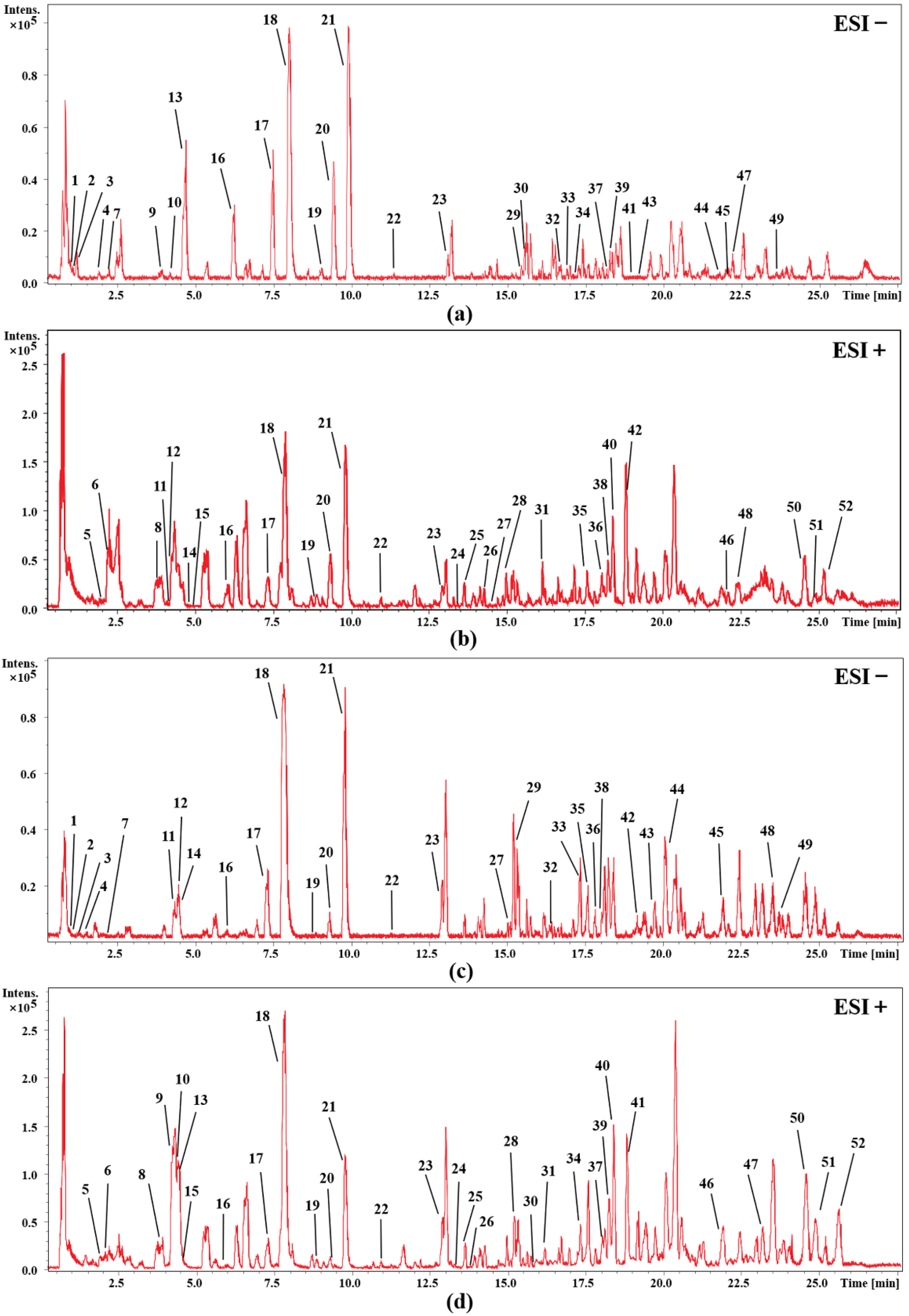
2.1. Identification of the Chemical Compositions by UHPLC-Q-TOF-MS
2.2. Cytotoxicity Results of L-02 Cells
2.3. General Observation and Body Weight
2.4. Urinalysis, Hematology and Biochemical Analysis
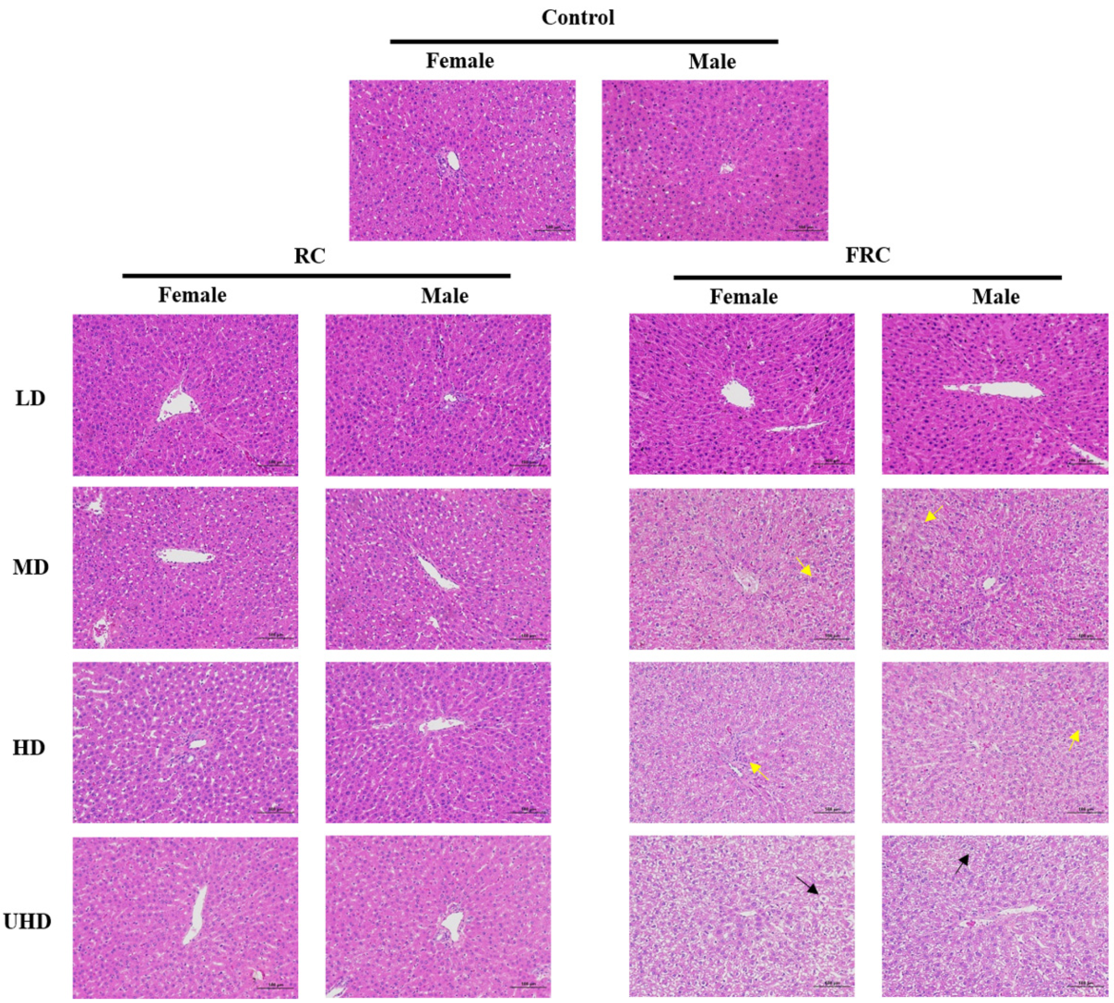
2.5. Organ Weights and Histopathological Changes
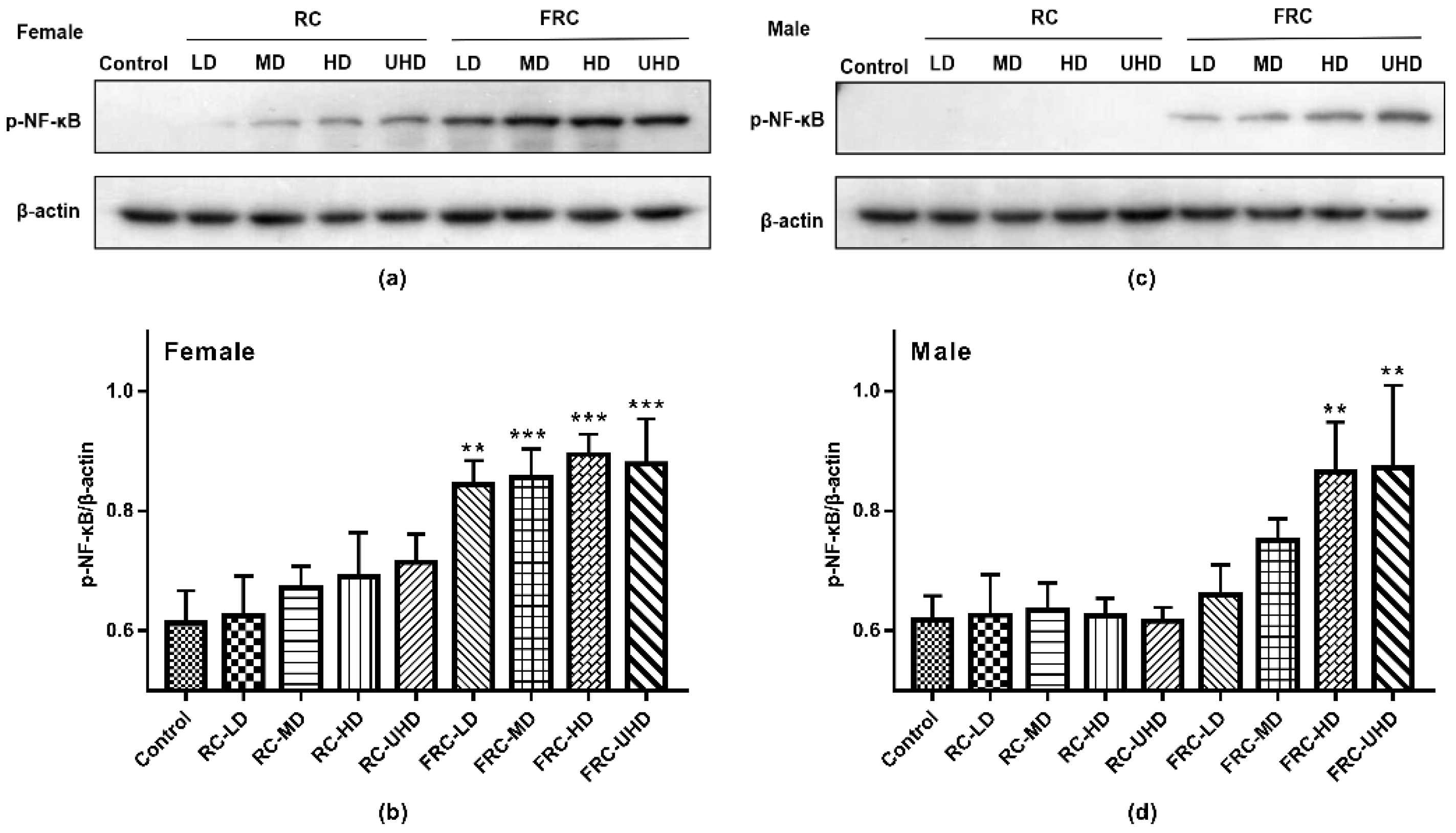
2.6. Effect on Protein Expression of p-NF-κB
3. Materials and Methods
3.1. Plant Material and Animals
3.2. Identification of Chemical Compositions in Crude Extract
3.3. In Vitro Hepatocyte Toxicity Test
3.4. Experimental Design for the Oral Toxicity Study
3.5. Urinalysis, Hematology and Biochemistry Analysis
3.6. Gross Findings, Organ Weights, and Histopathological Assessments
3.7. Western Blot Analysis
3.8. Statistical Analysis
4. Conclusions
Author Contributions
Funding
Institutional Review Board Statement
Informed Consent Statement
Data Availability Statement
Conflicts of Interest
Sample Availability
References
- Enbom, E.T.; Le, M.D.; Oesterich, L.; Rutgers, J.; French, S.W. Mechanism of hepatotoxicity due to black cohosh (Cimicifuga racemosa): Histological, immunohistochemical and electron microscopy analysis of two liver biopsies with clinical correlation. Exp. Mol. Pathol. 2014, 96, 279–283. [Google Scholar] [CrossRef] [PubMed]
- Teschke, R.; Schwarzenboeck, A. Suspected hepatotoxicity by Cimicifugae racemosae rhizoma (black cohosh, root): Critical analysis and structured causality assessment. Phytomedicine 2009, 16, 72–84. [Google Scholar] [CrossRef] [PubMed]
- Fabio, F.; Luigi, G.; Paolo, R.D.S. Black Cohosh Hepatic Safety: Follow-Up of 107 Patients Consuming a Special Cimicifuga racemosa rhizome Herbal Extract and Review of Literature. Evid.-Based Compl. Alt. 2011, 2011, 821392. [Google Scholar] [CrossRef] [Green Version]
- Lim, T.Y.; Considine, A.; Quaglia, A.; Shawcross, D.L. Subacute liver failure secondary to black cohosh leading to liver transplantation. BMJ Case Rep. 2013, 2013, bcr2013009325. [Google Scholar] [CrossRef] [PubMed] [Green Version]
- He, C.C.; Dai, Y.Q.; Hui, R.R.; Hua, J.; Chen, H.J.; Luo, Q.Y.; Li, J.X. NMR-based metabonomic approach on the toxicological effects of a Cimicifuga triterpenoid. J. Appl. Toxicol. 2012, 32, 88–97. [Google Scholar] [CrossRef] [PubMed]
- Lude, S.; Torok, M.; Dieterle, S.; Knapp, A.C.; Kaeufeler, R.; Jaggi, R.; Spornitz, U.; Krahenbuhl, S. Hepatic effects of Cimicifuga racemosa extract in vivo and in vitro. Cell. Mol. Life Sci. 2007, 64, 2848–2857. [Google Scholar] [CrossRef]
- Mazzanti, G.; Di Sotto, A.; Franchitto, A.; Mastrangelo, S.; Pezzella, M.; Vitalone, A.; Mammola, C.L. Effects of Cimicifuga racemosa extract on liver morphology and hepatic function indices. Phytomedicine 2008, 15, 1021–1024. [Google Scholar] [CrossRef]
- Naser, B.; Schnitker, J.; Minkin, M.J.; de Arriba, S.G.; Nolte, K.U.; Osmers, R. Suspected black cohosh hepatotoxicity: No evidence by meta-analysis of randomized controlled clinical trials for isopropanolic black cohosh extract. Menopause 2011, 18, 366–375. [Google Scholar] [CrossRef]
- Mahady, G.B.; Low Dog, T.; Barrett, M.L.; Chavez, M.L.; Gardiner, P.; Ko, R.; Marles, R.J.; Pellicore, L.S.; Giancaspro, G.I.; Sarma, D.N. United States Pharmacopeia review of the black cohosh case reports of hepatotoxicity. Menopause 2008, 15 Pt 1, 628–638. [Google Scholar] [CrossRef]
- Qin, R.-L.; Lv, C.-N.; Zhao, Y.; Zhao, Y.-D.; Yu, Y.; Lu, J.-C. Assessment of phenolics contents and antioxidant properties in Cimicifuga dahurica (Turcz.) Maxim during drying process. Ind. Crops Prod. 2017, 107, 288–296. [Google Scholar] [CrossRef]
- Guo, Y.; Yin, T.; Wang, X.; Zhang, F.; Pan, G.; Lv, H.; Wang, X.; Owoicho Orgah, J.; Zhu, Y.; Wu, H. Traditional uses, phytochemistry, pharmacology and toxicology of the genus Cimicifuga: A review. J. Ethnopharmacol. 2017, 209, 264–282. [Google Scholar] [CrossRef] [PubMed]
- Jia, H.; Wang, X.; Liu, W.; Qin, X.; Hu, B.; Ma, Q.; Lv, C.; Lu, J. Cimicifuga dahurica extract inhibits the proliferation, migration and invasion of breast cancer cells MDA-MB-231 and MCF-7 in vitro and in vivo. J. Ethnopharmacol. 2021, 277, 114057. [Google Scholar] [CrossRef] [PubMed]
- Wu, L.; Chen, Z.-L.; Su, Y.; Wang, Q.-H.; Kuang, H.-X. Cycloartenol triterpenoid saponins from Cimicifuga simplex (Ranunculaceae) and their biological effects. Chin. J. Nat. Med. 2015, 13, 81–89. [Google Scholar] [CrossRef]
- Lu, Q.; Zhang, W.-Y.; Pan, D.-B.; Shi, D.-F.; Pang, Q.-Q.; Li, H.-B.; Yao, X.-J.; Yao, Z.-H.; Yu, Y.; Yao, X.-S. Phenolic acids and their glycosides from the rhizomes of Cimicifuga dahurica. Fitoterapia 2019, 134, 485–492. [Google Scholar] [CrossRef]
- Lourenço, A.M.; Máximo, P.; Ferreira, L.M.; Pereira, M.M.A. Indolizidine and quinolizidine alkaloids structure and bioactivity. In Studies in Natural Products Chemistry; Attaur, R., Ed.; Elsevier: Amsterdam, The Netherlands, 2002; Volume 27, pp. 233–298. [Google Scholar]
- Miao, L.-Y.; Chu, T.T.H.; Li, P.; Jiang, Y.; Li, H.-J. Cimicifuga heracleifolia is therapeutically similar to black cohosh in relieving menopausal symptoms: Evidence from pharmacological and metabolomics studies. Chin. J. Nat. Med. 2019, 17, 435–445. [Google Scholar] [CrossRef]
- Fan, M.; Qin, K.; Ding, F.; Huang, Y.; Wang, X.; Cai, B. Identification and differentiation of major components in three different “Sheng-ma” crude drug species by UPLC/Q-TOF-MS. Acta Pharm. Sin. B 2017, 7, 185–192. [Google Scholar] [CrossRef] [PubMed]
- Hu, L.; Song, X.; Nagai, T.; Yamamoto, M.; Dai, Y.; He, L.; Kiyohara, H.; Yao, X.; Yao, Z. Chemical profile of Cimicifuga heracleifolia Kom. And immunomodulatory effect of its representative bioavailable component, cimigenoside on Poly(I:C)-induced airway inflammation. J. Ethnopharmacol. 2021, 267, 113615. [Google Scholar] [CrossRef]
- Bain, B.J. Structure and function of red and white blood cells and platelets. Medicine 2021, 49, 183–188. [Google Scholar] [CrossRef]
- Parente, J. Diagnostics for White Blood Cell Abnormalities: Leukocytosis and Leukopenia. Physician Assist. Clin. 2019, 4, 625–635. [Google Scholar] [CrossRef]
- Yun, J.-W.; You, J.-R.; Kim, Y.-S.; Cho, E.-Y.; Kim, S.-H.; Yoon, J.-H.; Kwon, E.; Chung, D.H.; Kim, Y.T.; Jang, J.-J.; et al. Pre-clinical in vitro and in vivo safety evaluation of Cimicifuga heracleifolia. Regul. Toxicol. Pharm. 2015, 73, 303–310. [Google Scholar] [CrossRef]
- Xie, K.; Chen, C.H.; Tsai, S.P.; Lu, P.J.; Wu, H.; Zeng, Y.; Ye, Y.; Tu, H.; Wen, C.; Huang, M.; et al. Loss of Life Expectancy by 10 Years or More From Elevated Aspartate Aminotransferase: Finding Aspartate Aminotransferase a Better Mortality Predictor for All-Cause and Liver-Related than Alanine Aminotransferase. Am. J. Gastroenterol. 2019, 114, 1478–1487. [Google Scholar] [CrossRef] [PubMed]
- Xu, J.; Xia, Y.; Li, S.; Cheng, X.; Hu, S.; Gao, Y.; Zhou, X.; Wang, G.; Zheng, Q. A retrospective pilot study to examine the potential of aspartate aminotransferase to alanine aminotransferase ratio as a predictor of postoperative acute kidney injury in patients with hepatocellular carcinoma. Ann. Clin. Biochem. 2019, 56, 357–366. [Google Scholar] [CrossRef] [PubMed]
- Lee, T.H.; Kim, W.R.; Poterucha, J.J. Evaluation of Elevated Liver Enzymes. Clin. Liver Dis. 2012, 16, 183–198. [Google Scholar] [CrossRef] [PubMed]
- Siddique, A.; Kowdley, K.V. Approach to a Patient with Elevated Serum Alkaline Phosphatase. Clin. Liver Dis. 2012, 16, 199–229. [Google Scholar] [CrossRef] [PubMed] [Green Version]
- McDaniel, M.J. Hepatic Function Testing: The ABCs of the Liver Function Tests. Physician Assist. Clin. 2019, 4, 541–550. [Google Scholar] [CrossRef]
- Domínguez-Pérez, M.; Simoni-Nieves, A.; Rosales, P.; Nuño-Lámbarri, N.; Rosas-Lemus, M.; Souza, V.; Miranda, R.U.; Bucio, L.; Uribe Carvajal, S.; Marquardt, J.U.; et al. Cholesterol burden in the liver induces mitochondrial dynamic changes and resistance to apoptosis. J. Cell. Physiol. 2019, 234, 7213–7223. [Google Scholar] [CrossRef] [PubMed]
- Manoeuvrier, G.; Bach-Ngohou, K.; Batard, E.; Masson, D.; Trewick, D. Diagnostic performance of serum blood urea nitrogen to creatinine ratio for distinguishing prerenal from intrinsic acute kidney injury in the emergency department. BMC Nephrol. 2017, 18, 173. [Google Scholar] [CrossRef] [PubMed] [Green Version]
- Mckay, L.I.; Cidlowski, J.A. Cross-talk between nuclear factor-kappa B and the steroid hormone receptors: Mechanisms of mutual antagonism. Mol. Endocrinol. 1998, 12, 45–56. [Google Scholar] [CrossRef] [Green Version]
- Tak, P.P.; Firestein, G.S. NF-kappaB: A key role in inflammatory diseases. J. Clin. Investig. 2001, 107, 7–11. [Google Scholar] [CrossRef]


| NO. | tR (min) | Measured Mass (m/z) | Extraction Mass (m/z) | Formula | Error (ppm) | Fragment Ions (m/z) | Identified Compounds |
|---|---|---|---|---|---|---|---|
| 1 | 1.10 | 271.0454 [M − H]− | 271.0459 | C11H12O8 | 1.9 | 253.0340, 181.0497, 123.0458 | Fukiic acid |
| 2 | 1.21 | 315.1079 [M − H]− | 315.1085 | C14H20O8 | 2.1 | 153.0566, 123.0451 | Cimidahurinine |
| 3 | 1.24 | 255.0506 [M − H]− | 255.0510 | C11H12O7 | 1.8 | 193.0487, 179.0334, 165.0553 | Piscidic acid |
| 4 | 1.85 | 315.1097 [M − H]− | 315.1085 | C14H20O8 | −3.7 | 153.0542, 123.0441 | Cimidahurine |
| 5 | 2.08 | 379.0985 [M + Na]+ | 379.1000 | C16H20O9 | 3.8 | 177.0530 | cis-Ferulic acid-4-O-β-d-gal |
| 6 | 2.12 | 379.0982 [M + Na]+ | 379.1000 | C16H20O9 | 4.8 | 177.0529 | trans-Ferulic acid-4-O-β-d-gal |
| 7 | 2.19 | 179.0348 [M − H]− | 179.0350 | C9H8O4 | 0.9 | 135.0454 | Caffeic acid |
| 8 | 4.09 | 476.1902 [M + H]+ | 476.1915 | C24H29NO9 | 2.7 | 314.1370, 177.0524 | trans-feruloyl tyramine-4-O-β-d-glu |
| 9 | 4.12 | 193.0501 [M − H]− | 193.0506 | C10H10O4 | 2.9 | 178.0231, 134.0359 | Ferulic acid |
| 10 | 4.17 | 193.0509 [M − H]− | 193.0506 | C10H10O4 | −1.2 | 178.0244, 134.0368 | Isoferulic acid |
| 11 | 4.23 | 307.1163 [M + H]+ | 307.1176 | C16H18O6 | 4.2 | 289.1053, 259.0590, 235.0583 | Cimifugin |
| 12 | 4.36 | 457.0719 [M + Na]+ | 457.0741 | C20H18O11 | 4.8 | 295.0396, 163.0372 | Cimicifugic acid G |
| 13 | 4.73 | 433.0769 [M − H]− | 433.0776 | C20H18O11 | 1.7 | 271.0450, 179.0344, 135.0452 | Fukinolic acid |
| 14 | 4.81 | 506.2011 [M + H]+ | 506.2021 | C25H31NO10 | 1.9 | 344.1466, 177.0532 | Isocimicifugamide |
| 15 | 4.83 | 506.2003 [M + H]+ | 506.2021 | C25H31NO10 | 3.5 | 344.1456, 177.0530 | Cimicifugamide |
| 16 | 6.07 | 441.0755 [M + Na]+ | 411.0792 | C20H18O10 | 3.4 | 279.0453, 163.0378 | Cimicifugic acid D |
| 6.25 | 417.0813 [M − H]− | 417.0827 | 3.5 | 179.0342, 135.0447, | |||
| 17 | 7.39 | 471.0865 [M + Na]+ | 471.0898 | C21H20O11 | 3.3 | 295.0390, 177.0525 | Cimicifugic acid A |
| 7.47 | 447.0916 [M − H]− | 447.0933 | 3.9 | 253.0341, 181.0499, 165.0556, 109.0286 | |||
| 18 | 7.94 | 471.0864 [M + Na]+ | 471.0898 | C21H20O12 | 3.3 | 295.0398, 177.0528 | Cimicifugic acid B |
| 8.01 | 447.0912 [M − H]− | 447.0933 | 4.6 | 253.0343, 181.0496, 165.0544, 109.0288 | |||
| 19 | 8.92 | 485.1025 [M + Na]+ | 485.1054 | C22H22O11 | 3.0 | 279.0461, 207.0623 | 2-isoferuloyl fukinolic acid-1-melyl esier |
| 9.12 | 461.1083 [M − H]− | 461.1089 | 1.4 | 253.0601, 193.0499, 165.0547, 149.0607 | |||
| 20 | 9.39 | 455.0918 [M + Na]+ | 455.0949 | C21H20O10 | 3.1 | 279.0453, 177.0533 | 2-feruloyl-piscidic acid |
| 9.55 | 431.0974 [M − H]− | 431.0984 | 2.3 | 237.0396, 193.0494, 165.0544 | |||
| 21 | 9.86 | 431.0969 [M − H]− | 431.0984 | C21H20O10 | 3.5 | 237.0395, 193.0491, 165.0543 | 2-isoferuloyl-piscidic acid |
| 9.88 | 455.0919 [M + Na]+ | 455.0949 | 3.0 | 279.0458, 177.0524 | |||
| 22 | 10.99 | 485.1034 [M + Na]+ | 485.1054 | C22H22O11 | 2.1 | 279.0432, 207.0601 | 2-feruloyl-fukinolic acid-1-melyl esier |
| 11.37 | 461.1085 [M − H]− | 461.1089 | 0.9 | 253.0339, 181.0503, 109.0280 | |||
| 23 | 12.94 | 367.076 [M + Na]+ | 367.0788 | C18H16O7 | 2.8 | 299.4364, 177.0667 | 4′-methoxyl-3′-hydroxy-carboxy benzoyl isoferulic acid anhyelride |
| 13.07 | 343.0814 [M − H]− | 343.0823 | 2.8 | 193.0497, 134.0364 | |||
| 24 | 13.52 | 657.3595 [M + Na]+ | 657.3609 | C35H54O10 | 2.2 | 617.3627, 467.3116 | 12β-O-7,8-Didehydroxycimigenol-3-O-β-d-xyl |
| 25 | 13.59 | 659.3772 [M + Na]+ | 659.3766 | C35H56O10 | −0.9 | 619.3786, 487.3391, 469.3289 | (22R)-22β-Hydroxycimigenol-3-O-β-d-xyl |
| 26 | 14.32 | 703.3993 [M + Na]+ | 703.4028 | C37H60O11 | 5.0 | 663.4060, 645.3944, 513.3543, 495.3408, 453.3217 | 24-epi-O-Acetylhydrosheng manol-3-O-β-ara |
| 27 | 14.47 | 701.3843 [M + Na]+ | 701.3871 | C37H58O11 | 4.0 | 679.4008, 661.3910, 601.3685, 451.3176 | 12β-Acetylcimigenol-3-O-β-d-xyl |
| 28 | 14.99 | 745.3728 [M + Na]+ | 745.3770 | C38H58O13 | 4.5 | 745.3728 | Cimilactone K |
| 29 | 15.57 | 198.0916 [M − H]− | 198.0924 | C13H13NO | 4.4 | 198.0916 | (E)-3-(3-methyl-2-butenylidene)-2-indolinone |
| 30 | 15.59 | 198.0922 [M − H]− | 198.0924 | C13H13NO | 1.3 | 198.0922 | (Z)-3-(3-methyl-2-butenylidene)-2-indolinone |
| 31 | 16.16 | 787.3830 [M + Na]+ | 787.3875 | C40H60O14 | 4.8 | 585.3597, 435.3159 | 24-epi-24-O-Acetyl-7,8-didehydroshengmanol-3-O-(2′-O-malonyl)-β-d-xyl |
| 32 | 16.70 | 635.3796 [M − H]− | 635.3801 | C35H56O10 | 0.8 | 577.3356 | 7β-Hydroxycimigenol-3-O-β-D-xyl |
| 33 | 16.92 | 637.3948 [M − H]− | 637.3957 | C35H58O10 | 1.5 | 579.3510 | Beesioside E |
| 34 | 17.09 | 679.4091 [M − H]− | 679.4063 | C37H60O11 | −4.1 | 619.3796 | 24-O-acetylhydro-shengmanol-3-O-β-d-xyl |
| 35 | 17.61 | 643.3798 [M + Na]+ | 643.3817 | C35H56O9 | 1.8 | 585.3713, 435.3190 | Cimigenol-3-O-β-d-xyl (Cimigenoside) |
| 36 | 18.06 | 683.3700 [M + Na]+ | 683.3766 | C37H56O10 | 4.9 | 643.3609, 435.3288 | 25-O-Acetyl-7,8-didehydrocimigenol-3-O-β-d-xyl |
| 37 | 18.08 | 635.3789 [M − H]− | 635.3801 | C35H56O10 | 1.8 | 577.3359 | 12β-hydroxycimigenol-3-O-α-L-ara |
| 38 | 18.26 | 643.3790 [M + Na]+ | 643.3817 | C35H56O9 | 2.7 | 585.3726, 435.3295 | Cimigenol-3-O-α-L-ara (Cimiracemoside C) |
| 39 | 18.30 | 637.3957 [M − H]− | 637.3957 | C35H58O10 | 0.1 | 579.3521 | Beesioside B |
| 40 | 18.40 | 703.3967 [M + Na]+ | 703.4028 | C37H60O11 | 2.7 | 645.3944, 513.3544 | 24-epi-24-O-Acetylhydroshengmanol-3-O-β-d-xyl |
| 41 | 18.76 | 679.4069 [M − H]− | 679.4063 | C37H60O11 | −1.0 | 619.3770 | 24-O-acetylhydroshengmanol-3-O-β-D-xyl |
| 42 | 18.83 | 701.3828 [M + Na]+ | 701.3871 | C37H58O11 | 4.5 | 451.3172, 274.2716 | 24-O-Acetyl-7,8-didehydro-hydroshengmanol-3-O-β-d-xyl |
| 43 | 19.11 | 707.3979 [M − H]− | 707.4012 | C38H60O12 | 4.7 | 661.3903, 619.3691, 469.3570 | 24-Epi-24-O-acetyl-7,8-dehydro cohosh alcohol-3-O-β-d-gal |
| 44 | 21.77 | 659.3785 [M − H]− | 659.3801 | C37H56O10 | 2.4 | 617.3720, 559.3198 | 27-Deoxy Arcot hormone |
| 45 | 21.97 | 661.3938 [M − H]− | 661.3957 | C37H58O10 | 2.9 | 619.3715, 601.3583 | 23-O-Acetyl alcohol cimicifuga-3-O-β-xyl |
| 46 | 22.11 | 511.3374 [M + Na]+ | 511.3394 | C30H48O5 | 1.9 | 453.3342 | 24-epi-Cimigenol |
| 47 | 22.18 | 677.3889 [M − H]− | 677.3906 | C37H58O11 | 2.6 | 617.3732 | 7,8-Deoxy cohosh alcohol-24-O-acetylalcohol-ara |
| 48 | 22.45 | 511.3368 [M + Na]+ | 511.3394 | C30H48O5 | 2.6 | 435.3219 | Cimigenol |
| 49 | 23.56 | 665.3882 [M − H]− | 665.3906 | C36H58O11 | 3.7 | 619.3203 | 12β-Hydroxy cohosh alcohol-3-O-β-d-gal |
| 50 | 24.56 | 863.4360 [M + Na]+ | 863.4400 | C43H68O16 | 3.9 | 803.7323, 643.3770, 572.4235, 435.3229 | Heracleifolinoside F |
| 51 | 24.82 | 569.3434 [M + Na]+ | 569.3449 | C32H50O7 | 1.5 | 529.3466, 511.3389 | 24-O-Acetyl-7,8-didehydro-hydroshengmanol |
| 52 | 25.12 | 571.3590 [M + Na]+ | 571.3605 | C32H52O7 | 1.5 | 513.3530, 453.3352 | 24-O-Acetylhydroshengmanol |
| NO. | tR (min) | Measured Mass (m/z) | Extraction Mass (m/z) | Formula | Error (ppm) | Fragment Ions (m/z) | Identified Compounds |
|---|---|---|---|---|---|---|---|
| 1 | 1.07 | 271.0457 [M − H]− | 271.0459 | C11H12O8 | 0.8 | 253.0379, 181.0493, 123.0428 | Fukiic acid |
| 2 | 1.12 | 315.1079 [M − H]− | 315.1085 | C14H20O8 | 2.0 | 153.0553, 123.0437 | Cimidahurinine |
| 3 | 1.16 | 255.0507 [M − H]− | 255.0510 | C11H12O7 | 1.8 | 193.0467, 179.0293, 165.0559 | Piscidic acid |
| 4 | 1.19 | 315.1082 [M − H]− | 315.1085 | C14H20O8 | 1.1 | 153.0560, 123.0433 | Cimidahurine |
| 5 | 2.00 | 379.0991 [M + Na]+ | 379.1000 | C16H20O9 | 2.4 | 177.0541 | cis-Ferulic acid-4-O-β-d-gal |
| 6 | 2.15 | 379.0990 [M + Na]+ | 379.1000 | C16H20O9 | 2.6 | 177.0537 | trans-Ferulic acid-4-O-β-d-gal |
| 7 | 2.17 | 179.0350 [M − H]− | 179.0350 | C9H8O4 | −0.1 | 135.0431 | Caffeic acid |
| 8 | 4.00 | 476.1899 [M + H]+ | 476.1915 | C24H29NO9 | 3.5 | 314.1380, 177.0548 | trans-feruloyl tyramine-4-O-β-d-glu |
| 9 | 4.33 | 457.0734 [M + Na]+ | 457.0741 | C20H18O11 | 1.7 | 295.0406, 163.0382 | Cimicifugic acid G |
| 10 | 4.39 | 307.1167 [M + H]+ | 307.1176 | C16H18O6 | 3.0 | 289.1044, 259.0595, 235.0592 | Cimifugin |
| 11 | 4.39 | 433.0758 [M − H]− | 433.0776 | C20H18O11 | 4.3 | 271.0476, 179.0342, 135.0449 | Fukinolic acid |
| 12 | 4.40 | 193.0503 [M − H]− | 193.0506 | C10H10O4 | 1.7 | 178.0308, 134.0359 | Ferulic acid |
| 13 | 4.46 | 506.2009 [M + H]+ | 506.2021 | C25H31NO10 | 2.2 | 344.1520, 177.0540 | Isocimicifugamide |
| 14 | 4.52 | 193.0497 [M − H]− | 193.0506 | C10H10O4 | 4.7 | 178.0261, 134.0380 | Isoferulic acid |
| 15 | 4.69 | 506.2006 [M + H]+ | 506.2021 | C25H31NO10 | 2.8 | 344.1492, 177.0557 | Cimicifugamide |
| 16 | 6.06 | 441.0783 [M + Na]+ | 411.0792 | C20H18O10 | 2.2 | 279.0482,163.0377 | Cimicifugic acid D |
| 6.07 | 417.0821 [M − H]− | 417.0827 | 1.5 | 255.0465, 193.0519, 179.0347 | |||
| 17 | 7.27 | 447.0920 [M − H]− | 447.0933 | C21H20O11 | 2.8 | 253.0341, 181.0495, 165.0538, 109.0294 | Cimicifugic acid A |
| 7.37 | 471.0896 [M + Na]+ | 471.0898 | 0.4 | 295.0435, 177.0549 | |||
| 18 | 7.87 | 471.0895 [M + Na]+ | 471.0898 | C21H20O12 | 0.5 | 295.0417, 177.0548 | Cimicifugic acid B |
| 7.88 | 447.0918 [M − H]− | 447.0933 | 3.3 | 253.0348, 181.0500, 165.0550, 109,0291 | |||
| 19 | 8.91 | 461.1077 [M − H]− | 461.1089 | C22H22O11 | 2.7 | 165.0536, 193.0495, 233.0624 | 2-isoferuloyl fukinolic acid-1-melyl esier |
| 8.92 | 485.1059 [M + Na]+ | 485.1054 | −1.0 | 279.0494, 207.0615 | |||
| 20 | 9.35 | 455.0941 [M + Na]+ | 455.0949 | C21H20O10 | 1.6 | 367.0784, 279.0461 | 2-feruloyl-piscidic acid |
| 9.40 | 431.0978 [M − H]− | 431.0984 | 1.4 | 237.0418, 193.0560, 165.0560 | |||
| 21 | 9.74 | 431.0964 [M − H]− | 431.0984 | C21H20O10 | 4.6 | 237.0398, 193.0500, 165.0548 | 2-isoferuloyl-piscidic acid |
| 9.87 | 455.0947 [M + Na]+ | 455.0949 | 0.3 | 279.0473, 177.0547 | |||
| 22 | 11.29 | 485.1047 [M + Na]+ | 485.1054 | C22H22O11 | 1.5 | 279.0456, 207.0672 | 2-feruloyl-fukinolic acid-1-melyl esier |
| 11.41 | 461.1072 [M − H]− | 461.1089 | 3.8 | 253.0352, 181.0496, 109.0289 | |||
| 23 | 12.90 | 343.0809 [M − H]− | 343.0823 | C18H16O7 | 4.1 | 193.0499, 134.0363 | 4′-methoxyl-3′-hydroxy-carboxy benzoyl isoferulic acid anhyelride |
| 12.92 | 367.0768 [M + Na]+ | 367.0788 | 2.7 | 299.4364,177.0667 | |||
| 24 | 13.48 | 657.3593 [M + Na]+ | 657.3609 | C35H54O10 | 2.4 | 617.3645, 467.3129 | 12β-O-7,8-Didehydroxycimigenol-3-O-β-d-xyl |
| 25 | 13.70 | 659.3736 [M + Na]+ | 659.3766 | C35H56O10 | 4.5 | 451.3189, 177.0521 | (22R)-22β-Hydroxycimigenol-3-O-β-d-xyl |
| 26 | 13.76 | 703.4055 [M + Na]+ | 703.4028 | C37H60O11 | −3.9 | 663.4041, 645.3946, 513.3544, 495.3244, 453.3280 | 24-epi-O-Acetylhydrosheng manol-3-O-β-ara |
| 27 | 15.18 | 198.0920 [M − H]− | 198.0924 | C13H13NO | 2.5 | 198.0913 | (E)-3-(3-methyl-2-butenylidene)-2-indolinone |
| 28 | 15.24 | 701.3838 [M + Na]+ | 701.3871 | C37H58O11 | 4.8 | 679.4007, 661.3891, 601.3702, 451.3158 | 12β-Acetylcimigenol-3-O-β-d-xyl |
| 29 | 15.26 | 198.0924 [M − H]− | 198.0924 | C13H13NO | 0.4 | 198.0924 | (Z)-3-(3-methyl-2-butenylidene)-2-indolinone |
| 30 | 15.89 | 745.3764 [M + Na]+ | 745.3770 | C38H58O13 | 0.7 | 671.3720, 513.3649, 435.3291 | Cimilactone K |
| 31 | 16.24 | 787.3838 [M + Na]+ | 787.3875 | C40H60O14 | 4.7 | 647.3384, 526.3150, 429.2978 | 24-epi-24-O-Acetyl-7,8-didehydroshengmanol-3-O-(2′-O-malonyl)-β-d-xyl |
| 32 | 16.44 | 635.3794 [M − H]− | 635.3801 | C35H56O10 | 1.1 | 577.3356 | 7β-Hydroxycimigenol-3-O-β-D-xyl |
| 33 | 17.30 | 637.3927 [M − H]− | 637.3957 | C35H58O10 | 4.7 | 579.3483 | Beesioside E |
| 34 | 17.37 | 643.3786 [M + Na]+ | 643.3817 | C35H56O9 | 3.8 | 585.3712, 435.3242 | Cimigenol-3-O-β-d-xyl (Cimigenoside) |
| 35 | 17.52 | 679.4043 [M − H]− | 679.4063 | C37H60O11 | 2.9 | 619.377 | 24-O-acetylhydro-shengmanol-3-O-β-d-xyl |
| 36 | 17.79 | 635.3777 [M − H]− | 635.3801 | C35H56O10 | 3.7 | 577.3352 | 12β-hydroxycimigenol-3-O-α-l-ara |
| 37 | 18.06 | 683.3731 [M + Na]+ | 683.3766 | C37H56O10 | 5.0 | 511.3385, 453.3369 | 25-O-Acetyl-7,8-didehydrocimigenol-3-O-β-d-xyl |
| 38 | 18.09 | 637.3928 [M − H]− | 637.3957 | C35H58O10 | 4.6 | 579.3481 | Beesioside B |
| 39 | 18.28 | 643.3803 [M + Na]+ | 643.3817 | C35H56O9 | 2.0 | 585.3746, 435,3225 | Cimigenol-3-O-α-l-ara (Cimiracemoside C) |
| 40 | 18.42 | 703.4001 [M + Na]+ | 703.4028 | C37H60O11 | 3.8 | 645.3962, 513.3558 | 24-epi-24-O-Acetylhydroshengmanol-3-O-β-d-xyl |
| 41 | 18.81 | 701.3828 [M + Na]+ | 701.3871 | C37H58O11 | 5.4 | 451.3210 | 24-O-Acetyl-7,8-didehydro-hydroshengmanol-3-O-β-d-xyl |
| 42 | 19.17 | 679.4031 [M − H]− | 679.4063 | C37H60O11 | 4.7 | 619.3784 | 24-O-acetylhydroshengmanol-3-O-β-d-xyl |
| 43 | 19.63 | 707.3988 [M − H]− | 707.4012 | C38H60O12 | 3.4 | 661.3891, 619.3727, 469.3341 | 24-Epi-24-O-acetyl-7,8-dehydro cohosh alcohol-3-O-β-D-gal |
| 44 | 20.15 | 659.3770 [M − H]− | 659.3801 | C37H56O10 | 2.4 | 617.3614, 559.3276 | 27-Deoxy Arcot hormone |
| 45 | 21.92 | 661.3937 [M − H]− | 661.3957 | C37H58O11 | 3.0 | 619.3892, 601.3476 | 23-O-Acetyl alcohol cimicifuga-3-O-β-xyl |
| 46 | 21.96 | 511.3379 [M + Na]+ | 511.3394 | C30H48O5 | 2.9 | 453.3355, 471.3932 | 24-epi-Cimigenol |
| 47 | 23.19 | 511.3396 [M + Na]+ | 511.3394 | C30H48O5 | −0.5 | 453.3340, 435.3219 | Cimigenol |
| 48 | 23.39 | 677.3897 [M − H]− | 677.3906 | C37H58O11 | 1.4 | 617.3685 | 7,8-Deoxy cohosh alcohol-24-O-acetylalcohol-ara |
| 49 | 23.61 | 665.3908 [M − H]− | 665.3906 | C36H58O11 | −0.3 | 619.3825 | 12β-Hydroxy cohosh alcohol-3-O-β-d-gal |
| 50 | 24.62 | 643.3783 [M + Na]+ | 643.3817 | C35H56O9 | 5.2 | 529.3487, 453.3347 | Cimiaceroside B |
| 51 | 24.92 | 569.3432 [M + Na]+ | 569.3449 | C32H50O7 | 2.9 | 529.3481, 511.3442 | 24-O-Acetyl-7,8-didehydro-hydroshengmanol |
| 52 | 25.55 | 571.3580 [M + Na]+ | 571.3605 | C32H52O7 | 4.4 | 531.3659, 453.3344 | 24-O-Acetylhydroshengmanol |
| Group | Control | RC | FRC | ||||||
|---|---|---|---|---|---|---|---|---|---|
| 0 | LD | MD | HD | UHD | LD | MD | HD | UHD | |
| Females | |||||||||
| WBC (Cell/μL) | - | - | - | - | - | - | - | - | - |
| KET (mmol/L) | - | - | - | - | - | - | - | - | - |
| NIT | - | - | - | - | - | - | - | - | - |
| URO (μmol/L) | Normal | Normal | Normal | Normal | Normal | Normal | Normal | Normal | Normal |
| BIL (μmol/L) | - | - | - | - | - | - | - | - | - |
| PRO (g/L) | +3 | +3 | +2 | +3 | +3 | +3 | +3 | +3 | +2 |
| GLU | - | - | - | - | - | - | - | - | - |
| SG | 1.005 | 1.005 | 1.010 | 1.005 | 1.005 | 1.005 | 1.010 | 1.010 | 1.020 |
| BLD (Cell/μL) | - | - | - | - | - | - | - | - | - |
| pH | 7.5 | 7.5 | 7.5 | 7.5 | 7.5 | 7.5 | 7.5 | 7.5 | 7.0 |
| Vc (mmol/L) | ± | ± | - | ± | ± | ± | ± | ± | ± |
| Males | |||||||||
| WBC (Cell/μL) | - | - | - | - | - | - | - | - | - |
| KET (mmol/L) | - | - | - | - | - | - | - | - | - |
| NIT | - | - | - | - | - | - | - | - | - |
| URO (μmol/L) | Normal | Normal | Normal | Normal | Normal | Normal | Normal | Normal | Normal |
| BIL (μmol/L) | - | - | - | - | - | - | - | - | - |
| PRO (g/L) | +3 | +3 | +3 | +3 | +3 | +2 | +3 | +3 | +2 |
| GLU | - | - | - | - | - | - | - | - | - |
| SG | 1.005 | 1.005 | 1.010 | 1.005 | 1.005 | 1.005 | 1.005 | 1.010 | 1.015 |
| BLD (Cell/μL) | - | - | - | - | - | - | - | - | - |
| pH | 7.5 | 7.0 | 7.5 | 7.5 | 7.5 | 7.0 | 7.5 | 7.0 | 7.0 |
| Vc (mmol/L) | ± | ± | ± | ± | ± | ± | ± | ± | ± |
| Group | Control | RC | FRC | ||||||
|---|---|---|---|---|---|---|---|---|---|
| 0 | LD | MD | HD | UHD | LD | MD | HD | UHD | |
| Females | |||||||||
| WBC (109 cells/L) | 7.4 ± 0.3 | 6.8 ± 0.6 | 7.2 ± 1.0 | 7.9 ± 1.4 | 5.2 ± 0.5 | 10.5 ± 1.3 * | 10.3 ± 0.6 * | 10.4 ± 0.7 ** | 10.9 ± 1.2 ** |
| RBC (1012 cells/L) | 6.9 ± 0.7 | 6.2 ± 0.3 | 6.4 ± 0.5 | 6.4 ± 0.6 | 6.5 ± 0.2 | 6.7 ± 1.0 | 6.1 ± 0.3 | 6.9 ± 0.4 | 6.5 ± 1.1 |
| HGB (g/L) | 131.7 ± 8.3 | 130.5 ± 8.6 | 129.2 ± 9.6 | 126.4 ± 10.4 | 124.8 ± 4.5 | 132.5 ± 9.7 | 124.3 ± 6.0 | 138.0 ± 3.7 | 128.0 ± 14.9 |
| HCT (%) | 43.6 ± 2.0 | 42.6 ± 1.9 | 43.1 ± 2.4 | 42.1 ± 2.7 | 42.0 ± 2.1 | 44.2 ± 2.4 | 42.1 ± 2.2 | 43.9 ± 2.4 | 44.6 ± 1.8 |
| PLT (1011 cells/L) | 13.2 ± 1.5 | 12.4 ± 0.8 | 14.3 ± 1.1 | 12.0 ± 1.2 | 13.9 ± 2.5 | 11.6 ± 2.4 | 11.6 ± 1.1 | 11.9 ± 1.8 | 11.9 ± 2.7 |
| MCV (fL) | 65.6 ± 2.0 | 68.4 ± 1.0 | 67.7 ± 1.5 | 66.3 ± 2.5 | 64.7 ± 2.6 | 66.3 ± 2.4 | 68.6 ± 1.8 | 67.0 ± 2.0 | 67.1 ± 2.2 |
| MCH (pg) | 19.2 ± 1.0 | 21.0 ± 0.3 | 20.3 ± 0.5 | 19.9 ± 0.4 | 19.3 ± 0.4 | 20.6 ± 0.7 | 20.3 ± 0.4 | 20.1 ± 0.7 | 19.7 ± 0.9 |
| MCHC (g/L) | 301.7 ± 5.5 | 306.0 ± 7.3 | 300.0 ± 6.9 | 300.0 ± 6.2 | 297.5 ± 5.3 | 299.8 ± 6.6 | 295.8 ± 3.9 | 299.2 ± 4.2 | 298.6 ± 5.0 |
| Neutrophils (%) | 11.1 ± 1.7 | 10.8 ± 4.4 | 11.3 ± 4.3 | 11.4 ± 2.6 | 14.2 ± 3.1 | 15.5 ± 5.2 | 15.9 ± 4.2 | 17.5 ± 5.5 | 18.9 ± 3.4 |
| Eosinophils (%) | 2.3 ± 0.2 | 2.6 ± 0.8 | 1.5 ± 0.2 | 2.2 ± 1.1 | 2.8 ± 0.4 | 3.4 ± 0.9 | 3.3 ± 1.0 | 2.2 ± 0.2 | 2.1 ± 0.3 |
| Basophils (%) | 0.5 ± 0.2 | 0.5 ± 0.1 | 0.4 ± 0.2 | 0.5 ± 0.3 | 0.5 ± 0.1 | 0.5 ± 0.1 | 0.3 ± 0.1 | 0.5 ± 0.2 | 0.4 ± 0.2 |
| Lymphocytes (%) | 76.8 ± 12.8 | 88.1 ± 3.8 | 86.0 ± 4.4 | 85.1 ± 3.2 | 71.2 ± 13.0 | 75.4 ± 10.2 | 78.9 ± 3.9 | 85.2 ± 6.6 | 83.3 ± 2.9 |
| Monocytes (%) | 0.9 ± 0.5 | 0.5 ± 0.1 | 0.7 ± 0.3 | 0.7 ± 0.2 | 0.7 ± 0.1 | 0.9 ± 0.1 | 0.8 ± 0.2 | 0.8 ± 0.2 | 0.7 ± 0.2 |
| Reticulocytes (%) | 12.1 ± 2.3 | 13.8 ± 1.7 | 12.0 ± 1.4 | 11.7 ± 2.5 | 10.7 ± 0.8 | 14.9 ± 1.7 | 10.4 ± 0.8 | 12.1 ± 1.8 | 11.5 ± 1.5 |
| Males | |||||||||
| WBC (109 cells/L) | 7.4 ± 1.0 | 8.5 ± 1.2 | 8.8 ± 0.6 | 9.0 ± 0.9 | 8.9 ± 0.6 | 8.8 ± 1.0 | 9.0 ± 0.3 | 9.5 ± 0.5 | 10.4 ± 1.0 ** |
| RBC (1012 cells/L) | 6.8 ± 0.7 | 7.8 ± 0.5 | 7.9 ± 0.3 | 7.6 ± 0.4 | 7.5 ± 0.6 | 7.0 ± 1.4 | 7.4 ± 0.6 | 7.3 ± 0.7 | 7.7 ± 0.2 |
| HGB (g/L) | 146.0 ± 4.4 | 149.0 ± 5.3 | 143.7 ± 2.3 | 141.5 ± 3.7 | 137.3 ± 5.1 | 142.0 ± 4.6 | 141.7 ± 4.6 | 144.7 ± 5.8 | 141.7 ± 5.7 |
| HCT (%) | 42.1 ± 2.0 | 47.0 ± 2.6 | 47.8 ± 0.3 | 46.6 ± 1.8 | 45.9 ± 2.1 | 48.4 ± 3.1 | 47.6 ± 4.1 | 47.4 ± 3.9 | 47.4 ± 2.2 |
| PLT (1011 cells/L) | 12.0 ± 1.8 | 12.1 ± 1.5 | 13.0 ± 1.5 | 14.2 ± 1.7 | 11.7 ± 1.7 | 11.0 ± 1.5 | 12.1 ± 1.9 | 11.2 ± 1.1 | 11.2 ± 1.5 |
| MCV (fL) | 60.6 ± 1.3 | 60.5 ± 1.6 | 60.4 ± 2.4 | 61.0 ± 1.6 | 61.2 ± 2.0 | 64.2 ± 4.3 | 64.7 ± 4.3 | 64.8 ± 1.6 | 65.1 ± 3.9 |
| MCH (pg) | 18.6 ± 1.3 | 18.7 ± 0.4 | 18.2 ± 0.4 | 18.6 ± 0.6 | 18.4 ± 0.9 | 20.4 ± 1.7 | 19.7 ± 1.1 | 19.3 ± 0.5 | 18.6 ± 0.4 |
| MCHC (g/L) | 299.0 ± 5.6 | 309.2 ± 4.7 | 301.3 ± 6.4 | 309.3 ± 6.4 | 294.5 ± 5.2 | 308.5 ± 2.6 | 304.7 ± 5.0 | 297.7 ± 1.2 | 296.3 ± 3.5 |
| Neutrophils (%) | 14.6 ± 2.1 | 17.9 ± 3.3 | 16.2 ± 2.7 | 17.2 ± 1.7 | 17.8 ± 5.7 | 17.6 ± 3.8 | 15.1 ± 5.0 | 20.0 ± 7.2 | 19.3 ± 6.3 |
| Eosinophils (%) | 4.8 ± 1.1 | 3.4 ± 1.6 | 2.7 ± 1.3 | 3.1 ± 1.9 | 2.4 ± 1.2 | 2.7 ± 1.8 | 2.4 ± 0.6 | 3.7 ± 1.2 | 3.4 ± 1.1 |
| Basophils (%) | 0.3 ± 0.1 | 0.6 ± 0.2 | 0.4 ± 0.1 | 0.4 ± 0.1 | 0.5 ± 0.1 | 0.6 ± 0.1 | 0.6 ± 0.2 | 0.5 ± 0.4 | 0.4 ± 0.2 |
| Lymphocytes (%) | 74.5 ± 6.9 | 72.0 ± 12.3 | 79.7 ± 2.2 | 75.4 ± 6.3 | 76.1 ± 7.2 | 76.6 ± 5.8 | 80.7 ± 4.8 | 79.6 ± 5.5 | 80.6 ± 2.2 |
| Monocytes (%) | 1.0 ± 0.3 | 0.8 ± 0.5 | 0.9 ± 0.1 | 0.8 ± 0.3 | 1.1 ± 0.2 | 1.2 ± 1.0 | 1.0 ± 0.6 | 0.8 ± 0.2 | 0.8 ± 0.2 |
| Reticulocytes (%) | 6.8 ± 1.5 | 7.6 ± 0.8 | 7.8 ± 0.5 | 9.0 ± 2.2 | 9.6 ± 1.4 | 9.8 ± 1.9 | 9.7 ± 2.1 | 9.7 ± 0.7 | 9.9 ± 2.3 |
| Group | Control | RC | FRC | ||||||
|---|---|---|---|---|---|---|---|---|---|
| 0 | LD | MD | HD | UHD | LD | MD | HD | UHD | |
| Females | |||||||||
| ALP (U/L) | 38.2 ± 9.0 | 33.7 ± 10.6 | 35.9 ± 11.9 | 36.3 ± 11.8 | 34.3 ± 12.4 | 39.8 ± 10.3 | 32.4 ± 6.5 | 39.9 ± 7.2 | 41.0 ± 11.1 |
| ALT (U/L) | 34.8 ± 2.5 | 42.5 ± 4.8 | 42.7 ± 3.7 | 42.4 ± 5.4 | 42.8 ± 5.2 | 46.0 ± 6.2 ** | 39.3 ± 2.8 | 43.0 ± 6.0 * | 63.1 ± 8.4 *** |
| AST (U/L) | 182.1 ± 28.1 | 168.9 ± 17.7 | 174.5 ± 23.2 | 208.5 ± 56.7 | 215.7 ± 42.4 | 248.0 ± 42.4 * | 237.7 ± 41.4 * | 227.3 ± 67.5 | 185.5 ± 20.5 |
| UREA (mmol/L) | 6.7 ± 0.5 | 6.2 ± 0.4 | 6.5 ± 0.6 | 7.1 ± 0.4 | 6.9 ± 0.4 | 7.3 ± 0.7 | 8.0 ± 0.7 ** | 7.4 ± 0.7 | 7.5 ± 0.9 |
| CREA (Umol/L) | 25.8 ± 2.2 | 26.9 ± 6.2 | 25.2 ± 2.4 | 26.5 ± 2.0 | 25.9 ± 4.5 | 28.1 ± 4.3 | 27.4 ± 6.8 | 27.6 ± 2.0 | 29.0 ± 6.5 |
| TBIL (Umol/L) | 5.8 ± 1.7 | 5.4 ± 1.4 | 6.1 ± 1.4 | 5.7 ± 1.0 | 6.4 ± 1.1 | 6.1 ± 1.4 | 5.7 ± 0.9 | 5.9 ± 1.4 | 5.6 ± 1.4 |
| GLU (mmol/L) | 3.0 ± 0.2 | 3.3 ± 0.7 | 3.3 ± 0.4 | 3.3 ± 0.4 | 2.8 ± 0.3 | 3.0 ± 0.6 | 2.9 ± 0.5 | 2.8 ± 0.7 | 3.6 ± 0.5 |
| CHOL (mmol/L) | 2.0 ± 0.2 | 1.8 ± 0.2 | 1.8 ± 0.2 | 1.9 ± 0.1 | 1.9 ± 0.3 | 2.0 ± 0.2 | 2.2 ± 0.2 | 2.2 ± 0.2 | 2.5 ± 0.3 ** |
| TG (mmol/L) | 0.6 ± 0.1 | 0.5 ± 0.1 | 0.5 ± 0.1 | 0.6 ± 0.1 | 0.6 ± 0.0 | 0.6 ± 0.0 | 0.6 ± 0.1 | 0.6 ± 0.1 | 0.5 ± 0.0 |
| TP (g/L) | 60.7 ± 4.7 | 57.7 ± 2.8 | 61.0 ± 5.3 | 63.0 ± 2.6 | 63.2 ± 3.7 | 63.7 ± 2.5 | 63.1 ± 1.9 | 65.4 ± 3.0 | 69.4 ± 5.0 |
| ALB (g/L) | 39.7 ± 2.7 | 40.4 ± 3.5 | 40.8 ± 2.5 | 41.0 ± 1.2 | 41.4 ± 2.3 | 41.7 ± 0.9 | 40.7 ± 0.9 | 41.8 ± 1.4 | 44.7 ± 2.7 |
| Males | |||||||||
| ALP (U/L) | 58.9 ± 9.7 | 69.8 ± 12.4 | 66.8 ± 14.3 | 71.0 ± 8.2 | 57.1 ± 11.3 | 68.2 ± 17.7 | 58.4 ± 8.8 | 64.3 ± 9.6 | 90.8 ± 3.4 *** |
| ALT (U/L) | 42.0 ± 7.0 | 55.1 ± 8.4 | 48.1 ± 6.1 | 52.5 ± 2.0 | 45.5 ± 6.0 | 54.9 ± 9.3 | 48.6 ± 4.7 | 48.5 ± 8.0 | 80.4 ± 11.9 *** |
| AST (U/L) | 201.2 ± 11.4 | 241.5 ± 71.7 | 202.1 ± 33.7 | 234.8 ± 51.0 | 148.1 ± 54.8 | 262.7 ± 35.8 | 216.2 ± 32.4 | 192.1 ± 48.4 | 194.3 ± 35.7 |
| UREA (mmol/L) | 5.4 ± 0.3 | 6.2 ± 0.7 | 6.0 ± 0.4 | 6.2 ± 0.4 | 6.0 ± 0.7 | 7.2 ± 0.4 *** | 6.3 ± 0.7 * | 6.3 ± 0.7 | 7.5 ± 0.9 *** |
| CREA (Umol/L) | 16.3 ± 3.6 | 15.1 ± 3.3 | 17.4 ± 3.3 | 20.8 ± 4.2 | 21.7 ± 4.4 | 21.7 ± 3.5 | 25.0 ± 3.5 ** | 19.5 ± 3.7 | 20.1 ± 2.6 |
| TBIL (Umol/L) | 5.2 ± 0.6 | 5.6 ± 0.8 | 5.4 ± 1.1 | 5.8 ± 1.3 | 5.7 ± 1.1 | 6.1 ± 0.6 | 6.6 ± 1.0 | 6.4 ± 1.1 | 7.6 ± 1.7 * |
| GLU (mmol/L) | 4.0 ± 0.7 | 2.9 ± 0.8 | 3.4 ± 0.4 | 3.5 ± 0.7 | 3.3 ± 0.7 | 2.9 ± 0.7 | 3.6 ± 0.2 | 3.6 ± 0.6 | 3.5 ± 0.5 |
| CHOL (mmol/L) | 1.6 ± 0.2 | 1.7 ± 0.3 | 1.8 ± 0.2 | 1.8 ± 0.1 | 1.7 ± 0.2 | 1.8 ± 0.1 | 1.9 ± 0.2 | 1.8 ± 0.3 | 1.9 ± 0.3 * |
| TG (mmol/L) | 0.5 ± 0.1 | 0.5 ± 0.1 | 0.4 ± 0.1 | 0.6 ± 0.1 | 0.5 ± 0.1 | 0.7 ± 0.1 | 0.6 ± 0.1 | 0.5 ± 0.1 | 0.4 ± 0.0 |
| TP (g/L) | 57.3 ± 5.8 | 64.1 ± 3.7 | 65.5 ± 5.6 | 63.7 ± 1.4 | 60.6 ± 3.1 | 63.5 ± 5.2 | 66.0 ± 4.1 | 67.3 ± 5.3 | 72.4 ± 4.8 |
| ALB (g/L) | 36.4 ± 1.8 | 40.9 ± 1.6 | 41.5 ± 2.5 | 40.0 ± 1.1 | 39.0 ± 1.5 | 40.3 ± 2.4 | 41.4 ± 1.7 | 42.9 ± 2.9 | 45.2 ± 1.5 |
| Group | Control | RH | FRC | |||||||
|---|---|---|---|---|---|---|---|---|---|---|
| 0 | LD | MD | HD | UHD | LD | MD | HD | UHD | ||
| Females | ||||||||||
| Heart | (g) | 0.9 ± 0.12 | 0.88 ± 0.06 | 0.88 ± 0.09 | 0.89 ± 0.07 | 1.19 ± 0.35 | 1.04 ± 0.15 | 0.91 ± 0.08 | 1.00 ± 0.20 | 1.05 ± 0.20 |
| (%BW) | 0.36 ± 0.02 | 0.33 ± 0.02 | 0.33 ± 0.05 | 0.34 ± 0.01 | 0.38 ± 0.07 | 0.39 ± 0.08 | 0.35 ± 0.04 | 0.38 ± 0.08 | 0.38 ± 0.08 | |
| Liver | (g) | 5.56 ± 0.58 | 5.56 ± 0.49 | 5.25 ± 0.27 | 7.55 ± 0.44 | 8.87 ± 1.28 | 7.90 ± 1.57 | 7.99 ± 1.10 | 7.89 ± 1.15 | 7.66 ± 1.10 |
| (%BW) | 2.24 ± 0.17 | 2.12 ± 0.17 | 1.96 ± 0.18 | 2.93 ± 0.24 | 3.21 ± 0.46 | 2.87 ± 0.40 | 3.09 ± 0.38 | 2.99 ± 0.41 | 2.81 ± 0.46 | |
| Spleen | (g) | 0.43 ± 0.06 | 0.50 ± 0.10 | 0.46 ± 0.07 | 0.59 ± 0.07 | 0.53 ± 0.12 | 0.45 ± 0.10 | 0.53 ± 0.08 | 0.49 ± 0.09 | 0.55 ± 0.07 |
| (%BW) | 0.17 ± 0.01 | 0.19 ± 0.05 | 0.17 ± 0.02 | 0.23 ± 0.04 | 0.19 ± 0.05 | 0.17 ± 0.03 | 0.21 ± 0.04 | 0.19 ± 0.03 | 0.20 ± 0.03 | |
| Lung | (g) | 1.27 ± 0.47 | 1.16 ± 0.13 | 1.02 ± 0.11 | 1.03 ± 0.11 | 1.20 ± 0.07 | 1.48 ± 0.36 | 1.14 ± 0.05 | 1.41 ± 0.60 | 1.23 ± 0.15 |
| (%BW) | 0.52 ± 0.22 | 0.44 ± 0.05 | 0.38 ± 0.05 | 0.40 ± 0.05 | 0.44 ± 0.03 | 0.56 ± 0.15 | 0.44 ± 0.04 | 0.53 ± 0.22 | 0.45 ± 0.07 | |
| Kidney | (g) | 1.72 ± 0.16 | 1.64 ± 0.19 | 1.57 ± 0.14 | 1.74 ± 0.19 | 2.04 ± 0.20 | 1.96 ± 0.20 | 1.28 ± 0.72 | 1.87 ± 0.14 | 1.86 ± 0.24 |
| (%BW) | 0.70 ± 0.07 | 0.62 ± 0.06 | 0.58 ± 0.05 | 0.67 ± 0.04 | 0.74 ± 0.07 | 0.74 ± 0.10 | 0.66 ± 0.04 | 0.71 ± 0.03 | 0.68 ± 0.09 | |
| Stomach | (g) | 1.59 ± 0.27 | 1.35 ± 0.10 | 1.49 ± 0.11 | 1.37 ± 0.25 | 1.48 ± 0.27 | 1.54 ± 0.15 | 1.43 ± 0.12 | 1.48 ± 0.09 | 1.63 ± 0.26 |
| (%BW) | 0.64 ± 0.07 | 0.51 ± 0.04 | 0.56 ± 0.06 | 0.53 ± 0.09 | 0.54 ± 0.09 | 0.58 ± 0.05 | 0.55 ± 0.03 | 0.56 ± 0.04 | 0.59 ± 0.07 | |
| Brain | (g) | 1.67 ± 0.15 | 1.77 ± 0.26 | 1.74 ± 0.09 | 1.79 ± 0.16 | 1.74 ± 0.17 | 1.48 ± 0.56 | 1.62 ± 0.14 | 1.74 ± 0.16 | 1.72 ± 0.21 |
| (%BW) | 0.68 ± 0.11 | 0.67 ± 0.10 | 0.65 ± 0.07 | 0.69 ± 0.08 | 0.63 ± 0.08 | 0.62 ± 0.10 | 0.63 ± 0.04 | 0.66 ± 0.04 | 0.63 ± 0.08 | |
| Adrenal gland | (g) | 0.18 ± 0.27 | 0.08 ± 0.01 | 0.07 ± 0.01 | 0.07 ± 0.01 | 0.10 ± 0.03 | 0.08 ± 0.01 | 0.08 ± 0.01 | 0.08 ± 0.01 | 0.09 ± 0.03 |
| (%BW) | 0.03 ± 0.00 | 0.03 ± 0.00 | 0.03 ± 0.01 | 0.03 ± 0.00 | 0.04 ± 0.01 | 0.03 ± 0.01 | 0.03 ± 0.00 | 0.03 ± 0.00 | 0.03 ± 0.01 | |
| Thymus | (g) | 0.23 ± 0.12 | 0.22 ± 0.06 | 0.21 ± 0.04 | 0.17 ± 0.04 | 0.21 ± 0.07 | 0.24 ± 0.12 | 0.17 ± 0.05 | 0.16 ± 0.03 | 0.25 ± 0.05 |
| (%BW) | 0.09 ± 0.04 | 0.08 ± 0.02 | 0.08 ± 0.02 | 0.07 ± 0.01 | 0.08 ± 0.03 | 0.09 ± 0.05 | 0.06 ± 0.02 | 0.06 ± 0.01 | 0.09 ± 0.02 | |
| Genitals | (g) | 0.63 ± 0.17 | 0.69 ± 0.20 | 0.46 ± 0.08 | 0.56 ± 0.09 | 0.70 ± 0.28 | 0.82 ± 0.39 | 0.46 ± 0.05 | 0.59 ± 0.22 | 0.67 ± 0.12 |
| (%BW) | 0.25 ± 0.06 | 0.26 ± 0.07 | 0.17 ± 0.03 | 0.22 ± 0.02 | 0.25 ± 0.10 | 0.31 ± 0.16 | 0.18 ± 0.03 | 0.22 ± 0.08 | 0.24 ± 0.05 | |
| Males | ||||||||||
| Heart | (g) | 1.31 ± 0.10 | 1.29 ± 0.10 | 1.38 ± 0.15 | 9.51 ± 0.81 | 1.32 ± 0.12 | 1.35 ± 0.10 | 1.34 ± 0.14 | 1.36 ± 0.08 | 1.33 ± 0.24 |
| (%BW) | 0.32 ± 0.03 | 0.30 ± 0.01 | 0.32 ± 0.03 | 0.28 ± 0.03 | 0.32 ± 0.04 | 0.31 ± 0.01 | 0.32 ± 0.02 | 0.26 ± 0.14 | 0.32 ± 0.05 | |
| Liver | (g) | 7.93 ± 0.53 | 7.77 ± 0.65 | 7.49 ± 1.02 | 9.51 ± 0.81 | 10.68 ± 1.44 | 10.19 ± 2.23 | 11.13 ± 1.01 | 10.53 ± 0.81 | 12.03 ± 2.93 |
| (%BW) | 1.91 ± 0.07 | 1.82 ± 0.15 | 1.72 ± 0.16 | 2.17 ± 0.13 | 2.59 ± 0.43 | 2.33 ± 0.35 | 2.65 ± 0.16 | 2.52 ± 0.11 | 2.92 ± 0.55 | |
| Spleen | (g) | 0.54 ± 0.04 | 0.56 ± 0.10 | 0.63 ± 0.10 | 0.59 ± 0.09 | 0.67 ± 0.12 | 0.68 ± 0.19 | 0.64 ± 0.12 | 0.64 ± 0.13 | 0.71 ± 0.11 |
| (%BW) | 0.36 ± 0.05 | 0.31 ± 0.02 | 0.32 ± 0.04 | 0.14 ± 0.03 | 0.16 ± 0.02 | 0.16 ± 0.04 | 0.33 ± 0.04 | 0.31 ± 0.01 | 0.41 ± 0.13 | |
| Lung | (g) | 1.50 ± 0.27 | 1.32 ± 0.13 | 1.39 ± 0.18 | 1.42 ± 0.21 | 1.72 ± 0.95 | 1.60 ± 0.34 | 1.40 ± 0.17 | 1.28 ± 0.13 | 1.71 ± 0.64 |
| (%BW) | 0.13 ± 0.01 | 0.13 ± 0.03 | 0.14 ± 0.02 | 0.32 ± 0.04 | 0.42 ± 0.27 | 0.37 ± 0.06 | 0.15 ± 0.02 | 0.15 ± 0.03 | 0.17 ± 0.02 | |
| Kidney | (g) | 2.60 ± 0.24 | 2.71 ± 0.24 | 2.59 ± 0.15 | 2.67 ± 0.25 | 2.90 ± 0.28 | 2.85 ± 0.46 | 2.83 ± 0.18 | 2.85 ± 0.28 | 2.87 ± 0.36 |
| (%BW) | 0.63 ± 0.05 | 0.64 ± 0.05 | 0.59 ± 0.04 | 0.61 ± 0.04 | 0.70 ± 0.08 | 0.65 ± 0.07 | 0.67 ± 0.02 | 0.68 ± 0.07 | 0.70 ± 0.04 | |
| Stomach | (g) | 1.69 ± 0.36 | 1.63 ± 0.38 | 1.79 ± 0.30 | 1.88 ± 0.26 | 1.89 ± 0.15 | 1.81 ± 0.23 | 2.00 ± 0.44 | 2.18 ± 0.23 | 2.02 ± 0.19 |
| (%BW) | 0.40 ± 0.06 | 0.38 ± 0.08 | 0.41 ± 0.07 | 0.43 ± 0.05 | 0.46 ± 0.04 | 0.42 ± 0.04 | 0.48 ± 0.11 | 0.52 ± 0.04 | 0.50 ± 0.03 | |
| Brain | (g) | 1.75 ± 0.30 | 1.89 ± 0.13 | 1.85 ± 0.18 | 1.90 ± 0.12 | 1.70 ± 0.51 | 1.72 ± 0.38 | 1.94 ± 0.10 | 1.80 ± 0.24 | 1.83 ± 0.29 |
| (%BW) | 0.42 ± 0.06 | 0.43 ± 0.04 | 0.43 ± 0.04 | 0.44 ± 0.03 | 0.41 ± 0.12 | 0.45 ± 0.02 | 0.41 ± 0.09 | 0.43 ± 0.06 | 0.45 ± 0.07 | |
| Adrenal gland | (g) | 0.06 ± 0.02 | 0.07 ± 0.02 | 0.06 ± 0.02 | 0.06 ± 0.01 | 0.07 ± 0.02 | 0.05 ± 0.00 | 0.07 ± 0.01 | 0.06 ± 0.02 | 0.06 ± 0.02 |
| (%BW) | 0.01 ± 0.00 | 0.02 ± 0.00 | 0.01 ± 0.00 | 0.01 ± 0.00 | 0.02 ± 0.01 | 0.01 ± 0.00 | 0.02 ± 0.00 | 0.01 ± 0.00 | 0.01 ± 0.01 | |
| Thymus | (g) | 0.18 ± 0.10 | 0.26 ± 0.08 | 0.16 ± 0.08 | 0.14 ± 0.05 | 0.21 ± 0.12 | 0.26 ± 0.11 | 0.19 ± 0.07 | 0.19 ± 0.03 | 0.02 ± 0.08 |
| (%BW) | 0.04 ± 0.02 | 0.06 ± 0.02 | 0.04 ± 0.02 | 0.03 ± 0.01 | 0.05 ± 0.03 | 0.06 ± 0.02 | 0.05 ± 0.02 | 0.04 ± 0.01 | 0.04 ± 0.03 | |
| Genitals | (g) | 3.32 ± 0.26 | 2.91 ± 0.30 | 3.15 ± 0.22 | 3.32 ± 0.36 | 3.07 ± 0.24 | 3.16 ± 0.16 | 3.18 ± 0.08 | 2.75 ± 1.13 | 2.98 ± 0.35 |
| (%BW) | 0.80 ± 0.06 | 0.73 ± 0.09 | 0.68 ± 0.09 | 0.76 ± 0.09 | 0.73 ± 0.07 | 0.76 ± 0.04 | 0.73 ± 0.08 | 0.77 ± 0.03 | 0.73 ± 0.11 | |
Publisher’s Note: MDPI stays neutral with regard to jurisdictional claims in published maps and institutional affiliations. |
© 2022 by the authors. Licensee MDPI, Basel, Switzerland. This article is an open access article distributed under the terms and conditions of the Creative Commons Attribution (CC BY) license (https://creativecommons.org/licenses/by/4.0/).
Share and Cite
Yu, Y.; Tan, J.; Nie, J.; Lv, C.; Lu, J. Fibrous Roots of Cimicifuga Are at Risk of Hepatotoxicity. Molecules 2022, 27, 938. https://doi.org/10.3390/molecules27030938
Yu Y, Tan J, Nie J, Lv C, Lu J. Fibrous Roots of Cimicifuga Are at Risk of Hepatotoxicity. Molecules. 2022; 27(3):938. https://doi.org/10.3390/molecules27030938
Chicago/Turabian StyleYu, Yang, Jialiang Tan, Jianing Nie, Chongning Lv, and Jincai Lu. 2022. "Fibrous Roots of Cimicifuga Are at Risk of Hepatotoxicity" Molecules 27, no. 3: 938. https://doi.org/10.3390/molecules27030938
APA StyleYu, Y., Tan, J., Nie, J., Lv, C., & Lu, J. (2022). Fibrous Roots of Cimicifuga Are at Risk of Hepatotoxicity. Molecules, 27(3), 938. https://doi.org/10.3390/molecules27030938