

Ratiometric Singlet Oxygen Sensor Based on BODIPY-DPA Dyad

 , , , , and
Abstract
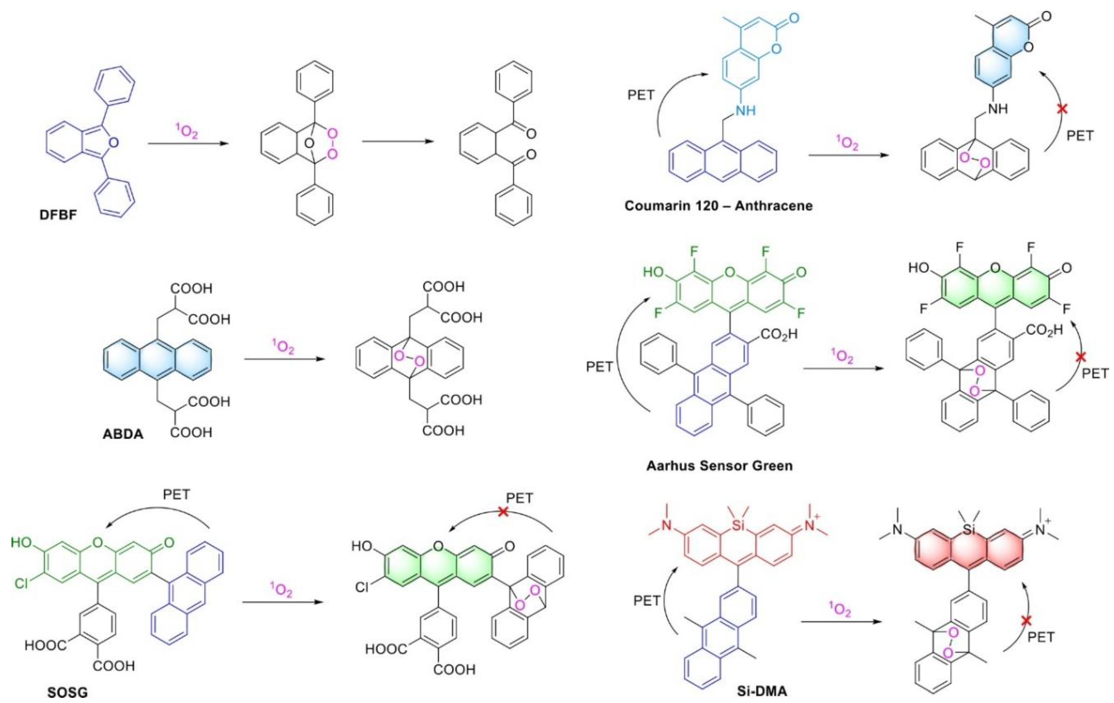
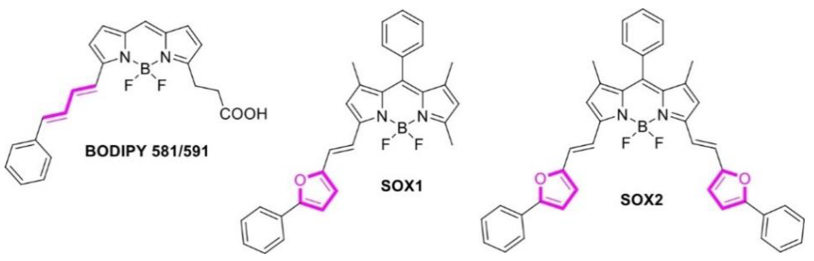
1. Introduction
2. Results and Discussion
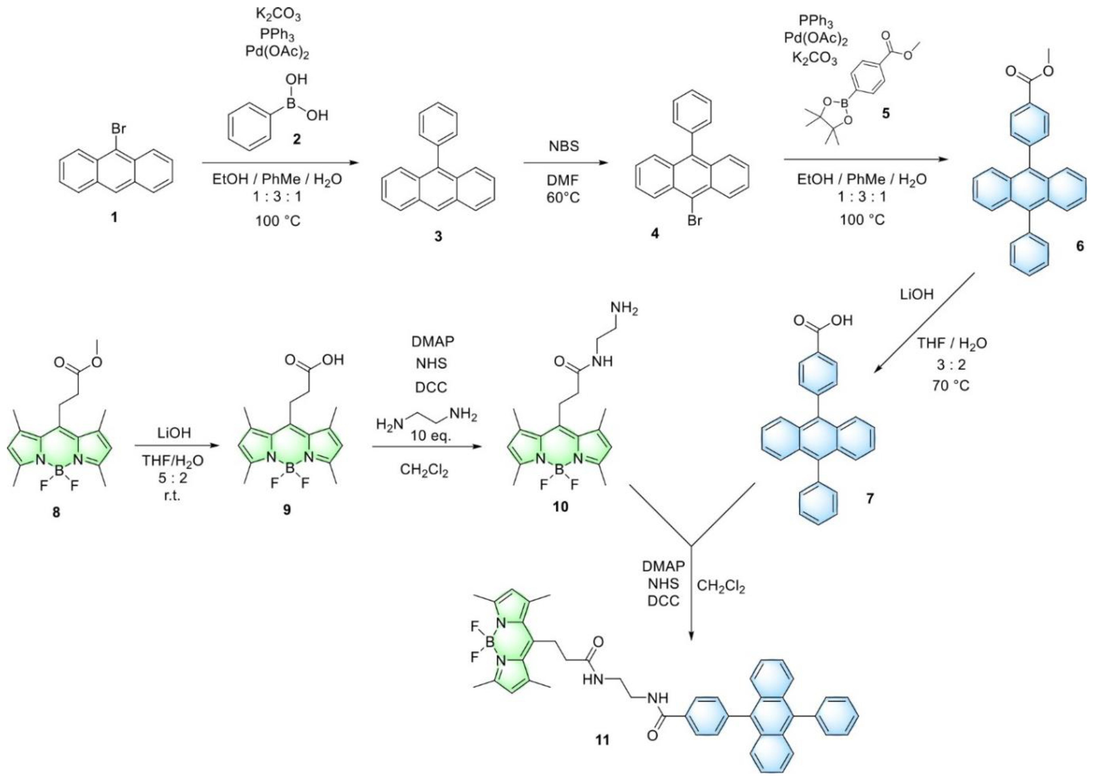
2.1. Synthesis of BODIPY-DPA Dyad
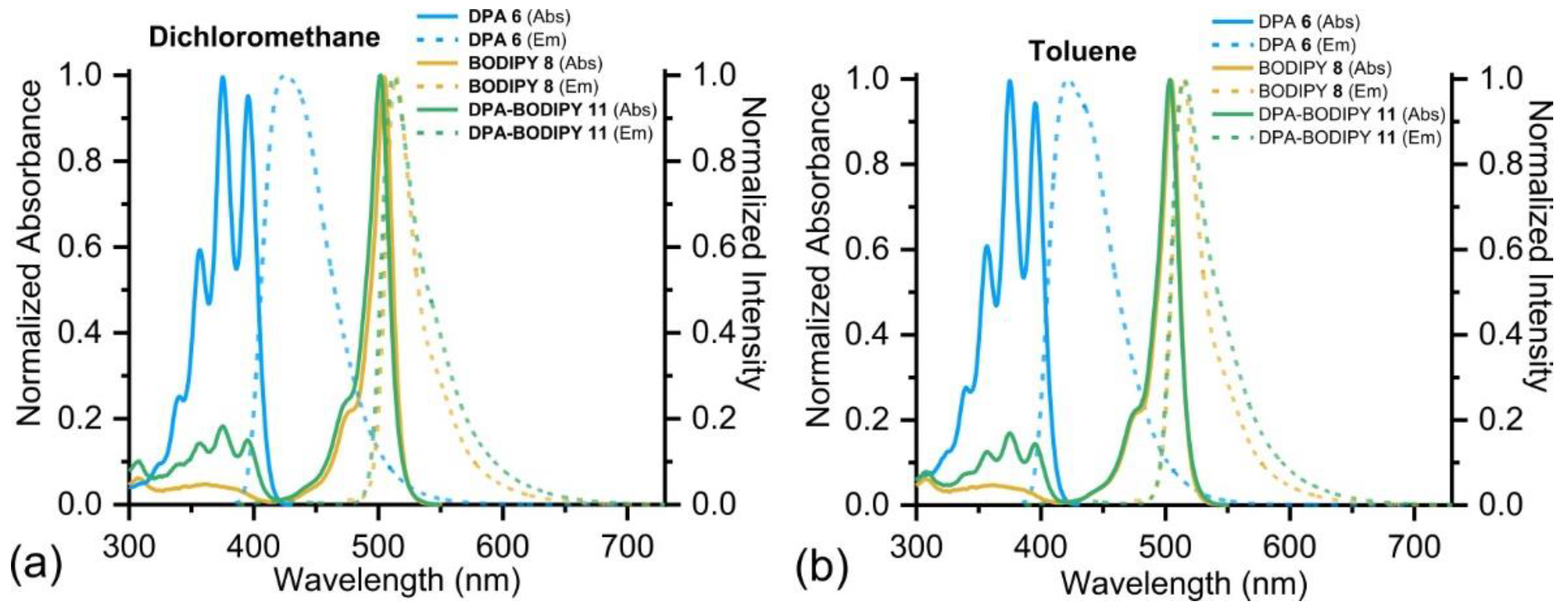
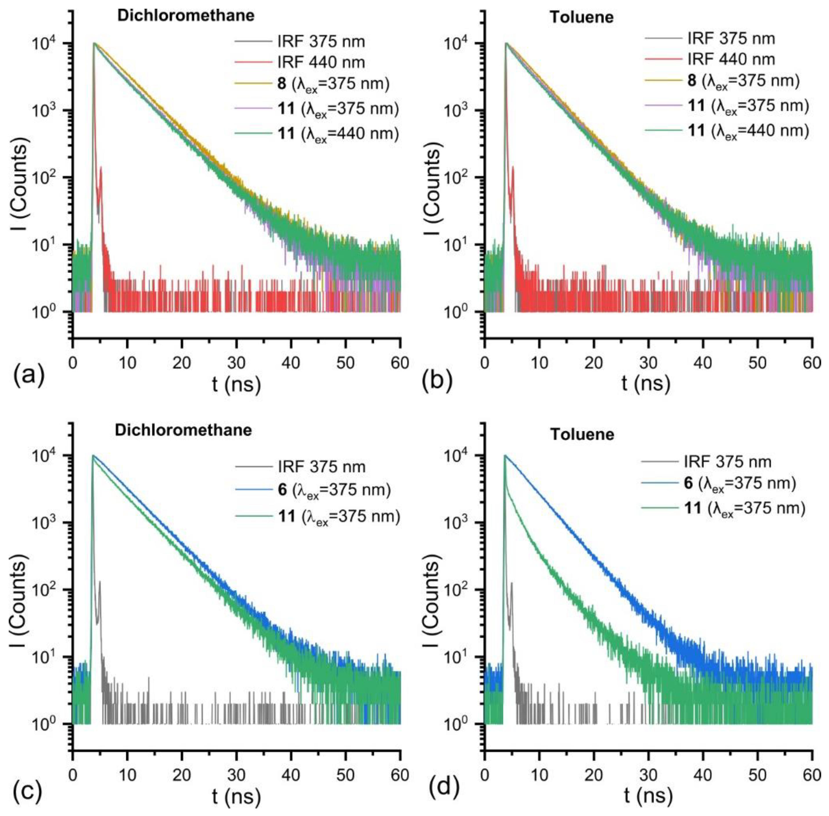
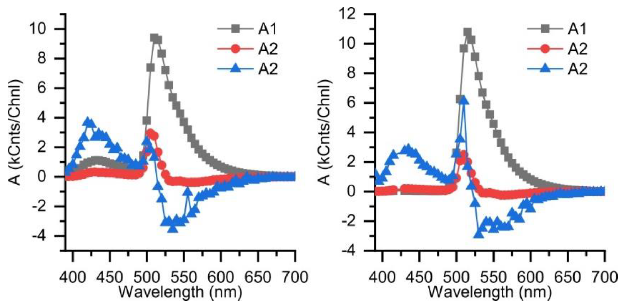
2.2. Optical Properties of the Synthesized Compounds
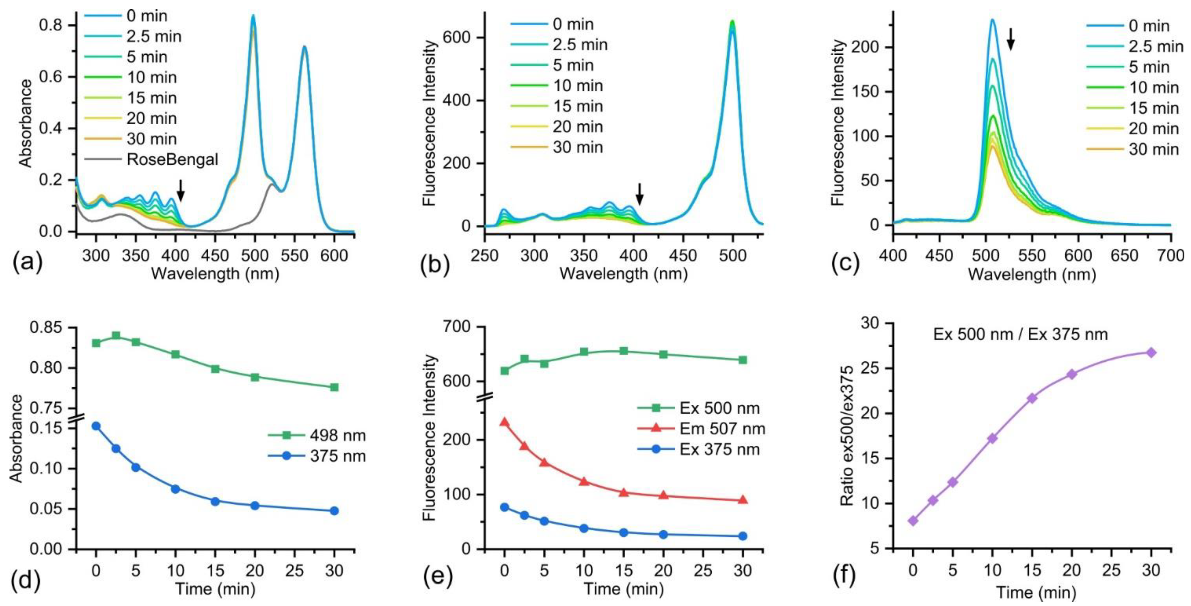
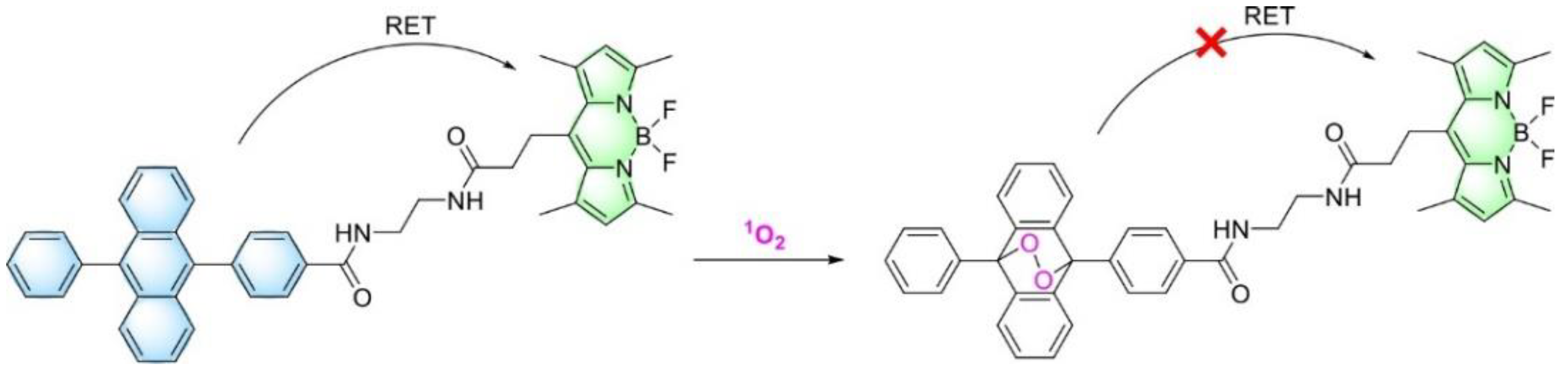
2.3. Singlet Oxygen Detection
3. Materials and Methods
4. Conclusions
Supplementary Materials
Author Contributions
Funding
Institutional Review Board Statement
Informed Consent Statement
Data Availability Statement
Conflicts of Interest
Appendix A


References
- Apel, K.; Hirt, H. Reactive Oxygen Species: Metabolism, Oxidative Stress, and Signal Transduction. Annu. Rev. Plant Biol. 2004, 55, 373–399. [Google Scholar] [CrossRef] [PubMed]
- Forrester, S.J.; Kikuchi, D.S.; Hernandes, M.S.; Xu, Q.; Griendling, K.K. Reactive Oxygen Species in Metabolic and Inflammatory Signaling. Circ. Res. 2018, 122, 877–902. [Google Scholar] [CrossRef] [PubMed]
- Romo-González, M.; Ijurko, C.; Hernández-Hernández, Á. Reactive Oxygen Species and Metabolism in Leukemia: A Dangerous Liaison. Front. Immunol. 2022, 13, 889875. [Google Scholar] [CrossRef] [PubMed]
- Agostinis, P.; Berg, K.; Cengel, K.A.; Foster, T.H.; Girotti, A.W.; Gollnick, S.O.; Hahn, S.M.; Hamblin, M.R.; Juzeniene, A.; Kessel, D.; et al. Photodynamic Therapy of Cancer: An Update. CA Cancer J. Clin. 2011, 61, 250–281. [Google Scholar] [CrossRef]
- Chilakamarthi, U.; Giribabu, L. Photodynamic Therapy: Past, Present and Future. Chem. Rec. 2017, 17, 775–802. [Google Scholar] [CrossRef]
- Lan, M.; Zhao, S.; Liu, W.; Lee, C.; Zhang, W.; Wang, P. Photosensitizers for Photodynamic Therapy. Adv. Healthc. Mater. 2019, 8, 1900132. [Google Scholar] [CrossRef]
- Kashyap, A.; Ramasamy, E.; Ramalingam, V.; Pattabiraman, M. Supramolecular Control of Singlet Oxygen Generation. Molecules 2021, 26, 2673. [Google Scholar] [CrossRef]
- Gomes, A.; Fernandes, E.; Lima, J.L.F.C. Fluorescence Probes Used for Detection of Reactive Oxygen Species. J. Biochem. Biophys. Methods 2005, 65, 45–80. [Google Scholar] [CrossRef]
- Krajczewski, J.; Rucińska, K.; Townley, H.E.; Kudelski, A. Role of Various Nanoparticles in Photodynamic Therapy and Detection Methods of Singlet Oxygen. Photodiagn. Photodyn. Ther. 2019, 26, 162–178. [Google Scholar] [CrossRef]
- Ohyashiki, T.; Nunomura, M.; Katoh, T. Detection of Superoxide Anion Radical in Phospholipid Liposomal Membrane by Fluorescence Quenching Method Using 1,3-Diphenylisobenzofuran. Biochim. Et Biophys. Acta (BBA)-Biomembr. 1999, 1421, 131–139. [Google Scholar] [CrossRef]
- Entradas, T.; Waldron, S.; Volk, M. The Detection Sensitivity of Commonly Used Singlet Oxygen Probes in Aqueous Environments. J. Photochem. Photobiol. B Biol. 2020, 204, 111787. [Google Scholar] [CrossRef] [PubMed]
- Gollmer, A.; Arnbjerg, J.; Blaikie, F.H.; Pedersen, B.W.; Breitenbach, T.; Daasbjerg, K.; Glasius, M.; Ogilby, P.R. Singlet Oxygen Sensor Green®: Photochemical Behavior in Solution and in a Mammalian Cell: Photochemistry and Photobiology. Photochem. Photobiol. 2011, 87, 671–679. [Google Scholar] [CrossRef] [PubMed]
- Dallas, P.; Velasco, P.Q.; Lebedeva, M.; Porfyrakis, K. Detecting the Photosensitization from Fullerenes and Their Dyads with Gold Nanoparticles with Singlet Oxygen Sensor Green. Chem. Phys. Lett. 2019, 730, 130–137. [Google Scholar] [CrossRef]
- Liu, H.; Carter, P.J.H.; Laan, A.C.; Eelkema, R.; Denkova, A.G. Singlet Oxygen Sensor Green Is Not a Suitable Probe for 1O2 in the Presence of Ionizing Radiation. Sci. Rep. 2019, 9, 8393. [Google Scholar] [CrossRef] [PubMed]
- Pedersen, S.K.; Holmehave, J.; Blaikie, F.H.; Gollmer, A.; Breitenbach, T.; Jensen, H.H.; Ogilby, P.R. Aarhus Sensor Green: A Fluorescent Probe for Singlet Oxygen. J. Org. Chem. 2014, 79, 3079–3087. [Google Scholar] [CrossRef]
- Kim, S.; Tachikawa, T.; Fujitsuka, M.; Majima, T. Far-Red Fluorescence Probe for Monitoring Singlet Oxygen during Photodynamic Therapy. J. Am. Chem. Soc. 2014, 136, 11707–11715. [Google Scholar] [CrossRef]
- Sasikumar, D.; Kohara, R.; Takano, Y.; Yuyama, K.; Biju, V. Kinetics of Singlet Oxygen Sensing Using 9-Substituted Anthracene Derivatives. J. Chem. Sci. 2019, 131, 5. [Google Scholar] [CrossRef]
- Martynov, V.I.; Pakhomov, A.A.; Deyev, I.E.; Petrenko, A.G. Genetically Encoded Fluorescent Indicators for Live Cell PH Imaging. Biochim. Biochim. Et Biophys. Acta (BBA)-Gen. Subj. 2018, 1862, 2924–2939. [Google Scholar] [CrossRef]
- Song, D.; Cho, S.; Han, Y.; You, Y.; Nam, W. Ratiometric Fluorescent Probes for Detection of Intracellular Singlet Oxygen. Org. Lett. 2013, 15, 3582–3585. [Google Scholar] [CrossRef]
- Martynov, V.I.; Pakhomov, A.A. BODIPY Derivatives as Fluorescent Reporters of Molecular Activities in Living Cells. Russ. Chem. Rev. 2021, 90, 1213–1262. [Google Scholar] [CrossRef]
- Hwang, T.-Y.; Choi, Y.; Song, Y.; Eom, N.S.A.; Kim, S.; Cho, H.-B.; Myung, N.V.; Choa, Y.-H. A Noble Gas Sensor Platform: Linear Dense Assemblies of Single-Walled Carbon Nanotubes (LACNTs) in a Multi-Layered Ceramic/Metal Electrode System (MLES). J. Mater. Chem. C 2018, 6, 972–979. [Google Scholar] [CrossRef]
- Cheng, H.; Cao, X.; Zhang, S.; Zhang, K.; Cheng, Y.; Wang, J.; Zhao, J.; Zhou, L.; Liang, X.; Yoon, J. BODIPY as Multifunctional Theranostic Reagent in Biomedicine: Self-Assembly, Properties and Applications. Adv. Mater. 2022, 2207546. [Google Scholar] [CrossRef] [PubMed]
- Kolemen, S.; Akkaya, E.U. Reaction-Based BODIPY Probes for Selective Bio-Imaging. Coord. Chem. Rev. 2018, 354, 121–134. [Google Scholar] [CrossRef]
- Loudet, A.; Burgess, K. BODIPY Dyes and Their Derivatives: Syntheses and Spectroscopic Properties. Chem. Rev. 2007, 107, 4891–4932. [Google Scholar] [CrossRef] [PubMed]
- Lu, H.; Mack, J.; Yang, Y.; Shen, Z. Structural Modification Strategies for the Rational Design of Red/NIR Region BODIPYs. Chem. Soc. Rev. 2014, 43, 4778–4823. [Google Scholar] [CrossRef] [PubMed]
- Poddar, M.; Misra, R. Recent Advances of BODIPY Based Derivatives for Optoelectronic Applications. Coord. Chem. Rev. 2020, 421, 213462. [Google Scholar] [CrossRef]
- Antina, E.; Bumagina, N.; Marfin, Y.; Guseva, G.; Nikitina, L.; Sbytov, D.; Telegin, F. BODIPY Conjugates as Functional Compounds for Medical Diagnostics and Treatment. Molecules 2022, 27, 1396. [Google Scholar] [CrossRef]
- Kamkaew, A.; Lim, S.H.; Lee, H.B.; Kiew, L.V.; Chung, L.Y.; Burgess, K. BODIPY Dyes in Photodynamic Therapy. Chem. Soc. Rev. 2013, 42, 77–88. [Google Scholar] [CrossRef]
- Kue, C.S.; Ng, S.Y.; Voon, S.H.; Kamkaew, A.; Chung, L.Y.; Kiew, L.V.; Lee, H.B. Recent Strategies to Improve Boron Dipyrromethene (BODIPY) for Photodynamic Cancer Therapy: An Updated Review. Photochem. Photobiol. Sci. 2018, 17, 1691–1708. [Google Scholar] [CrossRef]
- Nguyen, V.-N.; Ha, J.; Cho, M.; Li, H.; Swamy, K.M.K.; Yoon, J. Recent Developments of BODIPY-Based Colorimetric and Fluorescent Probes for the Detection of Reactive Oxygen/Nitrogen Species and Cancer Diagnosis. Coord. Chem. Rev. 2021, 439, 213936. [Google Scholar] [CrossRef]
- Malacarne, M.C.; Gariboldi, M.B.; Caruso, E. BODIPYs in PDT: A Journey through the Most Interesting Molecules Produced in the Last 10 Years. Int. J. Mol. Sci. 2022, 23, 10198. [Google Scholar] [CrossRef] [PubMed]
- Prieto-Montero, R.; Prieto-Castañeda, A.; Sola-Llano, R.; Agarrabeitia, A.R.; García-Fresnadillo, D.; López-Arbeloa, I.; Villanueva, A.; Ortiz, M.J.; Moya, S.; Martínez-Martínez, V. Exploring BODIPY Derivatives as Singlet Oxygen Photosensitizers for PDT. Photochem. Photobiol. 2020, 96, 458–477. [Google Scholar] [CrossRef] [PubMed]
- Drummen, G.P.C.; Gadella, B.M.; Post, J.A.; Brouwers, J.F. Mass Spectrometric Characterization of the Oxidation of the Fluorescent Lipid Peroxidation Reporter Molecule C11-BODIPY581/591. Free. Radic. Biol. Med. 2004, 36, 1635–1644. [Google Scholar] [CrossRef] [PubMed]
- Kaya, S.; Ismaiel, Y.A.; Kwon, N.; Kim, G.; Bila, J.L.; Yoon, J.; Seven, O.; Akkaya, E.U. Imaging of Intracellular Singlet Oxygen with Bright BODIPY Dyes. Dye. Pigment. 2021, 188, 109158. [Google Scholar] [CrossRef]
- Laguerre, M.; Lecomte, J.; Villeneuve, P. Evaluation of the Ability of Antioxidants to Counteract Lipid Oxidation: Existing Methods, New Trends and Challenges. Prog. Lipid Res. 2007, 46, 244–282. [Google Scholar] [CrossRef]
- Zhu, J.; Zou, J.; Zhang, J.; Sun, Y.; Dong, X.; Zhang, Q. An Anthracene Functionalized BODIPY Derivative with Singlet Oxygen Storage Ability for Photothermal and Continuous Photodynamic Synergistic Therapy. J. Mater. Chem. B 2019, 7, 3303–3309. [Google Scholar] [CrossRef]
- Mahmood, Z.; Taddei, M.; Rehmat, N.; Bussotti, L.; Doria, S.; Guan, Q.; Ji, S.; Zhao, J.; Di Donato, M.; Huo, Y.; et al. Color-Tunable Delayed Fluorescence and Efficient Spin–Orbit Charge Transfer Intersystem Crossing in Compact Carbazole-Anthracene-Bodipy Triads Employing the Sequential Electron Transfer Approach. J. Phys. Chem. C 2020, 124, 5944–5957. [Google Scholar] [CrossRef]
- Callaghan, S.; Filatov, M.A.; Savoie, H.; Boyle, R.W.; Senge, M.O. In Vitro Cytotoxicity of a Library of BODIPY-Anthracene and -Pyrene Dyads for Application in Photodynamic Therapy. Photochem. Photobiol. Sci. 2019, 18, 495–504. [Google Scholar] [CrossRef]
- Callaghan, S.; Vindstad, B.E.; Flanagan, K.J.; Melø, T.B.; Lindgren, M.; Grenstad, K.; Gederaas, O.A.; Senge, M.O. Structural, Photophysical, and Photobiological Studies on BODIPY-Anthracene Dyads. ChemPhotoChem 2021, 5, 131–141. [Google Scholar] [CrossRef]
- Pakhomov, A.A.; Kononevich, Y.N.; Stukalova, M.V.; Svidchenko, E.A.; Surin, N.M.; Cherkaev, G.V.; Shchegolikhina, O.I.; Martynov, V.I.; Muzafarov, A.M. Synthesis and Photophysical Properties of a New BODIPY-Based Siloxane Dye. Tetrahedron Lett. 2016, 57, 979–982. [Google Scholar] [CrossRef]
- Kononevich, Y.N.; Belova, A.S.; Ionov, D.S.; Sazhnikov, V.A.; Pakhomov, A.A.; Alfimov, M.V.; Muzafarov, A.M. Novel DBMBF 2 -BODIPY Dyads Connected via a Flexible Linker: Synthesis and Photophysical Properties. New J. Chem. 2022, 46, 12739–12750. [Google Scholar] [CrossRef]
- Pakhomov, A.A.; Kim, E.E.; Kononevich, Y.N.; Ionov, D.S.; Maksimova, M.A.; Khalchenia, V.B.; Maksimov, E.G.; Anisimov, A.A.; Shchegolikhina, O.I.; Martynov, V.I.; et al. Modulation of the Photophysical Properties of Multi-BODIPY-Siloxane Conjugates by Varying the Number of Fluorophores. Dye. Pigment. 2022, 203, 110371. [Google Scholar] [CrossRef]


| Compound | Solvent | λabs (nm) | ε (M−1 cm−1) | λem (nm) | Φf | ΦET6 | ΦET7 |
|---|---|---|---|---|---|---|---|
| DPA-COOMe 6 | Dichloromethane | 375 | 12,000 | 425 | 0.83 1 | ||
| Toluene | 375 | 12,000 | 421 | 0.74 1 | |||
| BODIPY-COOMe 8 | Dichloromethane | 502 | 78,300 | 511 | 0.86 2 | ||
| Toluene | 505 | 79,100 | 514 | 0.95 2 | |||
| DPA-BODIPY 11 | Dichloromethane | 375 (D) 502 (B) | - | 424 (D) 512 (B) | 0.76 (B) 3 0.01 (D) 4 0.60 (T) 5 | 0.99 | 0.72 |
| Toluene | 375 (D) 504 (B) | - | 425 (D) 515 (B) | 0.90 (B) 3 0.01 (D) 4 0.66 (T) 5 | 0.99 | 0.65 |
| Compound | Solvent | τ (ns) at 431 nm, λex = 375 nm | τ (ns) at 511 nm, λex = 375 nm | τ (ns) at 511 nm, λex = 440 nm | Global Fitting | |
|---|---|---|---|---|---|---|
| τ (ns), λex = 375 nm | τ (ns), λex = 440 nm | |||||
| DPA-COOMe 6 | Dichloromethane | 5.25 | ||||
| Toluene | 4.57 | |||||
| BODIPY-COOMe 8 | Dichloromethane | 5.3 | 5.34 | |||
| Toluene | 4.9 | 4.94 | ||||
| DPA-BODIPY 11 | Dichloromethane | 5.24 (37%) 1.54 (8%) 0.07 (55%) | 5.28 (79%) 0.96 (21%) | 5.3 (74%) 0.96 (26%) | 5.29 0.98 0.05 | 5.26 0.63 |
| Toluene | 4.8 (3%) 1.74 (7%) 0.06 (90%) | 4.96 (84%) 0.80 (16%) | 4.93 (74%) 0.74 (26%) | 4.92 1.27 0.06 | 4.93 0.5 | |
Publisher’s Note: MDPI stays neutral with regard to jurisdictional claims in published maps and institutional affiliations. |
© 2022 by the authors. Licensee MDPI, Basel, Switzerland. This article is an open access article distributed under the terms and conditions of the Creative Commons Attribution (CC BY) license (https://creativecommons.org/licenses/by/4.0/).
Share and Cite
Pakhomov, A.A.; Belova, A.S.; Khchoyan, A.G.; Kononevich, Y.N.; Ionov, D.S.; Maksimova, M.A.; Frolova, A.Y.; Alfimov, M.V.; Martynov, V.I.; Muzafarov, A.M. Ratiometric Singlet Oxygen Sensor Based on BODIPY-DPA Dyad. Molecules 2022, 27, 9060. https://doi.org/10.3390/molecules27249060
Pakhomov AA, Belova AS, Khchoyan AG, Kononevich YN, Ionov DS, Maksimova MA, Frolova AY, Alfimov MV, Martynov VI, Muzafarov AM. Ratiometric Singlet Oxygen Sensor Based on BODIPY-DPA Dyad. Molecules. 2022; 27(24):9060. https://doi.org/10.3390/molecules27249060
Chicago/Turabian StylePakhomov, Alexey A., Anastasia S. Belova, Arevik G. Khchoyan, Yuriy N. Kononevich, Dmitriy S. Ionov, Margarita A. Maksimova, Anastasiya Yu. Frolova, Mikhail V. Alfimov, Vladimir I. Martynov, and Aziz M. Muzafarov. 2022. "Ratiometric Singlet Oxygen Sensor Based on BODIPY-DPA Dyad" Molecules 27, no. 24: 9060. https://doi.org/10.3390/molecules27249060
APA StylePakhomov, A. A., Belova, A. S., Khchoyan, A. G., Kononevich, Y. N., Ionov, D. S., Maksimova, M. A., Frolova, A. Y., Alfimov, M. V., Martynov, V. I., & Muzafarov, A. M. (2022). Ratiometric Singlet Oxygen Sensor Based on BODIPY-DPA Dyad. Molecules, 27(24), 9060. https://doi.org/10.3390/molecules27249060