


Antitrypanosomal, Antitopoisomerase-I, and Cytotoxic Biological Evaluation of Some African Plants Belonging to Crassulaceae; Chemical Profiling of Extract Using UHPLC/QTOF-MS/MS

 , ,
, ,  , , , , and
Abstract
:1. Introduction
2. Results and Discussions
2.1. Total Phenolic Content (TPC) and Total Flavonoids Contents (TFC) Assay
2.2. Antitrypanosomal Examination
2.3. Cytotoxic Examination
2.4. Topo I Inhibitory Activity
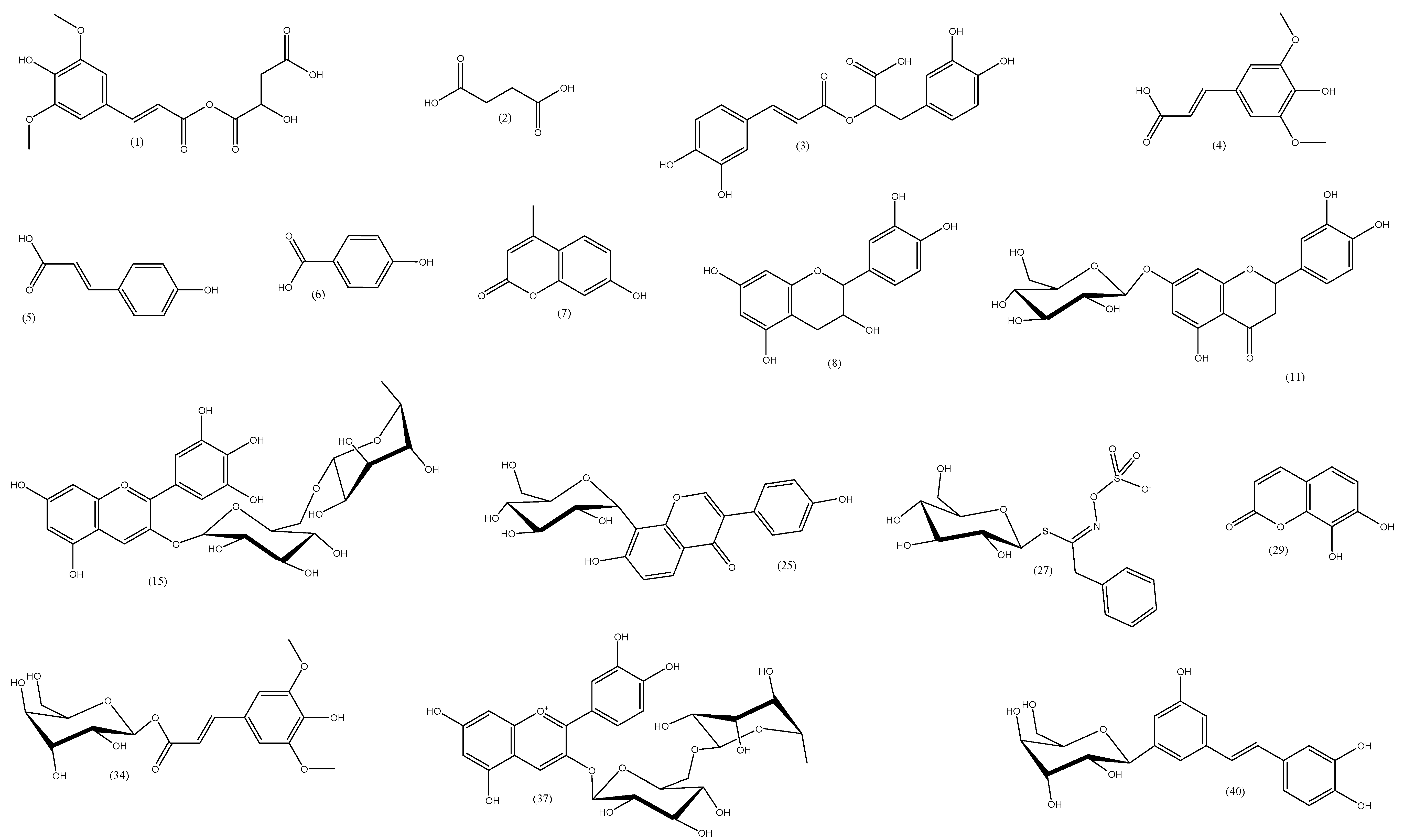
2.5. LC–MS/MS Assay
2.5.1. Identification of Flavonoids

| P | Tentative Assignment | RT (min) | Chemical Formula | Precursor m/z | MS [−] MS/MS m/z | MS [+] MS/MS m/z | Error (ppm) | Ref. |
|---|---|---|---|---|---|---|---|---|
| 1 | Sinapoyl malate | 0.446 | C15H16O9 | 339.0567 | 339.0567 [M-H]−, 223.04 [M-H-malate]− | 0.3 | [34] | |
| 2 | Succinic acid | 0.533 | C4H6O4 | 116.9861 | 116.9861 [M-H]−, 73.03 [M-H-CO2]− | 0 | [35] | |
| 3 | Rosmarinic acid | 0.751 | C18H16O8 | 359.0998 | 359.0998 | 1.6 | [36] | |
| 4 | Sinapic acid | 0.791 | C11H12O5 | 223.046 | 223.046 [M-H]− | 1 | [37] | |
| 5 | P-coumaric acid | 0.967 | C9H8O3 | 163.0402 | 163.0402 [M-H]−, 119.05 [M-H-COOH]− | 1 | [37] | |
| 6 | P-hydroxybenzoic acid | 1.032 | C7H6O3 | 137.0245 | 137.0245 [M-H]−, 93.03 [M-H-CO2]− | 0.8 | [37] | |
| 7 | 7-hydroxy-4-methylcoumarin (Hymecromon) | 1.237 | C10H8O3 | 175.0974 | 175.0974 [M-H]−, 130.97 [M-H-CO2]− | 2.3 | [38] | |
| 8 | (+) Epicatechin | 1.715 | C15H14O6 | 289.1311 | 289.1311 [M-H]− | −5.7 | [37,39] | |
| 9 | Kaempferol-7-neohesperidoside | 1.731 | C27H30O15 | 593.1486 | 593.1486 [M-H]−, 447.68 [M-H-Rhamnosyl]− | 2.3 | [40] | |
| 10 | Luteolin-6-C-glucoside (Isoorientin) | 2.841 | C21H20O11 | 449.1082 | 449.1082 [M+H]+ | −0.2 | [41] | |
| 11 | Eriodictyol-7-o-glucoside | 3.071 | C21H22O11 | 449.0722 | 449.0722 [M-H]−, 287.02 [M-glucose-H]− | 0.7 | [42] | |
| 12 | Apigenin-6-C-glucoside -7-O-glucoside (Saponarin) | 3.079 | C27H30O15 | 595.1672 | 595.1672 [M+H]+ | −0.7 | [43] | |
| 13 | Myricitrin | 3.189 | C21H20O12 | 463.0877 | 463.0877 [M-H]− | 0.9 | [44] | |
| 14 | Vitexin-2″-o-rhamnoside | 3.213 | C27H30O14 | 577.1559 | 577.1559 [M-H]−, 457.11, 413.07, 293.05 | 0.5 | [45] | |
| 15 | Delphinidin-3-o-(6″-o-alpha-rhamnopyranosyl-β-glucopyranoside) | 3.333 | C27H31O16 | 609.1464 | 609.1464 [M-2H]− | −0.3 | [46] | |
| 16 | Apigenin 8-c-glucoside (vitexin) | 3.386 | C21H20O10 | 431.0981 | 431.0981 [M-H]− | 0 | [41] | |
| 17 | Syringetin-3-o-galactoside | 3.614 | C23H24O13 | 507.1167 | 507.1167 [M-H]− | −3.2 | [47] | |
| 18 | Kaempferol-3-glucuronide | 3.688 | C21H18O12 | 461.1086 | 461.1086 [M-H]− | 0.3 | [48] | |
| 19 | Quercetin-3-d-xyloside (Reynoutrin) | 3.745 | C20H18O11 | 433.0757 | 433.0757 [M-H]− | 4.3 | [49] | |
| 20 | Baicalein-7-o-glucuronide (Baicalin) | 3.862 | C21H18O11 | 445.1104 | 445.1104 [M-H]− | 0.8 | [50] | |
| 21 | Kaempferol-3,7-O-bis-α-L-rhamnoside (Kaempferitrin) | 4.043 | C27H30O14 | 579.1714 | 579.1714 [M+H]+ | 0.3 | [51,52] | |
| 22 | Quercetin-7-o-rhamnoside | 4.105 | C21H20O11 | 447.0931 | 447.0931 [M-H]− | 0 | [39] | |
| 23 | Rhoifolin | 4.389 | C27H30O14 | 577.1563 | 577.1563 [M-H]− | −0.2 | [53] | |
| 24 | Myricetin | 4.463 | C15H10O8 | 317.0298 | 317.0298 [M-H]− | 1.6 | [52,54] | |
| 35 | Daidzein-8-c-glucoside (Puerarin) | 4.774 | C24H40O5 | 415.196 | 415.196 [M-H]− | 0.9 | [55] | |
| 26 | Luteolin | 5.592 | C15H10O6 | 285.0396 | 285.0396 [M-H]− | 2.8 | [45,52] | |
| 27 | Benzyl glucosinolate | 5.793 | C14H19NO9S2 | 408.1458 | 408.1458 [M-H]− | −5.1 | [56] | |
| 28 | Quercetin | 5.835 | C15H10O7 | 303.0484 | 303.0484 [M+H]+ | 1.2 | [45] | |
| 29 | 7, 8-Dihydroxycoumarin (Daphnetin) | 6.006 | C9H6O4 | 177.0553 | 177.0553 [M-H]− | 1.4 | [57] | |
| 30 | Apigenin | 6.519 | C15H10O5 | 269.0456 | 269.0456 [M-H]− | 0.4 | [45,52] | |
| 31 | 3 5 7-trihydroxy-4′-methoxyflavone (Kaempferide) | 6.786 | C16H12O6 | 299.0561 | 299.0561 [M-H]− | −0.3 | [58] | |
| 32 | Kaempferol-3-o-α-l-arabinoside | 7.333 | C20H18O10 | 417.1781 | 417.1781 [M-H]− | −3.3 | [59] | |
| 33 | Kaempferol-3-o-α-l-rhamnoside (kaempferin) | 9.919 | C21H20O10 | 431.1713 | 431.1713 [M-H]− | −0.5 | [52,60] | |
| 34 | 1-O-β-D-glucopyranosyl sinapate | 9.921 | C17H22O10 | 387.1808 | 387.1808 [M+H]+ | −0.9 | [61] | |
| 35 | Gossypin | 11.194 | C21H20O13 | 481.1479 | 481.1479 [M+H]+ | 0.6 | [62] | |
| 36 | Quercetin-4′-glucoside | 11.271 | C21H20O12 | 465.1524 | 465.1524 [M+H]+ | 0.2 | [63] | |
| 37 | cyanidin-3-O-rutinoside | 11.872 | C27H31O15 | 595.2518 | 595.2518 [M]+ | 0.7 | [64] | |
| 38 | Luteolin-8-C-glucoside | 12.651 | C21H20O11 | 449.1575 | 449.1575 [M+H]+ | 0.4 | [41] | |
| 39 | Acacetin-7-O-neohesperidoside (Fortunellin) | 12.885 | C28H32O14 | 593.2344 | 593.2344 [M+H]+ | 1.7 | [65] | |
| 40 | E-3,4,5′-trihydroxy-3′-glucopyranosylstilbene (Astringin) | 13.795 | C20H22O9 | 405.2042 | 405.2042 [M-H]− | 2.2 | [66] |
2.5.2. Phenolic Acids Identification
2.5.3. Other Metabolites
3. Materials and Methods
3.1. Plant Materials and Extraction
3.2. Total Phenolic Contents
3.3. Total Flavonoid Contents
3.4. Assay of Antitrypanosomal Activity
3.5. Cytotoxic Assay
3.6. Topo I Assay
3.7. LC–MS/MS
3.7.1. Chemicals
3.7.2. Sample Preparation
3.7.3. Instruments and Acquisition Method
3.7.4. Data Processing
4. Conclusions
Author Contributions
Funding
Institutional Review Board Statement
Informed Consent Statement
Data Availability Statement
Conflicts of Interest
References
- Bakshi, R.P.; Sang, D.; Morrell, A.; Cushman, M.; Shapiro, T.A. Activity of indenoisoquinolines against African trypanosomes. Antimicrob. Agents Chemother. 2009, 53, 123–128. [Google Scholar] [CrossRef] [PubMed] [Green Version]
- Ibrahim, M.A.; Mohammed, A.; Isah, M.B.; Aliyu, A.B. Anti-trypanosomal activity of African medicinal plants: A review update. J. Ethnopharmacol. 2014, 154, 26–54. [Google Scholar] [CrossRef] [PubMed]
- Nwodo, N.J.; Ibezim, A.; Ntie-Kang, F.; Adikwu, M.U.; Mbah, C.J. Anti-trypanosomal activity of Nigerian plants and their constituents. Molecules 2015, 20, 7750–7771. [Google Scholar] [CrossRef] [PubMed] [Green Version]
- International Agency for Research on Cancer. Latest global cancer data: Cancer burden rises to 18.1 million new cases and 9.6 million cancer deaths in 2018. Int. Agency Res. Cancer Lyon Fr. 2018. [Google Scholar]
- Greenwell, M.; Rahman, P. Medicinal plants: Their use in anticancer treatment. Int. J. Pharm. Sci. Res. 2015, 6, 4103. [Google Scholar] [PubMed]
- Alves-Silva, J.M.; Romane, A.; Efferth, T.; Salgueiro, L. North African medicinal plants traditionally used in cancer therapy. Front. Pharmacol. 2017, 8, 383. [Google Scholar] [CrossRef] [Green Version]
- Bawm, S.; Tiwananthagorn, S.; San Lin, K.; Hirota, J.; Irie, T.; Htun, L.L.; Maw, N.N.; Myaing, T.T.; Phay, N.; Miyazaki, S. Evaluation of Myanmar medicinal plant extracts for antitrypanosomal and cytotoxic activities. J. Vet. Med. Sci. 2010, 72, 525–528. [Google Scholar] [CrossRef] [Green Version]
- Muzitano, M.F.; Bergonzi, M.C.; De Melo, G.O.; Lage, C.L.S.; Bilia, A.R.; Vincieri, F.F.; Rossi-Bergmann, B.; Costa, S.S. Influence of cultivation conditions, season of collection and extraction method on the content of antileishmanial flavonoids from Kalanchoe pinnata. J. Ethnopharmacol. 2011, 133, 132–137. [Google Scholar] [CrossRef]
- Singh, N.; Kaushik, N.K.; Mohanakrishnan, D.; Tiwari, S.K.; Sahal, D. Antiplasmodial activity of medicinal plants from Chhotanagpur plateau, Jharkhand, India. J. Ethnopharmacol. 2015, 165, 152–162. [Google Scholar] [CrossRef]
- Al-Snafi, A.E. The Chemical constituents and pharmacological effects of Bryophyllum calycinum. A review. J. Pharma Sci. Res. 2013, 4, 171–176. [Google Scholar]
- Eid, O.; Gonaid, M. Crassulaceae (chemistry and pharmacology)-A review. Future J. Pharm. Sci. 2018, 4, 234–240. [Google Scholar] [CrossRef]
- Meinke, P.T.; Liberator, P. Histone deacetylase: A target for antiproliferative and antiprotozoal agents. Curr. Med. Chem. 2001, 8, 211–235. [Google Scholar] [CrossRef] [PubMed]
- Wang, J.C. Cellular roles of DNA topoisomerases: A molecular perspective. Nat. Rev. Mol. Cell Biol. 2002, 3, 430. [Google Scholar] [CrossRef]
- Lodish, H.; Berk, A.; Zipursky, S.L.; Matsudaira, P.; Baltimore, D.; Darnell, J. The Role of Topoisomerases in DNA Replication; WH Freeman: Sydney, Australia, 2000. [Google Scholar]
- Pommier, Y.; Leo, E.; Zhang, H.; Marchand, C. DNA topoisomerases and their poisoning by anticancer and antibacterial drugs. Chem. Biol. 2010, 17, 421–433. [Google Scholar] [CrossRef] [Green Version]
- Hogan, M.A.; McKinney, D.S. Comprehensive Review for NCLEX-PN; Pearson: London, UK, 2012. [Google Scholar]
- Bodley, A.L.; Shapiro, T.A. Molecular and cytotoxic effects of camptothecin, a topoisomerase I inhibitor, on trypanosomes and Leishmania. Proc. Natl. Acad. Sci. USA 1995, 92, 3726–3730. [Google Scholar] [CrossRef] [Green Version]
- Zuma, A.A.; Cavalcanti, D.P.; Maia, M.C.; de Souza, W.; Motta, M.C.M. Effect of topoisomerase inhibitors and DNA-binding drugs on the cell proliferation and ultrastructure of Trypanosoma cruzi. Int. J. Antimicrob. Agents 2011, 37, 449–456. [Google Scholar] [CrossRef] [PubMed] [Green Version]
- Otero, E.; Vergara, S.; Robledo, S.M.; Cardona, W.; Carda, M.; Vélez, I.D.; Rojas, C.; Otálvaro, F. Synthesis, leishmanicidal and cytotoxic activity of triclosan-chalcone, triclosan-chromone and triclosan-coumarin hybrids. Molecules 2014, 19, 13251–13266. [Google Scholar] [CrossRef] [Green Version]
- Eggli, U. Illustrated Handbook of Succulent Plants: Dicotyledons; Springer Science & Business Media: Berlin, Germany, 2002. [Google Scholar]
- Xu, T.; Wang, Z.; Lei, T.; Lv, C.; Wang, J.; Lu, J. New flavonoid glycosides from Sedum aizoon L. Fitoterapia 2015, 101, 125–132. [Google Scholar] [CrossRef]
- Stevens, J.F.; Hart, H.T.; Van Ham, R.C.; Elema, E.T.; Van Den Ent, M.M.; Wildeboer, M.; Zwaving, J.H. Distribution of alkaloids and tannins in the Crassulaceae. Biochem. Syst. Ecol. 1995, 23, 157–165. [Google Scholar] [CrossRef]
- Melero, C.P.; Medarde, M.; San Feliciano, A. A short review on cardiotonic steroids and their aminoguanidine analogues. Molecules 2000, 5, 51–81. [Google Scholar] [CrossRef] [Green Version]
- van Maarseveen, C.; Jetter, R. Composition of the epicuticular and intracuticular wax layers on Kalanchoe daigremontiana (Hamet et Perr. de la Bathie) leaves. Phytochemistry 2009, 70, 899–906. [Google Scholar] [CrossRef] [PubMed]
- Akihisa, T.; Kokke, W.; Tamura, T.; Matsumoto, T. Sterols ofKalanchoe pinnata: First report of the isolation of both C-24 epimers of 24-alkyl-Δ25-sterols from a higher plant. Lipids 1991, 26, 660–665. [Google Scholar] [CrossRef]
- Lindroth, R.L.; Batzli, G.O. Plant phenolics as chemical defenses: Effects of natural phenolics on survival and growth of prairie voles (Microtus ochrogaster). J. Chem. Ecol. 1984, 10, 229–244. [Google Scholar] [CrossRef] [PubMed]
- Azmi, A.S.; Bhat, S.H.; Hanif, S.; Hadi, S.M. Plant polyphenols mobilize endogenous copper in human peripheral lymphocytes leading to oxidative DNA breakage: A putative mechanism for anticancer properties. FEBS Lett. 2006, 580, 533–538. [Google Scholar] [CrossRef] [Green Version]
- Koide, T.; Nose, M.; Inoue, M.; Ogihara, Y.; Yabu, Y.; Ohta, N. Trypanocidal effects of gallic acid and related compounds. Planta Med. 1998, 64, 27–30. [Google Scholar] [CrossRef]
- Tasdemir, D.; Kaiser, M.; Brun, R.; Yardley, V.; Schmidt, T.J.; Tosun, F.; Rüedi, P. Antitrypanosomal and antileishmanial activities of flavonoids and their analogues: In vitro, in vivo, structure-activity relationship, and quantitative structure-activity relationship studies. Antimicrob. Agents Chemother. 2006, 50, 1352–1364. [Google Scholar] [CrossRef] [Green Version]
- Hafez, H.M.; El Deeb, S.; Mahmoud Swaif, M.; Ismail Ibrahim, R.; Ali Kamil, R.; Salman Abdelwahed, A.; Ehab Ibrahim, A. Micellar Organic-solvent Free HPLC Design of Experiment for the Determination of Ertapenem and Meropenem; Assessment using GAPI, AGREE and Analytical Eco-scale models. Microchem. J. 2022, 185, 108262. [Google Scholar] [CrossRef]
- El-Hela, A.A.; Hegazy, M.M.; Abbass, H.S.; Ahmed, A.H.; Bakr, M.S.A.; Elkousy, R.H.; Ibrahim, A.E.; El Deeb, S.; Sayed, O.M.; Gad, E.S. Dinebra retroflexa Herbal Phytotherapy: A Simulation Study Based on Bleomycin-Induced Pulmonary Fibrosis Retraction Potential in Swiss Albino Rats. Medicina 2022, 58, 1719. [Google Scholar] [CrossRef]
- Zhao, X.; Zhang, S.; Liu, D.; Yang, M.; Wei, J. Analysis of Flavonoids in Dalbergia odorifera by Ultra-Performance Liquid Chromatography with Tandem Mass Spectrometry. Molecules 2020, 25, 389. [Google Scholar] [CrossRef] [Green Version]
- Negri, G.; Tabach, R. Saponins, tannins and flavonols found in hydroethanolic extract from Periandra dulcis roots. Rev. Bras. De Farmacogn. 2013, 23, 851–860. [Google Scholar] [CrossRef] [Green Version]
- Lake, J.A.; Field, K.J.; Davey, M.P.; Beerling, D.J.; Lomax, B.H. Metabolomic and physiological responses reveal multi-phasic acclimation of Arabidopsis thaliana to chronic UV radiation. Plant Cell Environ. 2009, 32, 1377–1389. [Google Scholar] [CrossRef] [PubMed]
- Jestilä, J.S.; Uggerud, E. Unimolecular dissociation of anions derived from succinic acid (H2Su) in the gas phase: HSu− and ClMgSu−. Relationship to CO2 fixation. Eur. J. Mass Spectrom. 2018, 24, 33–42. [Google Scholar] [CrossRef] [PubMed]
- Sik, B.; Kapcsándi, V.; Székelyhidi, R.; Hanczné, E.L.; Ajtony, Z. Recent advances in the analysis of rosmarinic acid from herbs in the Lamiaceae family. Nat. Prod. Commun. 2019, 14, 1934578X19864216. [Google Scholar] [CrossRef] [Green Version]
- Sun, J.; Liang, F.; Bin, Y.; Li, P.; Duan, C. Screening non-colored phenolics in red wines using liquid chromatography/ultraviolet and mass spectrometry/mass spectrometry libraries. Molecules 2007, 12, 679–693. [Google Scholar] [CrossRef] [PubMed] [Green Version]
- Tine, Y.; Renucci, F.; Costa, J.; Wélé, A.; Paolini, J. A method for LC-MS/MS profiling of coumarins in Zanthoxylum zanthoxyloides (Lam.) B. Zepernich and Timler extracts and essential oils. Molecules 2017, 22, 174. [Google Scholar] [CrossRef] [PubMed] [Green Version]
- Xiao, Y.C.; Liu, L.T.; Bian, J.J.; Yan, C.Q.; Ye, L.; Zhao, M.X.; Huang, Q.S.; Wang, W.; Liang, K.; Shi, Z.F. Identification of multiple constituents in shuganjieyu capsule and rat plasma after oral administration by ultra-performance liquid chromatography coupled with electrospray ionization and ion trap mass spectrometry. Acta Chromatogr. 2018, 30, 95–102. [Google Scholar] [CrossRef]
- Cuyckens, F.; Ma, Y.L.; Pocsfalvi, G.; Claeysi, M. Tandem mass spectral strategies for the structural characterization of flavonoid glycosides. Analusis 2000, 28, 888–895. [Google Scholar] [CrossRef] [Green Version]
- da Silva Mathias, M.; Rodrigues de Oliveira, R. Differentiation of the phenolic chemical profiles of Cecropia pachystachya and Cecropia hololeuca. Phytochem. Anal. 2019, 30, 73–82. [Google Scholar] [CrossRef] [Green Version]
- De Beer, D.; Schulze, A.E.; Joubert, E.; De Villiers, A.; Malherbe, C.J.; Stander, M.A. Food ingredient extracts of Cyclopia subternata (Honeybush): Variation in phenolic composition and antioxidant capacity. Molecules 2012, 17, 14602–14624. [Google Scholar] [CrossRef] [Green Version]
- Ibrahim, R.M.; El-Halawany, A.M.; Saleh, D.O.; El Naggar, E.M.B.; El-Shabrawy, A.E.-R.O.; El-Hawary, S.S. HPLC-DAD-MS/MS profiling of phenolics from Securigera securidaca flowers and its anti-hyperglycemic and anti-hyperlipidemic activities. Rev. Bras. De Farmacogn. 2015, 25, 134–141. [Google Scholar] [CrossRef] [Green Version]
- Hwang, I.-W.; Chung, S.-K. Isolation and identification of myricitrin, an antioxidant flavonoid, from daebong persimmon peel. Prev. Nutr. Food Sci. 2018, 23, 341. [Google Scholar] [CrossRef] [PubMed]
- Li, H.; Song, F.; Xing, J.; Tsao, R.; Liu, Z.; Liu, S. Screening and structural characterization of α-glucosidase inhibitors from hawthorn leaf flavonoids extract by ultrafiltration LC-DAD-MS n and SORI-CID FTICR MS. J. Am. Soc. Mass Spectrom. 2009, 20, 1496–1503. [Google Scholar] [CrossRef] [PubMed]
- Bochi, V.C.; Godoy, H.T.; Giusti, M.M. Anthocyanin and other phenolic compounds in Ceylon gooseberry (Dovyalis hebecarpa) fruits. Food Chem. 2015, 176, 234–243. [Google Scholar] [CrossRef] [PubMed] [Green Version]
- De Rosso, M.; Panighel, A.; Vedova, A.D.; Gardiman, M.; Flamini, R. Characterization of non-anthocyanic flavonoids in some hybrid red grape extracts potentially interesting for industrial uses. Molecules 2015, 20, 18095–18106. [Google Scholar] [CrossRef] [Green Version]
- Kumar, S.; Singh, A.; Kumar, B. Identification and characterization of phenolics and terpenoids from ethanolic extracts of Phyllanthus species by HPLC-ESI-QTOF-MS/MS. J. Pharm. Anal. 2017, 7, 214–222. [Google Scholar] [CrossRef]
- Jang, G.H.; Kim, H.W.; Lee, M.K.; Jeong, S.Y.; Bak, A.R.; Lee, D.J.; Kim, J.B. Characterization and quantification of flavonoid glycosides in the Prunus genus by UPLC-DAD-QTOF/MS. Saudi J. Biol. Sci. 2018, 25, 1622–1631. [Google Scholar] [CrossRef]
- Tong, L.; Wan, M.; Zhang, L.; Zhu, Y.; Sun, H.; Bi, K. Simultaneous determination of baicalin, wogonoside, baicalein, wogonin, oroxylin A and chrysin of Radix scutellariae extract in rat plasma by liquid chromatography tandem mass spectrometry. J. Pharm. Biomed. Anal. 2012, 70, 6–12. [Google Scholar] [CrossRef]
- Ibrahim, L.F.; Elkhateeb, A.; Marzouk, M.M.; Hussein, S.R.; Abdel-Hameed, E.-S.S.; Kassem, M.E.S. Flavonoid investigation, LC–ESI-MS profile and cytotoxic activity of Raphanus raphanistrum L.(Brassicaceae). J. Chem. Pharm. Res. 2016, 8, 786–793. [Google Scholar]
- Tahir, N.I.; Shaari, K.; Abas, F.; Parveez, G.K.A.; Ishak, Z.; Ramli, U.S. Characterization of apigenin and luteolin derivatives from oil palm (Elaeis guineensis Jacq.) leaf using LC–ESI-MS/MS. J. Agric. Food. Chem. 2012, 60, 11201–11210. [Google Scholar] [CrossRef]
- Brito, A.; Ramirez, J.E.; Areche, C.; Sepúlveda, B.; Simirgiotis, M.J. HPLC-UV-MS profiles of phenolic compounds and antioxidant activity of fruits from three citrus species consumed in Northern Chile. Molecules 2014, 19, 17400–17421. [Google Scholar] [CrossRef] [Green Version]
- Saldanha, L.L.; Vilegas, W.; Dokkedal, A.L. Characterization of flavonoids and phenolic acids in Myrcia bella cambess. Using FIA-ESI-IT-MSn and HPLC-PAD-ESI-IT-MS combined with NMR. Molecules 2013, 18, 8402–8416. [Google Scholar] [CrossRef] [PubMed] [Green Version]
- Zhao, W.; Shang, Z.; Li, Q.; Huang, M.; He, W.; Wang, Z.; Zhang, J. Rapid screening and identification of daidzein metabolites in rats based on uhplc-ltq-orbitrap mass spectrometry coupled with data-mining technologies. Molecules 2018, 23, 151. [Google Scholar] [CrossRef] [PubMed] [Green Version]
- Castro-Vargas, H.I.; Baumann, W.; Parada-Alfonso, F. Valorization of agroindustrial wastes: Identification by LC-MS and NMR of benzylglucosinolate from papaya (Carica papaya L.) seeds, a protective agent against lipid oxidation in edible oils. Electrophoresis 2016, 37, 1930–1937. [Google Scholar] [CrossRef] [PubMed]
- Venditti, A.; Sanna, C.; Lorenzetti, L.M.; Ballero, M.; Bianco, A. New coumarinyl ethers in Daphne oleoides Schreb. Collected from Sardinia island. Chem. Biodivers. 2017, 14, e1700072. [Google Scholar] [CrossRef] [PubMed]
- Chen, F.; Li, H.-L.; Li, Y.-H.; Tan, Y.-F.; Zhang, J.-Q. Quantitative analysis of the major constituents in Chinese medicinal preparation SuoQuan formulae by ultra fast high performance liquid chromatography/quadrupole tandem mass spectrometry. Chem. Cent. J. 2013, 7, 131. [Google Scholar] [CrossRef] [PubMed] [Green Version]
- Topalovic, A.; Mikulic-Petkovsek, M.; Perovic, N.; Trifunovic, S.; Knezevic, M. Phenolic composition of the leaf of grapevine cv.’Cardinal’. Poljopr. I Sumar. 2006, 52, 5. [Google Scholar]
- Amaral, J.S.; Ferreres, F.; Andrade, P.B.; Valentão, P.; Pinheiro, C.; Santos, A.; Seabra, R. Phenolic profile of hazelnut (Corylus avellana L.) leaves cultivars grown in Portugal. Nat. Prod. Res. 2005, 19, 157–163. [Google Scholar] [CrossRef]
- Oszmiański, J.; Kolniak-Ostek, J.; Wojdyło, A. Application of ultra performance liquid chromatography-photodiode detector-quadrupole/time of flight-mass spectrometry (UPLC-PDA-Q/TOF-MS) method for the characterization of phenolic compounds of Lepidium sativum L. sprouts. Eur. Food Res. Technol. 2013, 236, 699–706. [Google Scholar] [CrossRef] [Green Version]
- Petsalo, A.; Jalonen, J.; Tolonen, A. Identification of flavonoids of Rhodiola rosea by liquid chromatography-tandem mass spectrometry. J. Chromatogr. A 2006, 1112, 224–231. [Google Scholar] [CrossRef]
- Mullen, W.; Graf, B.A.; Caldwell, S.T.; Hartley, R.C.; Duthie, G.G.; Edwards, C.A.; Lean, M.E.J.; Crozier, A. Determination of flavonol metabolites in plasma and tissues of rats by HPLC−radiocounting and tandem mass spectrometry following oral ingestion of [2-14C] quercetin-4′-glucoside. J. Agric. Food Chem. 2002, 50, 6902–6909. [Google Scholar] [CrossRef]
- Fanali, C.; Dugo, L.; D’Orazio, G.; Lirangi, M.; Dachà, M.; Dugo, P.; Mondello, L. Analysis of anthocyanins in commercial fruit juices by using nano-liquid chromatography-electrospray-mass spectrometry and high-performance liquid chromatography with UV-vis detector. J. Sep. Sci. 2011, 34, 150–159. [Google Scholar] [CrossRef] [PubMed]
- Ilboudo, O.; Ouédraogo, I.W.; Tapsoba, I.; Gerbaux, P.; Coulibaly, Y.L.B. Analysis of flavonoid diglycosides in leaves of Mentha piperita L by MALDI-MS/MS and LC-MS. Nat. Prod: Indian J. 2012, 8, 321. [Google Scholar]
- Gabaston, J.; Richard, T.; Biais, B.; Waffo-Teguo, P.; Pedrot, E.; Jourdes, M.; Corio-Costet, M.-F.; Mérillon, J.-M. Stilbenes from common spruce (Picea abies) bark as natural antifungal agent against downy mildew (Plasmopara viticola). Ind. Crops Prod. 2017, 103, 267–273. [Google Scholar] [CrossRef]
- Calzada, F.; Meckes, M.; Cedillo-Rivera, R. Antiamoebic and antigiardial activity of plant flavonoids. Planta Med. 1999, 65, 78–80. [Google Scholar] [CrossRef] [PubMed]
- Marin, C.; Boutaleb-Charki, S.; Diaz, J.G.; Huertas, O.; Rosales, M.J.; Pérez-Cordon, G.; Guitierrez-Sánchez, R.; Sánchez-Moreno, M. Antileishmaniasis activity of flavonoids from Consolida oliveriana. J. Nat. Prod. 2009, 72, 1069–1074. [Google Scholar] [CrossRef] [PubMed]
- Muzitano, M.F.; Tinoco, L.W.; Guette, C.; Kaiser, C.R.; Rossi-Bergmann, B.; Costa, S.S. The antileishmanial activity assessment of unusual flavonoids from Kalanchoe pinnata. Phytochemistry 2006, 67, 2071–2077. [Google Scholar] [CrossRef]
- Mamani-Matsuda, M.; Rambert, J.; Malvy, D.; Lejoly-Boisseau, H.; Daulouède, S.; Thiolat, D.; Coves, S.; Courtois, P.; Vincendeau, P.; Mossalayi, M.D. Quercetin induces apoptosis of Trypanosoma brucei gambiense and decreases the proinflammatory response of human macrophages. Antimicrob. Agents Chemother. 2004, 48, 924–929. [Google Scholar] [CrossRef] [Green Version]
- Fazel Nabavi, S.; Sureda, A.; Daglia, M.; Izadi, M.; Rastrelli, L.; Mohammad Nabavi, S. Flavonoids and Chagas’ Disease: The Story So Far! Curr. Top. Med. Chem. 2017, 17, 460–466. [Google Scholar] [CrossRef]
- Mostafa, A.E.; Atef, A.; Mohammad, A.-E.I.; Jacob, M.; Cutler, S.J.; Ross, S.A. New secondary metabolites from Dodonaea viscosa. Phytochem. Lett. 2014, 8, 10–15. [Google Scholar] [CrossRef]
- Ainsworth, E.A.; Gillespie, K.M. Estimation of total phenolic content and other oxidation substrates in plant tissues using Folin–Ciocalteu reagent. Nat. Protoc. 2007, 2, 875–877. [Google Scholar] [CrossRef]
- Chang, C.-C.; Yang, M.-H.; Wen, H.-M.; Chern, J.-C. Estimation of total flavonoid content in propolis by two complementary colorimetric methods. J. Food Drug Anal. 2002, 10, 10–38212. [Google Scholar]
- Räz, B.; Iten, M.; Grether-Bühler, Y.; Kaminsky, R.; Brun, R. The Alamar Blue® assay to determine drug sensitivity of African trypanosomes (Tb rhodesiense and Tb gambiense) in vitro. Acta Trop. 1997, 68, 139–147. [Google Scholar] [CrossRef] [PubMed]
- Borenfreund, E.; Puerner, J.A. Toxicity determined in vitro by morphological alterations and neutral red absorption. Toxicol. Lett. 1985, 24, 119–124. [Google Scholar] [CrossRef] [PubMed]
- Hegazy, M.M.; Metwaly, A.M.; Mostafa, A.E.; Radwan, M.M.; Mehany, A.B.M.; Ahmed, E.; Enany, S.; Magdeldin, S.; Afifi, W.M.; ElSohly, M.A. Biological and chemical evaluation of some African plants belonging to Kalanchoe species: Antitrypanosomal, cytotoxic, antitopoisomerase I activities and chemical profiling using ultra-performance liquid chromatography/quadrupole-time-of-flight mass spectrometer. Pharmacogn. Mag. 2021, 17, 6. [Google Scholar]
- Fayek, N.M.; Farag, M.A.; Abdel Monem, A.R.; Moussa, M.Y.; Abd-Elwahab, S.M.; El-Tanbouly, N.D. Comparative metabolite profiling of four citrus peel cultivars via ultra-performance liquid chromatography coupled with quadrupole-time-of-flight-mass spectrometry and multivariate data analyses. J. Chromatogr. Sci. 2019, 57, 349–360. [Google Scholar] [CrossRef]
- Hafez, H.M.; El Deeb, S.; Naji, E.A.A.; Aziz, Z.A.; Mahmood, A.S.; Khalil, N.I.; Ibrahim, A.E. Design of an Experimental Study for the Simultaneous Determination of Cefepime, Piperacillin and Tazobactam Using Micellar Organic Solvent-Free HPLC. Separations 2022, 9, 215. [Google Scholar] [CrossRef]
- Kanehisa, M.; Goto, S. KEGG: Kyoto encyclopedia of genes and genomes. Nucleic Acids Res. 2000, 28, 27–30. [Google Scholar] [CrossRef]

| Plant Name | Total Phenolics (mg GAE/100 gm Fresh Weight) | Total Flavonoids (mg QE/100 gm Fresh Weight) |
|---|---|---|
| C. convolute | 113.1 ± 4.8 | 23.6 ± 1.7 |
| C. erosula | 117.6 ± 5.8 | 25.9 ± 1.4 |
| C. mesembryanthemoides | 130.2 ± 6.2 | 28.2 ± 1.8 |
| C. obliqa | 89.5 ± 3.9 | 25.2 ± 2 |
| C. ovata | 110.3 ± 6.1 | 30.4 ± 1.8 |
| C. portulacaria | 114.2 ± 4.1 | 29.3 ± 1.9 |
| S. anacampseros | 139.9 ± 7.6 | 33.5 ± 2.1 |
| S. nussbaumerianum | 150.2 ± 5.9 | 35.4 ± 2.3 |
| S. sieboldii | 170.1 ± 9.1 | 40.2 ± 2.7 |
| Plant Name | HCT-116 Human Colon Carcinoma | HEPG-2 Human Hepatocyte Carcinoma | MCF-7 Human Breast Adenocarcinoma |
|---|---|---|---|
| C. convolute | 12.05 ± 1.82 | 13.25 ± 1.14 | 11.15 ± 0.87 |
| C. erosula | 16.51 ± 0.68 | 15.96 ± 1.08 | 17.24 ± 0.36 |
| C. mesembryanthemoides | 15.16 ± 0.18 | 10.33 ± 0.78 | 13.45 ± 0.76 |
| C. obliqa | 37.44 ± 0.38 | 38.15 ± 0.48 | 40.37 ± 0.57 |
| C. ovate | 30.65 ± 0.45 | 27.24 ± 1.05 | 28.25 ± 0.58 |
| C. portulacaria | 44.58 ± 1.59 | 40.14 ± 1.48 | 41.55 ± 2.57 |
| S. anacampseros | 13.96 ± 1.57 | 11.25 ± 1.9 | 10.17 ± 1.78 |
| S. nussbaumerianum | 28.88 ± 0.54 | 33.18 ± 1.24 | 34.25 ± 0.74 |
| S. sieboldii | 28.18 ± 0.24 | 22.05 ± 0.66 | 26.47 ± 0.85 |
| Basic Structure | 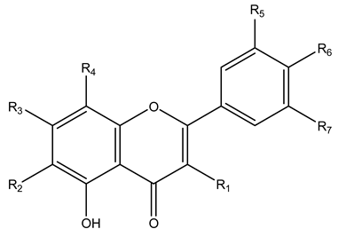 | ||||||
|---|---|---|---|---|---|---|---|
| Comp. No. | R1 | R2 | R3 | R4 | R5 | R6 | R7 |
| 9 | OH | H | -O-neohesperidoside | H | OH | H | |
| 10 | H | -C-β-D-Glucopyranoside | OH | H | OH | OH | H |
| 12 | H | -C-β-D-Glucopyranoside | -O-β-D-Glucopyranoside | H | H | OH | H |
| 13 | -O-α-L-rhamnopyranoside | H | OH | H | OH | OH | OH |
| 14 | H | H | -O-β-D-Glucopyranoside | -C-β-D Glucopyranoside | H | OH | H |
| 16 | H | H | OH | -C-β-D-Glucopyranoside | H | OH | H |
| 17 | -O-β-D-Glucopyranoside | H | OH | H | OCH3 | OH | OCH3 |
| 18 | -O- glucuronide | H | OH | H | H | OH | H |
| 19 | -O-D-xyloside | H | OH | H | OH | OH | H |
| 20 | H | OH | -O-β-D-Glucopyranoside | H | H | H | H |
| 21 | -O-α-L-rhamnopyranoside | H | -O-α-L-rhamnopyranoside | H | H | OH | H |
| 22 | OH | H | -O-β-D-rhamnopyranoside | H | OH | OH | H |
| 23 | H | H | -O-neohesperidoside | H | H | OH | H |
| 24 | OH | H | OH | H | OH | OH | OH |
| 26 | H | H | OH | H | OH | OH | H |
| 28 | OH | H | OH | H | OH | OH | H |
| 30 | H | H | OH | H | H | OH | H |
| 31 | OH | H | OH | H | H | OCH3 | H |
| 32 | o-α-L-arabinoside | H | OH | H | H | OH | H |
| 33 | -O-α-L-rhamnopyranoside | H | OH | H | H | OH | H |
| 35 | OH | H | OH | -O-α-D-Glucopyranoside | OH | OH | H |
| 36 | OH | H | OH | H | -O-β-D-Glucopyranoside | OH | H |
| 38 | H | H | OH | -C-β-D-Glucopyranoside | OH | OH | H |
| 39 | H | H | -O-neohesperidoside | H | H | OCH3 | H |
Publisher’s Note: MDPI stays neutral with regard to jurisdictional claims in published maps and institutional affiliations. |
© 2022 by the authors. Licensee MDPI, Basel, Switzerland. This article is an open access article distributed under the terms and conditions of the Creative Commons Attribution (CC BY) license (https://creativecommons.org/licenses/by/4.0/).
Share and Cite
Hegazy, M.M.; Afifi, W.M.; Metwaly, A.M.; Radwan, M.M.; Abd-Elraouf, M.; Mehany, A.B.M.; Ahmed, E.; Enany, S.; Ezzeldin, S.; Ibrahim, A.E.; et al. Antitrypanosomal, Antitopoisomerase-I, and Cytotoxic Biological Evaluation of Some African Plants Belonging to Crassulaceae; Chemical Profiling of Extract Using UHPLC/QTOF-MS/MS. Molecules 2022, 27, 8809. https://doi.org/10.3390/molecules27248809
Hegazy MM, Afifi WM, Metwaly AM, Radwan MM, Abd-Elraouf M, Mehany ABM, Ahmed E, Enany S, Ezzeldin S, Ibrahim AE, et al. Antitrypanosomal, Antitopoisomerase-I, and Cytotoxic Biological Evaluation of Some African Plants Belonging to Crassulaceae; Chemical Profiling of Extract Using UHPLC/QTOF-MS/MS. Molecules. 2022; 27(24):8809. https://doi.org/10.3390/molecules27248809
Chicago/Turabian StyleHegazy, Mostafa M., Wael M. Afifi, Ahmed M. Metwaly, Mohamed M. Radwan, Muhamad Abd-Elraouf, Ahmed B. M. Mehany, Eman Ahmed, Shymaa Enany, Shahd Ezzeldin, Adel E. Ibrahim, and et al. 2022. "Antitrypanosomal, Antitopoisomerase-I, and Cytotoxic Biological Evaluation of Some African Plants Belonging to Crassulaceae; Chemical Profiling of Extract Using UHPLC/QTOF-MS/MS" Molecules 27, no. 24: 8809. https://doi.org/10.3390/molecules27248809
APA StyleHegazy, M. M., Afifi, W. M., Metwaly, A. M., Radwan, M. M., Abd-Elraouf, M., Mehany, A. B. M., Ahmed, E., Enany, S., Ezzeldin, S., Ibrahim, A. E., El Deeb, S., & Mostafa, A. E. (2022). Antitrypanosomal, Antitopoisomerase-I, and Cytotoxic Biological Evaluation of Some African Plants Belonging to Crassulaceae; Chemical Profiling of Extract Using UHPLC/QTOF-MS/MS. Molecules, 27(24), 8809. https://doi.org/10.3390/molecules27248809