Article

Structure-Activity Relationship Studies Based on 3D-QSAR CoMFA/CoMSIA for Thieno-Pyrimidine Derivatives as Triple Negative Breast Cancer Inhibitors

Yonsei Institute of Pharmaceutical Sciences, College of Pharmacy, Yonsei University, Incheon 21983, Republic of Korea
*
Author to whom correspondence should be addressed.
Molecules 2022, 27(22), 7974; https://doi.org/10.3390/molecules27227974
Submission received: 12 October 2022 / Revised: 4 November 2022 / Accepted: 15 November 2022 / Published: 17 November 2022
(This article belongs to the Special Issue Computational Approaches in Drug Discovery and Design)

Abstract

:
Triple-negative breast cancer (TNBC) is defined as a kind of breast cancer that lacks estrogen receptors (ER), progesterone receptors (PR), and human epidermal growth factor receptors (HER2). This cancer accounts for 10–15% of all breast cancers and has the features of high invasiveness and metastatic potential. The treatment regimens are still lacking and need to develop novel inhibitors for therapeutic strategies. Three-dimensional quantitative structure-activity relationship (3D-QSAR) analyses, based on a series of forty-seven thieno-pyrimidine derivatives, were performed to identify the key structural features for the inhibitory biological activities. The established comparative molecular field analysis (CoMFA) presented a leave-one-out cross-validated correlation coefficient q2 of 0.818 and a determination coefficient r2 of 0.917. In comparative molecular similarity indices analysis (CoMSIA), a q2 of 0.801 and an r2 of 0.897 were exhibited. The predictive capability of these models was confirmed by using external validation and was further validated by the progressive scrambling stability test. From these results of validation, the models were determined to be statistically reliable and robust. This study could provide valuable information for further optimization and design of novel inhibitors against metastatic breast cancer.

1. Introduction

Breast cancer is the most frequently diagnosed cancer that occurs from the epithelium of the ducts or lobules in the breast glandular tissue [1,2]. In 2020, 2.3 million women were diagnosed with breast cancer, and 685,000 of them leading to deaths around the world [3]. There are four subtypes of breast cancer based on the expression of the estrogen receptor (ER), progesterone receptor (PR), and human epidermal growth factor receptor 2 (HER2) [4,5,6,7,8,9]. Triple-negative breast cancer (TNBC), accounting for 10–15% of all breast cancers, is regarded as a heterogeneous and aggressive type of breast cancer with negative expression of ERs, PRs, and HER2s. Compared to receptor-positive breast cancers, TNBC is associated with poorer prognosis and higher rates of recurrence [10].
The vascular endothelial growth factor (VEGF) family participates in the formation of new blood and lymphatic vessels in tumor-related tissue, which leads to the spread of tumor cells [11,12]. VEGF receptor-3 (VEGFR3), as the primary factor in tumor lymphatic angiogenesis, is overexpressed in human breast cancer. The tumor-associated lymphangiogenesis and lymphatic metastasis for breast cancer can be suppressed by the inhibition of VEGFR3 signaling [13,14]. It has been reported that anti-VEGFR3 therapy combined with taxanes targeting lymph nodes has a significant effect on TNBC patients [15].
In the studies by Li et al., N-(4-chloro-3-(trifluoromethyl)phenyl)-4-(6-(4-(4-methylpiperazin-1-yl)phenyl)thieno [2,3-d]pyrimidin-4-yl)piperazine-1-carboxamide 42, as the novel small molecule VEGFR3 inhibitor, possessed a suitable potency on lymphatic cells and TNBC cells. The most active compound 42 also exhibited higher selectivity (>100) for VEGFR3 compared to other subfamily members such as VEGFR1 and VEGFR2. Based on the interaction analysis of the binding mode between the most active compound 42 and VEGFR3, the Asn934, Arg940, and Arg984 amino acid residues of VEGFR3 were explained to play a key role in the selectivity of the most active compound 42. The urea NH of the most active compound 42 showed the hydrogen bonding interaction with the carbonyl oxygen of Leu851. The urea oxygen of the most active compound 42 interacted with the amide NH of Asn934 through two hydrogen bonds. The 4-chloro-3-(trifluoromethyl)phenyl group of the most active compound 42 displayed a hydrophobic interaction with Phe929, Ala983, and Leu1044. The benzene ring of the N-methyl-4-(p-phenyl)piperazine group of the most active compound 42 formed a hydrophobic interaction with Arg940 by the π–cation interaction. The urea group between the rings A and B, the benzene ring E, and the N-methyl-4-(p-phenyl)piperazine group of the most active compound 42 were necessary to increase the biological activity [16].
The lymphangiogenic inhibitors targeting the VEGFRs are displayed in Figure 1. Cabozantinib and Sorafenib are FDA-approved inhibitors targeting VEGFRs against renal cell carcinoma [17,18,19,20]. Sunitinib is FDA-approved targeting VEGFRs for renal cell carcinoma and presents the reduction of tumor lymphatic and blood vessels against breast cancer in preclinical status [21]. Targeting VEGFR2/3, Lenvatinib is FDA-approved for thyroid cancer and presents the suppression of lymph node metastasis against breast cancer in a preclinical study [22]. In the case of targeting VEGFR3, there are MAZ51 against melanoma and SAR131675 against diabetic nephropathy in preclinical status [23,24].
For the analysis of the structural compound patterns governing the biological activity, computer-aided research is known as a relatively fast and economical technique compared to the time-consuming and costly method of in vitro activity determination. Among structure-activity relationship (SAR) analytical studies, three-dimensional quantitative structure-activity relationship (3D-QSAR) analysis is one of the most useful computational approaches for developing potent and effective inhibitors [25,26]. The 3D-QSAR, using the ligand-based alignment of compounds, is widely used for the identification of effective structural features associated with biological activities. In the 3D-QSAR methods, comparative molecular field analysis (CoMFA) and comparative molecular similarity indices analysis (CoMSIA) by using known biological activities can provide, not only, helpful guidance but also a prediction of the bioactivity for the design of novel potent inhibitors [27,28].
To date, no VEGFR3 selective small molecule inhibitor has been approved by the FDA. It is necessary to develop a highly selective VEGFR3 inhibitor with an anti-VEGFR3 therapeutic effect on breast cancer patients. In the present study, the 3D-QSAR was performed to analyze the correlation between the structural features and the inhibitory activities of the thieno-pyrimidine derivatives as VEGFR3 inhibitors against TNBC [16]. The obtained information from 3D-QSAR analysis could be helpful for the construction of novel potent inhibitors against breast cancer.

2. Results and Discussion

2.1. 3D-QSAR

The models were generated with all possible combinations of the steric and electrostatic fields for CoMFA and with all possible combinations of steric, electrostatic, hydrophobic, hydrogen bond donor, and hydrogen bond acceptor fields for the CoMSIA. To build the models, the cross-validated partial least square (PLS) method, which provided a leave-one-out cross-validated correlation coefficient q2 and determination coefficient r2, was used [29]. Based on the basic requirement of the criteria proposed by Golbraikh and Tropsha, the model satisfying q2 > 0.5 and r2 > 0.6 is considered to be acceptable and predictive [30,31]. Among all constructed models, some models could not meet the criteria of q2 > 0.5 and r2 > 0.6, which indicates an unacceptable 3D-QSAR model.
As shown in Table 1, CoMFA_SE and CoMSIA_SEHDA models satisfied the q2 > 0.5 and r2 > 0.6 criteria. The CoMFA model presented a q2 of 0.818, r2 of 0.917, standard error of estimate (SEE) of 8.142, fisher test value (F) of 114.235, predictive correlation coefficient (r2pred) of 0.794, and an optimum number of components (ONC) of 3. The contributions of steric and electrostatic fields were 67.7% and 32.3%, respectively. For the CoMSIA model, the statistical results exhibited a q2 of 0.801, r2 of 0.897, SEE of 9.057, F value of 90.340, r2pred of 0.762, and an ONC of 3. The field contributions of steric, electrostatic, hydrophobic, hydrogen bond donor, and hydrogen bond acceptor were 29.5, 29.8, 29.8, 6.5, and 4.4%, respectively. To assess the predictive capability for the generated CoMFA and CoMSIA models, r2pred was calculated with the test set compounds. The detailed results for all possible combination models are presented in Table S1 (Supplementary Materials).

2.1.1. CoMFA Studies

Scatter Plots

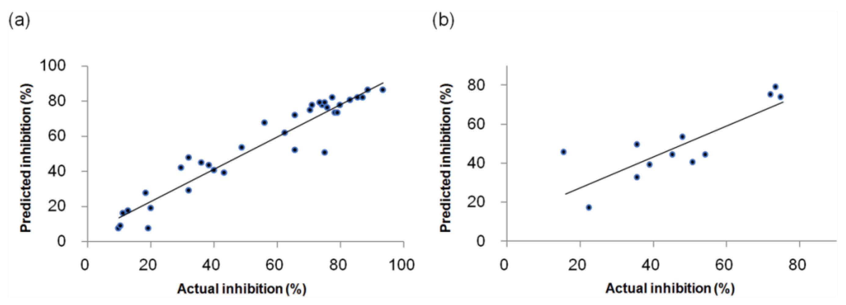
The scatter plots between the actual and predicted inhibitory activities of the training and test sets for the CoMFA model are shown in Figure 2. The data distribution of the training and test set compounds are fit well to the line Y = X, which represents that the constructed CoMFA model shows the predictive capability.

Progressive Scrambling Stability Test

To validate the robustness of the CoMFA model, the progressive scrambling stability test was performed and the obtained results are shown in Table 2 [32]. The inhibitory activities as the dependent variable were randomly shuffled and a new QSAR model was built. In the stable model, the slope (dq2′/dr2yy′) would be less than 1.20. The best model was found to be the 3-components model with the highest q2 value of 0.691, the lowest cSDEP (calculated cross-validated standard error of prediction) of 15.640, and the slope value of 1.102. The 3-components, as the ideal number of ONC, was identified, which was consistent with having an ONC of 3 in the CoMFA model. It indicates that the developed CoMFA model is robust and statistically reliable.

Contour Map Analysis

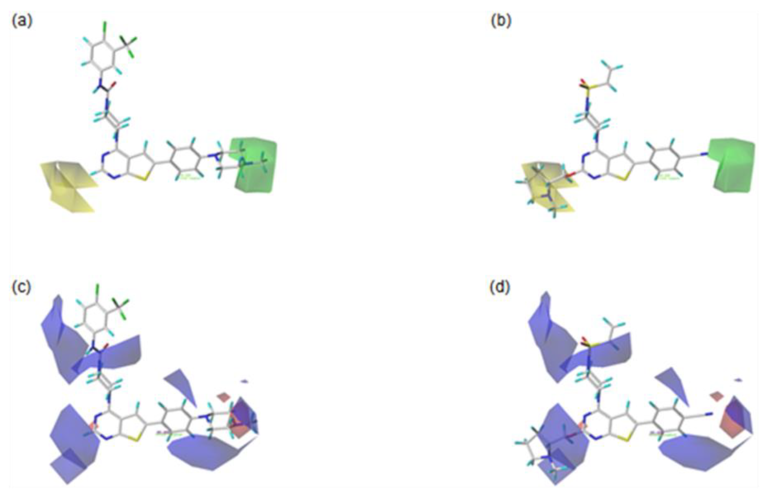
For the visualization and the explanation of each field effect, the CoMFA contour maps created by the StDev*Coeff mapping option are superimposed with the most active compound 42, and the least active compound 20 in Figure 3. The contributions for the favorable and unfavorable regions in each field are displayed by 80% and 20%, respectively.
In the steric contour maps, as shown in Figure 3a for the most active compound 42 and 3b for the least active compound 20, the green contour block means that the presence of the bulky steric groups in this area would be favorable for the biological activity of a compound, while the yellow block represents that the bulky groups would be unfavorable. The green contour block covering the F ring of the most active compound 42 in Figure 3a indicates that the bulky group in this area would be favorable to increase the bioactivity of a compound. For example, compound 5 with the bulky methyl morpholine group on the green region is more biologically active than compound 3 with the methoxy group. In addition, this could explain why compound 6 with the bulkier methyl piperazine group on the green area is more active than compound 5 with the morpholine group. Compared to compound 4, with no substituent on the yellow block, compounds 20 and 21, with the large substituents on this block, are less active. Moreover, compound 22 with the bulky substituted group on the yellow area shows lower biological activity than compound 7, with no substituted group in this region. This could explain why the compounds 23, 24, 25, 26, 45, 46, and 47 with the large substituent on the yellow region show the reduced activities.
As shown in Figure 3c for the most active compound 42 and Figure 3d for the least active compound 20, the blue contours represent that the electropositive groups would be favorable to increase the activity of a compound, while the red contours indicate that the electronegative groups would be unfavorable to the activity of a compound. Compared to compound 6, with no substituent group on the blue contour, compound 25, with the electronegative groups on this block, is less active. This could explain why compounds 20, 21, 22, 23, 24, 25, 26, 45, 46, and 47 present low activities due to the presence of the electronegative substituents on the blue contour region. The compounds 4, 5, and 6, with electronegative groups on the red contour area, show the higher bioactivities than compound 1, without substituents at the corresponding region.

2.1.2. CoMSIA Studies

Scatter Plots

In Figure 4, the scatter plots between the actual and predicted inhibitory activities of the training, and test sets for the CoMSIA model, are shown. The data correlation of the training and test sets shows a good fit along the fitted line, which indicates that the developed CoMSIA model has a predictable ability.

Progressive Scrambling Stability Test

The progressive scrambling stability test was carried out for the additional validation of the CoMSIA model. As shown in Table 3, the obtained results of this validation demonstrate that the 3-components model was found to be the best model with the highest q2 value of 0.674, the lowest cSDEP (calculated cross-validated standard error of prediction) of 16.106, and the slope value of 1.035. This ONC of 3 was identified and consistent with the ONC of 3 in the generated CoMSIA model, which means that the CoMSIA model is statistically stable and robust.

Contour Map Analysis

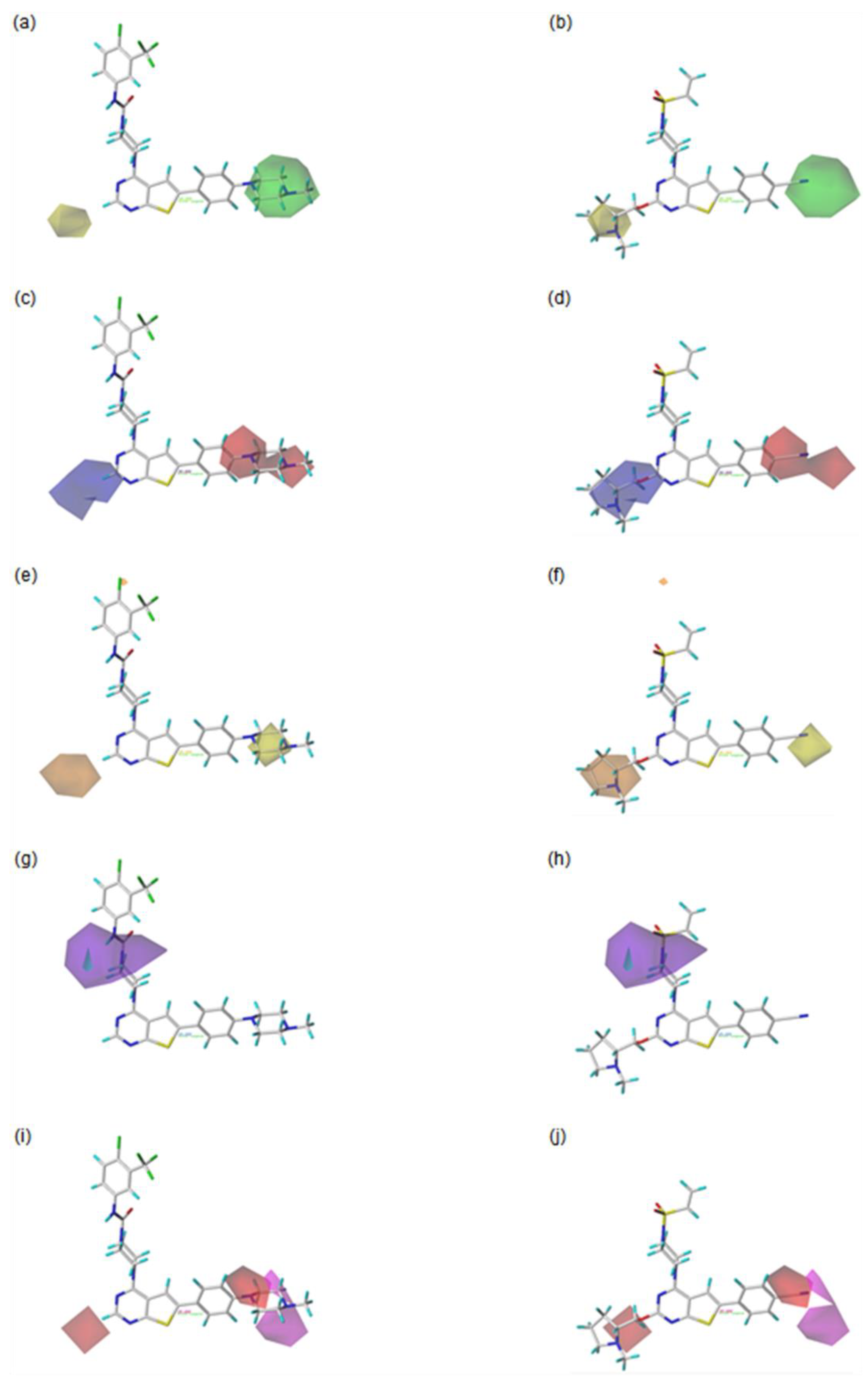
The CoMSIA steric and electrostatic contour maps are similar to those of the CoMFA discussed above. Therefore, the following discussion is focused on the contour maps of the hydrophobic, hydrogen bond donor, and hydrogen bond acceptor fields.
The contour maps for the hydrophobic field are shown in Figure 5e for the most active compound 42 and Figure 5f for the least active compound 20. The orange contour blocks indicate that the introduction of the hydrophobic substituent would be favorable for enhancing the biological activity of a compound, while the yellow contours mean that the hydrophobic groups would be unfavorable in this area. For example, compounds 20 and 21, with the hydrophilic groups including the N or O atom around the orange region, are less active than compound 4, with hydrogen at the corresponding position. This could explain why compounds 22, 23, 24, 25, 26, 45, 46, and 47, with the hydrophilic groups around the orange area, show low biological activities. On the other hand, compound 6 has higher bioactivity than compound 1. This might be because of the hydrophilic piperazine group of compound 6 on the yellow region, compared to compound 1 with an H atom at the corresponding location.
Figure 5g for the most active compound 42 and Figure 5h for the least active compound 20 display the hydrogen bond donor contour maps. The cyan contours indicate that the hydrogen bond donor group would be favored for improving the bioactivity of a compound, while the purple contours represent that the hydrogen bond donor group would be disfavored for bioactivity in this region. In the most active compound 42, the presence of the NH group as a hydrogen bond donor group toward the cyan contour block might be one of the factors that lead to high bioactivity.
In the hydrogen bond acceptor contour map, as shown in Figure 5i, for the most active compound 42 and Figure 5j, for the least active compound 20, the magenta contours represent the area where the hydrogen bond acceptor substituents are favorable for the compound’s bioactivity, while the red contours indicate the region where the hydrogen bond acceptor groups are unfavorable. For example, compound 42 with the N atom, next to the methyl group of the piperazine substituent at the F ring, as the hydrogen bond acceptor around the magenta contour area, has high bioactivity. On the other hand, compounds 20 and 21, with the N or O atom as the hydrogen bond acceptor around the disfavored red contour region, show lower biological activity than compound 4 with the H atom at the corresponding area. This could explain why the compounds 22, 23, 24, 25, 26, 45, 46, and 47 with the N or O atom as the hydrogen bond acceptor at the red contour region have low bioactivities.

2.1.3. Design of the Novel Potent Derivatives

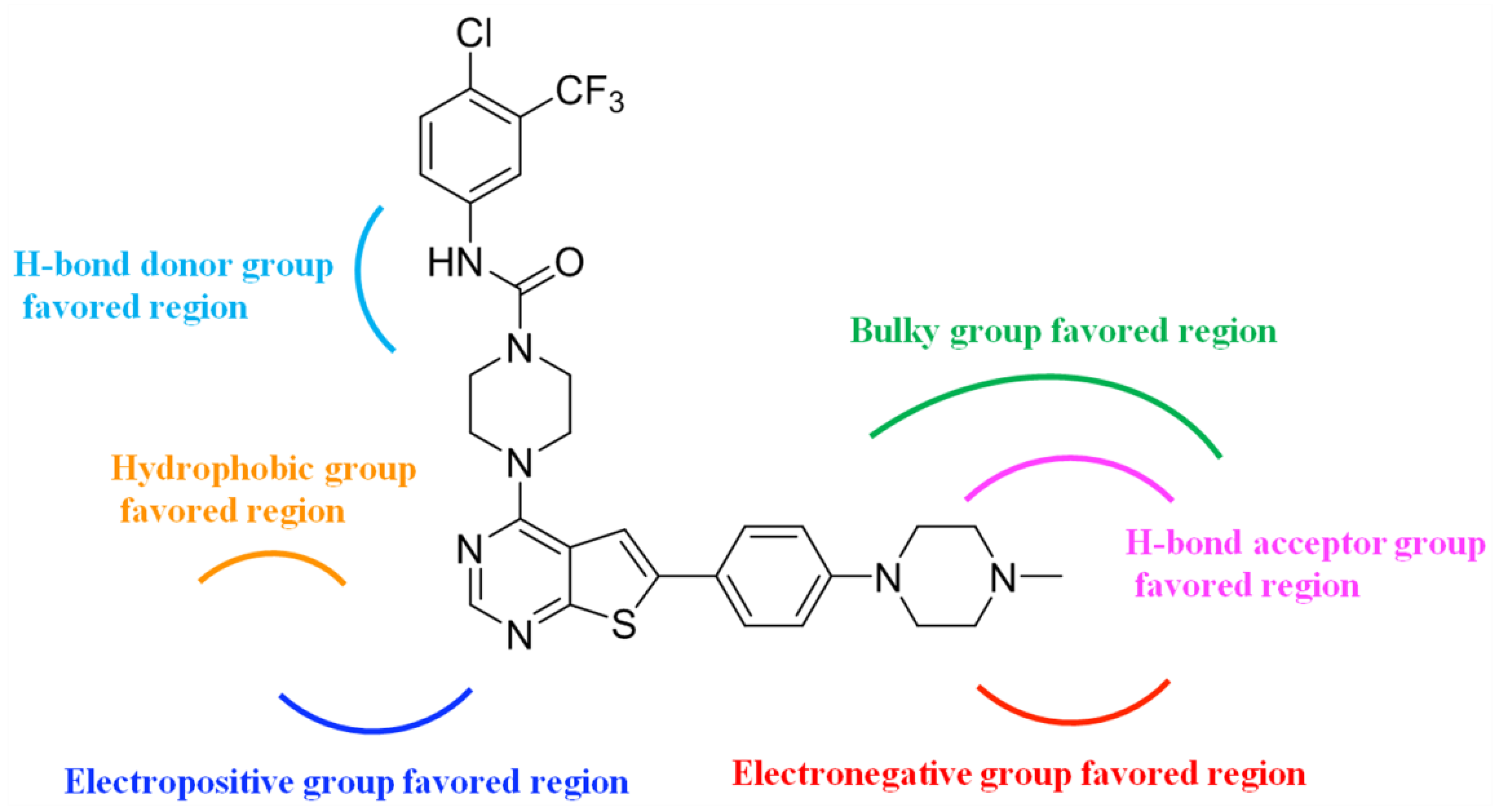
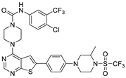
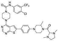
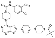
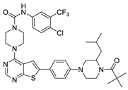
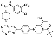
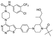
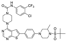
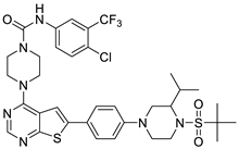
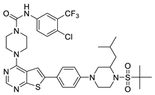
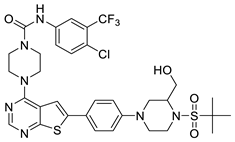
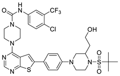
Based on the information provided by the developed 3D-QSAR models, CoMFA_SE and CoMSIA_SEHDA, we have designed a series of novel potent thieno-pyrimidine derivatives. The chemical structures and predicted biological activities of the newly designed inhibitors are presented in Table 4. The newly designed compounds N1N14 were aligned to the database using compound 42 as the template and their inhibitory activities were predicted. They showed higher bioactivities than that of the most active compound 42 in the series. The introduction of bulky substituents on the piperazine ring F and the modification of the bulky substituents, including the carbonyl group or the sulfonyl group at one of the N atoms of the piperazine ring F, could improve the inhibitory activities of the novel designed compounds N1N14 more than those of the most active compound 42 in the series.
To support drug development, we have also predicted the ADMET (absorption, distribution, metabolism, excretion, and toxicity) parameters, pharmacokinetic properties, drug-likeness nature, and synthetic accessibility of the novel designed compounds N1N14 using the online servers SwissADME [33] and pkCSM [34]. The detailed properties of the most active compound 42 and the newly designed inhibitors N1N14 are shown in Table 5. The rule-based Moriguchi log P (MLogP) value was calculated for the lipophilicity and the log S by estimated solubility (ESOL) was calculated for the solubility. The gastrointestinal (GI) absorption and the blood-brain barrier (BBB) permeant were also calculated. Toxicity was predicted by Ames positive test to assess the mutagenic potential of compounds to act as a carcinogen. Drug-likeness qualitatively evaluates the potential of a compound to be an oral drug with respect to bioavailability. Two or more Lipinski’s rule-of-five (Ro5) violations predict a compound as a non-orally available drug. Lipinski’s Ro5 violation defines four classes of molecules with the probabilities of 11%, 17%, 56%, and 85%. The novel designed inhibitors N1N14 presented the prediction results of the acceptable ADMET properties. To represent ease of synthesis, the synthetic accessibility as a score from 1 (very easy to make) to 10 (very difficult to make) is described. The SMILES codes for the most active compound 42 and the novel designed derivatives N1-N14 are shown in Table S2 (Supplementary Materials).

3. Materials and Methods

3.1. Dataset

A series of thieno-pyrimidine derivatives as selective VEGFR3 inhibitors identified by Li et al. were taken for the 3D-QSAR study [16]. The structures and their inhibitory activities are listed in Table 6. To develop the CoMFA and CoMSIA models, forty-seven thieno-pyrimidine analogs were used and divided into thirty-five compounds (74.5%) as the training set and twelve compounds (25.5%) as the test set, based on the structural and bioactive diversity by the DIVERSITY function of SYBYL-X 2.1.1 (Tripos, Inc., St. Louis, MO, USA). The compound structures in the test set sufficiently represented the diversity of the whole dataset. The training set was used for the construction of the model and the test set was used for the validation of the predicted power of the developed model.

3.2. Alignment

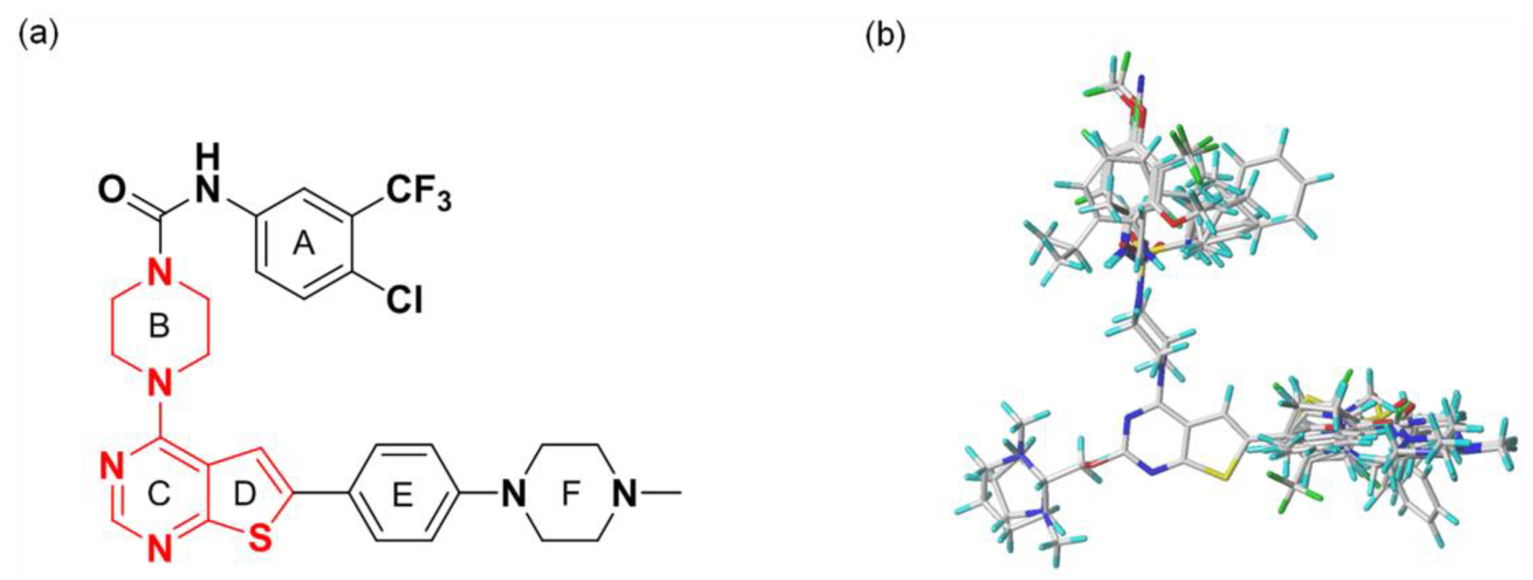
All compounds in the training and test sets were drawn in ChemDraw. All compounds were superimposed and aligned on the maximum common substructure of thieno-pyrimidine derivatives by using DISTIL RIGID alignment function in SYBYL-X 2.1.1 [35]. Based on the hypothesis that the common alignment core contributes equally to the bioactivities of the compounds, the conformational angles for maximum common substructures of the most active compound 42 as the template for the alignment were copied and applied to the remaining compounds in the whole dataset. Figure 6 shows the most active compound 42 as the template, and the structural alignment of the training and test sets for the 3D-QSAR study.

3.3. CoMFA Studies

The CoMFA was performed by the Lennard–Jones potential for the steric field and Coulombic potential for the electrostatic field in SYBYL-X 2.1.1. The aligned compounds were placed in a 3D cubic lattice of 2.0 Å grid spacing in the x, y, and z directions and these potentials were calculated for each compound. The sp3 carbon probe with the van der Waals radius of 1.52 Å and the point charge of +1.0 was used at each lattice point of the grid box for the calculation of the steric and electrostatic fields. To avoid overpower by large steric and electrostatic energy values, the default energy cutoff value was set to 30 kcal/mol. A default value of 0.3 was set as the attenuation coefficient. The regression analysis was performed using the cross-validated partial least square (PLS) method. To develop the final model by performing a PLS analysis, the first run was conducted with the cross-validation to find ONC, and then the final run yielded the non-cross-validated r2 value.

3.4. CoMSIA Studies

In this study, the CoMSIA model was developed with the steric, electrostatic, hydrophobic, hydrogen bond donor, and hydrogen bond acceptor fields in SYBYL-X 2.1.1. As the five descriptors, the default settings of the probe atom with 1.0 Å radius, +1.0 charge, +1.0 hydrophobicity, +1.0 hydrogen bond donor, and +1.0 hydrogen bond acceptor properties were used in the same grid box for the CoMFA calculation. The default attenuation coefficient was set to 0.3. In CoMSIA, the Gaussian function was used for the distance dependence between the probe atom and the molecule atoms to measure the relative attenuation of the field position for each atom in the lattice. The similarity indices were calculated at all grid points through the different shape of the Gaussian function. The use of the Gaussian function resulted in smoother sampling of the fields around the molecules compared to CoMFA.

3.5. Partial Least Squares Analysis

The partial least squares analysis (PLS) regression method was performed to analyze the linear correlation of CoMFA and CoMSIA as the independent variables to the biological activities as the dependent variables. The sample-distance partial least square (SAMPLS) method in SYBYL-X2.1.1 was used to speed up the cross-validation analysis. Cross-validation analysis was carried out by using the leave-one-out (LOO) method, which removes one compound from the training set and predicts its bioactivity from the constructed model using the rest compounds. Until all compounds have been removed once, this process is repeated to obtain the cross-validated correlation coefficient q2, the optimum number of components (ONC). To speed up the analysis and reduce noise, a minimum column filtering value was set to 2.0 Kcal/mol. Using the determined ONC, the final PLS model was constructed without cross-validation. The predictive ability of the developed 3D-QSAR models was confirmed by external validation using the test set compounds, which were not included in the training set. For the test set compounds, the optimization, alignment, and all other steps were the same as those of the training set compounds, and then their biological activities were predicted using the final model developed by the training set compounds. The predictive correlation coefficient r2pred value is obtained by this formula:
r2pred = (SD − PRESS)/(SD)
In the formula, SD represents the sum of the squared deviations between the inhibitory activities in the test set and the mean inhibitory activity in the training set, and PRESS represents the sum of the squared deviations between the actual and predicted inhibitory activities of the test set compounds.

3.6. Progressive Scrambling Stability Test

To further validate the robustness and stability of the developed model, the progressive scrambling stability test was carried out by the Scrambling Stability Test function in SYBYL-X 2.1.1 [32]. The sensitivity of the model to small systematic perturbations was determined by this test. The inhibitory activities of the training set compounds were randomly scrambled many times and the q2 values were generated using LOO cross-validation after every iteration. The unstable 3D-QSAR model is characterized by a greater slope (dq2′/dr2yy′) value than 1.2.

4. Conclusions

In the present study, 3D-QSAR analyses were performed to identify the key structural features for improving the biological activities of the thieno-pyrimidine derivatives as breast cancer inhibitors. The CoMFA (q2 = 0.818, r2 = 0.917) and CoMSIA (q2 = 0.801, r2 = 0.897) models, satisfying the criteria provided by Golbraikh and Tropsha, were selected. Statistical reliability and robustness of the selected CoMFA and CoMSIA models were confirmed by external validation with the test set compounds which did not exist in the training set. The established models were further validated by the progressive scrambling stability test and presented to be statistically stable and robust. The contour maps of CoMFA and CoMSIA provided key information to understand the SAR and to identify the structural requirement for enhancing the inhibitory bioactivity of compounds. Based on the information derived from the contour maps, the summarized design strategy of the principal SAR for the design of the new compounds, with potential therapeutic applications, is shown in Figure 7. These obtained results might be helpful in the design of novel inhibitors with improved inhibitory biological activity against metastatic breast cancer.

Supplementary Materials

The following supporting information can be downloaded at: https://www.mdpi.com/article/10.3390/molecules27227974/s1, Table S1: Statistical results of all possible field combinations for the CoMFA and CoMSIA models. Table S2. The SMILES codes for the most active compound 42 and the novel designed inhibitors N1N14.

Author Contributions

Conceptualization, J.-H.K.; methodology, J.-H.K.; software, J.-H.K.; formal analysis, J.-H.K.; investigation, J.-H.K.; data curation, J.-H.K.; visualization, J.-H.K.; supervision, J.-H.J.; funding acquisition, J.-H.J.; writing—original draft preparation, J.-H.K.; writing—review and editing, J.-H.K. and J.-H.J. All authors have read and agreed to the published version of the manuscript.

Funding

This research was funded by the National Research Foundation of Korea (grant number NRF-2018R1A6A1A03023718).

Institutional Review Board Statement

Not applicable.

Informed Consent Statement

Not applicable.

Data Availability Statement

Not applicable.

Acknowledgments

We are grateful for the financial support from the National Research Foundation of Korea (NRF-2018R1A6A1A03023718).

Conflicts of Interest

The authors declare no conflict of interest.

Sample Availability

Samples of the compounds are not available from the authors.

References

  1. Suleman, M.; Tahir Ul Qamar, M.; Saleem, S.; Ahmad, S.; Ali, S.S.; Khan, H.; Akbar, F.; Khan, W.; Alblihy, A.; Alrumaihi, F.; et al. Mutational Landscape of Pirin and Elucidation of the Impact of Most Detrimental Missense Variants that Accelerate the Breast Cancer Pathways: A Computational Modelling Study. Front. Mol. Biosci. 2021, 8, 692835. [Google Scholar] [CrossRef] [PubMed]
  2. Ghoncheh, M.; Pournamdar, Z.; Salehiniya, H. Incidence and Mortality and Epidemiology of Breast Cancer in the World. Asian Pac. J. Cancer Prev. 2016, 17, 43–46. [Google Scholar] [CrossRef] [PubMed] [Green Version]
  3. World Health Organization. Available online: http://www.who.int/news-room/fact-sheets/detail/breast-cancer (accessed on 26 March 2021).
  4. Medina, M.A.; Oza, G.; Sharma, A.; Arriaga, L.G.; Hernández, J.M.H.; Rotello, V.M.; Ramirez, J.T. Triple-Negative Breast Cancer: A Review of Conventional and Advanced Therapeutic Strategies. Int. J. Environ. Res. Public Health 2020, 17, 2078. [Google Scholar] [CrossRef] [PubMed] [Green Version]
  5. Ensenyat-Mendez, M.; Llinàs-Arias, P.; Orozco, J.I.J.; Iñiguez-Muñoz, S.; Salomon, M.P.; Sesé, B.; DiNome, M.L.; Marzese, D.M. Current Triple-Negative Breast Cancer Subtypes: Dissecting the Most Aggressive Form of Breast Cancer. Front. Oncol. 2021, 11, 681476. [Google Scholar] [CrossRef]
  6. Zhao, S.; Zuo, W.-J.; Shao, Z.-M.; Jiang, Y.-Z. Molecular subtypes and precision treatment of triple-negative breast cancer. Ann. Transl. Med. 2020, 8, 499. [Google Scholar] [CrossRef]
  7. Gupta, G.K.; Collier, A.L.; Lee, D.; Hoefer, R.A.; Zheleva, V.; Siewertsz van Reesema, L.L.; Tang-Tan, A.M.; Guye, M.L.; Chang, D.Z.; Winston, J.S.; et al. Perspectives on Triple-Negative Breast Cancer: Current Treatment Strategies, Unmet Needs, and Potential Targets for Future Therapies. Cancers 2020, 12, 2392. [Google Scholar] [CrossRef]
  8. Yin, L.; Duan, J.-J.; Bian, X.-W.; Yu, S.-C. Triple-negative breast cancer molecular subtyping and treatment progress. Breast Cancer Res. 2020, 22, 61. [Google Scholar] [CrossRef]
  9. Bianchini, G.; Balko, J.M.; Mayer, I.A.; Sanders, M.E.; Gianni, L. Triple-negative breast cancer: Challenges and opportunities of a heterogeneous disease. Nat. Rev. Clin. Oncol. 2016, 13, 674–690. [Google Scholar] [CrossRef]
  10. The American Cancer Society. Available online: http://www.cancer.org/cancer/breast-cancer/about/types-of-breast-cancer/triple-negative.html (accessed on 1 March 2022).
  11. Baker, E.; Whiteoak, N.; Hall, L.; France, J.; Wilson, D.; Bhaskar, P. Mammaglobin-A, VEGFR3, and Ki67 in Human Breast Cancer Pathology and Five Year Survival. Breast Cancer Basic Clin. Res. 2019, 13, 1–8. [Google Scholar] [CrossRef]
  12. Shibata, M.-A.; Shibata, E.; Tanaka, Y.; Shiraoka, C.; Kondo, Y. Soluble Vegfr3 gene therapy suppresses multi-organ metastasis in a mouse mammary cancer model. Cancer Sci. 2020, 111, 2837–2849. [Google Scholar] [CrossRef]
  13. Makinen, T.; Jussila, L.; Veikkola, T.; Karpanen, T.; Kettunen, M.I.; Pulkkanen, K.J.; Kauppinen, R.; Jackson, D.G.; Kubo, H.; Nishikawa, S.-I.; et al. Inhibition of lymphangiogenesis with resulting lymphedema in transgenic mice expressing soluble VEGF receptor-3. Nat. Med. 2001, 7, 199–205. [Google Scholar] [CrossRef] [PubMed]
  14. Mandriota, S.J.; Jussila, L.; Jeltsch, M.; Compagni, A.; Baetens, D.; Prevo, R.; Banerji, S.; Huarte, J.; Montesano, R.; Jackson, D.G.; et al. Vascular endothelial growth factor-C-mediated lymphangiogenesis promotes tumour metastasis. EMBO J. 2001, 20, 672–682. [Google Scholar] [CrossRef] [PubMed] [Green Version]
  15. Harris, A.R.; Perez, M.J.; Munson, J.M. Docetaxel facilitates lymphatic-tumor crosstalk to promote lymphangiogenesis and cancer progression. BMC Cancer 2018, 18, 718. [Google Scholar] [CrossRef] [PubMed] [Green Version]
  16. Li, Y.; Yang, G.; Zhang, J.; Tang, P.; Yang, C.; Wang, G.; Chen, J.; Liu, J.; Zhang, L.; Ouyang, L. Discovery, Synthesis, and Evaluation of Highly Selective Vascular Endothelial Growth Factor Receptor 3 (VEGFR3) Inhibitor for the Potential Treatment of Metastatic Triple-Negative Breast Cancer. J. Med. Chem. 2021, 64, 12022–12048. [Google Scholar] [CrossRef]
  17. Roskoski, R., Jr. Vascular endothelial growth factor (VEGF) and VEGF receptor inhibitors in the treatment of renal cell carcinomas. Pharmacol. Res. 2017, 120, 116–132. [Google Scholar] [CrossRef]
  18. Escudier, B.; Powles, T.; Motzer, R.J.; Olencki, T.; Arén Frontera, O.; Oudard, S.; Rolland, F.; Tomczak, P.; Castellano, D.; Appleman, L.J.; et al. Cabozantinib, a New Standard of Care for Patients with Advanced Renal Cell Carcinoma and Bone Metastases? Subgroup Analysis of the METEOR Trial. J. Clin. Oncol. 2018, 36, 765–772. [Google Scholar] [CrossRef]
  19. Bruix, J.; Cheng, A.-L.; Meinhardt, G.; Nakajima, K.; De Sanctis, Y.; Llovet, J. Prognostic Factors and Predictors of Sorafenib Benefit in Patients with Hepatocellular Carcinoma: Analysis of Two Phase III Studies. J. Hepatol. 2017, 67, 999–1008. [Google Scholar] [CrossRef] [Green Version]
  20. Gounder, M.M.; Mahoney, M.R.; Van Tine, B.A.; Ravi, V.; Attia, S.; Deshpande, H.A.; Gupta, A.A.; Milhem, M.M.; Conry, R.M.; Movva, S.; et al. Sorafenib for Advanced and Refractory Desmoid Tumors. N. Engl. J. Med. 2018, 379, 2417–2428. [Google Scholar] [CrossRef]
  21. Kodera, Y.; Katanasaka, Y.; Kitamura, Y.; Tsuda, H.; Nishio, K.; Tamura, T.; Koizumi, F. Sunitinib Inhibits Lymphatic Endothelial Cell Functions and Lymph Node Metastasis in a Breast Cancer Model through Inhibition of Vascular Endothelial Growth Factor Receptor 3. Breast Cancer Res. 2011, 13, R66. [Google Scholar] [CrossRef] [Green Version]
  22. Matsui, J.; Funahashi, Y.; Uenaka, T.; Watanabe, T.; Tsuruoka, A.; Asada, M. Multi-Kinase Inhibitor E7080 Suppresses Lymph Node and Lung Metastases of Human Mammary Breast Tumor MDA-MB-231 via Inhibition of Vascular Endothelial Growth Factor-Receptor (VEGF-R) 2 and VEGF-R3 Kinase. Clin. Cancer. Res. 2008, 14, 5459–5465. [Google Scholar] [CrossRef]
  23. Varney, M.L.; Singh, R.K. VEGF-C-VEGFR3/Flt4 axis regulates mammary tumor growth and metastasis in an autocrine manner. Am. J. Cancer Res. 2015, 5, 616–628. [Google Scholar] [PubMed]
  24. Alam, A.; Blanc, I.; Gueguen-Dorbes, G.; Duclos, O.; Bonnin, J.; Barron, P.; Laplace, M.-C.; Morin, G.; Gaujarengues, F.; Dol, F.; et al. SAR131675, a Potent and Selective VEGFR-3–TK Inhibitor with Antilymphangiogenic, Antitumoral, and Antimetastatic Activities. Mol. Cancer Ther. 2012, 11, 1637–1649. [Google Scholar] [CrossRef] [PubMed] [Green Version]
  25. Lorca, M.; Morales-Verdejo, C.; Vásquez-Velásquez, D.; Andrades-Lagos, J.; Campanini-Salinas, J.; Soto-Delgado, J.; Recabarren-Gajardo, G.; Mella, J. Structure-Activity Relationships Based on 3D-QSAR CoMFA/CoMSIA and Design of Aryloxypropanol-Amine Agonists with Selectivity for the Human β3-Adrenergic Receptor and Anti-Obesity and Anti-Diabetic Profiles. Molecules 2018, 23, 1191. [Google Scholar] [CrossRef] [PubMed] [Green Version]
  26. Srivastava, V.; Gupta, S.P.; Siddiqi, M.I.; Mishra, B.N. 3D-QSAR studies on quinazoline antifolate thymidylate synthase inhibitors by CoMFA and CoMSIA models. Eur. J. Med. Chem. 2010, 45, 1560–1571. [Google Scholar] [CrossRef]
  27. Cramer, R.D.; Patterson, D.E.; Bunce, J.D. Comparative Molecular Field Analysis (CoMFA). 1. Effect of Shape on Binding of Steroids to Carrier Proteins. J. Am. Chem. Soc. 1988, 110, 5959–5967. [Google Scholar] [CrossRef]
  28. Klebe, G.; Abraham, U.; Mietzner, T. Molecular Similarity Indices in a Comparative Analysis (CoMSIA) of Drug Molecules to Correlate and Predict Their Biological Activity. J. Med. Chem. 1994, 37, 4130–4146. [Google Scholar] [CrossRef]
  29. Luco, J.M.; Ferretti, F.H. QSAR Based on Multiple Linear Regression and PLS Methods for the Anti-HIV Activity of a Large Group of HEPT Derivatives. J. Chem. Inf. Comput. Sci. 1997, 37, 392–401. [Google Scholar] [CrossRef]
  30. Golbraikh, A.; Shen, M.; Xiao, Z.; Xiao, Y.-D.; Lee, K.-H.; Tropsha, A. Rational Selection of Training and Test Sets for the Development of Validated QSAR Models. J. Comput. Aided Mol. Des. 2003, 17, 241–253. [Google Scholar] [CrossRef]
  31. Golbraikh, A.; Tropsha, A. Beware of q2! J. Mol. Graph. Model. 2002, 20, 269–276. [Google Scholar] [CrossRef]
  32. Clark, R.D.; Fox, P.C. Statistical Variation in Progressive Scrambling. J. Comput. Aided Mol. Des. 2004, 18, 563–576. [Google Scholar] [CrossRef]
  33. Daina, A.; Michielin, O.; Zoete, V. SwissADME: A free web tool to evaluate pharmacokinetics, drug-likeness and medicinal chemistry friendliness of small molecules. Sci. Rep. 2017, 7, 42717. [Google Scholar] [CrossRef] [PubMed] [Green Version]
  34. Pires, D.E.V.; Blundell, T.L.; Ascher, D.B. pkCSM: Predicting Small-Molecule Pharmacokinetic and Toxicity Properties Using Graph-Based Signatures. J. Med. Chem. 2015, 58, 4066–4072. [Google Scholar] [CrossRef] [PubMed]
  35. Patel, S.; Patel, B.; Bhatt, H. 3D-QSAR studies on 5-hydroxy-6-oxo-1,6-dihydropyrimidine-4-carboxamide derivatives as HIV-1 integrase inhibitors. J. Taiwan Inst. Chem. Eng. 2016, 59, 61–68. [Google Scholar] [CrossRef]
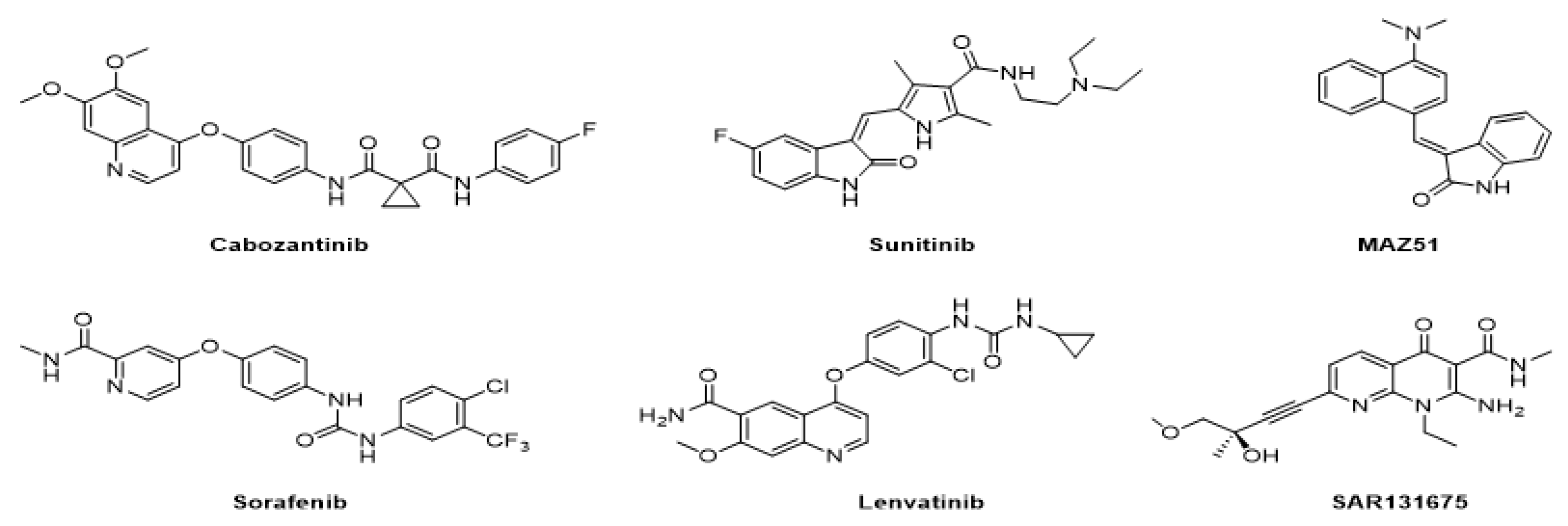
Figure 1. Inhibitors targeting VEGFRs.
Molecules 27 07974 g001
Figure 2. Scatter plots of the actual versus predicted inhibitory activities for the CoMFA model; (a) The training set; (b) The test set.
Molecules 27 07974 g002
Figure 3. Contour maps of CoMFA analysis with the most active compound 42 (left) and the least compound 20 (right). In each field, favored and disfavored areas are fixed with 80% and 20% contribution levels, respectively; (a,b) Steric field: favored regions are in green contours and disfavored regions are in yellow contours; (c,d) Electrostatic field: favored regions are in blue contours and disfavored regions are in red contours.
Molecules 27 07974 g003
Figure 4. Scatter plots of the actual versus predicted inhibitory activities for the CoMSIA model; (a) The training set; (b) The test set.
Molecules 27 07974 g004
Figure 5. Contour maps of CoMSIA with the most active compound 42 (left) and the least active compound 20 (right). In each field, favored and disfavored areas are fixed with 80% and 20% contribution levels, respectively; (a,b) Steric field: favored regions are in green contours and disfavored regions are in yellow contours; (c,d) Electrostatic field: favored regions are in blue contours and disfavored regions are in red contours; (e,f) Hydrophobic field: favored regions are in orange contours and disfavored regions are in yellow contours; (g,h) Hydrogen bond donor field: favored regions are in cyan contours and disfavored regions are in purple contours; (i,j) Hydrogen bond acceptor field: favored regions are in magenta contours and disfavored regions are in red contours.
Molecules 27 07974 g005
Figure 6. (a) The structure of the most active compound 42 as the template including the maximum common substructure in red; (b) The alignment of the training and test sets used in the 3D-QSAR model.
Molecules 27 07974 g006
Figure 7. The novel design strategy information obtained from the 3D-QSAR study.
Molecules 27 07974 g007
Table 1. Statistical results of the selected CoMFA and CoMSIA models.
Modelq2r2SEEFr2predONCField Contribution (%)
SEHDA
CoMFA_SE0.8180.9178.142114.2350.794367.732.3---
CoMSIA_SEHDA0.8010.8979.05790.3400.762329.529.829.86.54.4
Leave-one-out cross-validated correlation coefficient (q2), determination coefficient (r2), standard error of estimate (SEE), Fisher test value (F), predictive correlation coefficient (r2pred), optimum number of components (ONC), Steric (S), Electrostatic (E), Hydrophobic (H), Hydrogen bond donor (D), Hydrogen bond acceptor (A).
Table 2. Summary of progressive scrambling stability test for the CoMFA model.
No. of Componentsq2cSDEPdq2′/dr2yy′
10.62516.7630.881
20.67815.7101.058
30.69115.6401.102
40.65716.7221.260
50.65217.0731.266
The predictivity of the model (q2 = 1 − (cSDEP)2), the calculated cross-validated standard error (cSDEP), and the slope of q2 with respect to the correlation of the perturbed dependent variables against the unperturbed data (dq2′/dr2yy′).
Table 3. Summary of progressive scrambling stability test for the CoMSIA model.
No. of Componentsq2cSDEPdq2′/dr2yy′
10.42820.7080.709
20.64416.5800.819
30.67416.1061.035
40.63517.2421.162
50.59318.4411.165
The predictivity of the model (q2 = 1 − (cSDEP)2), the calculated cross-validated standard error (cSDEP), the slope of q2 with respect to the correlation of the perturbed dependent variables against the unperturbed data (dq2′/dr2yy′).
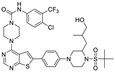
Table 4. The structures and the predicted inhibitory activities for the most active compound 42 and the newly designed inhibitors N1N14.
NoStructurePredicted
Inhibitory Activity (%)
42Molecules 27 07974 i00185.61
N1Molecules 27 07974 i00287.11
N2Molecules 27 07974 i00388.01
N3Molecules 27 07974 i00489.16
N4Molecules 27 07974 i00588.19
N5Molecules 27 07974 i00691.18
N6Molecules 27 07974 i00790.94
N7Molecules 27 07974 i00891.23
N8Molecules 27 07974 i00990.10
N9Molecules 27 07974 i01090.77
N10Molecules 27 07974 i01190.87
N11Molecules 27 07974 i01290.45
N12Molecules 27 07974 i01389.13
N13Molecules 27 07974 i01490.44
N14Molecules 27 07974 i01590.55
Table 5. The prediction results of ADMET parameters, pharmacokinetic properties, and drug-likeness of the most active compound 42 and the novel designed inhibitors N1N14.
NoLipophilicity
Log Po/w
(MLOGP)
Water
Solubility
Log S
(ESOL)
Pharmacokinetics Drug-Likeness
Lipinski Rule
Synthetic
Accessibility
GI
Absorption
BBB
Permeant
Toxicity
(AMES)
Categorical
(Yes/No)
424.05−7.21HighNoNoYes;
1 violation
4.25
N13.63−8.40LowNoNoYes;
1 violation
5.01
N22.97−7.88LowNoNoNo;
2 violation
5.70
N33.56−7.73HighNoNoNo;
2 violation
5.45
N44.34−8.01LowNoNoNo;
2 violation
4.66
N55.04−9.31LowNoYesNo;
2 violation
5.57
N63.92−8.08LowNoNoYes;
1 violation
5.63
N74.27−8.57LowNoNoNo;
2 violation
5.86
N83.90−8.19LowNoNoYes;
1 violation
5.37
N94.26−8.89LowNoNoNo;
2 violation
5.60
N104.43−9.13LowNoNoNo;
2 violation
5.73
N114.78−9.73LowNoNoNo;
2 violation
5.99
N123.14−7.55LowNoNoNo;
2 violation
5.43
N133.32−7.79LowNoNoNo;
2 violation
5.52
N143.67−8.39LowNoNoNo;
2 violation
6.03
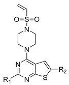
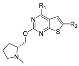
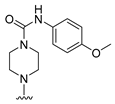
Table 6. Chemical structures and inhibitory activity values of thieno-pyrimidine derivatives.
Molecules 27 07974 i016Molecules 27 07974 i017Molecules 27 07974 i018
1–2122–26, 45–4627–44, 47
NoR1R2Actual
Inhibitory Activity
(1µM, %)
CoMFA
_SE
CoMSIA
_SEHDA
CoMFA ResidualCoMSIA
Residual
1 *HMolecules 27 07974 i01954.2344.4546.499.787.74
2HMolecules 27 07974 i02032.1047.9142.69−15.81−10.59
3HMolecules 27 07974 i02148.6853.1552.22−4.47−3.54
4HMolecules 27 07974 i02275.3650.8747.4824.5027.88
5HMolecules 27 07974 i02356.1667.3270.79−11.16−14.63
6HMolecules 27 07974 i02478.8772.8871.035.997.84
7HMolecules 27 07974 i02562.7061.5460.581.162.12
8HMolecules 27 07974 i02640.1139.9542.030.16−1.92
9HMolecules 27 07974 i02743.0738.2040.044.873.03
10 *HMolecules 27 07974 i02835.9149.6842.80−13.77−6.89
11HMolecules 27 07974 i02938.2842.5347.64−4.25−9.36
12HMolecules 27 07974 i03065.7151.8249.2313.8916.48
13HMolecules 27 07974 i03136.6045.0743.66−8.47−7.06
14 *HMolecules 27 07974 i03245.1843.3646.311.82−1.13
15 *HMolecules 27 07974 i03351.0239.5240.8111.5010.21
16 *HMolecules 27 07974 i03448.2852.3145.34−4.032.94
17 *HMolecules 27 07974 i03539.5938.5342.561.06−2.97
18HMolecules 27 07974 i03630.1841.6142.33−11.43−12.15
19 *HMolecules 27 07974 i03715.9945.4645.31−29.47−29.32
20Molecules 27 07974 i038Molecules 27 07974 i03910.016.725.823.294.19
21Molecules 27 07974 i040Molecules 27 07974 i04110.618.378.872.241.74
22 *Molecules 27 07974 i042Molecules 27 07974 i04322.8017.1018.835.703.97
23Molecules 27 07974 i044Molecules 27 07974 i04511.2016.1118.09−4.91−6.89
24Molecules 27 07974 i046Molecules 27 07974 i04719.227.747.5411.4811.68
25Molecules 27 07974 i048Molecules 27 07974 i04932.3428.4029.333.943.01
26Molecules 27 07974 i050Molecules 27 07974 i05118.3327.3228.64−8.99−10.31
27Molecules 27 07974 i052H66.1571.5370.31−5.38−4.16
28Molecules 27 07974 i053H82.9780.2178.642.764.33
29Molecules 27 07974 i054H79.2173.3071.675.917.54
30 *Molecules 27 07974 i055H72.3974.9473.26−2.55−0.87
31 *Molecules 27 07974 i056H75.3673.0771.732.293.63
32Molecules 27 07974 i057H76.4476.1676.190.280.25
33Molecules 27 07974 i058H80.4278.1278.412.302.01
34 *Molecules 27 07974 i059H73.5379.3778.08−5.84−4.55
35Molecules 27 07974 i060H89.2686.3482.412.926.86
36Molecules 27 07974 i061H85.4281.2280.994.204.43
37Molecules 27 07974 i062H87.1981.9481.345.255.85
38Molecules 27 07974 i063H74.7876.8477.65−2.05−2.87
39Molecules 27 07974 i064H71.6077.4278.20−5.82−6.60
40Molecules 27 07974 i065H70.5373.9175.48−3.38−4.95
41Molecules 27 07974 i066H73.7879.0779.41−5.29−5.63
42Molecules 27 07974 i067H93.4785.8485.617.637.86
43Molecules 27 07974 i068H75.0978.3280.09−3.23−4.99
44Molecules 27 07974 i069H77.8282.1283.61−4.30−5.79
45Molecules 27 07974 i070Molecules 27 07974 i07112.8017.5119.46−4.71−6.66
46Molecules 27 07974 i072Molecules 27 07974 i07320.0819.2019.080.881.00
47 *Molecules 27 07974 i074Molecules 27 07974 i07536.0931.6934.754.401.34
* Compounds in the test set.
Publisher’s Note: MDPI stays neutral with regard to jurisdictional claims in published maps and institutional affiliations.