Article

Nano-WSe2 Is Absorbable and Transformable by Rice Plants †

1
Department of Chemistry, College of Sciences, Northeastern University, Shenyang 110819, China
2
CAS Key Laboratory for Biomedical Effects of Nanomaterials and Nanosafety, CAS-HKU Joint Laboratory of Metallomics on Health and Environment, Beijing Metallomics Facility, National Consortium for Excellence in Metallomics, Institute of High Energy Physics, Chinese Academy of Sciences, Beijing 100049, China
3
University of Chinese Academy of Sciences, Beijing 100049, China
*
Authors to whom correspondence should be addressed.
Dedicated to Prof. Zhifang Chai on the occasion of his 80th birthday.
Molecules 2022, 27(22), 7826; https://doi.org/10.3390/molecules27227826
Submission received: 24 October 2022 / Revised: 10 November 2022 / Accepted: 11 November 2022 / Published: 13 November 2022
(This article belongs to the Special Issue Actinoids in Biologic Systems and Catalysis)

Abstract

:
As typical transition metal dichalcogenides (TMDC), tungsten selenide (WSe2) nanosheets (nano-WSe2) are widely used in various fields due to their layered structures and highly tunable electronic and magnetic properties, which results in the unwanted release of tungsten (W) and selenium (Se) into the environment. However, the environmental effects of nano-WSe2 in plants are still unclear. Herein, we evaluated the impacts and fate of nano-WSe2 and micro-WSe2 in rice plants (Oryza sativa L.). It was found that both nano-WSe2 and micro-WSe2 did not affect the germination of rice seeds up to 5000 mg/L but nano-WSe2 affected the growth of rice seedlings with shortened root lengths. The uptake and transportation of WSe2 was found to be size-dependent. Moreover, W in WSe2 was oxidized to tungstate while Se was transformed to selenocysteine, selenomethionine, SeIV and SeVI in the roots of rice when exposed to nano-WSe2, suggesting the transformation of nano-WSe2 in rice plants. The exposure to nano-WSe2 brought lipid peroxidative damage to rice seedlings. However, Se in nano-WSe2 did not contribute to the synthesis of glutathione peroxidase (GSH-Px) since the latter did not change when exposed to nano-WSe2. This is the first report on the impacts and fate of nano-WSe2 in rice plants, which has raised environmental safety concerns about the wide application of TMDCs, such as WSe2 nanosheets.

1. Introduction

Transition metal dichalcogenides (TMDCs) are a family of 2D nanosheets typically in the form of MX2, where M is the transition metal and X is the chalcogen. In recent years, TMDCs have attracted wide attention due to their layered structures and highly tunable electronic and magnetic properties [1]. TMDCs have been applied in the fields of energy [2], environment [3], biomedicine [4] and agriculture [5] due to their excellent physical, chemical, electrical and biological properties [6]. For example, molybdenum sulfide (MoS2) nanosheets have been applied in biomedicine, biosensing and bioimaging [7]. It was noticed that Mo in the MoS2 nanosheet could be used for the synthesis of molybdenum enzymes in animals [8], suggesting the bioavailability of MoS2 in animals. It was also found that MoS2 promoted rice growth by regulating chlorophyll synthesis and aquaporin gene expression [9].
Tungsten selenide (WSe2) nanosheet (nano-WSe2) is another typical TMDC, which has excellent mechanical, optical, electrical and magnetic properties [10]. It is used in photovoltaic devices and ultra-thin LEDs and is an additive in lubricants due to its flake structure with small friction coefficient [11]. The reconfiguration or doping on WSe2 with other atoms [12] enhances its applications in environmental engineering [13] and chemical engineering [14]. The wide application of nano-WSe2 increases the possibility of its release into the environment. The environmental toxicity of nano-WSe2 is still largely unknown. To date, only one study on the cytotoxicity of WSe2 has been reported, which found at least 50% cell death at a dose of 400 μg/mL [15].
Tungsten (W) is an essential element for archaea [16], such as Pyrococcus furiosus [17], and is linked to organic cofactors, such as tungsten cofactors [18]. It can replace Mo in the molybdenum pterin cofactor to reduce the severity of intestinal inflammation in mice with colitis [19]. However, W is also an environmental pollutant and has been associated with leukemia [20]. The increasing release of W to water and soil has attracted more and more attention since W was identified as an emerging environmental pollutant by the U.S. Environmental Protection Agency [21]. Selenium (Se) is an essential element for animals and human beings but is toxic to plants [22]. The indiscriminate incorporation of seleno-amino acids into proteins can impair protein functions and ultimately lead to physiological malfunction [23]. Se is involved in many enzymatic activities and the regulation of the physiological metabolism. For example, Se can reduce stress injury by down-regulating galactose metabolism and activating the metabolism of glutamate and glutamine [24] and enhances the production of heat shock proteins (HSPs) and glutathionase activity to alleviate heat stress-induced oxidative damage in vivo [25].
Plants are an important part of the ecosystem and play important roles in ecological balance [5]. Rice is one of the staple foods for human beings, which has also been applied as a model plant for ecotoxicological study [26]. The absorption and accumulation of toxic elements by crops will affect the food safety and even bring harm to human health [27]. Considering the low solubility of WSe2, it is generally believed that it is not bioavailable to plants; however, it is not clear whether WSe2 at nano size is bioavailable and transformable or not. Understanding the bioavailability and phytotoxicity of nano-WSe2 on crops is important for food safety and agricultural sustainability but such information is currently lacking.
To address these knowledge gaps, the impact and fate of nano-WSe2 in rice plants (Oryza sativa L.) was studied using WSe2 powder (micro-WSe2) for comparison in this study. The impacts on germination rate and growth of rice seedlings by nano-WSe2 or micro-WSe2 were first studied. Then the biological responses after exposure to nano-WSe2 or micro-WSe2 were compared. The distribution and transformation of W and Se in different tissues of rice plants were then examined. To the best of our knowledge, this is the first report on the impact and fate of nano-WSe2 on rice plants, which may shed light on the safe application of TMDCs.

2. Results and Discussion

2.1. The Size of Nano-WSe2 and Micro-WSe2

The SEM images for nano-WSe2 and micro-WSe2 is shown in Figure 1A,B. The size of nano-WSe2 is around 150 nm and it is around 2.5 μm for micro-WSe2. The particle size of nano-WSe2 through DLS is between 200–300 nm, and the particle size of micro-WSe2 is 1–3 μm (Figure 1C,D). It can be seen that both nano-WSe2 and micro-WSe2 are in the form of flakes, stacked layer by layer, which is consistent with other studies [13].

2.2. The Impacts on the Germination and Growth of Rice Plants by Nano-WSe2 or Micro-WSe2

2.2.1. The Impacts on Germination Rate of Rice Seeds

The germination rate when exposed to different concentrations of nano-WSe2 or micro-WSe2 is shown in Table 1. It can be seen both nano-WSe2 and micro-WSe2 had no significant difference on the germination rate of rice seeds under the studied concentrations up to 5000 mg/L. On the other hand, compared with the control group, the germination rate of seeds exposed to different concentrations both nano-WSe2 and micro-WSe2 was lowered, although not significantly different, suggesting a potential inhibition on the germination of rice seeds.

2.2.2. The Impacts on the Growth of Rice Seedlings

The lengths of roots, stems, and leaves in different groups are shown in Table 2. At the same concentration, the root length in nano-WSe2 group was shorter than that in the micro-WSe2 group. With increased concentration, the root length of rice exposed to nano-WSe2 became even shorter, especially at the concentration of 1000 and 5000 mg/L, and the root growth inhibition rate reached 30.94% and 66.67%, respectively, suggesting higher toxicity to roots by nano-WSe2 than micro-WSe2. Roots are an important site for the absorption, synthesis, and secretion of physiologically active substances required by rice [28]. In the nano-WSe2 group, it was found that the growth of the root system was restricted, which affected the growth and development of rice. When considering the impacts on the lengths of stems and leaves of rice, it can be seen that both nano-WSe2 and micro-WSe2 exposure generally led to decreased lengths, although not significantly different.

2.3. The Concentration of Se and W in Rice Plants after Exposure to Nano-WSe2 or Micro-WSe2

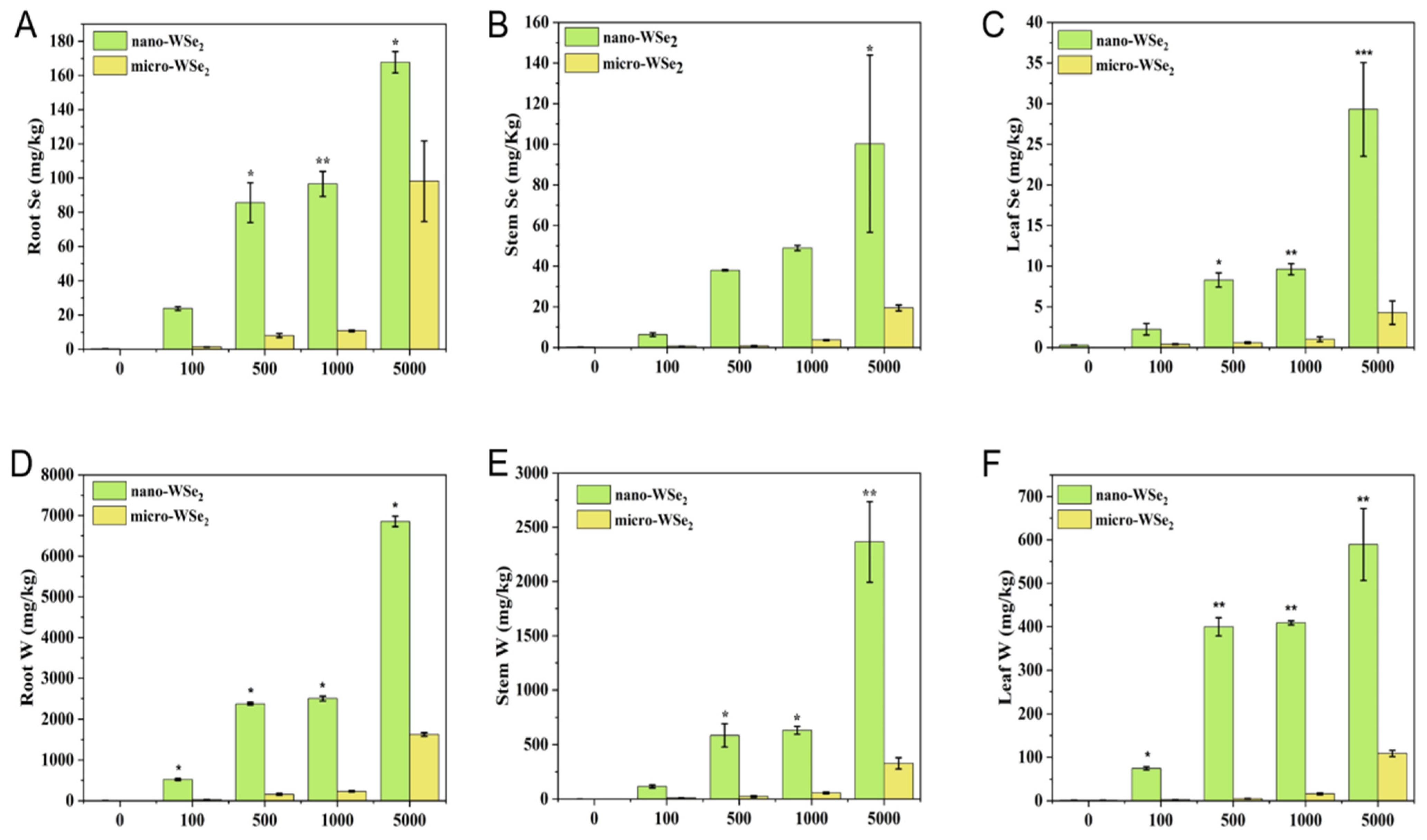
The concentration of Se and W in the roots, stems, and leaves of rice is shown in Figure 2. The content of Se and W in rice exposed to nano-WSe2 was significantly higher than that of micro-WSe2 at the same concentration, which indicated that nano-WSe2 was more easily absorbed by rice and transported to other parts of rice than micro-WSe2. On the other hand, the accumulation of Se and W was dose-dependent since higher concentration exposure led to higher Se and W levels in rice tissues. Studies have shown that the concentration of accumulated Se in most plants was lower than 100 mg/kg [29] and our results agree with this. Studies have found that W in plants growing in the W mine area can reach 1637 mg/kg or even 13,500 mg/kg [30], which is much higher than what we observed in this study. Moreover, the concentration of Se and W in rice was in the order of roots > stems > leaves, suggesting roots are the major accumulation sites in rice. The high accumulation of Se and W in roots may explain the shortened root length as previously mentioned (Table 2). The molar ratio of Se to W in different tissues of rice was calculated and is shown in Table 3. It can be seen that the molar ratio of Se to W in rice tissues was lower than the control and much less than 2 in all the tissues of rice due to the high accumulation of W in rice tissues.

2.4. The Levels of GSH-Px an MDA in Rice Roots, Stems, and Leaves after Exposure to by Nano-WSe2 or Micro-WSe2

In higher plants, Glutathione peroxidase (GSH-Px) is a sulfur-containing peroxidase that can scavenge H2O2, organic hydroperoxides, and lipid peroxides in the body and block ROS from causing further damage to the body [31]. Therefore, the GSH-Px activity can reflect the oxidative damage in plant. Moreover, Se is required for the synthesis of GSH-Px. Malondialdehyde (MDA) is the product of cell membrane lipid peroxidation, which can be used as an indicator for lipid peroxidation in plants [32].
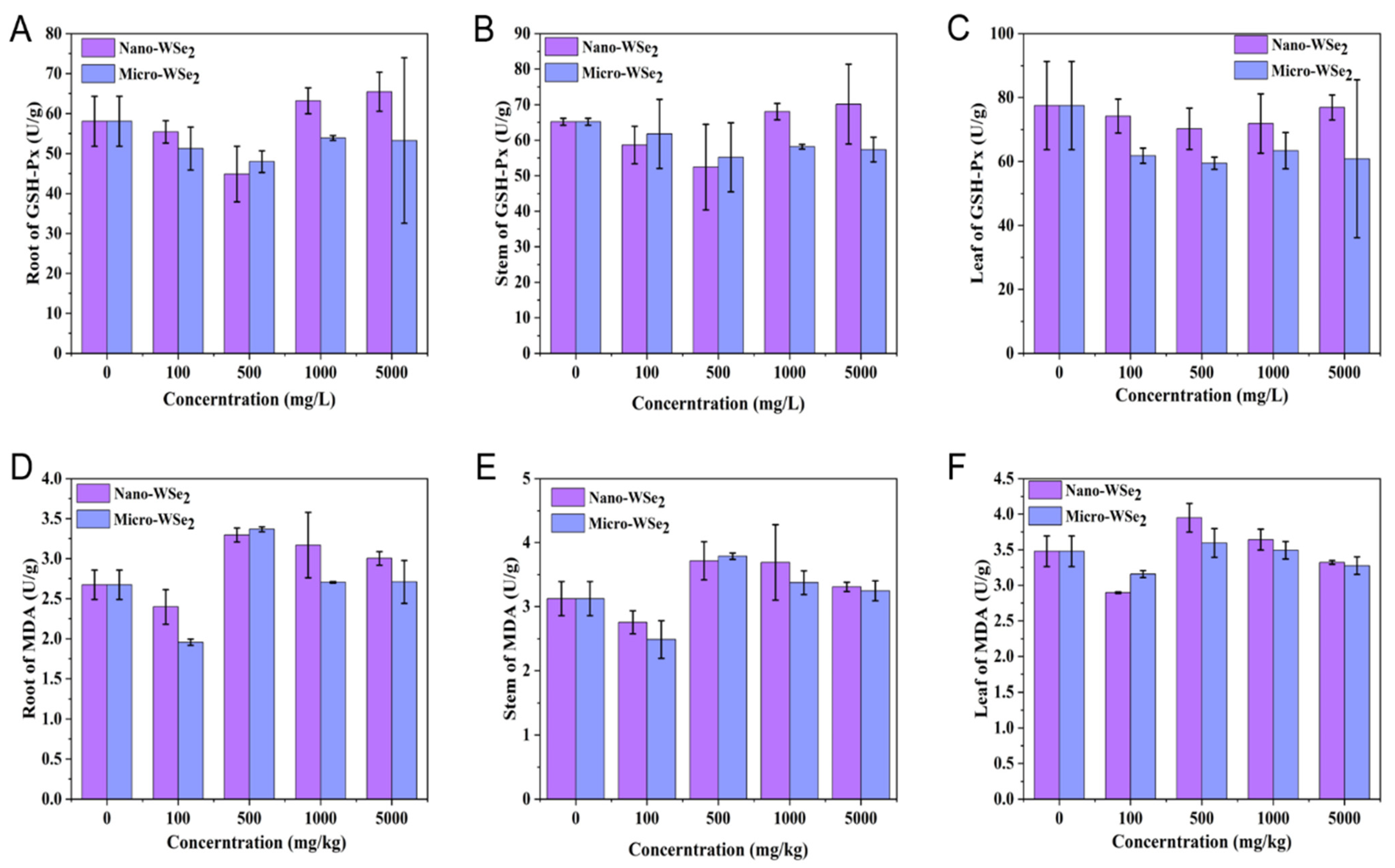
The levels of GSH-Px and MDA in roots, stems, and leaves are shown in Figure 3. From Figure 3A–C, no significant difference was found for GSH-Px levels in rice roots, stems, and leaves between the nano-WSe2 and micro-WSe2 groups. Many studies showed that Se can regulate the activity of antioxidant enzymes to a certain extent in higher plants such as wheat, rice, and cucumber [33]. It is known that Se is required for GSH-Px synthesis; however, no difference was found in this study when exposed to nano-WSe2 or micro-Wse2, suggesting that Se from nano-Wse2 or micro-Wse2 was not used for the formation of GSH-Px.
The levels of MDA in rice roots, stems, and leaves are shown in Figure 3D–F. It can be seen that the levels of MDA in rice cultivated by nano-WSe2 is higher than that in micro-WSe2, suggesting that nano-WSe2 brings more lipid peroxidation damage to rice than micro-WSe2. This agrees with the results that nano-WSe2 was more easily absorbed and transformed by rice than micro-WSe2.
Combined with the content of Se and W in rice and the values of GSH-Px and the level of MDA in rice, WSe2 was absorbed by rice and can be transported in rice, and there were peroxidative effects of WSe2 on rice. It was reported that the growth of plants was impaired when >3900 mg/kg in broccoli [21]. However, the concentration differences in hydroponic plants would be related to plant-specific tolerance mechanisms [34].

2.5. The Distribution and Transformation of Se and W in Rice Plants

2.5.1. The Distribution of Se and W in Rice Plants

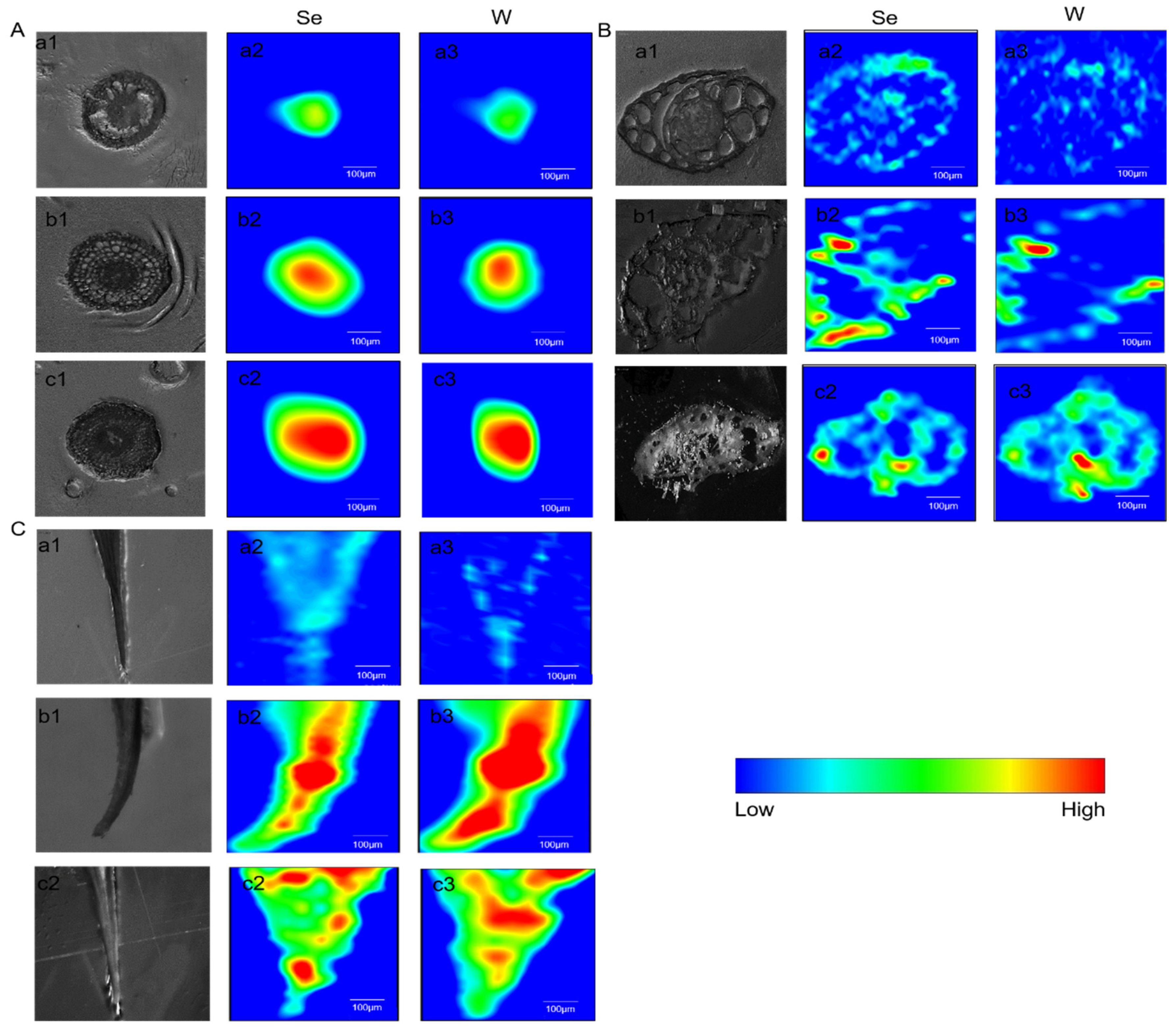
SRXRF is a multi-element analysis and in-situ elemental analysis technique with the advantages of high sensitivity and high spatial resolution [35]. The spatial distribution of Se and W in rice roots, stems, and leaves is shown in Figure 4. It can be seen that W and Se distributed mostly in the stele of rice roots, suggesting the absorption and transportation of W and Se from rhizosphere to the stele. The W and Se were transported upward along the vascular bundle and distributed in the stems and leaves, and also distributed in the veins of the rice leaves. Furthermore, W and Se were found coexisting in the tissues of rice. Combined with the element content, as found through ICP-MS, this confirmed that nano-WSe2 was more easily absorbed by rice than micro-WSe2.

2.5.2. The Transformation of Se and W in Rice Roots

The species of Se and W in rice roots are shown in Figure 5. From Figure 5A, higher levels of Se-Cys, Se-Met, and SeIV can be found in nano-WSe2 exposed rice roots, suggesting that nano-WSe2 is more easily transformed than micro-WSe2.
The oxidation state of tungsten in rice roots was studied by XAS. Figure 5B shows that the absorption edge of W in rice roots is right-shifted, suggesting that W has been oxidized to a higher valence than WSe2, most possibly to tungstate, which is a plant-available form [36]. This agrees with other studies that found W in rice was in the form of tungstate [37]. Studies have shown that the effects of tungstate on plants are bidirectional. For example, 1–50 mg/L tungstate in broccoli will not cause oxidative damage and is almost harmless, and it also has certain beneficial effects. At the dose of 100 mg/L, tungstate caused the reduction of germination rate, growth parameters, and some biochemical activities [36]. Moreover, studies have shown that low tungstate levels have a protective mechanism for the GSH-GSH-Px-GST system in plants and can maintain the amount of toxic reactive oxygen species and lipid peroxides under control plants [21].
These results suggested that WSe2 at the micro- and nano-scale could also be transformed by the rice plants, which raised the safety concerns about the application of TMDCs, such as nano-WSe2. However, it is still unclear how nano-WSe2 was transformed in these species, which deserves further study.

3. Materials and Methods

3.1. Reagents and Materials

WSe2 powder (99.9%) was purchased from Alfa Aesar (Shanghai, China), which was used as micro-WSe2. Nano-WSe2 was obtained from Xinchao New Materials Co. Ltd. (Rizhao, China). H2O2 (MOS) was obtained from Sinopharm (Shanghai, China). HNO3 (BV-III) was provided by Beijing Chemical Plants (Beijing, China). Sodium selenate (Na2SeO4), sodium selenite (Na2SeO3), selenocysteine (SeCys) and selenomethionine (SeMet) were used as standard for HPLC analysis, were commercially purchased from Sigma Aldrich (Merck KGaA, Darmstadt, Germany). Methanol was from Guangfu and ammonium citrate (98.5%) from Sigma. Deionized water was prepared by a Milli-Q system (Millipore, Bedford, MA, USA).

3.2. Characterization of the Micro-WSe2 and Nano-WSe2

The particle size was measured by a dynamic light scattering particle size analyzer (Zetasizer Nano ZS90, Malvern, UK) and Scanning Electron Microscope (SU8220, Tokyo, Japan).

3.3. Rice Germination and Hydroponic Cultivation

The germination of rice seeds follows Zhao et al. [38]. Briefly, the full-grained Y Liangyou 900 rice seeds were immersed in 30% hydrogen peroxide for 30 min for disinfection and then rinsed with deionized water to wash off the disinfectant followed by soaking in deionized water for 2 h. Fifty soaked seeds were placed into each petri dish with 5 mL nano-WSe2 or micro-WSe2 (0, 100, 500, 1000, or 5000 mg/L) with triplicate dishes in each dose. Previous studies showed that the cell death rate at 400 mg/L was 50% for nano-WSe2 [15]. Since the culture state of plants and cells is different, the high concentration up to 5000 mg/L was used in this study was higher than the previous used. The seeds were put in an artificial climate chamber at a temperature of 27 °C, light intensity of 0 (dark conditions), and humidity of between 50–70%. The germination rate of the seeds in each petri dish was counted on the 7th day.
The rice seedlings were then cultured with 5 mL nano-WSe2 or micro-WSe2 (0, 100, 500, 1000, or 5000 mg/L) in an artificial climate chamber. Hoagland’s solution was added at a fixed time each day. The daytime temperature was set at 27 °C with a light intensity of 300–350 μmol·m−2·s−1 for 14 h; the nighttime temperature was set at 20 °C with no light for 10 h. The relative humidity was controlled at 50–70%. On the 30th day, the rice plants were harvested and cleaned thoroughly using deionized water and kept in the refrigerator prior to further analysis. The lengths of roots, stems, and leaves were recorded in each group.

3.4. Analytical Method

3.4.1. Quantification of W and Se in Rice Plants

The concentration of W and Se in rice tissues was determined using an inductively coupled plasma-mass spectrometry (ICP-MS) (Thermo Elemental X7, Waltham, MA, USA) [39]. Each experiment was performed in triplicate. Citrus leaves (GBW10020/GSB-11) were used as the certified reference material, and the recovery rate of Se was 83%, while the recovery rate of W was 97% when using the tungsten standard solution.

3.4.2. Measurement of GSH-Px and MDA

The activity of glutathione peroxidase (GSH-Px) and the levels of malonaldehyde (MDA) in plant tissues were determined by a multi-function microplate reader (Infinite M200 Pro, TECAN Trading AG, Switzerland) using commercial assay kits (Nanjing Jian cheng Bioengineering Institute, Nanjing, China). Each experiment was performed in triplicate.

3.4.3. The Speciation of Se in Rice Tissues

Anion Exchange High Performance Liquid Chromatography Combined with Inductively Coupled Plasma Mass Spectrometry (AE-HPLC-ICP-MS) was used to analyze Se species in rice tissues [40]. A Hamilton PRP-Xl00 (250 mm × 4, 1 mm, 10 mm) was used for the separation of Se species [41]. The mobile solution was as follows: 0.5 mM ammonium citrate solution + 2% (v/v) methanol, pH 3.7, as mobile phase A and 100 mM ammonium citrate solution + 2% (v/v) methanol as mobile phase B with gradient elution conditions of 93% A + 7% B.
Plant samples were processed by enzymatic hydrolysis: 20 mg of root samples were weighed into a 15 mL centrifuge tube and 6 mg protease, 4 mg lipase, and 1 mL Tris-HCl (pH = 7.5) were added. The samples were shaken in a water bath shaker at 200 rpm for 24 h at 37 °C and were then centrifuged at 4000 rpm and 15 °C for 15 min; the supernatant was then collected after centrifugation. The above steps were repeated, and the supernatants obtained by centrifugation were mixed together. The supernatants were filtered with a 0.22 μm microporous membrane.

3.4.4. The Distribution and Transformation of W and Se in Rice Tissues

Synchrotron radiation X-ray fluorescence (SR-XRF) was used to study the distribution of W and Se in rice tissues. It was performed at beamline 4W1B, Beijing synchrotron Radiation Facility, which runs at the energy of 2.5 GeV with a current intensity of 150–250 mA. A polychromatic beam (pink beam) with the energy of 10–18 keV was used as the incident X-ray. The mapping was performed with a beam size of 50 × 50 µm2. A four-element Hitachi Vortex®-ME4 silicon drift detector coupled to a Quantum Detectors Xspress3 multi-channel analyzer system was used [42]. Spectral data were normalized by PyMCA [43] (Python multichannel analyzer) and then plotted by Origin 9.0 (OriginLab).
An X-ray absorption spectroscopy (XAS) experiment was performed at beamline 4B9A, Beijing synchrotron Radiation Facility. Elemental W and WSe2 were used as standard references. IFEFFIT Athena software (CARS, the Consortium for Advanced Radiation Sources at University of Chicago) was used for data analysis [44].

3.5. Statistical Analysis

Statistical tests were performed using the software Origin 9.0, and p < 0.05 was considered to be significant. All data were presented as mean ± standard deviations, and all data were analyzed using one-way ANOVA. Student’s t-test was used to compare means among groups.

4. Conclusions

The wide application of TMDCs leads to the increased possibility of their release into the environment. This study found that nano-WSe2 did not affect the germination of rice seeds up to 5000 mg/L but affected the growth of rice seedlings, especially at higher concentrations. Nano-WSe2 was much more easily absorbed and transported to other parts of rice than micro-WSe2. The high content of W and Se was found in roots than that in stems and leaves, suggesting most nano-WSe2 were blocked by roots. HPLC-ICP-MS and XAS found that nano-WSe2 was transformable by rice plants. In all, our results raised the safety concerns about the application of TMDCs such as nano-WSe2. However, it is still unclear how nano-WSe2 and micro-WSe2 were transformed in the plants, which deserves further study. In addition, the impacts on the growth of rice throughout its life cycle and whether the soil microbiota will be affected by nano-WSe2 have not been investigated. Therefore, further studies on the long-term effects of WSe2 on plants are also warranted.

Author Contributions

Conceptualization, Y.-F.L., X.T. and H.X.; methodology, X.T., Y.-L.Y., B.L. and Y.-F.L.; validation, Y.-L.Y. and Y.-F.L.; investigation, X.T., H.X., J.L. and L.C.; writing—original draft preparation, X.T., H.X., J.L. and L.C.; writing—review and editing Y.-F.L.; visualization, X.T. and H.X.; supervision, Y.-L.Y. and Y.-F.L.; project administration, Y.-F.L.; funding acquisition, Y.-F.L. All authors have read and agreed to the published version of the manuscript.

Funding

This research was funded by National Natural Science Foundation of China (11975247, 21922402).

Institutional Review Board Statement

Not applicable.

Informed Consent Statement

Not applicable.

Data Availability Statement

Not applicable.

Acknowledgments

The μ-XRF and XAS beam time was granted by the beamlines of 4W1B and 4B9A of Beijing Synchrotron Radiation Facility, Institute of High Energy Physics, Chinese Academy of Sciences. The staff members, including Dongliang Chen, Zhonghua Wu and Zhongjun Chen of 4W1B and 4B9A, are acknowledged for their support in measurements and data reduction.

Conflicts of Interest

The authors declare no competing financial interest.

Sample Availability

Samples of the compounds are available from the retailers and the authors.

References

  1. Nair, N.L.; Maniv, E.; John, C.; Doyle, S.; Orenstein, J.; Analytis, J.G. Electrical switching in a magnetically intercalated transition metal dichalcogenide. Nat. Mater. 2020, 19, 153–157. [Google Scholar] [CrossRef] [PubMed] [Green Version]
  2. Shi, Z.-T.; Kang, W.; Xu, J.; Sun, Y.-W.; Jiang, M.; Ng, T.-W.; Xue, H.-T.; Yu, D.Y.W.; Zhang, W.; Lee, C.-S. Hierarchical nanotubes assembled from MoS2-carbon monolayer sandwiched superstructure nanosheets for high-performance sodium ion batteries. Nano Engry 2016, 22, 27–37. [Google Scholar] [CrossRef]
  3. Gao, Y.; Chen, C.; Tan, X.; Xu, H.; Zhu, K. Polyaniline-modified 3D-flower-like molybdenum disulfide composite for efficient adsorption/photocatalytic reduction of Cr(VI). J. Colloid Interface Sci. 2016, 476, 62–70. [Google Scholar] [CrossRef]
  4. Yin, W.; Yu, J.; Lv, F.; Yan, L.; Zheng, L.R.; Gu, Z.; Zhao, Y. Functionalized Nano-MoS2 with Peroxidase Catalytic and Near-Infrared Photothermal Activities for Safe and Synergetic Wound Antibacterial Applications. ACS Nano 2016, 10, 11000–11011. [Google Scholar] [CrossRef] [PubMed]
  5. Zhao, L.; Lu, L.; Wang, A.; Zhang, H.; Huang, M.; Wu, H.; Xing, B.; Wang, Z.; Ji, R. Nano-Biotechnology in Agriculture: Use of Nanomaterials to Promote Plant Growth and Stress Tolerance. J. Agric. Food Chem. 2020, 68, 1935–1947. [Google Scholar] [CrossRef] [PubMed]
  6. Zhao, L.; Chen, S.; Tan, X.; Yan, X.; Zhang, W.; Huang, Y.; Ji, R.; White, J.C. Environmental implications of MoS2 nanosheets on rice and associated soil microbial communities. Chemosphere 2022, 291 Pt 1, 133004. [Google Scholar] [CrossRef]
  7. Sarkar, D.; Liu, W.; Xie, X.; Anselmo, A.C.; Mitragotri, S.; Banerjee, K. MoS2 Field-Effect Transistor for Next-Generation Label-Free Biosensors. ACS Nano 2014, 8, 3992–4003. [Google Scholar] [CrossRef]
  8. Cao, M.; Cai, R.; Zhao, L.; Guo, M.; Wang, L.; Wang, Y.; Zhang, L.; Wang, X.; Yao, H.; Xie, C.; et al. Molybdenum derived from nanomaterials incorporates into molybdenum enzymes and affects their activities in vivo. Nat. Nanotechnol. 2021, 16, 708–716. [Google Scholar] [CrossRef]
  9. Li, Y.; Jin, Q.; Yang, D.; Cui, J. Molybdenum Sulfide Induce Growth Enhancement Effect of Rice (Oryza sativa L.) through Regulating the Synthesis of Chlorophyll and the Expression of Aquaporin Gene. J. Agric. Food Chem. 2018, 66, 4013–4021. [Google Scholar] [CrossRef]
  10. Wen, B.; Zhu, Y.; Yudistira, D.; Boes, A.; Zhang, L.; Yidirim, T.; Liu, B.; Yan, H.; Sun, X.; Zhou, Y.; et al. Ferroelectric-Driven Exciton and Trion Modulation in Monolayer Molybdenum and Tungsten Diselenides. ACS Nano 2019, 13, 5335–5343. [Google Scholar] [CrossRef]
  11. Bertolazzi, S.; Gobbi, M.; Zhao, Y.; Backes, C.; Samorì, P. Molecular chemistry approaches for tuning the properties of two-dimensional transition metal dichalcogenides. Chem. Soc. Rev. 2018, 47, 6845–6888. [Google Scholar] [CrossRef] [PubMed] [Green Version]
  12. Huang, Y.; Zhao, Y.; Liu, Y.; Ye, R.; Chen, L.; Bai, G.; Xu, S. Erbium-doped tungsten selenide nanosheets with near-infrared II emission and photothermal conversion. Chem. Eng. J. 2021, 411, 128610. [Google Scholar] [CrossRef]
  13. Wang, W.; Gu, W.; Li, G.; Xie, H.; Wong, P.K.; An, T. Few-layered tungsten selenide as a co-catalyst for visible-light-driven photocatalytic production of hydrogen peroxide for bacterial inactivation. Environ. Sci. Nano 2020, 7, 3877–3887. [Google Scholar] [CrossRef]
  14. Lin, Y.T.; Zhang, X.Q.; Chen, P.H.; Chi, C.C.; Lin, E.C.; Rong, J.G.; Ouyang, C.; Chen, Y.F.; Lee, Y.H. Selective Growth of WSe2 with Graphene Contacts. Nanoscale Res. Lett. 2020, 15, 61. [Google Scholar] [CrossRef]
  15. Teo, W.Z.; Chng, E.L.; Sofer, Z.; Pumera, M. Cytotoxicity of exfoliated transition-metal dichalcogenides (MoS2, WS2, and WSe2) is lower than that of graphene and its analogues. Chemistry 2014, 20, 9627–9632. [Google Scholar] [CrossRef]
  16. Pushie, M.J.; George, G.N. Spectroscopic studies of molybdenum and tungsten enzymes. Coord. Chem. Rev. 2011, 255, 1055–1084. [Google Scholar] [CrossRef]
  17. Bevers, L.E.; Hagedoorn, P.-L.; Hagen, W.R. The bioinorganic chemistry of tungsten. Coord. Chem. Rev. 2009, 253, 269–290. [Google Scholar] [CrossRef]
  18. Seelmann, C.S.; Willistein, M.; Heider, J.; Boll, M. Tungstoenzymes: Occurrence, Catalytic Diversity and Cofactor Synthesis. Inorganics 2020, 8, 44. [Google Scholar] [CrossRef]
  19. Zhu, W.; Winter, M.G.; Byndloss, M.X.; Spiga, L.; Duerkop, B.A.; Hughes, E.R.; Buttner, L.; de Lima Romao, E.; Behrendt, C.L.; Lopez, C.A.; et al. Precision editing of the gut microbiota ameliorates colitis. Nature 2018, 553, 208–211. [Google Scholar] [CrossRef]
  20. Kumar, A.; Aery, N.C. Effect of tungsten on growth, biochemical constituents, molybdenum and tungsten contents in wheat. Plant Soil Environ. 2011, 57, 519–525. [Google Scholar] [CrossRef]
  21. Dawood, M.F.A.; Azooz, M.M. Insights into the oxidative status and antioxidative responses of germinating broccoli (Brassica oleracea var. italica L.) seeds in tungstate contaminated water. Chemosphere 2020, 261, 127585. [Google Scholar] [CrossRef] [PubMed]
  22. White, P.J. Selenium metabolism in plants. Biochim. Biophys. Acta Gen. Subj. 2018, 1862, 2333–2342. [Google Scholar] [CrossRef]
  23. Dimkovikj, A.; Fisher, B.; Hutchison, K.; Van Hoewyk, D. Stuck between a ROS and a hard place: Analysis of the ubiquitin proteasome pathway in selenocysteine treated Brassica napus reveals different toxicities during selenium assimilation. J. Plant Physiol. 2015, 181, 50–54. [Google Scholar] [CrossRef]
  24. Li, L.; Liu, Z.; Quan, J.; Lu, J.; Zhao, G.; Sun, J. Metabonomics analysis reveals the protective effect of nanoselenium against heat stress of rainbow trout (Oncorhynchus mykiss). J. Proteom. 2022, 259, 104545. [Google Scholar] [CrossRef] [PubMed]
  25. Liu, F.; Celi, P.; Cottrell, J.J.; Chauhan, S.S.; Leury, B.J.; Dunshea, F.R. Effects of a short-term supranutritional selenium supplementation on redox balance, physiology and insulin-related metabolism in heat-stressed pigs. J. Anim. Physiol. Anim. Nutr. 2018, 102, 276–285. [Google Scholar] [CrossRef] [PubMed]
  26. Yuanan, H.; He, K.; Sun, Z.; Chen, G.; Cheng, H. Quantitative source apportionment of heavy metal(loid)s in the agricultural soils of an industrializing region and associated model uncertainty. J. Hazard. Mater. 2020, 391, 122244. [Google Scholar] [CrossRef]
  27. Li, Z.; Liang, Y.; Hu, H.; Shaheen, S.M.; Zhong, H.; Tack, F.M.G.; Wu, M.; Li, Y.F.; Gao, Y.; Rinklebe, J.; et al. Speciation, transportation, and pathways of cadmium in soil-rice systems: A review on the environmental implications and remediation approaches for food safety. Environ. Int. 2021, 156, 106749. [Google Scholar] [CrossRef]
  28. Yang, J.-c.; Zhang, H.; Zhang, J.-h. Root Morphology and Physiology in Relation to the Yield Formation of Rice. J. Integr. Agric. 2012, 11, 920–926. [Google Scholar] [CrossRef]
  29. El-Ramady, H.; Abdalla, N.; Taha, H.S.; Alshaal, T.; El-Henawy, A.; Faizy, S.E.D.A.; Shams, M.S.; Youssef, S.M.; Shalaby, T.; Bayoumi, Y.; et al. Selenium and nano-selenium in plant nutrition. Environ. Chem. Lett. 2015, 14, 123–147. [Google Scholar] [CrossRef]
  30. Strigul, N.; Koutsospyros, A.; Arienti, P.; Christodoulatos, C.; Dermatas, D.; Braida, W. Effects of tungsten on environmental systems. Chemosphere 2005, 61, 248–258. [Google Scholar] [CrossRef]
  31. Niwa, Y.; Sasaki, Y. Plant self-defense mechanisms against oxidative injury and protection of the forest by planting trees of triploids and tetraploids. Ecotoxicol. Environ. Saf. 2003, 55, 70–81. [Google Scholar] [CrossRef]
  32. Liu, S.; Waqas, M.A.; Wang, S.H.; Xiong, X.Y.; Wan, Y.F. Effects of increased levels of atmospheric CO2 and high temperatures on rice growth and quality. PLoS ONE 2017, 12, e0187724. [Google Scholar] [CrossRef] [Green Version]
  33. Xu, H.; Yan, J.; Qin, Y.; Xu, J.; Shohag, M.J.I.; Wei, Y.; Gu, M. Effect of Different Forms of Selenium on the Physiological Response and the Cadmium Uptake by Rice under Cadmium Stress. Int. J. Environ. Res. Public Health 2020, 17, 6991. [Google Scholar] [CrossRef]
  34. Kennedy, A.J.; Johnson, D.R.; Seiter, J.M.; Lindsay, J.H.; Boyd, R.E.; Bednar, A.J.; Allison, P.G. Tungsten toxicity, bioaccumulation, and compartmentalization into organisms representing two trophic levels. Environ. Sci. Technol. 2012, 46, 9646–9652. [Google Scholar] [CrossRef]
  35. Gao, Y.; Liu, N.; Chen, C.; Luo, Y.; Li, Y.; Zhang, Z.; Zhao, Y.; Zhao, B.; Iida, A.; Chai, Z. Mapping technique for biodistribution of elements in a model organism, Caenorhabditis elegans, after exposure to copper nanoparticles with microbeam synchrotron radiation X-ray fluorescence. J. Anal. At. Spectrom. 2008, 23, 1121–1124. [Google Scholar] [CrossRef]
  36. Dawood, M.F.A.; Azooz, M.M. Concentration-dependent effects of tungstate on germination, growth, lignification-related enzymes, antioxidants, and reactive oxygen species in broccoli (Brassica oleracea var. italica L.). Environ. Sci. Pollut. Res. Int. 2019, 26, 36441–36457. [Google Scholar] [CrossRef]
  37. Shi, N.; Bai, T.; Wang, X.; Tang, Y.; Wang, C.; Zhao, L. Toxicological effects of WS2 nanomaterials on rice plants and associated soil microbes. Sci. Total Environ. 2022, 832, 154987. [Google Scholar] [CrossRef]
  38. Zhao, J.; Li, Y.; Li, Y.; Gao, Y.; Li, B.; Hu, Y.; Zhao, Y.; Chai, Z. Selenium modulates mercury uptake and distribution in rice (Oryza sativa L.), in correlation with mercury species and exposure level. Metallomics 2014, 6, 1951–1957. [Google Scholar] [CrossRef]
  39. Pan, M.Y.; Zang, Y.; Zhou, X.R.; Lu, Y.L.; Xiong, J.P.; Li, H.M.; Feng, L.X. Inductively coupled plasma mass spectrometry for metrometallomics: The study of quantitative metalloproteins. At. Spectrosc. 2021, 42, 262–270. [Google Scholar] [CrossRef]
  40. Li, H. Clinimetallomics: Arsenic Speciation in Urine from Patients with Arsenism by HPLC-ICP-MS. At. Spectrosc. 2021, 42, 278–281. [Google Scholar] [CrossRef]
  41. Hu, L.; Dong, Z.; Huang, X.; Li, Y.F.; Li, B.; Qu, L.; Wang, G.; Gao, Y.; Chen, C. Determination of selenium chemical species in serum after selenium intervention in people with long-term mercury exposure by anion exchange chromatography inductively coupled plasma mass spectrometry. Chin. J. Anal. Chem. 2011, 39, 466–470. [Google Scholar] [CrossRef]
  42. He, L.; Lu, Y.; Li, C.; Xie, H.; Zhao, J.; Wang, Y.; Wang, L.; Wang, X.; Wang, W.; Chen, D.; et al. Non-targeted metallomics through synchrotron radiation X-ray fluorescence with machine learning for cancer screening using blood samples. Talanta 2022, 245, 123486. [Google Scholar] [CrossRef]
  43. Solé, V.A.; Papillon, E.; Cotte, M.; Walter, P.; Susini, J. A multiplatform code for the analysis of energy-dispersive X-ray fluorescence spectra. Spectrochim. Acta Part B At. Spectrosc. 2007, 62, 63–68. [Google Scholar] [CrossRef]
  44. Li, Z.J.; Huang, Z.W.; Guo, W.L.; Wang, L.; Zheng, L.R.; Chai, Z.F.; Shi, W.Q. Enhanced Photocatalytic Removal of Uranium(VI) from Aqueous Solution by Magnetic TiO2/Fe3O4 and Its Graphene Composite. Environ. Sci. Technol. 2017, 51, 5666–5674. [Google Scholar] [CrossRef]
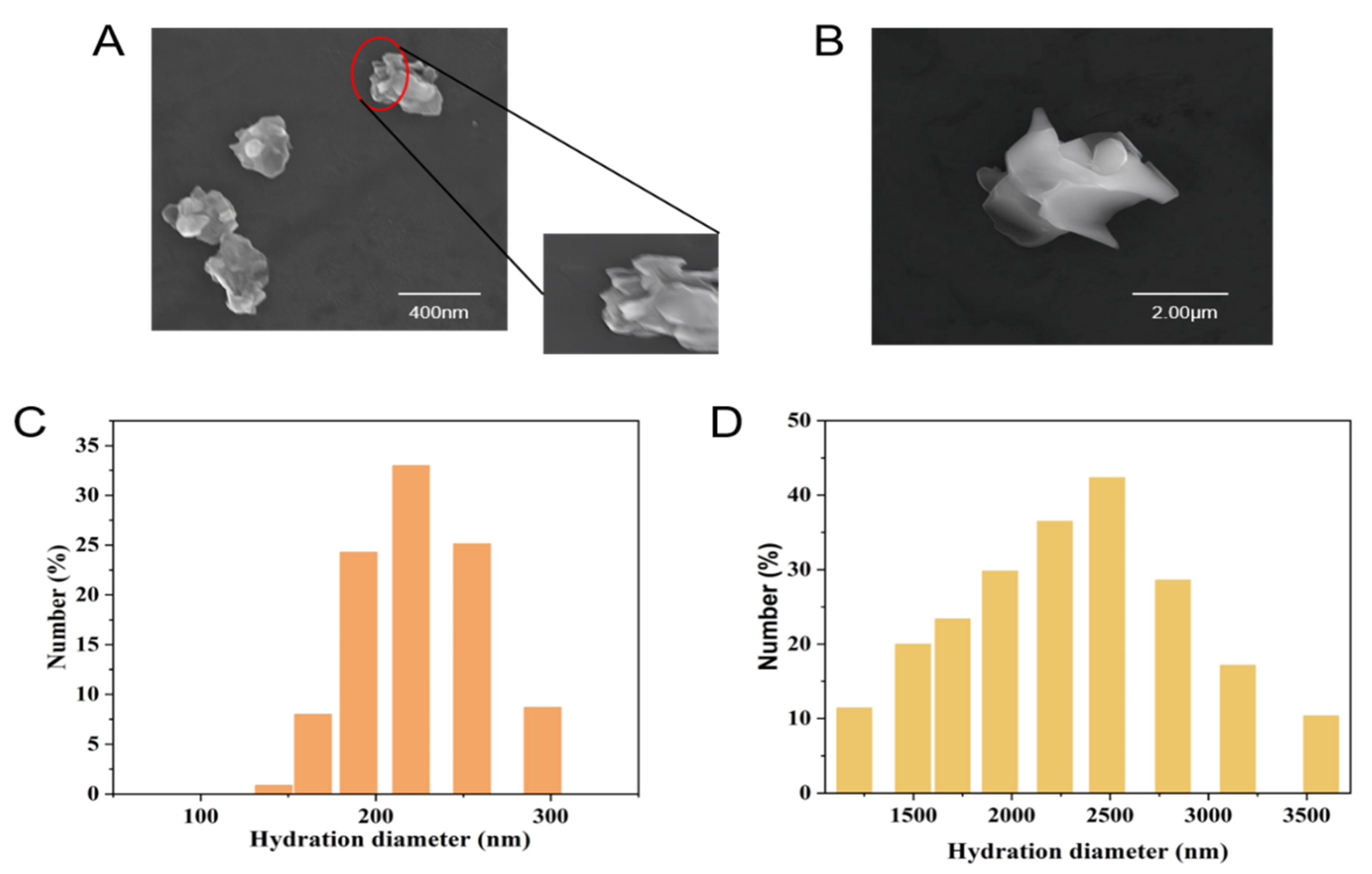
Figure 1. SEM images of (A) nano-WSe2 and (B) micro-WSe2 and DLS images of (C) nano-WSe2 and (D) micro-WSe2.
Molecules 27 07826 g001
Figure 2. Concentration of Se (AC) and W (DF) in rice roots, stems, and leaves after exposure to different concentrations of nano-WSe2 or micro-WSe2 (* p < 0.5, ** p < 0.05, *** p < 0.01, n = 3).
Molecules 27 07826 g002
Figure 3. GSH-Px (AC) and MDA (DF) in rice roots, stems, and leaves after exposure to different concentrations of nano-WSe2 or micro-WSe2.
Molecules 27 07826 g003
Figure 4. Spatial distribution of Se and W in rice (A) roots, (B) stems, and (C) leaves. (a1a3) the control group; (b1b3) the nano-WSe2 group; (c1c3) the micro-WSe2 group.
Molecules 27 07826 g004
Figure 5. The species of Se and W in rice roots exposed to 5000 mg/L nano-WSe2 or micro-WSe2. (A) The speciation of Se by AE-HPLC-ICP-MS. (B) The chemical forms of W by XAS.
Molecules 27 07826 g005
Table 1. The germination rate of rice seeds on the 7th day.
Concentration (mg/L)Germination Rate (%)
090.66 ± 0.01
(nano) 10081.33 ± 0.01
(micro) 10082.00 ± 0.07
(nano) 50085.33 ± 0.02
(micro) 50087.86 ± 0.07
(nano) 100081.33 ± 0.06
(micro) 100087.33 ± 0.03
(nano) 500086.67 ± 0.05
(micro) 500083.33 ± 0.03
Table 2. The length of roots, stems, and leaves of rice plants on the 30th day when exposed to different concentrations of nano-WSe2 or micro-WSe2 (* p < 0.05, ** p < 0.0001).
Concentration (mg/L)The Length of Root
(cm)
The Length of Stem
(cm)
The Length of Leaf
(cm)
06.69 ± 0.224.46 ± 1.228.59 ± 0.25
(nano) 1006.22 ± 0.284.13 ± 0.168.62 ± 0.71
(micro) 1006.30 ± 0.013.52 ± 0.288.21 ± 0.20
(nano) 5005.79 ± 0.523.60 ± 0.319.45 ± 0.21
(micro) 5007.17 ± 1.063.39 ± 0.047.78 ± 0.11
(nano) 10004.62 ± 0.22 *4.09 ± 0.328.47 ± 0.59
(micro) 10006.93 ± 0.163.34 ± 0.217.77 ± 0.55
(nano) 50002.23 ± 0.49 **3.54 ± 0.178.39 ± 0.31
(micro) 50005.97 ± 0.203.42 ± 0.026.76 ± 0.11
Table 3. Molar ratio of selenium and tungsten (nSe:nW) in rice.
Concentration (mg/kg)RootStemLeaf
00.140.210.80
(nano) 1000.110.130.07
(micro) 1000.110.140.40
(nano) 5000.080.140.05
(micro) 5000.120.100.18
(nano) 10000.060.070.05
(micro) 10000.110.140.15
(nano) 50000.060.110.09
(micro) 50000.140.140.09
Publisher’s Note: MDPI stays neutral with regard to jurisdictional claims in published maps and institutional affiliations.