Review

Can Natural Products Targeting EMT Serve as the Future Anticancer Therapeutics?

by
Sirajudheen Anwar
1,2,*,
Jonaid Ahmad Malik
3,4,
Sakeel Ahmed
5,
Verma Abhishek Kameshwar
6,
Jowaher Alanazi
1,2,
Abdulwahab Alamri
1,2 and
Nafees Ahemad
7
1
Department of Pharmacology and Toxicology, College of Pharmacy, University of Hail, Hail 81422, Saudi Arabia
2
Molecular Diagnostics Unit and Personalized Treatment, University of Hail, Hail 81422, Saudi Arabia
3
Department of Pharmacology and Toxicology, National Institute of Pharmaceutical Education and Research, Guwahati 781101, Assam, India
4
Department of Biomedical Engineering, Indian Institute of Technology Ropar, Rupnagar 140001, Punjab, India
5
Department of Pharmacology and Toxicology, National Institute of Pharmaceutical Education and Research, Ahmedabad 382355, Gujarat, India
6
Department of Pharmacology, Amrita School of Pharmacy, Amrita Vishwa Vidyapeetham, Kochi 641112, Kerala, India
7
School of Pharmacy, Monash University Malaysia, Jalan lagoon Selatan, Bandar Sunway, Petaling Jaya 47500, Selangor DE, Malaysia
*
Author to whom correspondence should be addressed.
Molecules 2022, 27(22), 7668; https://doi.org/10.3390/molecules27227668
Submission received: 4 October 2022 / Revised: 24 October 2022 / Accepted: 1 November 2022 / Published: 8 November 2022

Abstract

:
Cancer is the leading cause of death and has remained a big challenge for the scientific community. Because of the growing concerns, new therapeutic regimens are highly demanded to decrease the global burden. Despite advancements in chemotherapy, drug resistance is still a major hurdle to successful treatment. The primary challenge should be identifying and developing appropriate therapeutics for cancer patients to improve their survival. Multiple pathways are dysregulated in cancers, including disturbance in cellular metabolism, cell cycle, apoptosis, or epigenetic alterations. Over the last two decades, natural products have been a major research interest due to their therapeutic potential in various ailments. Natural compounds seem to be an alternative option for cancer management. Natural substances derived from plants and marine sources have been shown to have anti-cancer activity in preclinical settings. They might be proved as a sword to kill cancerous cells. The present review attempted to consolidate the available information on natural compounds derived from plants and marine sources and their anti-cancer potential underlying EMT mechanisms.

1. Introduction

Cancer is the leading cause of death globally [1]. According to the global demographic characteristics, it is expected to increase by approximately >20 million by 2025 [2]. The treatment paradigm improved in the past decade with the advancement in cancer research. Breast cancer (BC), colorectal cancer (CRC), lung cancer (LC), and prostate cancer (PC) are the most common types of cancers [1,3]. Various cellular pathways are involved in cancer development and progression. Several drug candidates are approved to target these pathways for their management [4]. One reason behind drug resistance is the process of Epithelial-mesenchymal transition (EMT) involved in cancer progression. EMT is an extremely regulated physiological process that has a significant role in tissue repair and embryogenesis [5]. During EMT, the cells undergo multiple morphologic, biological, and genetic rearrangements, leading to their mesenchymal phenotypes [6]. EMT is pathologically associated with fibrosis and cancer, leading to their progression. EMT has been linked to the formation of invasive and cancer stem cells in carcinomas [7].
EMT is initiated by EMT activating transcription factors (EMT-TFs), including SNAIL (SNAI1) and SLUG (SNAI2), the basic helix–loop–helix factors TWIST1 and TWIST2. As proven for SNAIL, TWIST, Zinc figure E-box binding homeobox 1 (ZEB1), and Zinc figure E-box binding homeobox 2 (ZEB2), these features can repress epithelial genes like the E-cadherin-producing CDH1 by binding to E-Box in their cognate promoter regions. Simultaneously, EMT-TFs activate genes associated with a mesenchymal phenotype, such as Vimentin (VIM), Fibronectin 1 (FN1), and N-Cadherin (CDH2). Several activities, however, are not common and are carried out by separate EMT- transcription factors (TFs) due to differences in coding sequences or protein size and structure [8]. An overview of the EMT pathway is shown in Figure 1. In other words, EMT is a biotic mechanism in which epithelial cells become polarized.
The heterogeneous mixture of cells like fibroblasts, endothelial cells, noncellular constituents, immune cells, extracellular matrix, cytokines, growth factors, and basement membrane is known as a tumor microenvironment (TME) [9]. EMT is essential for developing and initiating tumors and their recurrence and progression. In TME, the most abundant cells are cancer-associated fibroblasts (CAFs), which cross-talk with tumor cells, extracellular matrix (ECM), immune cells, and endothelial cells for cancer progression [9,10,11,12,13]. Several therapeutic agents are now being designed to target these CAFs [12,14]. Many natural agents have been identified to target CAF by altering the key signaling pathways, epigenetics, kinases, and enzymes. Targeting CAFs and altering the pathways affect cancer-stroma association in TME, resulting in decreased cancer progression. The natural compounds might have promised anti-cancer activity and are worth investigating against different tumors. The characteristic feature of carcinogenesis is ECM stiffness that supports the tumor cells. The crosslinking of ECM components like collagen with the tumor cells occurs via CAFs [15]. LOX-lysyl oxidase, the enzyme highly overexpressed in tumors derived from CAFs, acts as a collagen crosslinking initiator in several cancers like breast and gastric cancers, ultimately enhancing EMT, cell survival, invasion, drug resistance, and angiogenesis [16]. The ECM degrading enzymes like matrix metalloproteases (MMPs) and tissue inhibitors of metalloproteinase (TIMPs) inhibitors are altered by CAFs during angiogenesis and invasion, causing modulation of TME. The MMP2 and 9 are well investigated and highly associated with cancer growth and development [17,18]. The enzymes like metalloproteinases and disintegrin, associated with the MMPs super-family, are increased by CAFs, promoting cancer progression [19]. The CAFs also apply physical forces to pull out the epithelial basement membrane, causing the promotion of EMT in enzyme independent manner [20]. The CAFs promote EMT remolding and are promising therapeutic targets in halting EMT to prevent cancer metastasis. Natural compounds have proven to be the best alternatives to the current therapies against cancer. Many noteworthy examples are in front of us, where natural compounds have proven better than existing standard therapies against different ailments, such as cancer or infectious diseases. It is worth investigating the potential of natural compounds, whether from the marine, plant, or animal, against different types of cancers to find the solution for the growing deadly ailment in the world. Due to growing concerns regarding cancer metastasis, most therapies fail to cure, and the patients suffer greatly due to high toxicity. Cancer resistance is another challenge against current therapies, making cancer more complicated to manage. The natural compounds could be a game changer as anti-cancer therapy that specifically targets the EMT process and halts the process of cancer progression.

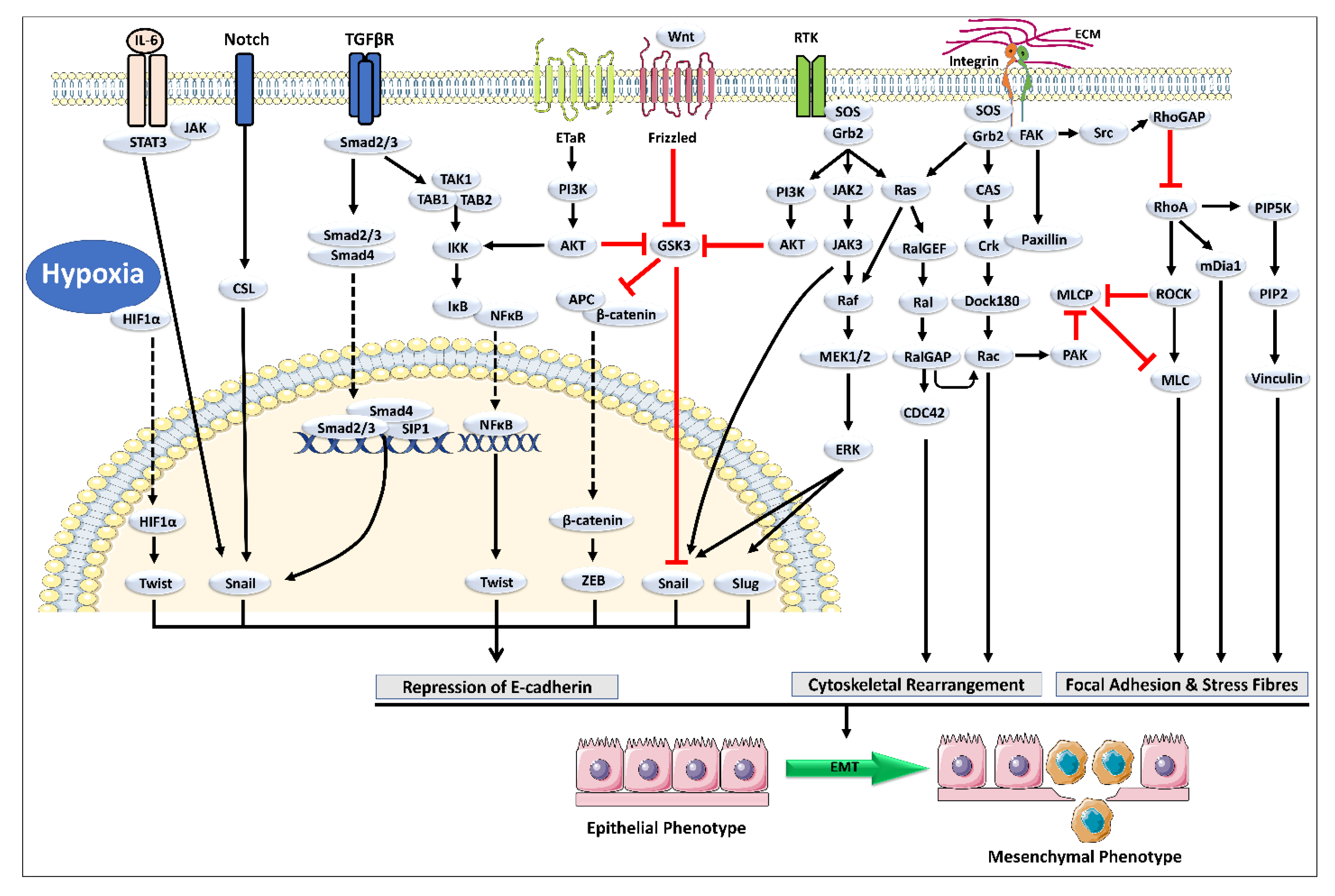
2. Cross-Talk between TGF-β and Other Signaling Pathways Mediating EMT

The signaling pathways cross-talk to form complex networks. Due to several cellular processes like apoptosis, differentiation, proliferation, and homeostasis, the Transforming growth factor β (TGF-β) cross-talk with various other signaling pathways during the EMT process (Figure 2) [21,22]. One of the mechanisms in which Akt activation and the phosphatase and tensin homolog (PTEN) dissociation from β-catenin are mainly responsible for the TGF-β mediated EMT process, where the displacement of β-catenin from adherent junctions occurs [23]. The other signaling pathway that cross-talks with TGF-β is Notch; Notch synergizes with TGF-β signals to enhance/inhibit its signaling activity depending on the input signal [21]. TGF-β signals activate the migration and inhibit the cell proliferation of endothelial cells. However, the Notch signals block the migration of bone morphogenetic protein (BMP) [24]. BMP stimulates the cell migration of endothelial cells; however, in the presence of Notch signaling, the migratory potential gets inhibited [24].
Interestingly, Notch signaling plays a crucial cross-talk in regulating migration by inducing gene expression. Notch dominates the BMP signaling; when the cell-to-cell contact is not there, endothelial cells are not in contact with the nearby cells to migrate until the new cell-to-cell attachment is set [21,24]. The TGF-β requires Notch signaling for the growth arrest in the epithelium; over thirty percent of the genes induced by TGF-β require Notch signaling [25]. The classy EMT marker, the TGF-β, also cross-talks with several other signaling pathways like Extracellular signal-regulating kinase (Erk), c-Jun N-terminal kinase (JNK), and p38. Erk, JNK, and p38 are indirectly regulating the TGF-β during EMT. However, TGF-β activates MAPK and Erk1/2 signaling pathways [26]. The cross-talk of TGF-β versus EGF signaling is the reason for activating Smad-dependent signaling and MAPK-mediated Erk1/2 [27]. The nuclear translocation of MAPK mediated by TGF-β is downregulated by the MAPK-Erk pathway that mediates nuclear exclusion and phosphorylation of Smad-2/3 [27]. During the initiation of EMT, the Akt and PTEN are also regulated by TGF-β. In addition, TGF-β cross-talk with ErbB signaling during the EMT development of breast cancer [28]. The TGF-β also regulates the phosphoinositide 3-kinase (PI3k)-Akt signaling pathways. Akt’s activity increases due to the induction of TGF-β-mediated functional activities like cell migration, epithelial to mesenchymal shift, cell survival, and cell growth [27,28,29]. Human epidermal growth factor receptor 2 (HER2)/RAS opposes the TGF-β-induced programmed cell death and cell arrest; however, it promotes migratory and invasive activities of TGF-β [30]. The EMT-associated cross-talk is validated by pharmacological inhibition of insulin-like growth factor-1R (IGF-1R), which prevents TGF-β-mediated EMT protein signatures [31]. The cross-talk of different pathways involved in EMT is demonstrated in Figure 2.
Figure 2. Illustrates the different pathways involved in initiating epithelial-mesenchymal transition (EMT) [32].
Molecules 27 07668 g002

3. Natural Chemical Agents as Potential Leads against Cancer

Among approved chemotherapeutic medications, 80% are bioactive natural compounds [33,34]. 70% of disease conditions, including cancer, are treated with the help of natural products [35]. The natural compounds induce cytotoxicity by targeting various oncogenic signaling [36]. Several marine-derived metabolites show anti-cancer activity in preclinical and clinical settings [37]. Marine-derived compounds include sulfated polysaccharides, sterols, carotenoids, and chitosan. Sulfated polysaccharides and carotenoids have effectively worked against cancer, acquired immune deficiency syndrome (AIDS), CVDs, and other acute and chronic disorders [35]. Various effective marine-derived compounds are summarized in Table 1. Plants, bacteria, animals, insects, and marine life are some of the key sources of natural chemicals with pharmacological and cytotoxic actions such as anti-proliferative, anti-angiogenic, apoptosis-generating, necrosis-inducing, and anti-inflammatory [38]. Secondary metabolites (Alkaloids, tannins, saponins, flavonoids, steroids) are molecules produced by plants at very minute levels that are therapeutically beneficial in various illnesses [39]. Animals and insects are also rich suppliers of enzymes with various pharmacological applications. Researchers have recently focused on natural substances to investigate their possible use in cancer treatment with lesser side effects. Still, they have only identified a few molecules with clinical efficacy demonstrated in Table 2 [40]. Compounds with anti-proliferative, anti-inflammatory, anti-angiogenic, and apoptotic-inducing properties can treat cancer and reduce the side effects of conventional cancer chemotherapy. They also have a high potential for future use as anti-cancer drugs.
Natural products can offer new hope in fighting EMT and increase therapeutic options worldwide. Drugs of natural origin, like traditional and Chinese medicine, are currently being investigated for various ailments [41]. The cost-effectiveness and richness of good therapeutic efficacy and safety were natural compounds’ true and popular features, considering them promising candidates against cancer [42]. The drugs against cancer at present share 1/3rd position share against cancer, meaning it is important to investigate more drugs from the natural origin against cancer [43]. Natural compounds have a high level of rigidity, enhancing the protein cross-talk more than synthetic drugs. They have diversity and versatile structure complexity, a unique natural feature making them the right candidates against cancer [43,44]. The signaling pathways that are responsible for cancer cell survival and TME maintenance are being halted by many natural compounds. The natural compounds have a comprehensive role in inhibiting tumor progression by blocking the survival pathways involved in EMT [9]. Many drugs have been used along with natural compounds, producing efficacious outcomes like decreasing drug resistance and toxicity [42,45]. Natural products can remold the TME [41]. Here we will show various candidates of natural origin demonstrating pharmacological activities against EMT and its associated factors.
Table 1. Marine and plants-derived compounds for anti-cancer activity.
Sr. NoDrug Product Source (Marine Origin)Mechanism of ActionIndicationFDA StatusReference
Marine Source
1Eribulin mesylate Sponge Halichondria okadaiKeeps the cytoskeleton’s growth cycle away from core aggregates tubulinMetastatic breast cancerApproved (Spain)[46]
2Brentuximab Vedotin Sea hare Dollabella Auricularia/ cyanobacteriaCell cycle arrest from G2 to M phaseHodgkin lymphoma Approved (USA.)[47]
3Cytarabine, Ara-C Sponge Cryptotheca cryptaInhibition of DNA SynthesisAcute lymphoblastic leukemiaApproved (USA.)[46]
4Halichondramide (HCA)Marine sponge Chondrosia corticataPhosphatase of regenerating liver-3 (PRL-3) and its downstream signaling pathway are suppressed.Prostate CancerApproved[48]
Plant Source
4IxabepiloneSoragium cellulosumCell-cycle arrest and apoptosis-inducer Hand-foot syndromeApproved[49]
5RomidepsinChromobacterium violaceumHistone deacetylase inhibitorsHematological toxicities like anemiaApproved[50]
6PodophyllotoxinsPodophyllum
(Berberidaceae)
Inhibit the polymerization
of tubulin, arresting the cell cycle in the metaphase
Ovarian cancer,
immunosuppressive ability
Approved[51]
7LigustrazineRhizome of Ligusticum wallichii.Inhibit SK-OV-3 and OVCAR-3 cell viability, proliferation, migration, and invasion.Ovarian cancerApproved[52]
Table 2. Potential NCEs with therapeutic effects against cancer.
Sr. NoNCE.SourceMechanism and Outcomes Method of ValidationPotential UseReference
1OregoninAlnus sibirica (AS)Anti-proliferative activity, Inhibition of NF-κB, induction of apoptosis, DNA MethylationMTT Assay, Western blotting, Flow, methylation-specific PCR, cytometryProstate cancer[53]
2Hirsutenone
3Hirsutanonol
4Chelerythrine chlorideChelidonium majus and Macleaya cordatacytotoxicity and anti-proliferative activityCell viability assaysNSCLC.[39]
5ThioholgamideStreptomyces sp. MUSC 136T.Caspase 3/7 Activation, membrane permeabilityMTT assayColon, breast, liver, and lung cancers[54]
67-deoxy-trans-dihydronarciclasinScadoxus pseudocaulusApoptosis inducer Cytotoxicity assayFollicular lymphoma[55]
74-(4-hydroxy-3-methoxyphenyl) curcumin Anti-proliferative, apoptosis-inducingMTT assay,
Western blotting analysis
Hepatic, colon, chronic myeloid leukemia, and lung cancer[56,57,58]

4. Potential NCE to Target EMT

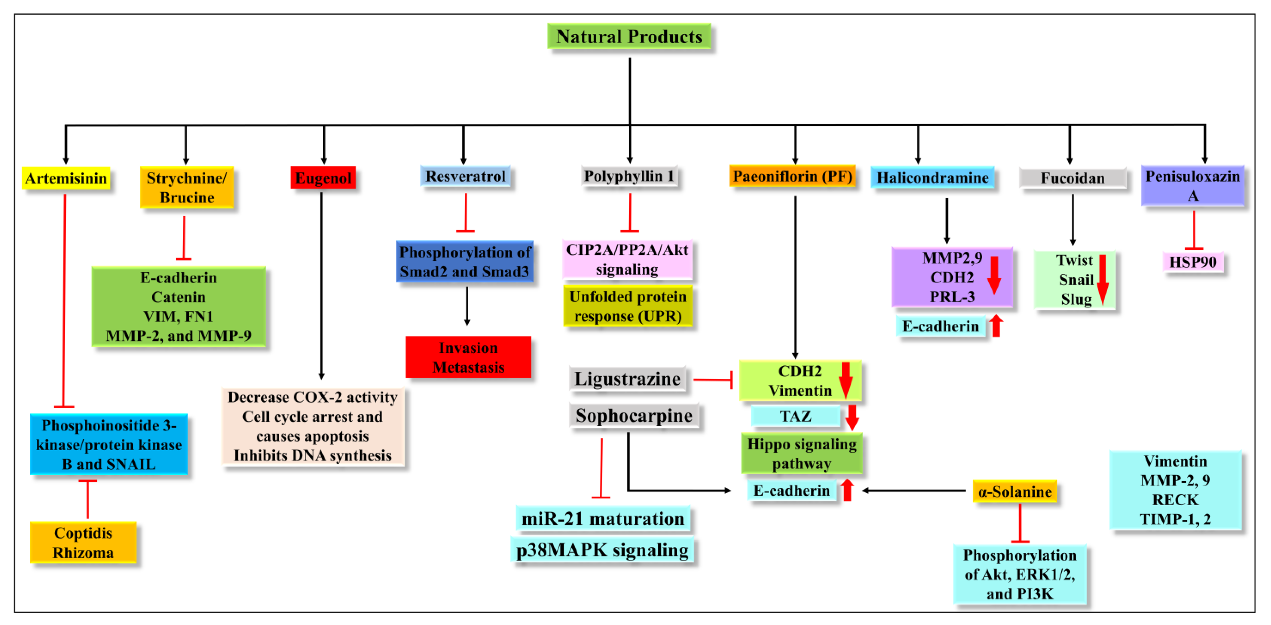
4.1. Artemisinin (ATM)

ATM is a sesquiterpene lactone isolated from sweet, warm wood, Artemisia annua. It is an antimalarial agent to treat multidrug-resistant falciparum malaria strains, mediated by producing organic peroxides [59]. ATM also has potent anti-cancer activity against CRC, BC, gastric cancer (GC), and cervix cancer (CC). Its anti-carcinogenic action is similar to antimalarial action in that free iron cleaves its endoperoxide bridge, releasing free radicals that cause cytotoxicity. ATM’s low toxicity and high specificity for cancer cells led to its development as an anti-cancer molecule. Dihydroartemisinic acid (DHA), an ATM derivative, reduced inflammation in a rat arthritis model by downregulating Interleukin-6 (IL-6). Additionally, DHA has anti-cancer properties. It can induce apoptosis in leukemic cells via noxa-mediated mechanisms [60].
Moreover, it inhibits GC cell invasion, migration, and proliferation by inhibiting the activation of phosphoinositide 3-kinase/protein kinase B and SNAIL. According to Sun et al., DHA’s anti-inflammatory and anti-cancer activities are mediated via microRNAs (miRNAs). DHA, for example, inhibits inflammation in vascular smooth muscle cells by regulating the miR-376b-3p/KLF pathway. The Jumonji and AT-rich interactive domain2 (ARID-2)/miR-7/miR-34a pathway inhibit prostate cancer cells by downregulating AXL tyrosine. In laryngeal cancer, miRNAs, in particular, play a function in EMT. For example, miR-217 inhibits EMT while miR-10b promotes it. miR-130b-3p is a tumor suppressor because it inhibits laryngeal cancer development, angiogenesis, migration, and invasion [61]. FoxM1, a member of the conserved forkhead box transcription factor family, is involved in cell cycle regulation, DNA damage repair, and apoptosis and has been associated with the development of breast, pancreas, and liver carcinomas. Nandi et al. hypothesized that FoxM1 was a critical inhibitory target of ATM in hepatocellular carcinoma (HCC) and that FoxM1 may play a role in the cell cycle triggered by DHA ATM-inhibited HCC cell survival and proliferation by attenuating FoxM1 and its transcription targets and interfering with FoxM1 trans-activation [62].

4.2. Strychnine/Brucine

Brucine is an alkaloid related to strychnine obtained from the Strychnic Nux-vomica tree. It has analgesic, anti-cancer, anti-inflammatory, antioxidant, and anti-venom properties [63]. In vitro, it inhibits the proliferation of Hela and K562 cell lines. Brucine also showed anti-metastasis action in MDA-MB-231 and Hs578-T-cells and inhibited invasive capacity and adhesion of MDA-MB-231 and Hs578-T-cells Matrigel, and preventing mRNA of E-cadherin, catenin, VIM, FN1, MMP-2, and MMP-9 in MDA-MB-231 cells [64]. These data collectively suggest that brucine might be a potential anti-cancer molecule; in vivo studies are still needed to confirm its anti-cancer potential.

4.3. Eugenol

A polypropanoid group of compounds is found in seeds of many plants, such as cloves, cinnamon, nutmeg, and bay leaves. It has antioxidant, anti-bacterial, anti-inflammatory, and anti-cancer activity and is widely used as a cosmetic, perfume, and culinary ingredient. It has anti-cancer potential due to its ability to increase reactive oxygen species (ROS) formation and apoptotic action, increase Cyt C’s release, and inhibit the EMT process, limiting the cells’ ability to metastasize [65]. It has shown anti-cancer activity against malignancies, including leukemia, lung, colon, colorectal, skin, gastric, breast, cervical, and prostate cancer, through the processes described below in Table 3.

4.4. Resveratrol

Resveratrol (RES) chemically trihydroxy stilbene is a polyphenol in grapes, berries, peanuts, and wine. RES has been shown to have cardioprotective, anti-inflammatory, and anti-aging effects. Studies also suggest that it also has anti-cancer properties. Moreover, RES has a regulatory role in EMT and the hedgehog (Hh) signaling pathway, which is critical for vertebrate development, homeostasis, and cancer. Hh is abnormally activated in breast, prostate, and pancreatic cancer (PC) and has a role in metastasis and invasion of GC via induction of the EMT. Hence, Hh signaling pathway is the center of attraction for anti-cancer activity. RES suppresses the Hh pathway, thus inhibiting cancer invasion and metastasis.
Moreover, RES has also been shown to block the Hh signaling pathway and EMT in malignancies [71]. A study proved that RES inhibited EMT in Glioblastoma (GBM) cells, as evidenced by morphological changes in the RES-treated G.B.M. cells. RES also inhibits EMT-mediated migration and invasion of GBM cells and EMT-induced stem cell-like properties in GBM cells [72]. A TGF-β/Smad signaling pathway is associated with the proliferation, differentiation, and migration of the cells and promotes results in invasion and metastasis. RES inhibited the penetration and metastasis by EMT-induced phosphorylation of Smad2 and Smad3 in a dose-dependent manner, suggesting the function of RES on EMT is related to Smad-dependent signaling [72].

4.5. Polyphyllin 1

Polyphyllin 1 (PP 1) demonstrated its anti-cancer activity via its apoptotic action and several pathways effectively against various cancers. Polyphyllin I induces apoptosis in HepF-2-Cells, and neural progenitor cells (NPC) cell lines [73]. The natural herb Paris polyphylla makes PP1 and has anti-cancer properties against various malignancies, including drug-resistant tumors. Paris polyphylla was recently found to inhibit CRC cells by activating autophagy and improving the efficiency of chemotherapy (Doxorubicin). By decreasing CIP2A/PP2A/Akt signaling, PP1 also reduced cisplatin-resistant GC cells. Liu et al. demonstrated that PP 1 has potent anti-cancer action on human non-small cell lung cancer (NSCLC) mediated by CHOP stabilization. PP1 induces ROS, and ER stress inhibits unfolded protein response (UPR) in cancer cells, subsequently increasing the levels of CHOP Via, accelerating CHOP gene expression. The UPR chaperone GRP78, restrained by PP1, is the main mechanism for CHOP stabilization [74].

4.6. Paeoniflorin (PF)

Paeoniflorin (PF) is a monoterpene glycoside derived from the root of Paeonia lactiflora. In the past, this plant’s roots were utilized in eastern medicine for pain, muscle spasms, inflammation, menstruation dysfunction, and degenerative illnesses for a long time [75,76,77,78]. Studies indicated that PF inhibits tumor growth, invasion, and metastasis in vivo and in vitro. In hypoxia-induced EMT in MDA-MB-231 BC cells, PF treatment resulted in a considerable increase in E-cadherin levels and a drop in CDH2 and Vimentin levels in the cells. Subsequently, it suppressed the EMT process by altering the expression of HIF-1, which is involved in hypoxia-driven EMT [79]. The hippo pathway plays a significant role in the progression of GC. This pathway is said to be dysregulated and thus contributes to gastric oncology and metastasis. Two important factors, yes associated protein (YAP1) and Transcriptional coactivator with PDZ-binding motif (TAZ), produce their metastatic effect via crosslinking with Notch, TGF-β, and Wnt/ β-catenin in GC. In GC, PF exerts its anti-cancer effect via regulation of the hippo signaling pathway and downregulating the effect of TAZ [80]. Further, there is a need to explore its potential in other cancers.

4.7. Halicondramine

Halicondramine (HCA) is a trisoxazole-containing macrolide from the marine sponge Chondrosia corticata [81]. It possesses antifungal and cytotoxic properties. It also has anti-proliferative activity against cancerous cells [82,83]. Modulation of the EMT is a key target for their action. Treatment with HCA significantly reduced the expression of MMP2 and 9, and CDH2. On the contrary, E-cadherin expression was significantly increased. HCA also inhibits the expression of PRL-3, a metastasis-associated marker, and PI3 kinase subunits p85 and p110 (PRL-3’s downstream targets). These findings imply that HCA inhibits EMT in human adenocarcinoma prostate cancer cells by modulating PRL-3 and downstream targets, such as PI3 kinase [48].

4.8. Ligustrazine

Ligustrazine (LSZ) is obtained from the rhizome of Ligusticum wallichii [84]. LSZ is shown to have anti-inflammatory, anti-fibrotic, antioxidant activity, and tumor-suppressing properties in numerous cancers, including LC, GC, BC, and melanoma [85]. LSZ showed anti-proliferative and anti-metastatic action [6]. LSZ increased E-cadherin expression while decreasing the mesenchymal indicators CDH2 and VIM expression. LSZ inhibits EMT in SK-OV-3 cells via modulating miR-211 expression [86].

4.9. Fucoidan

The Fucoidan (FC) is a polysaccharide obtained from brown seaweeds and has shown anti-proliferative action on BC cells, such as 4T1 and MDA-MB-231. It also lowered metastatic lung nodules in female Balb/c mice with 4T1 xenografts. The TGFRs molecular network is critical in controlling EMT in cancer cells. It was observed that FC efficiently reverses TGFR-induced EMT morphological alterations, increases epithelial markers, decreases mesenchymal markers and transcriptional repressor expression Twist, Snail, and Slug. Furthermore, fucoidan suppresses migration and invasion during EMT, implying that TGFR-mediated signaling is involved in BC cells [87].

4.10. Penisuloxazin A

Penisuloxazin A (PNSA) is a fungal mycotoxin that belongs to a new epipolythiodiketopiperazines (ETPs) possessing a rare 3H-spiro[benzofuran-2,2′-piperazine] ring system. PNSA prevented MDA-MB-231 cell adhesion to coated Matrigels containing several ECM components. Furthermore, after PNSA therapy, there is a transition from spindle-shaped or polygonal mesenchymal to flat polygonal epithelial-like cell morphology. These suggest that PNSA can prevent EMT in MDA-MB-231 cells [88]. PNSA is also considered a potent heat shock protein 90 (HSP90) inhibitor, a well-known N-terminal inhibitor binding to the ATP pocket of HSP90 in preventing BC cell metastasis. Multiple signaling pathways critical for cancer cell proliferation and metastasis can be disrupted by inhibiting HSP90 [89].

4.11. Sophocarpine

Sophocarpinr (SC) is one of the most active components of Sophora alopecuroides L, a tetracyclic quinolizidine alkaloid. SC has shown various pharmacological actions, including immunoregulatory, anti-inflammatory, and anti-nociceptive [90]. SC has also been shown to preserve heart function from ischemic reperfusion by inhibiting NF-kB and reducing hepatocyte steatosis via activation of the AMPK signaling pathway. Furthermore, in head and neck squamous cell carcinoma (HNSCC) cells, SC has shown anti-proliferative and anti-metastatic by inhibiting dicer-catalyzed miR-21 maturation and activation of the p38MAPK signaling pathway. SC also reduced the HNSCC tumor’s growth in vivo by reversing the EMT in cancer cells. In UM-SCC-22B and UM-SCC-47 cells, SC treatment reduced the expression of the Ki-67 and VIM while increasing E-cadherin’s expression [91]. These findings suggest that SC could be a promising lead drug for HNSCC.

4.12. Renieramycin M

Renieramycin M (RM) (22-Boc-Gly-RM), produced by Xestospongia sp., is a semi-synthetic amino ester derivative of the bistetrahydroisoquinoline alkaloid. Studies suggested RM-mediated inhibition of anchorage-independent development and sensitization of detachment-induced cell death in human lung cancer cells [92]. A semi-synthetic derivative of RM with a hydroquinone amino ester extension was synthesized to retain cytotoxicity with increase cancer selectivity [93]. It hinders the phosphorylation of FAK and Akt molecules, which upregulate TIMP2 and TIMP3 and downregulate MMPs expression. The inhibition of the p-FAK/p-Akt signal also marks the downregulation of CDH2 and Rac1-GTP and the upregulation of E-cadherin, where the regulation of cytoskeleton regulatory protein (Rac1-GTP), MMP-associated molecules (TIMP2, TIMP3) [94]. The mechanism of action of RM is demonstrated in Figure 3.

4.13. Luteolin

Luteolin (LT) (3,4,5,7-tetrahydroxy flavone) is a flavonoid in many plants including broccoli, carrots, perilla leaves, seeds, and celery. It possesses anti-allergy, anti-inflammatory, anti-cancer, antioxidant, and anti-microbial properties [96,97,98]. In various cancers (including lung, GBM, BC, CRC, PC), LT inhibits cell proliferation and tumor growth, promotes cancer cell apoptosis and cell cycle arrest, reduces drug resistance, and reduces cancer cell invasiveness and metastasis [99]. LT can also stop EMT from occurring, shrinking in the cytoskeleton, increasing the expression of E-cadherin, and decreasing the expression of CDH2, Snail, and VIM [100]. LT inhibits the Smad 2/3 pathway and the Wnt/-catenin pathway by inhibiting the synthesis of Snail and Slug by downregulating the production of β-catenin. Doing so prevents metastasis by upregulating CDH2, Zo 1, and claudin 1 and downregulating CDH2, fibronectin, VIM, and MMP-2 [99,101]. The mechanism of the action of LT is demonstrated in Figure 4.

4.14. Carnosic Acid

Carnosic acid (CA), a polyphenolic diterpene found in rosemary (Rosmarinus officinalis), has anti-cancer, anti-viral, and anti-inflammatory activities. CA suppresses cancer cell migration and proliferation while lowering vascular endothelial growth factor expression. In leukemia and CRC cells, CA also causes cell cycle arrest at the G2/M phase by downregulating cyclin expression and has been shown to trigger apoptotic cell death in human NB and PrC cells [102]. CA inhibits EMT and cell migration in B16F10 cells in a dose-dependent manner. It prevents Src/AKT phosphorylation and, therefore, activation. It decreases the secretion of uPAc, MMP-9, and TIMP-1, whereas it increases the secretion of TIMP-2 and has no effect on the secretion of MMP-2 and plasminogen activator inhibitor-1 (PAI-1). It is also responsible for the decrease in the expression of Snail and Slug but does not affect the expression of Twist in B16F10 melanoma cells [95]. The mechanism of action of CA is demonstrated in Figure 3 and Figure 4.

4.15. N-Phenethylacetamide

N-Phenethylacetamide (NPA) is found in the Aquamarina Sp. (MC085). Three compounds, two diketopiperazines [cyclo(L-Pro-L-Leu) (1) and cyclo(L-Pro-L-Ile) (2), and one NPA (3)] isolated with anti-cancer activity. By altering TGF-induced E.M.T., NPA inhibits the TGF/Smad pathway and suppresses A549 cell metastasis. It prevents Snail and Slug expression by inhibiting Smad 2/3 phosphorylation. It also suppresses Snail and Slug, which upregulates the epithelial markers E-cadherin, Zo-1, and claudin-1 while downregulating VIM, FN1, CDH2, and MMP-2 expression, preventing metastasis [101]. The mechanism of NPA is shown in Figure 4.

4.16. α-Solanine

α-Solanin (AS), a steroidal glycoalkaloid obtained from nightshade (Solanum nigrum Linn.), suppresses tumor cell growth and causes apoptosis in colon, liver, cervical, lymphoma, and stomach cancer cells. However, the mechanism by which it blocks cancer cell metastasis remains unknown. An animal model of BC induces cell death and inhibits cell proliferation and angiogenesis, resulting in chemotherapeutic actions [103,104]. It also increases E-cadherin expression, reducing VIM expression and cell invasion, which inhibits EMT. It also decreases extracellular inducer of matrix metalloproteinase (EMMPRIN), MMP-2, and MMP-9, increasing Cysteine-rich protein with Kazal motifs (RECK), TIMP-1, and TIMP-2 mRNA expression levels. It downregulated the phosphorylation of Akt, ERK1/2, and PI3K. Furthermore, it increases tumor suppressor miR-138 expression while decreasing oncogenic miR-21 expression [6,105].

4.17. Baicalein, Wogonin (WG), and Oroxylin-A (ORA)

Baicalein (BAI), wogonin (WG), and oroxylin-A (ORA) are present in a plant, namely Scutellaria baicalensis [105]. It has been reported that the extract of Scutellaria baicalensis has anti-tumor activity. A study reported that using the combination of BAI (65.8%), WG (21.2%), and ORA (13.0%) compounds against A549 lung adenoma cancer cells inhibited the EMT process significantly [105]. The Total Flavonoid Aglycones Extract (TFAE) isolated from Scutellaria baicalensis has shown inhibition against tumors by inducing apoptosis, mainly BAI, WG, and ORA [106]. It was reported that the TFAE of Scutellaria baicalensis has inhibited the EMT of A549 cells via PI3K/AKT-TWIST1 axis [105].

4.18. Coptidis Rhizoma

It was reported that the extract of Coptidis Rhizoma (CR) could inhibit the EMT process via the TGF-β signaling pathway [107]. It has been shown that 30% ethanol extract of Coptidis Rhizoma can inhibit cell migration and invasion via blocking E-cadherin and decreasing expression of vimentin, Snail, and ZEB2 [107]. It has a potential anti-metastatic effect and can be a candidate against cancer. The different pathways and proteins targeted by all natural products discussed in the present review are summarized in Figure 5.

5. Advantages of Targeting EMT

EMT is recognized as playing a key role in developing cancer, metastasis, and chemotherapy resistance, and its crucial roles throughout cancer progression have recently been discovered and investigated. Although there is still debate about whether EMT causes cancer metastasis, its importance in cancer chemoresistance is becoming more widely recognized, with many EMT-related signaling pathways implicated in cancer cell chemoresistance [39]. Targeted cancer treatments have been an emerging field in the recent decade. Several monoclonal antibody therapies and small chemicals, particularly kinase inhibitors, have been discovered/synthesized and undergo clinical trials with improved anti-cancer effectiveness. While many targeted therapeutic medications demonstrated encouraging preliminary clinical outcomes, such as enhanced overall survival, a significant percentage of patients who received targeted therapy acquired drug resistance following long-term treatment [108]. As a result, cancer drug resistance will determine the success of forthcoming targeted treatment medications. Drug resistance can be caused by various mechanisms, including drug efflux, drug metabolism, and drug target mutations [1,109]. The function of EMT in cancer therapy resistance has recently been explored. In the early 1990s, a relationship between EMT and cancer cell treatment resistance was proposed. Heckford et al. discovered that EMT occurred in two Adriamycin-resistant MCF-7 cells and vinblastine-resistant ZR-75-B cells [110]. Attempts have been devoted to targeting the ABC transporters to overcome drug resistance [111,112]. When it became obvious that EMT plays a critical role in drug resistance, scientists began exploring drugs targeting EMT to overcome drug resistance. Gupta et al. created EMT cells using E-cadherin shRNA and used this cell line to develop CSC-selective small molecule inhibitors. Using high-throughput screening, they discovered an antibiotic named Salinomycin that eliminated breast CSCs preferentially [113]. Salinomycin also reduced EMT caused by doxorubicin exposure and improved doxorubicin sensitivity in HCC cells [114]. It inhibited the expression and operation of drug efflux pumps in BC cells, resulting in a considerable reduction in doxorubicin resistance [115]. In addition to Salinomycin, several minor pharmacologic inhibitors of EMT have been discovered and tested in vitro and in vivo cancer treatment resistance models. Mocetinostat, a histone deacetylase (HDAC) inhibitor that restored miR-203 and decreased ZEB1 (EMT-TF) expression, reversed EMT in drug-resistant cancer cells and sensitized them to the chemotherapeutic agent docetaxel [116]. Curcumin, a component of curry, was discovered to sensitize 5-fluorouracil-resistant colorectal cancer cells via inhibiting EMT via miRNA [117]. According to Namba et al., EMT mediated by the Akt/GSK3/Snail1 pathway was a critical signaling event in acquiring gemcitabine resistance in PC cells. The anti-viral zidovudine inhibited these signaling pathways, restoring gemcitabine sensitivity in cancer cells. Co-administered zidovudine with gemcitabine reduced tumor growth and prevented cancer cells from establishing the EMT phenotype in mice with a gemcitabine-resistant pancreatic tumor xenograft [118]. Oncologists have recently focused on metformin since it has anti-cancer and chemopreventive qualities independent of anti-hyperglycemic effects [119,120]. Hirsch et al. later discovered that metformin targets BCSCs [121]. According to follow-up studies, metformin lowers CSCs by targeting EMT Metformin triggered transcriptional re-programming of BCSCs by lowering major EMT-TFs such as SNAIL2, Twist1, and ZEB1, according to Vazquez-Martin and colleagues [121]. Metformin has been shown to prevent EMT in lung cancer by inhibiting the IL-6/STAT3 axis in lung adenocarcinoma [122]. Although the direct molecular target of metformin in suppressing EMT is unknown, the Stimulation of AMPK may play a significant role in the drug’s anti-EMT activity [123,124]. Metformin is being studied in over 200 human clinical studies for cancer therapy because of its possible CSC, anti-cancer properties, and favorable safety profile [125]. As a result, targeting EMT has been viewed as a unique strategy for combating cancer treatment resistance. In addition to the small compounds that have been created, a lot of pharmacological screening is being done to find new EMT inhibitors. Chua et al. created an EMT spot migration recognition method that can be utilized for high-content screening to screen small molecule EMT inhibitors that target certain growth factors. Scientists could undertake high throughput screening of small compounds utilizing enhanced screening platforms thanks to advancements in EMT and CSC biology [126]. Aref et al. also created a microfluidic device that mimics the 3D tumor microenvironment by including tumor cell spheroids and an adjacent endothelial monolayer. This approach is very beneficial in identifying EMT medicines active in a complex in vivo tumor microenvironment with several cell kinds interacting [127,128]. As a result, targeting EMTs with the natural chemical entity is viewed as a unique and innovative approach to combating cancer treatment resistance.

6. Targeting EMT Process by Molecular Docking (MD)

MD is a promising approach for estimating the interaction between biological molecules, such as proteins and ligands [129,130]. In the last decade, MD has emerged as a promising tool in identifying lead molecules against different ailments. MD is applied to cancer stem cells (CSC) associated metabolic and signaling pathways. Different metabolic pathways participate in CSC survival concerning cancer progression and alterations [130]. This is why metabolic re-programming is considered one of the cancer symbols [131].
Natural products are considered the grounds of multi-targeting molecules. Alkaloids, a class of natural producers, are one of the promising molecules that have the strength to combat CSCs via MD [130,131]. The drugs like salasonine and tylophorine have shown that they altered the Hedgehog (Hh) pathways and exerted anti-cancer effects on CSCs [130,131]. The targeting of overexpressed receptors in cancer tissues has demonstrated anti-cancer potential by natural products earlier through MD and experimental approaches [130,131]. It is reported that natural products represent a rich source of therapeutically active compounds which can interact with numerous cellular targets and minimize the side effects [132]. MD may help us find the natural lead molecules against the EMT process. Different receptors play a role in the EMT process, so it will be more important to screen out natural compounds against those receptors to find lead molecules.

7. Future Prospective

EMT has long been suspected of contributing to cancer therapy resistance. It became clear when scientists observed strong parallels in gene expression profiles and marker expression between EMT cells and CSCs. The resistance of CSCs against pharmacotherapy is a major challenge and poses a significant threat to cancer patients. The pathways involved in EMT have been deciphered by scientists, and have developed several methodologies to investigate the phenotypes in EMT. These understandings form the basis for drug screening against EMT-mediated cancer. The miRNA and some chemical agents demonstrate inhibitory activity against EMT, but no currently available miRNAs in clinical settings can solve this problem. So natural compounds showing good results can be the game changers in mitigating the EMT process. Several small chemical agents have been discovered to help drug-resistant cancer cells targeting EMT become more chemo-sensitive. Many of these, including Ligustrazine Penisuloxazin A, Halichondramide, Sophocarpine, Fucoidan, and Diketopiperazines, were investigated in human clinical trials with standard chemotherapies or targeted treatments.
Furthermore, EMT inhibitors’ long-term safety is unknown. This is especially true if EMT inhibitors activate the MET pathway, which has been linked to cancer metastasis. Moreover, studies need to be conducted to investigate the link of EMT with different pathways involved in Cancers. There is a need to develop nanoformulations targeting the cancerous to avoid off-target side effects. Moreover, with the advancement of the compounds screening assays, more novel natural compounds need to be explored to find a more potent EMT inhibitor. Novel in vitro and in vivo approaches should be developed to reduce translational failure [133].

8. Conclusions

The part of EMT-MET in cancer cell dispersion and distant metastasis has been acknowledged. Recent research suggests that EMT may not be required for cancer cell metastasis despite its importance in chemoresistance. However, this is controversial because of the EMT phenotype’s variability and adaptability. Only a fraction of EMT populations may be controlled by Fsp1. Finally, EMT is a critical cancer cell characteristic contributing to medication resistance. Inhibitors of this biological mechanism will be suitable “partners” for chemotherapy or targeted therapy medications, allowing current cancer therapies to achieve better clinical outcomes. Natural products play a dynamic role in controlling the process of EMT in cancer; many assays have been performed to prove their activity. EMT is a progression where cells can lose their epithelial properties like E-cadherin and a few more and gain mesenchymal properties like CDH2, VIM, FN1, etc. EMT has long been investigated for its involvement in cancer treatment resistance and metastasis. Assay techniques to analyze EMT phenotypic and drug screening have been created based on a better knowledge of natural chemicals and critical signaling pathways in EMT. Natural compounds that downregulate EMT phenotype and, as a result, drug resistance and metastasis have been identified, allowing scientists to discover natural compounds as EMT inhibitors capable of improving chemosensitivity of drug-resistant cancer cells while inhibiting metastasis. However, additional research is needed to fully comprehend the significance of EMT in cancer treatment resistance, cell proliferation, invasion, metastasis, and natural chemicals and their involvement in blocking EMT.

Author Contributions

Conceptualization: S.A. (Sirajudheen Anwar) and J.A.M.; Methodology: J.A.M. and S.A. (Sirajudheen Anwar); Software: S.A. (Sakeel Ahmed); Validation: J.A.M., S.A. (Sakeel Ahmed) and S.A. (Sirajudheen Anwar). Writing—original draft preparation: J.A.M., V.A.K. and S.A. (Sakeel Ahmed); Review and editing: S.A. (Sirajudheen Anwar), J.A., A.A. and N.A.; project administration: S.A. (Sirajudheen Anwar); Funding acquisition: S.A. (Sirajudheen Anwar). All authors have read and agreed to the published version of the manuscript.

Funding

This research has been funded from Research Deanship in University of Ha’il—Saudi Arabia through project number MDR-22 031.

Institutional Review Board Statement

Not applicable.

Informed Consent Statement

Not applicable.

Data Availability Statement

Not applicable.

Acknowledgments

Authors extend their appreciation to project fund from Research Deanship in University of Ha’il—Saudi Arabia through project number MDR-22 031.

Conflicts of Interest

Authors do not have any conflict of interest, personal or financial.

Sample Availability

Not applicable.

References

  1. Malik, J.A.; Ahmed, S.; Jan, B.; Bender, O.; Al Hagbani, T.; Alqarni, A.; Anwar, S. Drugs repurposed: An advanced step towards the treatment of breast cancer and associated challenges. Biomed. Pharmacother. 2021, 145, 112375. [Google Scholar] [CrossRef] [PubMed]
  2. Ferlay, J.; Soerjomataram, I.; Dikshit, R.; Eser, S.; Mathers, C.; Rebelo, M.; Parkin, D.M.; Forman, D.; Bray, F. Cancer incidence and mortality worldwide: Sources, methods and major patterns in GLOBOCAN 2012. Int. J. Cancer 2015, 136, E359–E386. [Google Scholar] [CrossRef] [PubMed]
  3. Ferlay, J.; Steliarova-Foucher, E.; Lortet-Tieulent, J.; Rosso, S.; Coebergh, J.W.W.; Comber, H.; Forman, D.; Bray, F. Cancer incidence and mortality patterns in Europe: Estimates for 40 countries in 2012. Eur. J. Cancer 2013, 49, 1374–1403. [Google Scholar] [CrossRef] [PubMed] [Green Version]
  4. Sanchez-Vega, F.; Mina, M.; Armenia, J.; Chatila, W.K.K.; Luna, A.; La, K.C.C.; Dimitriadoy, S.; Liu, D.L.; Kantheti, H.S.; Saghafinia, S.; et al. Oncogenic Signaling Pathways in The Cancer Genome Atlas. Cell 2018, 173, 321–337.e310. [Google Scholar] [CrossRef] [Green Version]
  5. Kalluri, R.; Weinberg, R.A. The basics of epithelial-mesenchymal transition. J. Clin. Investig. 2009, 119, 1420–1428. [Google Scholar] [CrossRef] [Green Version]
  6. Avila-Carrasco, L.; Majano, P.; Sánchez-Toméro, J.A.; Selgas, R.; López-Cabrera, M.; Aguilera, A.; González-Mateo, G.T. Natural Plants Compounds as Modulators of Epithelial-to-Mesenchymal Transition. Front. Pharmacol. 2019, 10, 715. [Google Scholar] [CrossRef] [Green Version]
  7. Gundamaraju, R.; Lu, W.; Paul, M.K.; Jha, N.K.; Gupta, P.K.; Ojha, S.; Chattopadhyay, I.; Rao, P.V.; Ghavami, S. Autophagy and EMT in cancer and metastasis: Who controls whom? Biochim. Biophys. Acta-Mol. Basis Dis. 2022, 1868, 166431. [Google Scholar] [CrossRef]
  8. Brabletz, S.; Schuhwerk, H.; Brabletz, T.; Stemmler, M.P. Dynamic EMT: A multi-tool for tumor progression. EMBO J. 2021, 40, e108647. [Google Scholar] [CrossRef]
  9. Chiu, K.-J.; Chiou, H.-Y.C.; Huang, C.-H.; Lu, P.-C.; Kuo, H.-R.; Wang, J.-W.; Lin, M.-H. Natural Compounds Targeting Cancer-Associated Fibroblasts against Digestive System Tumor Progression: Therapeutic Insights. Biomedicines 2022, 10, 713. [Google Scholar] [CrossRef]
  10. Chen, Y.; McAndrews, K.M.; Kalluri, R. Clinical and therapeutic relevance of cancer-associated fibroblasts. Nat. Rev. Clin. Oncol. 2021, 18, 792–804. [Google Scholar] [CrossRef]
  11. Biffi, G.; Tuveson, D.A. Diversity and Biology of Cancer-Associated Fibroblasts. Physiol. Rev. 2021, 101, 147–176. [Google Scholar] [CrossRef] [PubMed]
  12. Chen, X.; Song, E. Turning foes to friends: Targeting cancer-associated fibroblasts. Nat. Rev. Drug Discov. 2019, 18, 99–115. [Google Scholar] [CrossRef] [PubMed]
  13. Liao, Z.; Tan, Z.W.; Zhu, P.; Tan, N.S. Cancer-associated fibroblasts in tumor microenvironment—Accomplices in tumor malignancy. Cell. Immunol. 2019, 343, 103729. [Google Scholar] [CrossRef] [PubMed]
  14. Ermakov, M.S.; Nushtaeva, A.A.; Richter, V.A.; Koval, O. Cancer-associated fibroblasts and their role in tumor progression. Vavilov J. Genet. Breed. 2022, 26, 14–21. [Google Scholar] [CrossRef] [PubMed]
  15. Nissen, N.I.; Karsdal, M.; Willumsen, N. Collagens and Cancer associated fibroblasts in the reactive stroma and its relation to Cancer biology. J. Exp. Clin. Cancer Res. 2019, 38, 115. [Google Scholar] [CrossRef] [Green Version]
  16. Wang, T.-H.; Hsia, S.-M.; Shieh, T.-M. Lysyl Oxidase and the Tumor Microenvironment. Int. J. Mol. Sci. 2016, 18, 62. [Google Scholar] [CrossRef] [Green Version]
  17. Najafi, M.; Farhood, B.; Mortezaee, K. Extracellular matrix (ECM) stiffness and degradation as cancer drivers. J. Cell. Biochem. 2019, 120, 2782–2790. [Google Scholar] [CrossRef]
  18. Tune, B.X.J.; Sim, M.S.S.; Poh, C.L.; Mac Guad, R.; Woon, C.K.; Hazarika, I.; Das, A.; Gopinath, S.C.B.; Rajan, M.; Sekar, M.; et al. Matrix Metalloproteinases in Chemoresistance: Regulatory Roles, Molecular Interactions, and Potential Inhibitors. J. Oncol. 2022, 2022, 3249766. [Google Scholar] [CrossRef]
  19. Mochizuki, S.; Ao, T.; Sugiura, T.; Yonemura, K.; Shiraishi, T.; Kajiwara, Y.; Okamoto, K.; Shinto, E.; Okada, Y.; Ueno, H. Expression and Function of a Disintegrin and Metalloproteinases in Cancer-Associated Fibroblasts of Colorectal Cancer. Digestion 2020, 101, 18–24. [Google Scholar] [CrossRef]
  20. Glentis, A.; Oertle, P.; Mariani, P.; Chikina, A.; El Marjou, F.; Attieh, Y.; Zaccarini, F.; Lae, M.; Loew, D.; Dingli, F.; et al. Cancer-associated fibroblasts induce metalloprotease-independent cancer cell invasion of the basement membrane. Nat. Commun. 2017, 8, 924. [Google Scholar] [CrossRef]
  21. Lindsey, S.; Langhans, S.A. Crosstalk of Oncogenic Signaling Pathways during Epithelial–Mesenchymal Transition. Front. Oncol. 2014, 4, 358. [Google Scholar] [CrossRef]
  22. Peng, D.; Fu, M.; Wang, M.; Wei, Y.; Wei, X. Targeting TGF-β signal transduction for fibrosis and cancer therapy. Mol. Cancer 2022, 21, 104. [Google Scholar] [CrossRef]
  23. Yu, M.; Trobridge, P.; Wang, Y.; Kanngurn, S.; Morris, S.M.; Knoblaugh, S.; Grady, W.M. Inactivation of TGF-β signaling and loss of PTEN cooperate to induce colon cancer in vivo. Oncogene 2014, 33, 1538–1547. [Google Scholar] [CrossRef] [Green Version]
  24. Holderfield, M.T.; Hughes, C.C. Crosstalk Between Vascular Endothelial Growth Factor, Notch, and Transforming Growth Factor-β in Vascular Morphogenesis. Circ. Res. 2008, 102, 637–652. [Google Scholar] [CrossRef] [Green Version]
  25. Niimi, H.; Pardali, K.; Vanlandewijck, M.; Heldin, C.-H.; Moustakas, A. Notch signaling is necessary for epithelial growth arrest by TGF-β. J. Cell Biol. 2007, 176, 695–707. [Google Scholar] [CrossRef] [Green Version]
  26. Bhaskaran, N.; Souchelnytskyi, S. Systemic analysis of TGFβ proteomics revealed involvement of Plag1/CNK1/RASSF1A/Src network in TGFβ1-dependent activation of Erk1/2 and cell proliferation. Proteomics 2008, 8, 4507–4520. [Google Scholar] [CrossRef] [PubMed]
  27. Guo, X.; Wang, X.-F. Signaling cross-talk between TGF-β/BMP and other pathways. Cell Res. 2009, 19, 71–88. [Google Scholar] [CrossRef] [PubMed]
  28. Kretzschmar, M.; Doody, J.; Timokhina, I.; Massague, J. A mechanism of repression of TGFbeta / Smad signaling by oncogenic Ras. Genes Dev. 1999, 13, 804–816. [Google Scholar] [CrossRef]
  29. Zhang, W.-J.; Luo, C.; Huang, C.; Pu, F.-Q.; Zhu, J.-F.; Zhu, Z.-M. PI3K/Akt/GSK-3β signal pathway is involved in P2X7 receptor-induced proliferation and EMT of colorectal cancer cells. Eur. J. Pharmacol. 2021, 899, 174041. [Google Scholar] [CrossRef]
  30. Ueda, Y.; Wang, S.; Dumont, N.; Yi, J.Y.; Koh, Y.; Arteaga, C.L. Overexpression of HER2 (erbB2) in Human Breast Epithelial Cells Unmasks Transforming Growth Factor β-induced Cell Motility. J. Biol. Chem. 2004, 279, 24505–24513. [Google Scholar] [CrossRef] [PubMed]
  31. Vazquez-Martin, A.; Cufí, S.; Oliveras-Ferraros, C.; Torres-Garcia, V.Z.; Corominas-Faja, B.; Cuyàs, E.; Bonavia, R.; Visa, J.; Martín-Castillo, B.; Barrajón-Catalán, E.; et al. IGF-1R/epithelial-to-mesenchymal transition (EMT) cross-talk suppresses the erlotinib-sensitizing effect of EGFR exon 19 deletion mutations. Sci. Rep. 2013, 3, srep02560. [Google Scholar] [CrossRef] [PubMed] [Green Version]
  32. Imodoye, S.O.; Adedokun, K.A.; Muhammed, A.O.; Bello, I.O.; Muhibi, M.A.; Oduola, T.; Oyenike, M.A. Understanding the Complex Milieu of Epithelial-Mesenchymal Transition in Cancer Metastasis: New Insight Into the Roles of Transcription Factors. Front. Oncol. 2021, 11, 4360. [Google Scholar] [CrossRef]
  33. Newman, D.J.; Cragg, G.M. Natural products as sources of new drugs from 1981 to 2014. J. Nat. Prod. 2016, 79, 629–661. [Google Scholar] [CrossRef] [Green Version]
  34. Aware, C.B.; Patil, D.N.; Suryawanshi, S.S.; Mali, P.R.; Rane, M.R.; Gurav, R.G.; Jadhav, J.P. Natural bioactive products as promising therapeutics: A review of natural product-based drug development. S. Afr. J. Bot. 2022. [Google Scholar] [CrossRef]
  35. Dongare, P.N.; Motule, A.S.; More, M.P.; Patinge, P.A.; Bakal, R. An overview on anti-cancer drugs from marine source. World J. Pharm. Res. 2021, 10, 950–956. [Google Scholar] [CrossRef]
  36. Nobili, S.; Lippi, D.; Witort, E.; Donnini, M.; Bausi, L.; Mini, E.; Capaccioli, S. Natural compounds for cancer treatment and prevention. Pharmacol. Res. 2009, 59, 365–378. [Google Scholar] [CrossRef]
  37. Newman, D.J.; Cragg, G.M. Marine-Sourced Anti-Cancer and Cancer Pain Control Agents in Clinical and Late Preclinical Development. Mar. Drugs 2014, 12, 255–278. [Google Scholar] [CrossRef] [PubMed] [Green Version]
  38. Park, S.-H.; Kim, M.; Lee, S.; Jung, W.; Kim, B. Therapeutic Potential of Natural Products in Treatment of Cervical Cancer: A Review. Nutrients 2021, 13, 154. [Google Scholar] [CrossRef]
  39. Heng, W.S.; Cheah, S.-C. Chelerythrine Chloride Downregulates β-Catenin and Inhibits Stem Cell Properties of Non-Small Cell Lung Carcinoma. Molecules 2020, 25, 224. [Google Scholar] [CrossRef] [Green Version]
  40. El-Adl, K.; Sakr, H.M.; Yousef, R.G.; Mehany, A.B.; Metwaly, A.M.; Elhendawy, M.A.; Radwan, M.M.; ElSohly, M.A.; Abulkhair, H.S.; Eissa, I.H. Discovery of new quinoxaline-2(1H)-one-based anticancer agents targeting VEGFR-2 as inhibitors: Design, synthesis, and anti-proliferative evaluation. Bioorganic Chem. 2021, 114, 105105. [Google Scholar] [CrossRef]
  41. Park, S.-A.; Surh, Y.-J. Modulation of tumor microenvironment by chemopreventive natural products. Ann. New York Acad. Sci. 2017, 1401, 65–74. [Google Scholar] [CrossRef]
  42. Sauter, E.R. Cancer prevention and treatment using combination therapy with natural compounds. Expert Rev. Clin. Pharmacol. 2020, 13, 265–285. [Google Scholar] [CrossRef]
  43. Chamberlin, S.R.; Blucher, A.; Wu, G.; Shinto, L.; Choonoo, G.; Kulesz-Martin, M.; McWeeney, S. Natural Product Target Network Reveals Potential for Cancer Combination Therapies. Front. Pharmacol. 2019, 10, 557. [Google Scholar] [CrossRef]
  44. Atanasov, A.G.; Zotchev, S.B.; Dirsch, V.M.; Supuran, C.T. Natural products in drug discovery: Advances and opportunities. Nat. Rev. Drug Discov. 2021, 20, 200–216. [Google Scholar] [CrossRef]
  45. Dasari, S.; Njiki, S.; Mbemi, A.; Yedjou, C.G.; Tchounwou, P.B. Pharmacological Effects of Cisplatin Combination with Natural Products in Cancer Chemotherapy. Int. J. Mol. Sci. 2022, 23, 1532. [Google Scholar] [CrossRef]
  46. Pereira, R.B.; Evdokimov, N.M.; Lefranc, F.; Valentaõ, P.; Kornienko, A.; Pereira, D.M.; Andrade, P.B.; Gomes, N.G.M. Marine-derived anti-cancer agents: Clinical benefits, innovative mechanisms, and new targets. Mar. Drugs 2019, 17, 329. [Google Scholar] [CrossRef] [PubMed] [Green Version]
  47. Saeed, A.F.; Su, J.; Ouyang, S. Marine-derived drugs: Recent advances in cancer therapy and immune signaling. Biomed. Pharmacother. 2021, 134, 111091. [Google Scholar] [CrossRef]
  48. Shin, Y.; Kim, G.D.; Jeon, J.-E.; Shin, J.; Lee, S.K. Antimetastatic Effect of Halichondramide, a Trisoxazole Macrolide from the Marine Sponge Chondrosia corticata, on Human Prostate Cancer Cells via Modulation of Epithelial-to-Mesenchymal Transition. Mar. Drugs 2013, 11, 2472–2485. [Google Scholar] [CrossRef] [PubMed] [Green Version]
  49. Sarici, F.; Aksoy, S. Efficacy and safety of ixabepilone monotherapy and ixabepilone-capecitabine combination in patients with heavily pretreated metastatic breast cancer. Int. J. Hematol. Oncol. 2021, 31, 85–91. [Google Scholar] [CrossRef]
  50. Mehta-Shah, N.; Lunning, M.A.; Moskowitz, A.J.; Boruchov, A.M.; Ruan, J.; Lynch, P.; Hamlin, P.A.; Leonard, J.; Matasar, M.J.; Myskowski, P.L.; et al. Romidepsin and lenalidomide-based regimens have efficacy in relapsed/refractory lymphoma: Combined analysis of two phase I studies with expansion cohorts. Am. J. Hematol. 2021, 96, 1211–1222. [Google Scholar] [CrossRef] [PubMed]
  51. Shah, Z.; Gohar, U.; Jamshed, I.; Mushtaq, A.; Mukhtar, H.; Zia-Ui-Haq, M.; Toma, S.; Manea, R.; Moga, M.; Popovici, B. Podophyllotoxin: History, Recent Advances and Future Prospects. Biomolecules 2021, 11, 603. [Google Scholar] [CrossRef] [PubMed]
  52. Zhang, C.; Guan, D.; Jiang, M.; Liang, C.; Li, L.; Zhao, N.; Zha, Q.; Zhang, W.; Lu, C.; Zhang, G.; et al. efficacy of leflunomide combined with ligustrazine in the treatment of rheumatoid arthritis: Prediction with network pharmacology and validation in a clinical trial. Chin. Med. 2019, 14, 26. [Google Scholar] [CrossRef] [PubMed] [Green Version]
  53. Seonu, S.-Y.; Kim, M.-J.; Yin, J.; Lee, M.-W. Alnus sibirica Compounds Exhibiting Anti-Proliferative, Apoptosis-Inducing, and GSTP1 Demethylating Effects on Prostate Cancer Cells. Molecules 2021, 26, 3830. [Google Scholar] [CrossRef]
  54. Dahlem, C.; Siow, W.X.; Lopatniuk, M.; Tse, W.K.F.; Kessler, S.M.; Kirsch, S.H.; Hoppstädter, J.; Vollmar, A.M.; Müller, R.; Luzhetskyy, A.; et al. Thioholgamide A, a New Anti-Proliferative Anti-Tumor Agent, Modulates Macrophage Polarization and Metabolism. Cancers 2020, 12, 1288. [Google Scholar] [CrossRef] [PubMed]
  55. Laure, A.; Pagning, N.; Tamokou, J.; Muhammad, B.T.; Ngnokam, D.; Azefacktapondjou, L.; Ali, M.S.; Hameed, M.W. Potential anti-proliferative effects of chemical constituents and hemisynthetic derivatives from Scadoxus pseudocaulus (Amarillydaceae). Afr. Health Sci. 2020, 20, 469–475. [Google Scholar] [CrossRef]
  56. Deng, Y.; Chen, C.; Xiao, Z.; Huang, X.; Xu, J. Enhanced anti-hepatoma effect of a novel curcumin analog C086 via solid dispersion technology. Drug Deliv. 2020, 27, 927–937. [Google Scholar] [CrossRef] [PubMed]
  57. Wu, L.; Yu, J.; Chen, R.; Liu, Y.; Lou, L.; Wu, Y.; Huang, L.; Fan, Y.; Gao, P.; Huang, M.; et al. Dual Inhibition of Bcr-Abl and Hsp90 by C086 Potently Inhibits the Proliferation of Imatinib-Resistant CML Cells. Clin. Cancer Res. 2015, 21, 833–843. [Google Scholar] [CrossRef] [Green Version]
  58. Chen, C.; Liu, Y.; Chen, Y.; Xu, J. C086, a novel analog of curcumin, induces growth inhibition and down-regulation of NF?B in colon cancer cells and xenograft tumors. Cancer Biol. Ther. 2011, 12, 797–807. [Google Scholar] [CrossRef]
  59. Anibogwu, R.; De Jesus, K.; Pradhan, S.; Pashikanti, S.; Mateen, S.; Sharma, K. Extraction, Isolation and Characterization of Bioactive Compounds from Artemisia and Their Biological Significance: A Review. Molecules 2021, 26, 6995. [Google Scholar] [CrossRef]
  60. Zhao, X.; Zhong, H.; Wang, R.; Liu, D.; Waxman, S.; Zhao, L.; Jing, Y. Dihydroartemisinin and its derivative induce apoptosis in acute myeloid leukemia through Noxa-mediated pathway requiring iron and endoperoxide moiety. Oncotarget 2015, 6, 5582–5596. [Google Scholar] [CrossRef]
  61. Sun, Y.; Lu, X.; Li, H.; Li, X. Dihydroartemisinin inhibits IL-6-induced epithelial–mesenchymal transition in laryngeal squamous cell carcinoma via the miR-130b-3p/STAT3/β-catenin signaling pathway. J. Int. Med Res. 2021, 49, 030006052110094. [Google Scholar] [CrossRef]
  62. Nandi, D.; Cheema, P.S.; Singal, A.; Bharti, H.; Nag, A. Artemisinin Mediates Its Tumor-Suppressive Activity in Hepatocellular Carcinoma Through Targeted Inhibition of FoxM1. Front. Oncol. 2021, 11, 751271. [Google Scholar] [CrossRef]
  63. Lu, L.; Huang, R.; Wu, Y.; Jin, J.-M.; Chen, H.-Z.; Zhang, L.-J.; Luan, X. Brucine: A Review of Phytochemistry, Pharmacology, and Toxicology. Front. Pharmacol. 2020, 11, 377. [Google Scholar] [CrossRef]
  64. Li, M.; Li, P.; Zhang, M.; Ma, F. Brucine suppresses breast cancer metastasis via inhibiting epithelial mesenchymal transition and matrix metalloproteinases expressions. Chin. J. Integr. Med. 2018, 24, 40–46. [Google Scholar] [CrossRef]
  65. Fathy, M.; Fawzy, M.A.; Hintzsche, H.; Nikaido, T.; Dandekar, T.; Othman, E.M. Eugenol exerts apoptotic effect and modulates the sensitivity of HeLa cells to cisplatin and radiation. Eur. J. Mol. Clin. Med. 2020, 7, 3979. [Google Scholar] [CrossRef] [PubMed] [Green Version]
  66. Fangjun, L.; Zhijia, Y. Tumor suppressive roles of eugenol in human lung cancer cells. Thorac. Cancer 2018, 9, 25–29. [Google Scholar] [CrossRef]
  67. Petrocelli, G.; Farabegoli, F.; Valerii, M.; Giovannini, C.; Sardo, A.; Spisni, E. Molecules Present in Plant Essential Oils for Prevention and Treatment of Colorectal Cancer (CRC). Molecules 2021, 26, 885. [Google Scholar] [CrossRef] [PubMed]
  68. Manikandan, P.; Murugan, R.S.S.; Priyadarsini, R.V.; Vinothini, G.; Nagini, S. Eugenol induces apoptosis and inhibits invasion and angiogenesis in a rat model of gastric carcinogenesis induced by MNNG. Life Sci. 2010, 86, 936–941. [Google Scholar] [CrossRef] [PubMed]
  69. Kurnia, P.H.; Bachtiar, E.A.; Ria, Q.F.; Fadhil, F.; Anita, D. Eugenol isolated from Syzygium aromaticum inhibits HeLa cancer cell migration by altering epithelial-mesenchymal transition protein regulators. J. Appl. Pharm. Sci. 2021, 11, 49–53. [Google Scholar] [CrossRef]
  70. Al-Sharif, I.; Remmal, A.; Aboussekhra, A. Eugenol triggers apoptosis in breast cancer cells through E2F1/survivin down-regulation. BMC Cancer 2013, 13, 600. [Google Scholar] [CrossRef]
  71. Gao, Q.; Yuan, Y.; Gan, H.-Z.; Peng, Q. Resveratrol inhibits the hedgehog signaling pathway and epithelial-mesenchymal transition and suppresses gastric cancer invasion and metastasis. Oncol. Lett. 2015, 9, 2381–2387. [Google Scholar] [CrossRef] [PubMed] [Green Version]
  72. Song, Y.; Chen, Y.; Li, Y.; Lyu, X.; Cui, J.; Cheng, Y.; Zheng, T.; Zhao, L.; Zhao, G. Resveratrol Suppresses Epithelial-Mesenchymal Transition in GBM by Regulating Smad-Dependent Signaling. BioMed Res. Int. 2019, 2019, 1321973. [Google Scholar] [CrossRef] [Green Version]
  73. Ahmad, B.; Rehman, S.U.; Azizullah, A.; Khan, M.F.; Din, S.R.U.; Ahmad, M.; Ali, A.; Tahir, N.; Azam, N.; Gamallat, Y.; et al. Molecular mechanisms of anticancer activities of polyphyllin VII. Chem. Biol. Drug Des. 2021, 97, 914–929. [Google Scholar] [CrossRef] [PubMed]
  74. Liu, M.-M.; Zhu, M.-L.; Dong, R.-F.; Zhang, C.; Yang, L.; Kong, L.-Y.; Xia, Y.-Z. Polyphyllin I promotes cell death via suppressing UPR-mediated CHOP ubiquitination and degradation in non-small cell lung cancer. Chin. J. Nat. Med. 2021, 19, 255–266. [Google Scholar] [CrossRef]
  75. Chen, Y.; Wu, K.; Wood, W.G. Paeonia lactiflora Extract Attenuating Cerebral Ischemia and Arterial Intimal Hyperplasia Is Mediated by Paeoniflorin via Modulation of VSMC Migration and Ras MEK/ERK Signaling Pathway. Evid.-Based Complement. Altern. Med. 2013, 2013, 482428. [Google Scholar]
  76. Wang, X.; Feng, S.; Wang, Y.; Chen, N.; Wang, Z. Phytomedicine Paeoniflorin: A neuroprotective monoterpenoid glycoside with promising anti-depressive properties. Phytomedicine 2021, 90, 153669. [Google Scholar] [CrossRef] [PubMed]
  77. Zhang, L.; Wei, W. Anti-inflammatory and immunoregulatory effects of paeoniflorin and total glucosides of paeony. Pharmacol. Ther. 2019, 207, 107452. [Google Scholar] [CrossRef]
  78. Ji, Y.; Dou, Y.-N.; Zhao, Q.-W.; Zhang, J.; Yang, Y.; Wang, T.; Xia, Y.-F.; Dai, Y.; Wei, Z.-F. Paeoniflorin suppresses TGF-β mediated epithelial-mesenchymal transition in pulmonary fibrosis through a Smad-dependent pathway. Acta Pharmacol. Sin. 2016, 37, 794–804. [Google Scholar] [CrossRef] [Green Version]
  79. Zhou, Z.; Wang, S.; Song, C.; Hu, Z. Paeoniflorin prevents hypoxia-induced epithelial–mesenchymal transition in human breast cancer cells. OncoTargets Ther. 2016, 9, 2511–2518. [Google Scholar] [CrossRef] [Green Version]
  80. Niu, K.; Liu, Y.; Zhou, Z.; Wu, X.; Wang, H.; Yan, J. Antitumor Effects of Paeoniflorin on Hippo Signaling Pathway in Gastric Cancer Cells. J. Oncol. 2021, 2021, 4724938. [Google Scholar] [CrossRef]
  81. Chill, L.; Yosief, T.; Kashman, Y. Halichondramine, a New Tetracyclic Bipiperidine Alkaloid from the Marine Sponge Halichondria sp. J. Nat. Prod. 2002, 65, 1738–1741. [Google Scholar] [CrossRef] [PubMed]
  82. Halim, H.; Chunhacha, P.; Suwanborirux, K.; Chanvorachote, P. Anticancer and antimetastatic activities of renieramyein M, a marine tetrahydroisoquinoline alkaloid, in human non-small cell lung cancer cells. Anti-Cancer Res. 2011, 31, 193–201. [Google Scholar]
  83. Bae, S.Y.; Kim, G.D.; Jeon, J.-E.; Shin, J.; Lee, S.K. Anti-proliferative effect of (19Z)-halichondramide, a novel marine macrolide isolated from the sponge Chondrosia corticata, is associated with G2/M cell cycle arrest and suppression of mTOR signaling in human lung cancer cells. Toxicol. Vitr. 2013, 27, 694–699. [Google Scholar] [CrossRef]
  84. Chen, J.; Wang, W.; Wang, H.; Liu, X.; Guo, X. Combination treatment of ligustrazine piperazine derivate DLJ14 and adriamycin inhibits progression of resistant breast cancer through inhibition of the EGFR/PI3K/Akt survival pathway and induction of apoptosis. Drug Discov. Ther. 2014, 8, 33–41. [Google Scholar] [CrossRef] [PubMed] [Green Version]
  85. Xie, H.-J.; Zhao, J.; Zhuo-Ma, D.; Zhan-Dui, N.; Er-Bu, A.; Tsering, T. Inhibiting tumour metastasis by DQA modified paclitaxel plus ligustrazine micelles in treatment of non-small-cell lung cancer. Artif. Cells Nanomed. Biotechnol. 2019, 47, 3465–3477. [Google Scholar] [CrossRef] [PubMed]
  86. Zhang, H.; Ding, S.; Xia, L. Ligustrazine inhibits the proliferation and migration of ovarian cancer cells via regulating miR-211. Biosci. Rep. 2021, 41, BSR20200199. [Google Scholar] [CrossRef]
  87. Hsu, H.-Y.; Lin, T.-Y.; Hwang, P.-A.; Tseng, L.-M.; Chen, R.-H.; Tsao, S.-M.; Hsu, J. Fucoidan induces changes in the epithelial to mesenchymal transition and decreases metastasis by enhancing ubiquitin-dependent TGF receptor degradation in breast cancer. Carcinogenesis 2012, 34, 874–884. [Google Scholar] [CrossRef]
  88. Loh, C.Y.; Chai, J.Y.; Tang, T.F.; Wong, W.F.; Sethi, G.; Shanmugam, M.K.; Chong, P.P.; Looi, C.Y. The E-Cadherin and N-Cadherin Switch in Epithelial-to-Mesenchymal Transition: Signaling, Therapeutic Implications, and Challenges. Cells 2019, 8, 1118. [Google Scholar] [CrossRef] [Green Version]
  89. Zhang, A.; Qi, X.; Du, F.; Zhang, G.; Li, D.; Li, J. PNSA, A Novel C-Terminal Inhibitor of HSP90, Reverses Epithelial–Mesenchymal Transition and Suppresses Metastasis of Breast Cancer Cells In Vitro. Mar. Drugs 2021, 19, 117. [Google Scholar] [CrossRef] [PubMed]
  90. Gao, Y.; Li, G.; Li, C.; Zhu, X.; Li, M.; Fu, C.; Li, B. Anti-nociceptive and anti-inflammatory activity of sophocarpine. J. Ethnopharmacol. 2009, 125, 324–329. [Google Scholar] [CrossRef]
  91. Liu, W.; Zhang, B.; Chen, G.; Wu, W.; Zhou, L.; Shi, Y.; Zeng, Q.; Li, Y.; Sun, Y.; Deng, X.; et al. Targeting miR-21 with Sophocarpine Inhibits Tumor Progression and Reverses Epithelial-Mesenchymal Transition in Head and Neck Cancer. Mol. Ther. 2017, 25, 2129–2139. [Google Scholar] [CrossRef] [PubMed] [Green Version]
  92. Prateep, A.; Sumkhemthong, S.; Karnsomwan, W.; De-Eknamkul, W.; Chamni, S.; Chanvorachote, P.; Chaotham, C. Avicequinone B sensitizes anoikis in human lung cancer cells. J. Biomed. Sci. 2018, 25, 32. [Google Scholar] [CrossRef] [PubMed]
  93. Chamni, S.; Sirimangkalakitti, N.; Chanvorachote, P.; Suwanborirux, K.; Saito, N. Chemistry of renieramycins. Part 19: Semi-syntheses of 22-O-amino ester and hydroquinone 5-O-amino ester derivatives of renieramycin M and their cytotoxicity against non-small-cell lung cancer cell lines. Mar. Drugs 2020, 18, 418. [Google Scholar] [CrossRef] [PubMed]
  94. Oo, Y.; Nealiga, J.Q.L.; Suwanborirux, K.; Chamni, S.; Ecoy, G.A.U.; Pongrakhananon, V.; Chanvorachote, P.; Chaotham, C. 22-O-(N-Boc-l-glycine) ester of renieramycin M inhibits migratory activity and suppresses epithelial–mesenchymal transition in human lung cancer cells. J. Nat. Med. 2021, 75, 949–966. [Google Scholar] [CrossRef]
  95. Park, S.Y.; Song, H.; Sung, M.-K.; Kang, Y.-H.; Lee, K.W.; Park, J.H.Y. Carnosic Acid Inhibits the Epithelial-Mesenchymal Transition in B16F10 Melanoma Cells: A Possible Mechanism for the Inhibition of Cell Migration. Int. J. Mol. Sci. 2014, 15, 12698–12713. [Google Scholar] [CrossRef] [Green Version]
  96. Chung, J.G.; Wu, L.T.; Chang, S.H.; Lo, H.H.; Hsieh, S.E.; Li, Y.C. Inhibitory Actions of Berberine on Growth and Arylamine N-Acetyltransferase Activity in Strains of Helicobacter Pylori from Peptic Ulcer Patients. Int. J. Toxicol. 1999, 18, 35–40. [Google Scholar] [CrossRef]
  97. Lin, Y.; Shi, R.; Wang, X.; Shen, H.-M. Luteolin, a Flavonoid with Potential for Cancer Prevention and Therapy. Curr. Cancer Drug Targets 2008, 8, 634–646. [Google Scholar] [CrossRef]
  98. Chen, C.-Y.; Peng, W.-H.; Tsai, K.-D.; Hsu, S.-L. Luteolin suppresses inflammation-associated gene expression by blocking NF-κB and AP-1 activation pathway in mouse alveolar macrophages. Life Sci. 2007, 81, 1602–1614. [Google Scholar] [CrossRef]
  99. Lin, D.; Kuang, G.; Wan, J.; Zhang, X.; Li, H.; Gong, X.; Li, H. Luteolin suppresses the metastasis of triple-negative breast cancer by reversing epithelial-to-mesenchymal transition via downregulation of β-catenin expression. Oncol. Rep. 2016, 37, 895–902. [Google Scholar] [CrossRef] [Green Version]
  100. Imran, M.; Rauf, A.; Abu-Izneid, T.; Nadeem, M.; Shariati, M.A.A.; Khan, I.A.; Imran, A.; Orhan, I.E.E.; Rizwan, M.; Atif, M.; et al. luteolin, a flavonoid, as an anticancer agent: A review. Biomed. Pharmacother. 2019, 112, 108612. [Google Scholar] [CrossRef]
  101. Lee, M.J.; Kim, G.J.; Shin, M.-S.; Moon, J.; Kim, S.; Nam, J.-W.; Kang, K.S.; Choi, H. Chemical Investigation of Diketopiperazines and N-Phenethylacetamide Isolated from Aquimarina sp. MC085 and Their Effect on TGF-β-Induced Epithelial–Mesenchymal Transition. Appl. Sci. 2021, 11, 8866. [Google Scholar] [CrossRef]
  102. D’Alesio, C.; Bellese, G.; Gagliani, M.C.; Aiello, C.; Grasselli, E.; Marcocci, G.; Bisio, A.; Tavella, S.; Daniele, T.; Cortese, K.; et al. Cooperative antitumor activities of carnosic acid and Trastuzumab in ERBB2+ breast cancer cells. J. Exp. Clin. Cancer Res. 2017, 36, 154. [Google Scholar] [CrossRef] [PubMed]
  103. Lin, K.; Lin, C.; Kuo, S.; Lai, J.; Wang, Y.; You, H.; Hsu, M.; Chen, C.; Shiu, L. Carnosic acid impedes cell growth and enhances anti-cancer effects of carmustine and lomustine in melanoma. Biosci. Rep. 2018, 38, BSR20180005. [Google Scholar]
  104. Shen, K.-H.; Liao, A.C.-H.; Hung, J.-H.; Lee, W.-J.; Hu, K.-C.; Lin, P.-T.; Liao, R.-F.; Chen, P.-S. α-Solanine Inhibits Invasion of Human Prostate Cancer Cell by Suppressing Epithelial-Mesenchymal Transition and MMPs Expression. Molecules 2014, 19, 11896–11914. [Google Scholar] [CrossRef] [PubMed]
  105. Cao, H.-J.; Zhou, W.; Xian, X.-L.; Sun, S.-J.; Ding, P.-J.; Tian, C.-Y.; Tian, F.-L.; Jiang, C.-H.; Fu, T.-T.; Zhao, S.; et al. A Mixture of Baicalein, Wogonin, and Oroxylin-A Inhibits EMT in the A549 Cell Line via the PI3K/AKT-TWIST1-Glycolysis Pathway. Front. Pharmacol. 2022, 12, 4171. [Google Scholar] [CrossRef]
  106. Wang, Y.; Cao, H.-J.; Sun, S.-J.; Dai, J.-Y.; Fang, J.-W.; Li, Q.-H.; Yan, C.; Mao, W.-W.; Zhang, Y.-Y. Total flavonoid aglycones extract in Radix scutellariae inhibits lung carcinoma and lung metastasis by affecting cell cycle and DNA synthesis. J. Ethnopharmacol. 2016, 194, 269–279. [Google Scholar] [CrossRef] [PubMed]
  107. Kang, Y.-H.; Wang, J.-H.; Lee, J.-S.; Lee, N.-H.; Son, C.-G. Coptidis Rhizoma Suppresses Metastatic Behavior by Inhibiting TGF-β-Mediated Epithelial-Mesenchymal Transition in 5-FU-Resistant HCT116 Cells. Front. Pharmacol. 2022, 13, 2336. [Google Scholar] [CrossRef] [PubMed]
  108. Juchum, M.; Günther, M.; Laufer, S.A. Fighting cancer drug resistance: Opportunities and challenges for mutation-specific EGFR inhibitors. Drug Resist. Updat. 2015, 20, 12–28. [Google Scholar] [CrossRef] [PubMed]
  109. Malik, J.A.; Jan, R.; Ahmed, S.; Anwar, S. Breast Cancer Drug Repurposing a Tool for a Challenging Disease. In Drug Repurposing-Molecular Aspects and Therapeutic Applications; IntechOpen: London, UK, 2022. [Google Scholar] [CrossRef]
  110. Sommers, C.L.; Heckford, S.E.E.; Skerker, J.M.; Worland, P.; Torri, J.A.; Thompson, E.W.; Byers, S.W.; Gelmann, E.P. Loss of epithelial markers and acquisition of vimentin expression in adriamycin- and vinblastine-resistant human breast cancer cell lines. Cancer Res. 1992, 52, 5190–5197. [Google Scholar]
  111. Lancet, J.E.E.; Baer, M.R.; Duran, G.E.; List, A.; Fielding, R.; Marcelletti, J.F.; Multani, P.S.S.; Sikic, B.I. A phase I trial of continuous infusion of the multidrug resistance inhibitor zosuquidar with daunorubicin and cytarabine in acute myeloid leukemia. Leuk. Res. 2009, 33, 1055–1061. [Google Scholar] [CrossRef]
  112. Kühnle, M.; Egger, M.; Müller, C.; Mahringer, A.; Bernhardt, G.; Fricker, G.; König, B.; Buschauer, A. Potent and Selective Inhibitors of Breast Cancer Resistance Protein (ABCG2) Derived from the p-Glycoprotein (ABCB1) Modulator Tariquidar. J. Med. Chem. 2009, 52, 1190–1197. [Google Scholar] [CrossRef]
  113. Gupta, P.B.; Onder, T.T.; Jiang, G.; Tao, K.; Kuperwasser, C.; Weinberg, R.A.; Lander, E.S. Identification of Selective Inhibitors of Cancer Stem Cells by High-Throughput Screening. Cell 2009, 138, 645–659. [Google Scholar] [CrossRef] [Green Version]
  114. Zhou, Y.; Liang, C.; Xue, F.; Chen, W.; Zhi, X.; Feng, X.; Bai, X.; Liang, T. Salinomycin decreases doxorubicin resistance in hepatocellular carcinoma cells by inhibiting the β-catenin/TCF complex association via FOXO3a activation. Oncotarget 2015, 6, 10350–10365. [Google Scholar] [CrossRef]
  115. Hermawan, A.; Wagner, E.; Roidl, A. Consecutive salinomycin treatment reduces doxorubicin resistance of breast tumor cells by diminishing drug efflux pump expression and activity. Oncol. Rep. 2015, 35, 1732–1740. [Google Scholar] [CrossRef] [Green Version]
  116. Meidhof, S.; Brabletz, S.; Lehmann, W.; Preca, B.; Mock, K.; Ruh, M.; Schüler, J.; Berthold, M.; Weber, A.; Burk, U.; et al. ZEB 1-associated drug resistance in cancer cells is reversed by the class I HDAC inhibitor mocetinostat. EMBO Mol. Med. 2015, 7, 831–847. [Google Scholar] [CrossRef] [PubMed]
  117. Toden, S.; Okugawa, Y.; Jascur, T.; Wodarz, D.; Komarova, N.L.; Buhrmann, C.; Shakibaei, M.; Boland, C.R.; Goel, A. Curcumin mediates chemosensitization to 5-fluorouracil through miRNA-induced suppression of epithelial-to-mesenchymal transition in chemoresistant colorectal cancer. Carcinogenesis 2014, 36, 355–367. [Google Scholar] [CrossRef] [PubMed] [Green Version]
  118. Namba, T.; Kodama, R.; Moritomo, S.; Hoshino, T.; Mizushima, T. Zidovudine, an anti-viral drug, resensitizes gemcitabine-resistant pancreatic cancer cells to gemcitabine by inhibition of the Akt-GSK3β-Snail pathway. Cell Death Dis. 2015, 6, e1795. [Google Scholar] [CrossRef] [PubMed] [Green Version]
  119. Tomimoto, A.; Endo, H.; Sugiyama, M.; Fujisawa, T.; Hosono, K.; Takahashi, H.; Nakajima, N.; Nagashima, Y.; Wada, K.; Nakagama, H.; et al. metformin suppresses intestinal polyp growth in ApcMin/+ mice. Cancer Sci. 2008, 99, 2136–2141. [Google Scholar] [CrossRef] [PubMed]
  120. Jiralerspong, S.; Palla, S.L.; Giordano, S.H.; Meric-Bernstam, F.; Liedtke, C.; Barnett, C.M.; Hsu, L.; Hung, M.-C.; Hortobagyi, G.N.; Gonzalez-Angulo, A.M. Metformin and Pathologic Complete Responses to Neoadjuvant Chemotherapy in Diabetic Patients With Breast Cancer. J. Clin. Oncol. 2009, 27, 3297–3302. [Google Scholar] [CrossRef] [Green Version]
  121. Hirsch, H.A.; Iliopoulos, D.; Tsichlis, P.N.; Struhl, K. Metformin Selectively Targets Cancer Stem Cells, and Acts Together with Chemotherapy to Block Tumor Growth and Prolong Remission. Cancer Res. 2009, 69, 7507–7511. [Google Scholar] [CrossRef] [Green Version]
  122. Zhao, Z.; Cheng, X.; Wang, Y.; Han, R.; Li, L.; Xiang, T.; He, L.; Long, H.; Zhu, B.; He, Y. Metformin Inhibits the IL-6-Induced Epithelial-Mesenchymal Transition and Lung Adenocarcinoma Growth and Metastasis. PLoS ONE 2014, 9, e95884. [Google Scholar] [CrossRef] [Green Version]
  123. Qu, C.; Zhang, W.; Zheng, G.; Zhang, Z.; Yin, J.; He, Z. Metformin reverses multidrug resistance and epithelial–mesenchymal transition (EMT) via activating AMP-activated protein kinase (AMPK) in human breast cancer cells. Mol. Cell. Biochem. 2013, 386, 63–71. [Google Scholar] [CrossRef] [PubMed]
  124. Chou, C.-C.; Lee, K.-H.; Lai, I.-L.; Wang, D.; Mo, X.; Kulp, S.K.; Shapiro, C.L.; Chen, C.-S. AMPK Reverses the Mesenchymal Phenotype of Cancer Cells by Targeting the Akt–MDM2–Foxo3a Signaling Axis. Cancer Res. 2014, 74, 4783–4795. [Google Scholar] [CrossRef] [PubMed]
  125. Lv, J.; Shim, J.S. Existing drugs and their application in drug discovery targeting cancer stem cells. Arch. Pharmacal Res. 2015, 38, 1617–1626. [Google Scholar] [CrossRef] [PubMed]
  126. Chua, K.-N.; Sim, W.-J.; Racine, V.; Lee, S.-Y.; Goh, B.C.; Thiery, J.P. A Cell-Based Small Molecule Screening Method for Identifying Inhibitors of Epithelial-Mesenchymal Transition in Carcinoma. PLoS ONE 2012, 7, e33183. [Google Scholar] [CrossRef] [PubMed] [Green Version]
  127. Aref, A.R.R.; Huang, R.Y.-J.; Yu, W.-M.; Chua, K.N.; Sun, W.; Tu, T.-Y.; Bai, J.; Sim, W.-J.; Zervantonakis, I.; Thiery, J.P.; et al. Screening therapeutic EMT blocking agents in a three-dimensional microenvironment. Integr. Biol. 2013, 5, 381–389. [Google Scholar] [CrossRef] [PubMed] [Green Version]
  128. Bai, J.; Tu, T.-Y.; Kim, C.; Thiery, J.P.; Kamm, R.D. Identification of drugs as single agents or in combination to prevent carcinoma dissemination in a microfluidic 3D environment. Oncotarget 2015, 6, 36603–36614. [Google Scholar] [CrossRef] [Green Version]
  129. Alamri, A.; Rauf, A.; Khalil, A.A.; Alghamdi, A.; Alafnan, A.; Alshammari, A.; Alshammari, F.; Malik, J.A.; Anwar, S. In Silico Screening of Marine Compounds as an Emerging and Promising Approach against Estrogen Receptor Alpha-Positive Breast Cancer. BioMed Res. Int. 2021, 2021, 9734279. [Google Scholar] [CrossRef] [PubMed]
  130. Arjmand, B.; Hamidpour, S.K.; Alavi-Moghadam, S.; Yavari, H.; Shahbazbadr, A.; Tavirani, M.R.; Gilany, K.; Larijani, B. Molecular Docking as a Therapeutic Approach for Targeting Cancer Stem Cell Metabolic Processes. Front. Pharmacol. 2022, 13, 768556. [Google Scholar] [CrossRef] [PubMed]
  131. Jagust, P.; De Luxán-Delgado, B.; Parejo-Alonso, B.; Sancho, P. Metabolism-Based Therapeutic Strategies Targeting Cancer Stem Cells. Front. Pharmacol. 2019, 10, 203. [Google Scholar] [CrossRef] [Green Version]
  132. Taylor, W.F.; Jabbarzadeh, E. The use of natural products to target cancer stem cells. Am. J. Cancer Res. 2017, 7, 1588–1605. [Google Scholar] [PubMed]
  133. Anwar, S.; Saleem, H.; Khurshid, U.; Ansari, S.Y.; Alghamdi, S.; Al-Khulaidi, A.W.A.; Malik, J.A.; Ahemad, N.; Awadh Ali, N.A. Comparative phytochemical composition, oleuropein quantification, antioxidant and cytotoxic properties of Olea europaea L. leaves. Nat. Prod. Res. 2022. [Google Scholar] [CrossRef] [PubMed]
Figure 1. Hypoxia, growth factor, cytokines, and ECM activate the pathways that can trigger the EMT by activating EMT transcription factors (EMT-TFs), including SNAIL, SLUG, and the basic helix–loop–helix factors TWIST. As proven for SNAIL, TWIST, Zinc figure E-box binding homeobox 1 (ZEB1), and ZEB2, these features can repress epithelial genes like the E-cadherin-producing CDH1 by binding to E-Box in their cognate promoter regions. Simultaneously, EMT-TFs activate genes associated with a mesenchymal phenotype, such as Vimentin (VIM), Fibronectin 1 (FN1), and N-Cadherin (CDH2), etc. [8].
Molecules 27 07668 g001
Figure 3. Inhibition of the FAK/AKT signaling pathway and subsequently decrease EMT markers, CDH2 MMPs, etc., and increases epithelial marker, E-cadherin, which reduces cell invasion and migration by Renieramycin M and Carnosic Acid [94,95].
Molecules 27 07668 g003
Figure 4. Possible mechanisms of luteolin, Carnosic acid, and N-Phenethylacetamide inhibit EMT via different pathways [94,95].
Molecules 27 07668 g004
Figure 5. Illustration of the different pathways targeted by the natural products discussed in the present review.
Molecules 27 07668 g005
Table 3. Mechanisms of Eugenol for anti-apoptotic in various cancers.
Type of the TumorStudy TypeEffective DoseMechanismReferences
Lung cancerIn vitro1000 μMDecrease cycloxygenase-2 activity, which leads to cell cycle arrest in the S phase followed by cell death[66]
Colon cancerIn vitro800 μMBoosts the cytotoxic effects of cisplatin and doxorubicin synergistically.[67]
Gastric cancerIn vitroLow conc.Inhibits cancer growth by upregulating preinvasive and angiogenic molecules and favoring apoptosis via the mitochondrial pathway via altering Bcl-2 family proteins. [68]
Cervical cancerIn vitro50–200 μMPrevents the cell cycle and causes apoptosis, and inhibits DNA synthesis.[69]
Breast cancerIn vitro 2 μMSuppresses breast cancer-related oncogenes by downregulating E2F1 and its downstream anti-apoptotic target[70]
Publisher’s Note: MDPI stays neutral with regard to jurisdictional claims in published maps and institutional affiliations.

