
Benzo[k,l]xanthene Lignan-Loaded Solid Lipid Nanoparticles for Topical Application: A Preliminary Study

 , ,
, ,  ,
, 
Abstract
:1. Introduction
2. Results and Discussion
2.1. SLN Characterization
2.2. Encapsulation Efficiency
2.3. Drug Release
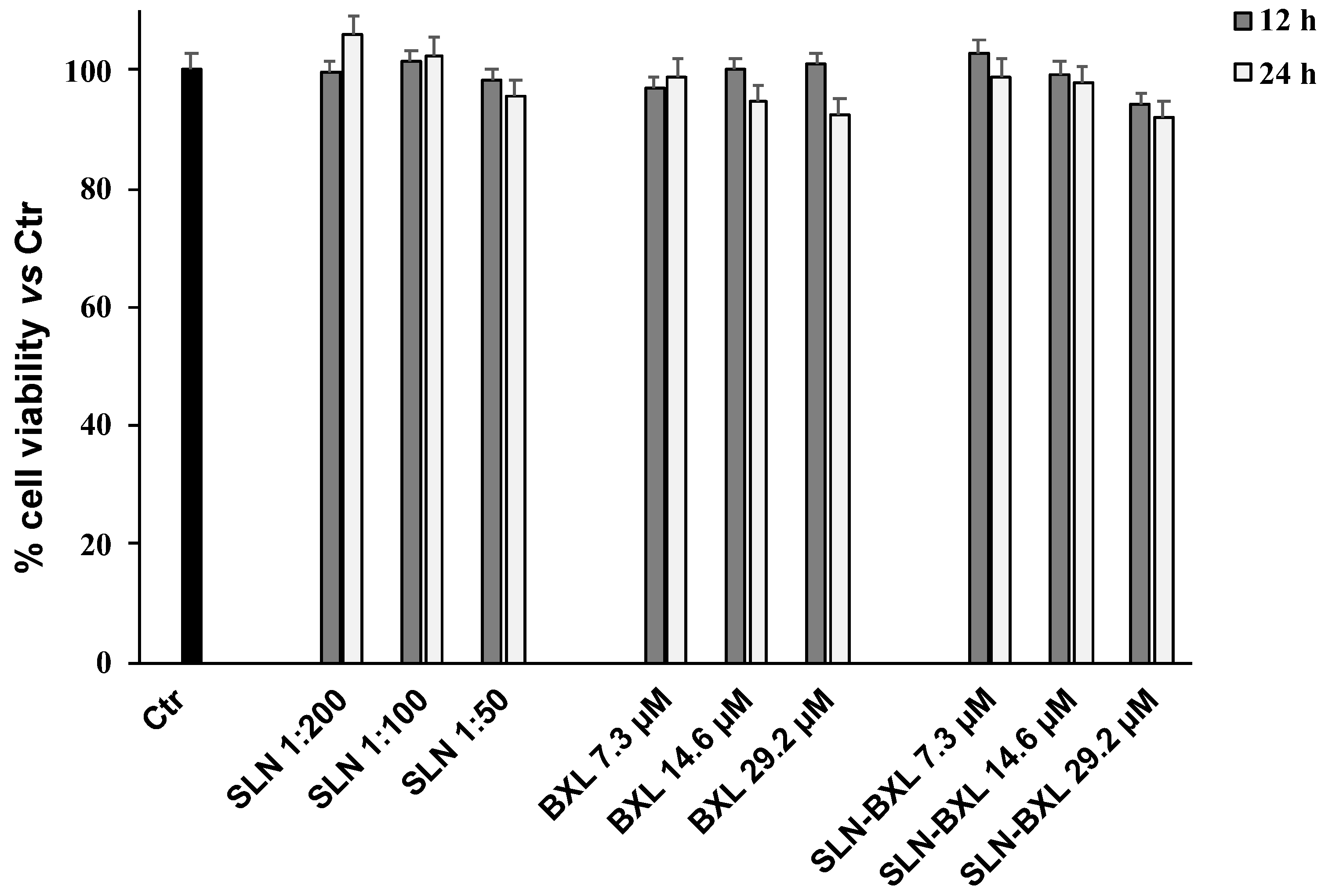
2.4. Cell Viability
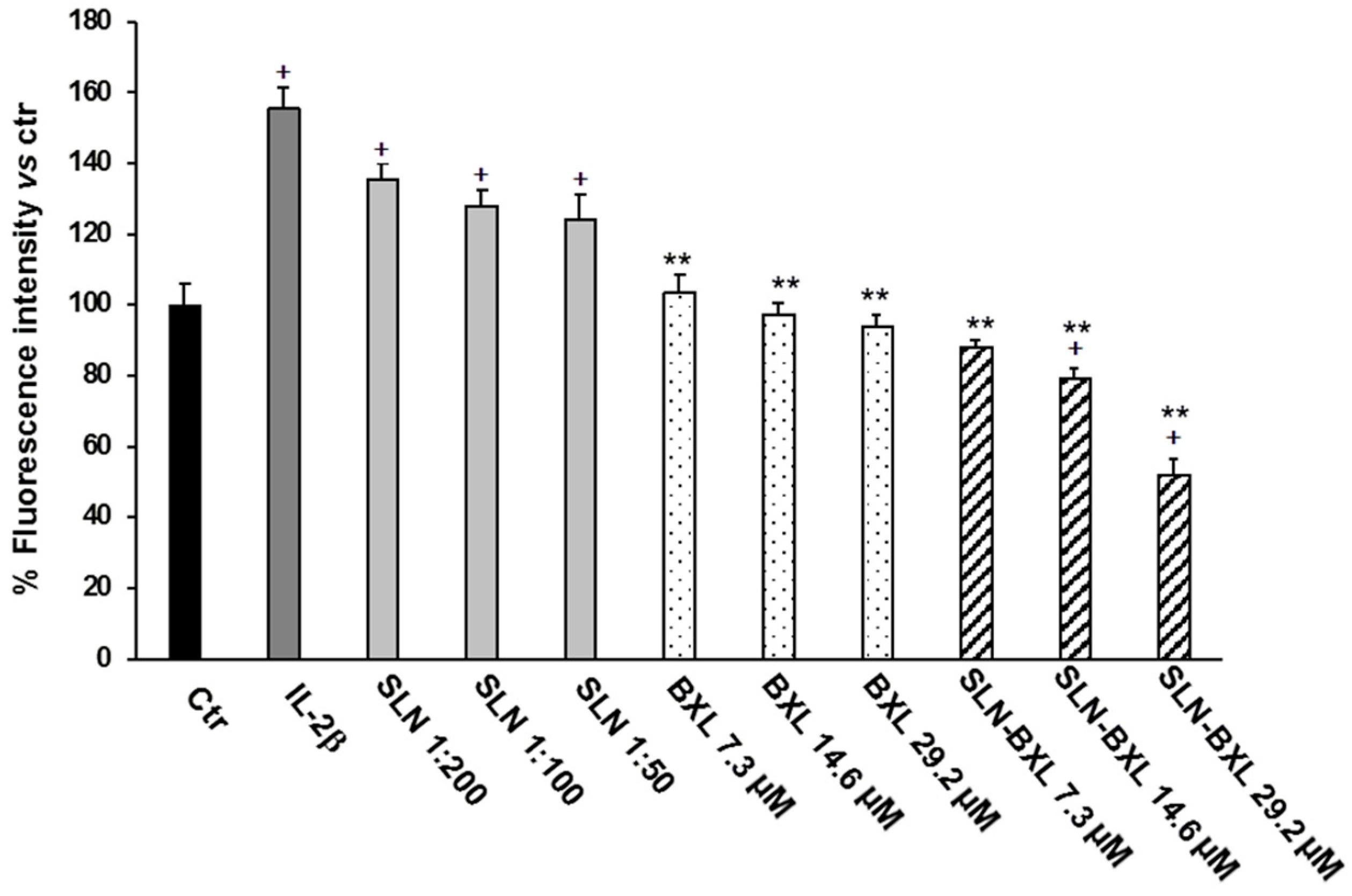
2.5. ROS Determination
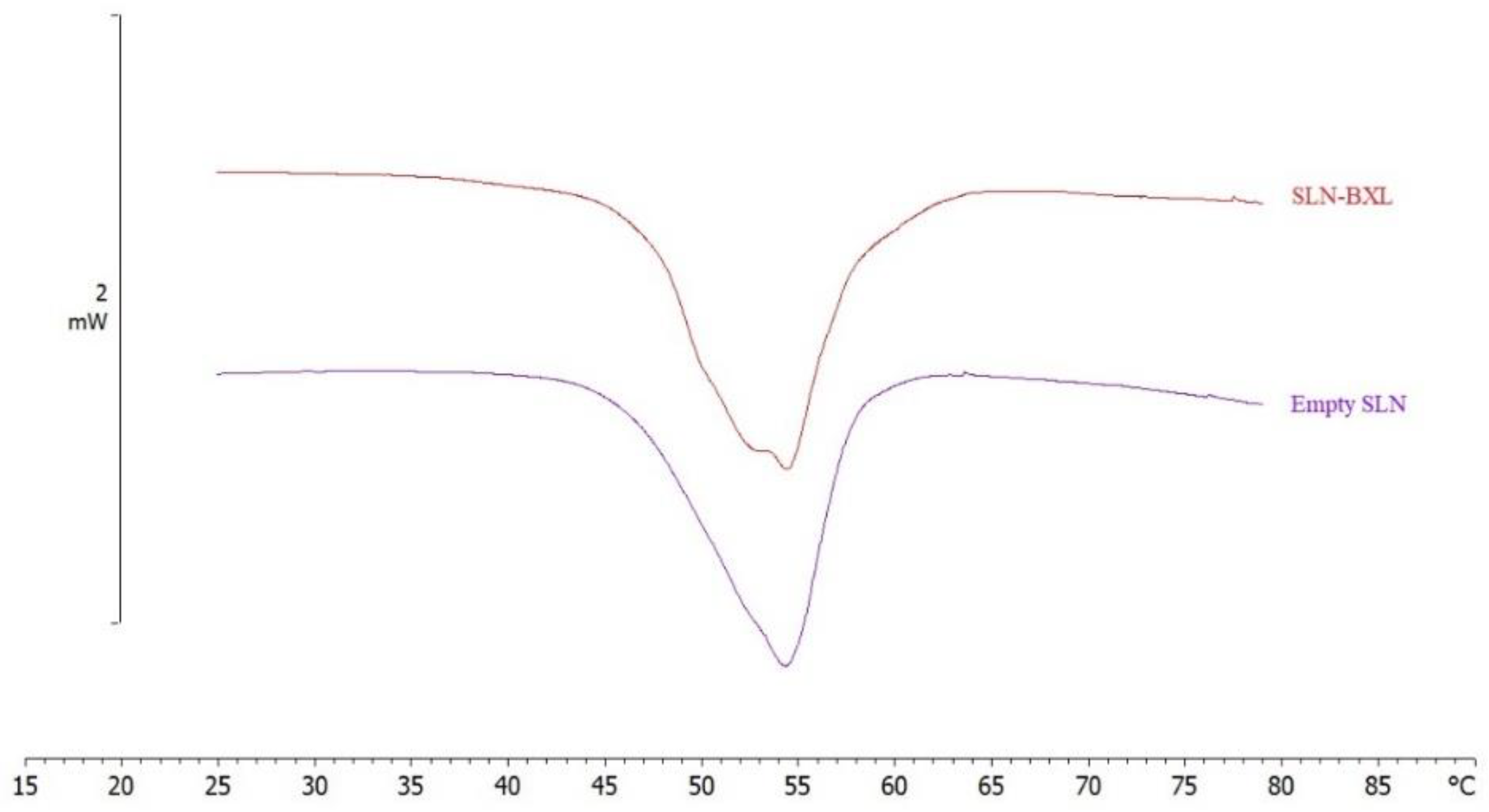
2.6. Empty SLN and SLN-BXL Calorimetric Analysis
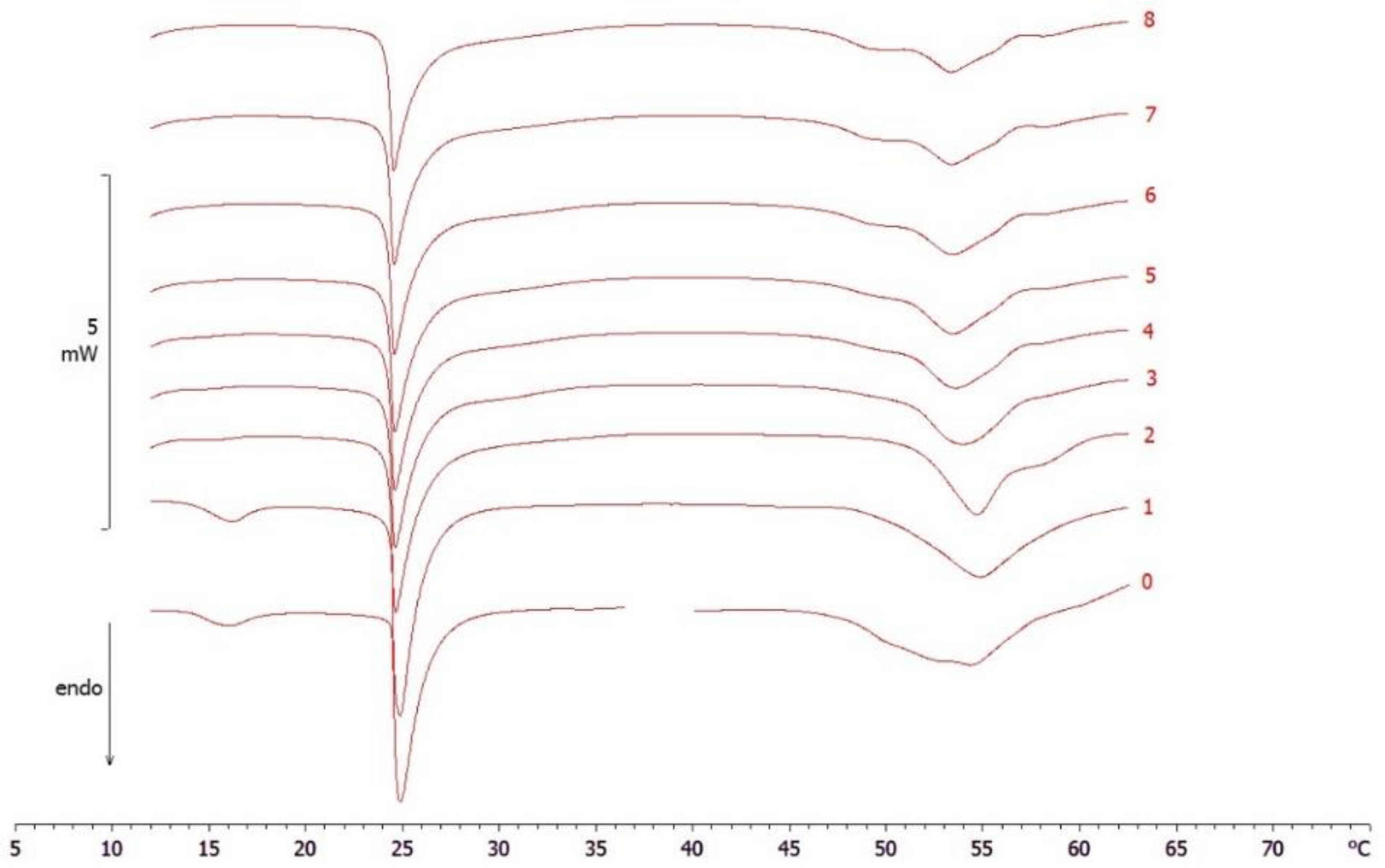
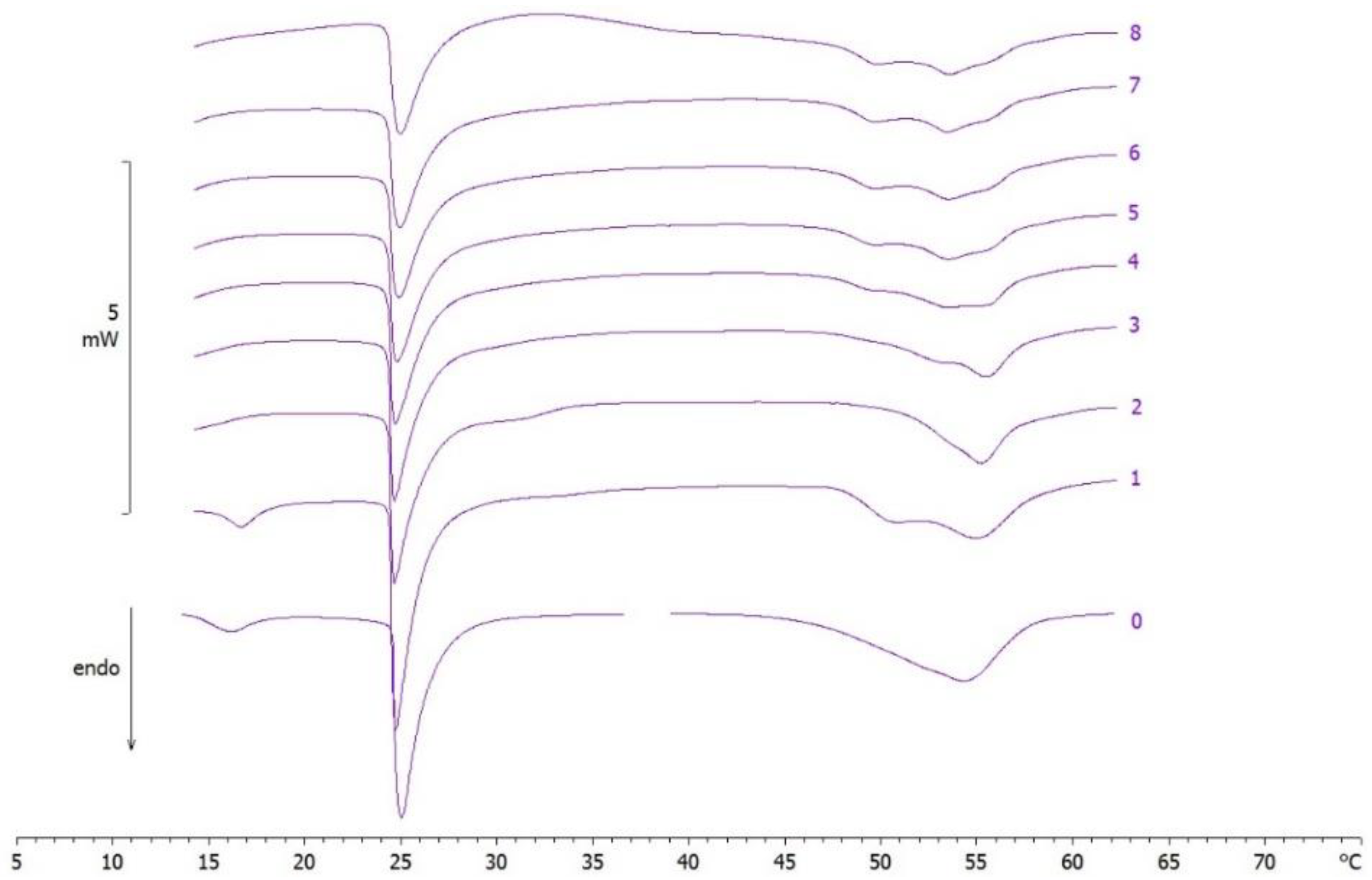
2.7. MLV/SLN Interaction Study
3. Materials and Methods
3.1. Materials
3.2. SLN Preparation
3.3. SLN Characterization
3.4. Encapsulation Efficiency
3.5. BXL Release from SLN
3.6. Cell Culture
3.7. MTT Bioassay
3.8. ROS Determination
3.9. Differential Scanning Calorimetry
3.10. Empty SLN and SLN-BXL Analysis
3.11. MLV/SLN Analysis
4. Conclusions
Supplementary Materials
Author Contributions
Funding
Institutional Review Board Statement
Informed Consent Statement
Data Availability Statement
Acknowledgments
Conflicts of Interest
Sample Availability
References
- Khavkin, J.; Ellis, D.A.F. Aging Skin: Histology, Physiology, and Pathology. Facial Plast. Surg. Clin. 2011, 19, 229–234. [Google Scholar] [CrossRef] [PubMed]
- Prow, T.W.; Grice, J.E.; Lin, L.L.; Faye, R.; Butler, M.; Becker, W.; Wurm, E.M.T.; Yoong, C.; Robertson, T.A.; Soyer, H.P.; et al. Nanoparticles and Microparticles for Skin Drug Delivery. Adv. Drug Deliv. Rev. 2011, 63, 470–491. [Google Scholar] [CrossRef] [PubMed]
- Garcês, A.; Amaral, M.H.; Sousa Lobo, J.M.; Silva, A.C. Formulations Based on Solid Lipid Nanoparticles (SLN) and Nanostructured Lipid Carriers (NLC) for Cutaneous Use: A Review. Eur. J. Pharm. Sci. 2018, 112, 159–167. [Google Scholar] [CrossRef]
- Sala, M.; Diab, R.; Elaissari, A.; Fessi, H. Lipid Nanocarriers as Skin Drug Delivery Systems: Properties, Mechanisms of Skin Interactions and Medical Applications. Int. J. Pharm. 2018, 535, 1–17. [Google Scholar] [CrossRef] [PubMed]
- Müller, R.H.; Radtke, M.; Wissing, S.A. Solid Lipid Nanoparticles (SLN) and Nanostructured Lipid Carriers (NLC) in Cosmetic and Dermatological Preparations. Adv. Drug Deliv. Rev. 2002, 54 (Suppl. 1), S131–S155. [Google Scholar] [CrossRef]
- Pardeike, J.; Hommoss, A.; Müller, R.H. Lipid Nanoparticles (SLN, NLC) in Cosmetic and Pharmaceutical Dermal Products. Int. J. Pharm. 2009, 366, 170–184. [Google Scholar] [CrossRef]
- Desai, P.; Patlolla, R.R.; Singh, M. Interaction of Nanoparticles and Cell-Penetrating Peptides with Skin for Transdermal Drug Delivery. Mol. Membr. Biol. 2010, 27, 247–259. [Google Scholar] [CrossRef] [PubMed]
- Mardhiah Adib, Z.; Ghanbarzadeh, S.; Kouhsoltani, M.; Yari Khosroshahi, A.; Hamishehkar, H. The Effect of Particle Size on the Deposition of Solid Lipid Nanoparticles in Different Skin Layers: A Histological Study. Adv. Pharm. Bull. 2016, 6, 31–36. [Google Scholar] [CrossRef]
- Bickers, D.R.; Athar, M. Oxidative Stress in the Pathogenesis of Skin Disease. J. Investig. Dermatol. 2006, 126, 2565–2575. [Google Scholar] [CrossRef] [PubMed]
- Genovese, C.; Pulvirenti, L.; Cardullo, N.; Muccilli, V.; Tempera, G.; Nicolosi, D.; Tringali, C. Bioinspired Benzoxanthene Lignans as a New Class of Antimycotic Agents: Synthesis and Candida Spp. Growth Inhibition. Nat. Prod. Res. 2020, 34, 1653–1662. [Google Scholar] [CrossRef]
- Tumir, L.-M.; Zonjić, I.; Žuna, K.; Brkanac, S.R.; Jukić, M.; Huđek, A.; Durgo, K.; Crnolatac, I.; Glavaš-Obrovac, L.; Cardullo, N.; et al. Synthesis, DNA/RNA-Interaction and Biological Activity of Benzo[k,l]Xanthene Lignans. Bioorg. Chem. 2020, 104, 104190. [Google Scholar] [CrossRef]
- Gerstmeier, J.; Kretzer, C.; Di Micco, S.; Miek, L.; Butschek, H.; Cantone, V.; Bilancia, R.; Rizza, R.; Troisi, F.; Cardullo, N.; et al. Novel Benzoxanthene Lignans That Favorably Modulate Lipid Mediator Biosynthesis: A Promising Pharmacological Strategy for Anti-Inflammatory Therapy. Biochem. Pharmacol. 2019, 165, 263–274. [Google Scholar] [CrossRef]
- Vijayakurup, V.; Carmela, S.; Carmelo, D.; Corrado, T.; Srinivas, P.; Gopala, S. Phenethyl Caffeate Benzo [Kl] Xanthene Lignan with DNA Interacting Properties Induces DNA Damage and Apoptosis in Colon Cancer Cells. Life Sci. 2012, 91, 1336–1344. [Google Scholar] [CrossRef] [PubMed]
- Capolupo, A.; Tosco, A.; Mozzicafreddo, M.; Tringali, C.; Cardullo, N.; Monti, M.C.; Casapullo, A. Proteasome as a New Target for Bio-Inspired Benzo[k,l]Xanthene Lignans. Chem. Weinh. Bergstr. Ger. 2017, 23, 8371–8374. [Google Scholar] [CrossRef] [PubMed]
- Di Micco, S.; Mazué, F.; Daquino, C.; Spatafora, C.; Delmas, D.; Latruffe, N.; Tringali, C.; Riccio, R.; Bifulco, G. Structural Basis for the Potential Antitumour Activity of DNA-Interacting Benzo [Kl] Xanthene Lignans. Org. Biomol. Chem. 2011, 9, 701–710. [Google Scholar] [CrossRef] [PubMed]
- Spatafora, C.; Barresi, V.; Bhusainahalli (Vedamurthy BM), V.; Micco, S.; Musso, N.; Riccio, R.; Bifulco, G.; Condorelli, D.; Tringali, C. Bio-Inspired Benzo[k,l]Xanthene Lignans: Synthesis, DNA-Interaction and Antiproliferative Properties. Org. Biomol. Chem. 2014, 12, 2686–2701. [Google Scholar] [CrossRef] [PubMed]
- Daina, A.; Michielin, O.; Zoete, V. SwissADME: A Free Web Tool to Evaluate Pharmacokinetics, Drug-Likeness and Medicinal Chemistry Friendliness of Small Molecules. Sci. Rep. 2017, 7, 42717. [Google Scholar] [CrossRef]
- Daina, A.; Michielin, O.; Zoete, V. ILOGP: A Simple, Robust, and Efficient Description of n-Octanol/Water Partition Coefficient for Drug Design Using the GB/SA Approach. J. Chem. Inf. Model. 2014, 54, 3284–3301. [Google Scholar] [CrossRef]
- Torrisi, C.; Cardullo, N.; Muccilli, V.; Tringali, C.; Castelli, F.; Sarpietro, M.G. Characterization and Interaction with Biomembrane Model of Benzo[k,l]Xanthene Lignan Loaded Solid Lipid Nanoparticles. Membranes 2022, 12, 615. [Google Scholar] [CrossRef]
- Gaspar, D.P.; Serra, C.; Lino, P.R.; Gonçalves, L.; Taboada, P.; Remuñán-López, C.; Almeida, A.J. Microencapsulated SLN: An Innovative Strategy for Pulmonary Protein Delivery. Int. J. Pharm. 2017, 516, 231–246. [Google Scholar] [CrossRef]
- Shnoudeh, A.J.; Hamad, I.; Abdo, R.W.; Qadumii, L.; Jaber, A.Y.; Surchi, H.S.; Alkelany, S.Z. Chapter 15—Synthesis, Characterization, and Applications of Metal Nanoparticles. In Biomaterials and Bionanotechnology; Tekade, R.K., Ed.; Advances in Pharmaceutical Product Development and Research; Academic Press: Cambridge, MA, USA, 2019; pp. 527–612. ISBN 978-0-12-814427-5. [Google Scholar]
- Barbosa, R.D.M.; Ribeiro, L.N.M.; Casadei, B.R.; da Silva, C.M.G.; Queiróz, V.A.; Duran, N.; de Araújo, D.R.; Severino, P.; de Paula, E. Solid Lipid Nanoparticles for Dibucaine Sustained Release. Pharmaceutics 2018, 10, 231. [Google Scholar] [CrossRef] [PubMed]
- Çankaya, G.; Akyol, S.; Genç, C.O.; Arslan, B.Ö.; Gökalp, M.U. A Novel and an Alternative in Vitro Release Test for Hydrophobic Diflorasone Diacetate Ointment with Dialysis Method by Using Reciprocating Cylinder, USP Apparatus 3. Glob. J. Pharm. Pharm. Sci. 2021, 8, 1–5. [Google Scholar] [CrossRef]
- Tomasello, B.; Malfa, G.A.; Acquaviva, R.; La Mantia, A.; Di Giacomo, C. Phytocomplex of a Standardized Extract from Red Orange (Citrus sinensis L. Osbeck) against Photoaging. Cells 2022, 11, 1447. [Google Scholar] [CrossRef]
- Cao, C.; Xiao, Z.; Wu, Y.; Ge, C. Diet and Skin Aging-From the Perspective of Food Nutrition. Nutrients 2020, 12, 870. [Google Scholar] [CrossRef]
- Bissett, D.L.; Chatterjee, R.; Hannon, D.P. Photoprotective Effect of Superoxide-Scavenging Antioxidants against Ultraviolet Radiation-Induced Chronic Skin Damage in the Hairless Mouse. Photodermatol. Photoimmunol. Photomed. 1990, 7, 56–62. [Google Scholar] [PubMed]
- Walde, P. Preparation of Vesicles (Liposomes). In Encyclopedia of Nanoscience and Nanotechnology; American Scientific Publishers: Stevenson Ranch, CA, USA, 2004; Volume 8, pp. 43–79. ISBN 1-58883-001-2. [Google Scholar]
- Pignatello, R.; Intravaia, V.D.; Puglisi, G. A Calorimetric Evaluation of the Interaction of Amphiphilic Prodrugs of Idebenone with a Biomembrane Model. J. Colloid Interface Sci. 2006, 299, 626–635. [Google Scholar] [CrossRef] [PubMed]
- Sarpietro, M.G.; Accolla, M.L.; Puglisi, G.; Castelli, F.; Montenegro, L. Idebenone Loaded Solid Lipid Nanoparticles: Calorimetric Studies on Surfactant and Drug Loading Effects. Int. J. Pharm. 2014, 471, 69–74. [Google Scholar] [CrossRef] [PubMed]
- Ricci, M.; Puglia, C.; Bonina, F.; Di Giovanni, C.; Giovagnoli, S.; Rossi, C. Evaluation of Indomethacin Percutaneous Absorption from Nanostructured Lipid Carriers (NLC): In Vitro and in Vivo Studies. J. Pharm. Sci. 2005, 94, 1149–1159. [Google Scholar] [CrossRef] [PubMed]
- Daré, R.G.; Costa, A.; Nakamura, C.V.; Truiti, M.C.T.; Ximenes, V.F.; Lautenschlager, S.O.S.; Sarmento, B. Evaluation of Lipid Nanoparticles for Topical Delivery of Protocatechuic Acid and Ethyl Protocatechuate as a New Photoprotection Strategy. Int. J. Pharm. 2020, 582, 119336. [Google Scholar] [CrossRef]
- Acquaviva, R.; Tomasello, B.; Di Giacomo, C.; Santangelo, R.; La Mantia, A.; Naletova, I.; Sarpietro, M.G.; Castelli, F.; Malfa, G.A. Protocatechuic Acid, a Simple Plant Secondary Metabolite, Induced Apoptosis by Promoting Oxidative Stress through HO-1 Downregulation and P21 Upregulation in Colon Cancer Cells. Biomolecules 2021, 11, 1485. [Google Scholar] [CrossRef] [PubMed]
- Tomasello, B.; Di Mauro, M.D.; Malfa, G.A.; Acquaviva, R.; Sinatra, F.; Spampinato, G.; Laudani, S.; Villaggio, G.; Bielak-Zmijewska, A.; Grabowska, W.; et al. Rapha Myr®, a Blend of Sulforaphane and Myrosinase, Exerts Antitumor and Anoikis-Sensitizing Effects on Human Astrocytoma Cells Modulating Sirtuins and DNA Methylation. Int. J. Mol. Sci. 2020, 21, 5328. [Google Scholar] [CrossRef] [PubMed]
- Torrisi, C.; Di Guardia, M.; Castelli, F.; Sarpietro, M.G. Naringenin Release to Biomembrane Models by Incorporation into Nanoparticles. Experimental Evidence Using Differential Scanning Calorimetry. Surfaces 2021, 4, 295–305. [Google Scholar] [CrossRef]
- Torrisi, C.; Morgante, A.; Malfa, G.; Acquaviva, R.; Castelli, F.; Pignatello, R.; Sarpietro, M.G. Sinapic Acid Release at the Cell Level by Incorporation into Nanoparticles: Experimental Evidence Using Biomembrane Models. Micro 2021, 1, 120–128. [Google Scholar] [CrossRef]


Publisher’s Note: MDPI stays neutral with regard to jurisdictional claims in published maps and institutional affiliations. |
© 2022 by the authors. Licensee MDPI, Basel, Switzerland. This article is an open access article distributed under the terms and conditions of the Creative Commons Attribution (CC BY) license (https://creativecommons.org/licenses/by/4.0/).
Share and Cite
Torrisi, C.; Cardullo, N.; Russo, S.; La Mantia, A.; Acquaviva, R.; Muccilli, V.; Castelli, F.; Sarpietro, M.G. Benzo[k,l]xanthene Lignan-Loaded Solid Lipid Nanoparticles for Topical Application: A Preliminary Study. Molecules 2022, 27, 5887. https://doi.org/10.3390/molecules27185887
Torrisi C, Cardullo N, Russo S, La Mantia A, Acquaviva R, Muccilli V, Castelli F, Sarpietro MG. Benzo[k,l]xanthene Lignan-Loaded Solid Lipid Nanoparticles for Topical Application: A Preliminary Study. Molecules. 2022; 27(18):5887. https://doi.org/10.3390/molecules27185887
Chicago/Turabian StyleTorrisi, Cristina, Nunzio Cardullo, Stefano Russo, Alfonsina La Mantia, Rosaria Acquaviva, Vera Muccilli, Francesco Castelli, and Maria Grazia Sarpietro. 2022. "Benzo[k,l]xanthene Lignan-Loaded Solid Lipid Nanoparticles for Topical Application: A Preliminary Study" Molecules 27, no. 18: 5887. https://doi.org/10.3390/molecules27185887
APA StyleTorrisi, C., Cardullo, N., Russo, S., La Mantia, A., Acquaviva, R., Muccilli, V., Castelli, F., & Sarpietro, M. G. (2022). Benzo[k,l]xanthene Lignan-Loaded Solid Lipid Nanoparticles for Topical Application: A Preliminary Study. Molecules, 27(18), 5887. https://doi.org/10.3390/molecules27185887