Article

Restoration of Hepatic and Intestinal Integrity by Phyllanthus amarus Is Dependent on Bax/Caspase 3 Modulation in Intestinal Ischemia-/Reperfusion-Induced Injury

by
Ayobami Oladele Afolabi
1,
Tunmise Maryanne Akhigbe
2,3,
Adeyemi Fatai Odetayo
3,4,
Davinson Chuka Anyogu
5,
Moses Agbomhere Hamed
3,6,7 and
Roland Eghoghosoa Akhigbe
1,3,*
1
Department of Physiology, Ladoke Akintola University of Technology, Ogbomoso 210214, Oyo State, Nigeria
2
Department of Agronomy, Osun State University, Osogbo 210001, Osun State, Nigeria
3
Reproductive Biology and Toxicology Research Laboratory, Oasis of Grace Hospital, Osogbo 210001, Osun State, Nigeria
4
Department of Physiology, University of Ilorin, Ilorin 240003, Kwara State, Nigeria
5
Department of Veterinary Pathology and Microbiology, University of Nigeria, Nsukka 410001, Enugu State, Nigeria
6
The Brainwill Laboratory, Osogbo 210001, Osun State, Nigeria
7
Department of Medical Laboratory Science, College of Medicine and Health Sciences, Afe Babalola University, Ado Ekiti 360001, Ekiti State, Nigeria
*
Author to whom correspondence should be addressed.
Molecules 2022, 27(16), 5073; https://doi.org/10.3390/molecules27165073
Submission received: 25 April 2022 / Revised: 25 May 2022 / Accepted: 27 May 2022 / Published: 9 August 2022

Abstract

:
Ethnopharmacological relevance: Oxidative stress is a key player in intestinal ischemia/reperfusion (I/R) injury (IIRI) with a tendency to trigger systemic inflammatory response, resulting in progressive distal organ injury. To date, the role of Bax/caspase 3 signaling in IIRI has not been reported. Furthermore, the discovery of a safe and effective drug remains pertinent in improving the outcome of IIRI. Therefore, this study investigated the role of Bax/caspase 3 signaling in intestinal I/R-induced intestinal and hepatic injury. In addition, the protective effect and possible associated mechanism of action of methanolic Phyllanthus amarus leaf extract (PA) against intestinal I/R-induced intestinal and hepatic injury were evaluated. Materials and methods: Fifty male Wistar rats were randomized into five groups (n = 10). The sham-operated group was received 0.5 mL of distilled water for seven days prior to the sham surgery, while the IIRI, febuxostat (FEB) + IIRI, low-dose PA (LDPA) + IIRI, and high-dose PA (HDPA) + IIRI groups underwent the I/R procedure. In addition to the procedure, IIRI, FEB + IIRI, LDPA + IIRI, and HDPA + IIRI received 0.5 mL of distilled water, 10 mg/kg of febuxostat, 200 mg/kg of PA, and 400 mg/kg of PA, respectively, for seven days prior to the I/R procedure. Results: Administration of methanolic Phyllanthus amarus leaf extracts attenuated the intestinal I/R-induced rise in intestinal and hepatic injury markers, malondialdehyde, nitric oxide, TNF-α, IL-6, and myeloperoxidase activities. In addition, Phyllanthus amarus ameliorated I/R-induced suppression of reduced glutathione, thiol and non-thiol proteins, and superoxide dismutase, catalase, and glutathione peroxidase activities in intestinal and hepatic tissues. These were coupled with the suppression of I/R-induced bacterial translocation, downregulation of I/R-induced activation of Bax/caspase 3 signaling, and improvement of I/R-induced distortion of intestinal and hepatic histoarchitecture by Phyllanthus amarus. Conclusion: Methanolic Phyllanthus amarus leaf extract protects against intestinal and hepatic injuries associated with intestinal I/R by suppressing oxidative-stress-mediated activation of Bax/caspase 3 signaling. The beneficial effects of Phyllanthus amarus may be ascribed to its constituent bioactive molecules, especially tannins, anthocyanin, alkaloids, and phenolics.

1. Introduction

Although virtually all organs are susceptible to ischemia reperfusion injury (IRI), intestinal ischemia–reperfusion injury (IIRI) has perhaps the greatest morbidity and mortality [1] among them all. This high morbidity and mortality are due to its propensity to affect virtually all organs, thus leading to multiple organ dysfunction syndrome (MODS) or, in extreme cases, multiple-organ failure (MOF) [2]. The mechanism by which this local event is transformed into life-threatening disorders has been the subject of numerous studies [3,4]. The pathophysiology of intestinal I/R-induced hepatic injury is complex and yet to be completely unraveled.
Restoration of blood flow results in biochemical and molecular metabolic alterations, which expose the tissues to damage mediated by free radicals [2] (generated by metabolic activities during the ischemic phase), leucocyte infiltration, inflammatory response, and apoptosis [5,6]. IIRI is especially lethal because of the disruption of the intestinal barrier, which results in increased permeability to toxins and bacteria, thereby allowing them to gain access to the general circulation [7]. The massive flux of intestinal microbial flora into portal circulation and, finally, into the systemic circulation [8,9] results in septicemia, which triggers a strong inflammatory response culminating in damage to distal organs such as the liver [10], lungs [11], kidney [12], heart [13], and even the brain [13]. Generally, the generation of reactive oxygen species plays a central role in the pathophysiological mechanism of IRI [5] through damage to cellular lipids [14], proteins [15], and DNA [16], which invariably facilitates apoptosis, eventually leading to cell death pathways [17]. Furthermore, ROS acting as a signaling molecule mediates the upregulation of pro-inflammatory cytokines, which disrupt intestinal barrier function [18], thereby promoting bacterial translocation. Although ROS-induced apoptosis has been shown to play a role in IIRI, there is a dearth of data reporting the role of Bcl-2-associated X protein (Bax) and cysteine-aspartic protease 3 (caspase 3) in I/R injury.
Caspase 3 may be activated via a death receptor (FAS, FAS-L), the endoplasmic reticulum, or the mitochondrial pathway (related to Bax) [19]. Upregulation of Bax triggers caspase 3 activation, which in turn induces apoptosis through chromosome condensation, cell membrane blebbing, and DNA fragmentation [20,21]. However, whether or not intestinal I/R-induced apoptosis involves Bax-mediated caspase 3 activation is not known.
Various molecules targeting ROS-mediated apoptosis have been employed in an attempt to protect against IRI. So far, no study has evaluated the role of Phyllanthus amarus (P. amarus) on intestinal I/R-induced injury. P. amarus has “slender, leaf-bearing branches, distichous leaves that are sub-sessile elliptic-oblong, rounded base and a branching annual glabrous herb that is about 30 to 60 cm high” [14] and belongs to the Euphorbiaceae family [15]. There are about 800 species of P. amarus, which are found in tropical and subtropical countries [16]. This herbal nutraceutical is known by various names. In Nigeria, it is called Eyin Olobe (Yoruba), Gyeron tsuntsaye (Hausa), and ngwu (Igbo). The leaves of P. amarus have been demonstrated to exert antioxidant, anti-inflammatory, analgesic, and antimicrobial properties [22,23]. It has also been reported to exhibit antidiabetic and antihypertensive activities [22] and to confer hepato-gastrointestinal protection [24,25].
Taking into consideration the medicinal values of P. amarus in folklore medicine, it is likely that P. amarus protects against I/R injury. Therefore, this study evaluated the role of Bax/caspase 3 signaling in intestinal I/R-induced intestinal and hepatic injury. In addition, the protective effect and the possible associated mechanism of action of methanolic P. amarus leaf extract against intestinal I/R-induced intestinal and hepatic injury was investigated.

2. Materials and Methods

2.1. Plant Collection

Fresh leaves of Phyllanthus amarus were collected in the California area, Ogbomoso, Oyo State, Nigeria. Identification and authentication of the plant were performed by Dr. Mrs. Ogundola of the Botany Unit, Department of Pure and Applied Biology, Ladoke Akintola University of Technology, Ogbomoso. The name of the plant was confirmed at http://www.theplantlist.org (accessed on 20 July 2021). A voucher specimen (LHO 615) was kept in the Herbarium at the Department of Botany, Ladoke Akintola University of Technology, Ogbomoso, Nigeria.

2.2. Preparation of Plant Extract

Preparation of plant extract has previously been reported [26,27]. The leaves of Phyllanthus amarus were air-dried for two weeks and pulverized using an electric blender. About 500 g of the obtained sample was soaked in 500 mL of 70% methanol for 72 h (3 days). The extract was filtered using mushin paper to separate the residue from the filtrate. The filtrate was poured inside a round-bottom flask of the Soxhlet apparatus and heated for one hour at a temperature of 60 °C, then poured inside a beaker and placed in a water bath for concentration at 100 °C for 24 h. The yield of the extract was 8.2%.

2.3. Phytochemical Analysis

Qualitative phytochemical analysis was performed using established methods described by Sofowora [28] and Trease et al. [29]. The secondary metabolites present were then quantified following established methods documented in previous studies [30,31,32,33,34].

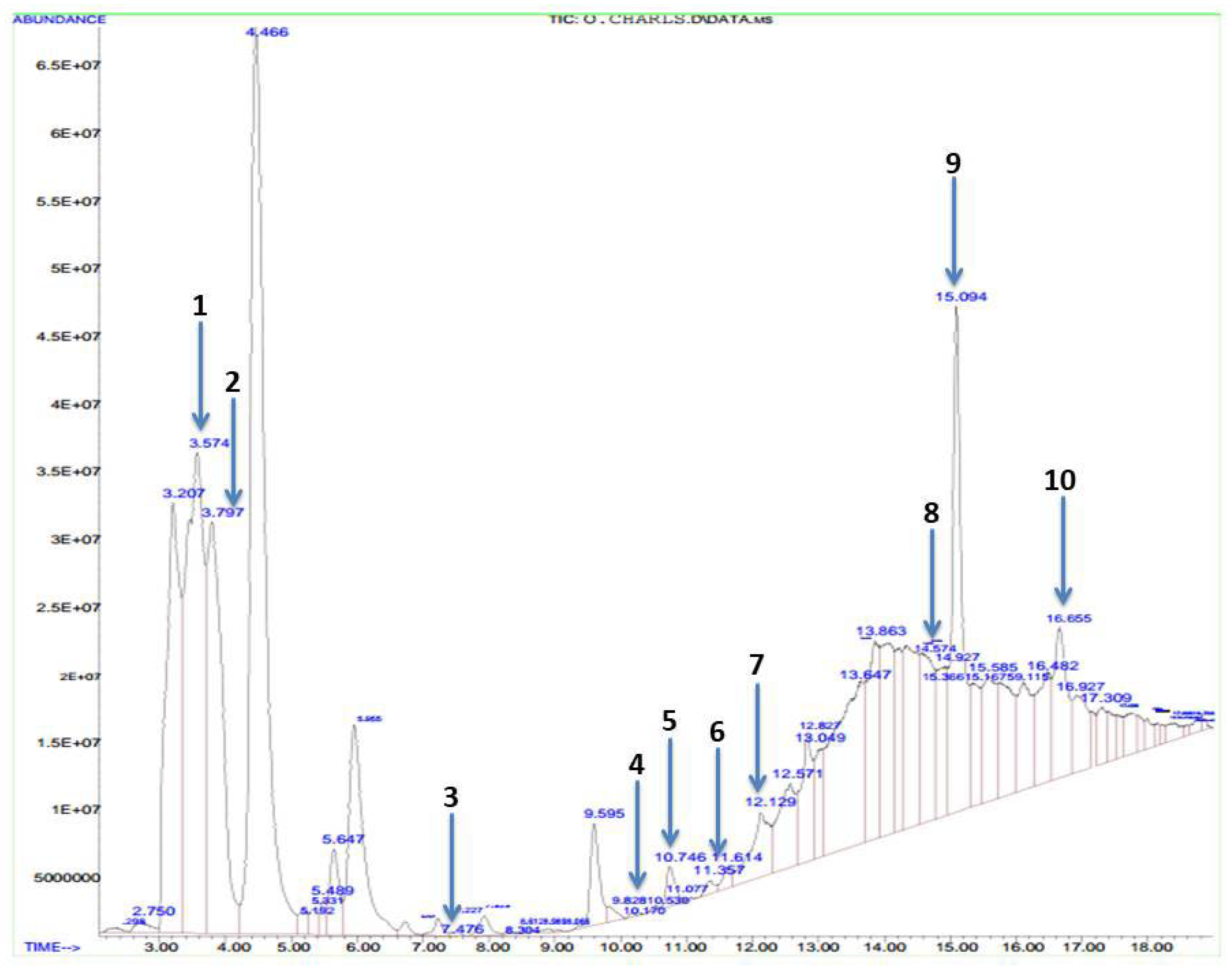
2.4. Gas Chromatography–Mass Spectrophotometric Analysis

The bioactive components of methanolic Phyllanthus amarus leaf extract were identified by gas chromatography–mass spectrophotometric (GC-MS) (PelkinEler Inc., Waltham, MA, USA) analysis following established methods [35]. The database of the National Institute of Standard and Technology (NIST) was employed in interpreting the mass spectrum of GC-MS to ascertain the name, molecular weight, and structure of the bioactive components of methanolic Phyllanthus amarus leaf extract.

2.5. Experimental Animals

This study was carried out in the animal house of the Department of Physiology, Ladoke Akintola University of Technology, Ogbomoso, Nigeria. Ethical approval was issued by The Ministry of Health, Oyo State, Nigeria (Approval Number AD13/479/44406). Fifty adult male Wistar rats of similar weight (190 ± 5 g) were used for the current study. The animal model using Wistar rats was adopted to mimic the clinical presentation of IIRI because this strain of rats has been established to share a close relationship to humans per genetics and biological and behavioral characteristics [36]. Animals were allowed free access to standard rat chow and water. Animals were humanely cared for using the Guidelines for the Care and Use of Laboratory Animals as published by the U.S. National Institutes of Health (NIH Publication No. 85-23, revised 1996).

2.6. Experimental Design

Animals were acclimatized for two weeks, then randomly assigned to five groups, each consisting of ten rats. The sham group was sham-operated and received 0.5 mL of distilled water for seven days prior to the sham surgery, while the IIRI, febuxostat (FEB) + IIRI, low-dose methanolic Phyllanthus amarus leaf extract (LDPA) + IIRI, and high-dose methanolic Phyllanthus amarus leaf extract (HDPA) + IIRI groups underwent the I/R procedure. In addition to the procedure, IIRI, FEB + IIRI, LDPA + IIRI, and HDPA + IIRI received 0.5 mL of distilled water, 10 mg/kg of febuxostat, 200 mg/kg of methanolic Phyllanthus amarus leaf extract, and 400 mg/kg of PA, respectively, for seven days prior to the I/R procedure. Febuxostat was used as a standard control drug with anti-inflammatory and antioxidant properties that protect against I/R injury [37,38]. The administration of the drugs was via gavage. The doses of febuxostat [39] and Phyllanthus amarus [40,41] were as previously reported.
IIRI was induced as previously reported [42] with some modifications. The rats were weighed and anesthetized with 10 mg/kg of Xylazine and 50 mg/kg of Ketamine. The abdomen was cleaned with 10% povidone iodine; a ventral midline incision was made on the abdomen, and the intestine was mobilized. About 5 cm of the ileum was measured proximally from the illio-cecal junction and a further 5 cm measured proximal to the first 5 cm. The most proximal five cm of ileum was folded on itself and twisted 720° clockwise. The twisted ileum was then anchored to the anterior abdominal wall by placing a 3-0 chromic suture around it and through the avascular part of the mesentery. The abdomen was then closed lightly with a 3-0 chromic suture, after which the animal was left for forty-five minutes. At the expiration of the 45 min of ischemia, the abdomen was reopened. Loss of palpitation was observed to confirm that ischemia had been successfully induced. Reperfusion was induced by removing the suture and untwisting the ileum for twenty-four hours.

2.7. Sacrifice and Tissue Collection

After 60 min of reperfusion, animals were euthanized and the intestine and liver were rapidly removed and weighed after separating the adhering structures.
Portions of the intestinal and hepatic tissues were homogenized in an appropriate volume of cold phosphate-buffered saline using a glass homogenizer. The homogenates were centrifuged at 10,000 rpm for 15 min in a cold centrifuge to obtain the supernatant fractions.
In addition, portions of the intestinal and hepatic tissues were obtained and fixed in 10% formalin-phosphate-buffered saline at 4 °C overnight. The samples were dehydrated and embedded in paraffin wax. About 5 µm-thick sections were cut and stained with hematoxylin-eosin and examined under a light microscope by a pathologist who was blinded to the study protocol. Photomicrographs were taken at ×100 and ×400 magnifications.
Formalin-fixed and paraffin-embedded intestinal tissues were sectioned at about 4 µm for immunohistochemistry for Bax protein expression, as earlier reported [43].

2.8. Biochemical Analyses

2.8.1. Tissue Injury Markers

The activities of aspartate transaminase (AST), alanine transferase (ALT), alanine phosphatase (ALP), and gamma-glutamyl transferase (GGT) in the hepatic tissue [44], as well as the activities of lactate dehydrogenase (LDH) in the intestinal and hepatic tissues were assayed by spectrophotometry, as previously reported [21].

2.8.2. Markers of Oxidative Stress and Antioxidant Levels

The intestinal and hepatic concentrations of malondialdehyde (MDA) and reduced glutathione (GSH) [5] and the activities of superoxide dismutase (SOD), catalase, and glutathione peroxidase (GPx) [17,45] in the intestinal and hepatic tissues were assayed by spectrophotometry, as previously reported.
Thiol and non-thiol proteins were assayed by colorimetry, as earlier reported [46,47].

2.8.3. Markers of Inflammation

The concentrations of nitric oxide (NO) and myeloperoxidase (MPO) activities in the intestinal and hepatic tissues were determined by colorimetric methods as earlier documented [17]. The intestinal and hepatic levels of tumor necrotic factor-α (TNF-α) and interleukin-6 (IL-6) were determined using ELISA kits (Elabscience Biotechnology Inc., Houston, TX, USA) according to the manufacturers’ guidelines.

2.8.4. Marker of Apoptosis

The activities of caspase 3 in the intestinal and hepatic tissues were assayed using ELISA kits (Elabscience Biotechnology Inc., Houston, TX, USA) according to the manufacturer’s guidelines.

2.9. Microbiological Analysis

Bacterial translocation was determined as previously reported [48] under strict sterile conditions. Briefly, some portions of the intestinal and hepatic tissues were cut into pieces using a sterile blade and put into 1 mL of Mueller–Hinton Broth. The samples were homogenized, and about 100 µL of each sample was inoculated into nutrient agar (NA), eosin-methylene blue (EMB), and Mac Conkey agar (MCCA) for colony counts. The cultures were incubated for 48 h and observed for the presence of growth under either aerobic or anaerobic conditions.

2.10. Histopathological Analysis

Intestinal and hepatic histomorphological appearance was scored using Chiu’s [49] (Supplementary Table S1) and Eckhoff’s score [50] (Supplementary Table S2), respectively.
Digital photomicrographs obtained from histopathology and immunohistochemistry were imported into the Image J software (NIH, Bethesda, MD, USA) with specific plugins for quantification of intestinal villi length and crypt depth, hepatic sinusoidal length and width, and Bax expression. The results were expressed as fold change relative to the normal control group [43].

2.11. Statistical Analysis

GraphPad Prism (Versions 7.00; Graph Pad Software, San Diego, CA 92108, USA) was used for data analysis. Analysis of variance (ANOVA) was used to compare the mean values across all the groups, and then, Tukey’s post hoc test was used for pairwise comparison. To ascertain intestinal–hepatic crosstalk, Pearson’s correlation was conducted between hepatic injury markers and intestinal histomorphological changes (Chiu’s score). Data are presented as means ± standard deviations.

3. Results

3.1. Bioactive Compounds of Methanolic Phyllanthus amarus Leaf Extract

Phytochemical analysis revealed that tannins (1.850 ± 0.030) and anthocyanin (1.820 ± 0.020) were the most abundant phytochemical compounds in methanolic P. amarus leaf extract, followed by alkaloids (1.607 ± 0.013), phenolics (1.130 ± 0.014), glycosides (0.863 ± 0.015), saponins (0.747 ± 0.017), and triterpenoids (0.140 ± 0.010) (Table 1).
Table 2 and Figure 1 show the organic constituents of methanolic P. amarus as revealed by GC-MS. It was observed that methyl stearate had the highest retention time of 16.655 min, while butanoic acid was found to have the least retention time of 3.574 min. The chemical properties of these organic constituents are shown in Table 2, and the chemical structures are provided in Supplementary Figure S1.

3.2. Organ Weight

As shown in Figure 2, I/R led to a significant increase in the absolute intestinal and hepatic weight compared with the animals in the sham group. The I/R-induced increase in intestinal and hepatic mass was significantly attenuated by FEB, LDPA, and HDPA.

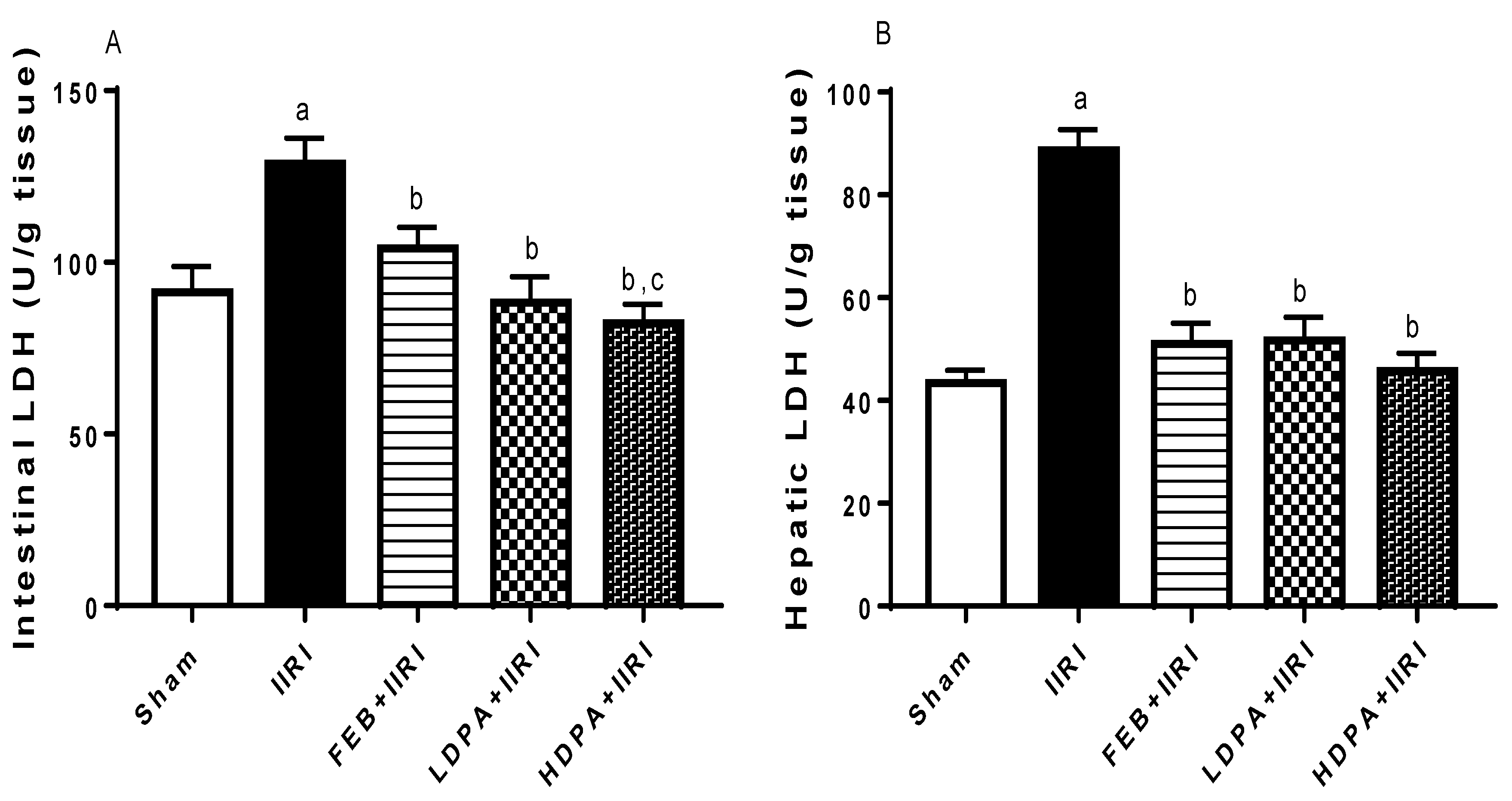
3.3. Hepatic Injury Markers

The effect of intestinal ischemia/reperfusion injury on tissue injury markers was also probed (Table 3). It was observed that intestinal I/R significantly elevated the activities of hepatic AST, ALT, ALP, and GGT compared with the sham group. The observed changes in hepatic injury markers following I/R were significantly ameliorated by FEB, LDPA, and HDPA. Although the effects of HDPA on ALT, ALP, and GGT were comparable with those of FEB, HDPA significantly reduced AST activity when compared with FEB.
In addition, I/R led to a marked increase in the activities of intestinal and hepatic LDH, as shown in Figure 3. The observed rise in intestinal and hepatic LDH was blunted by FEB, LDPA, and HDPA. When compared with FEB, HDPA caused a significant reduction in intestinal LDH activity.

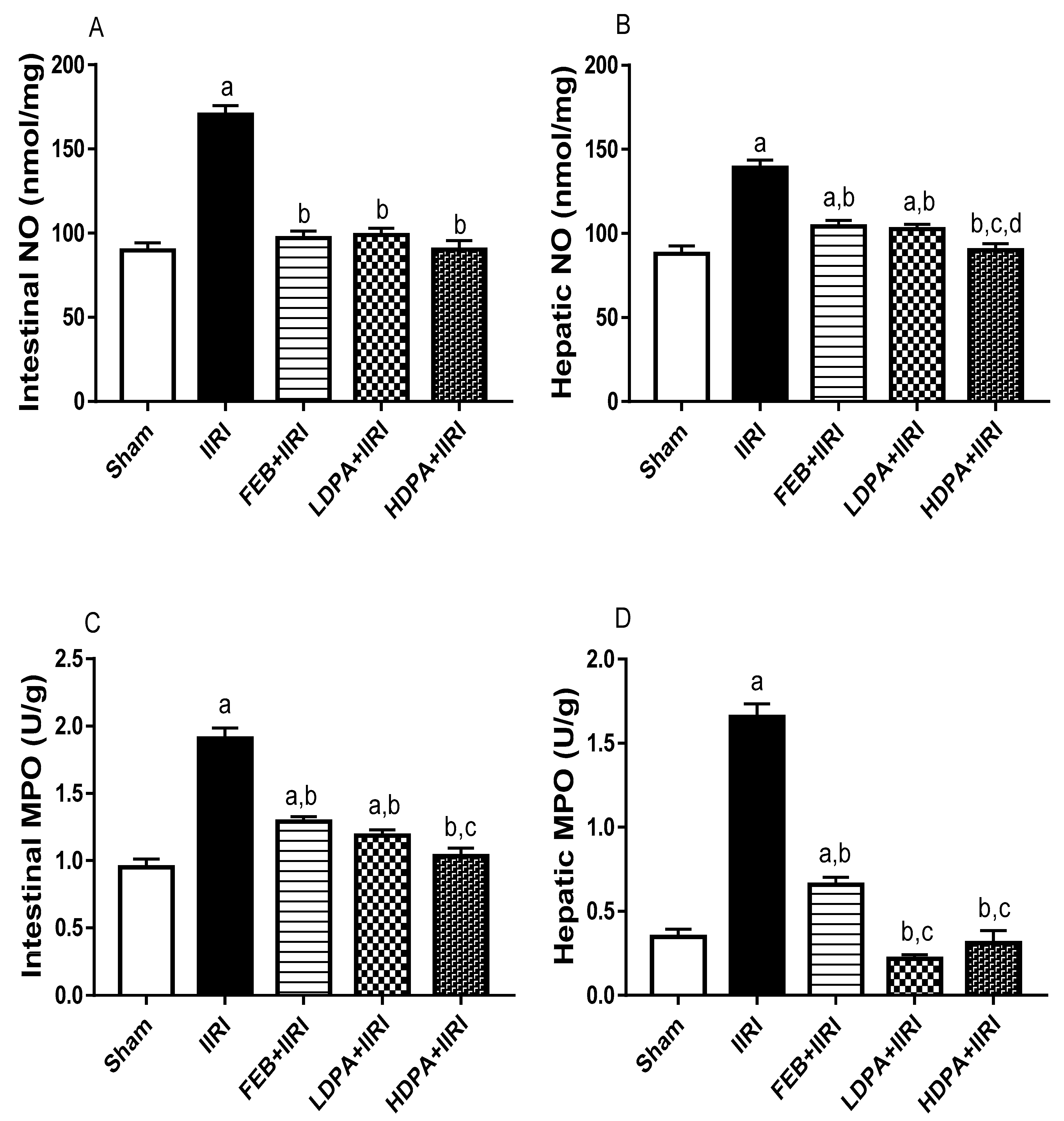
3.4. Markers of Oxidative Stress and Inflammation

I/R led to a significant increase in intestinal and hepatic MDA. This increase was significantly attenuated by FEB, LDPA, and HDPA. Furthermore, I/R led to a significant reduction in intestinal and hepatic GSH, thiol, and non-thiol proteins, as well as SOD, catalase, and GPx activities. The observed decrease in these parameters was abolished by FEB, LDPA, and HDPA treatments. When compared with other treatments, HDPA caused a significantly reduced intestinal MDA level and increased GSH and non-thiol protein levels and SOD activities (Table 4).
Furthermore, intestinal I/R led to a significant increase in intestinal and hepatic MPO activities and NO, TNF-α, and IL-6 levels compared with the sham-operated group. The I/R-led increase in these inflammatory markers was significantly prevented by the FEB, LDPA, and HDPA treatments. When compared with other treatments, HDPA caused significantly reduced intestinal and hepatic MPO activities and hepatic NO and TNF-α levels (Figure 4).

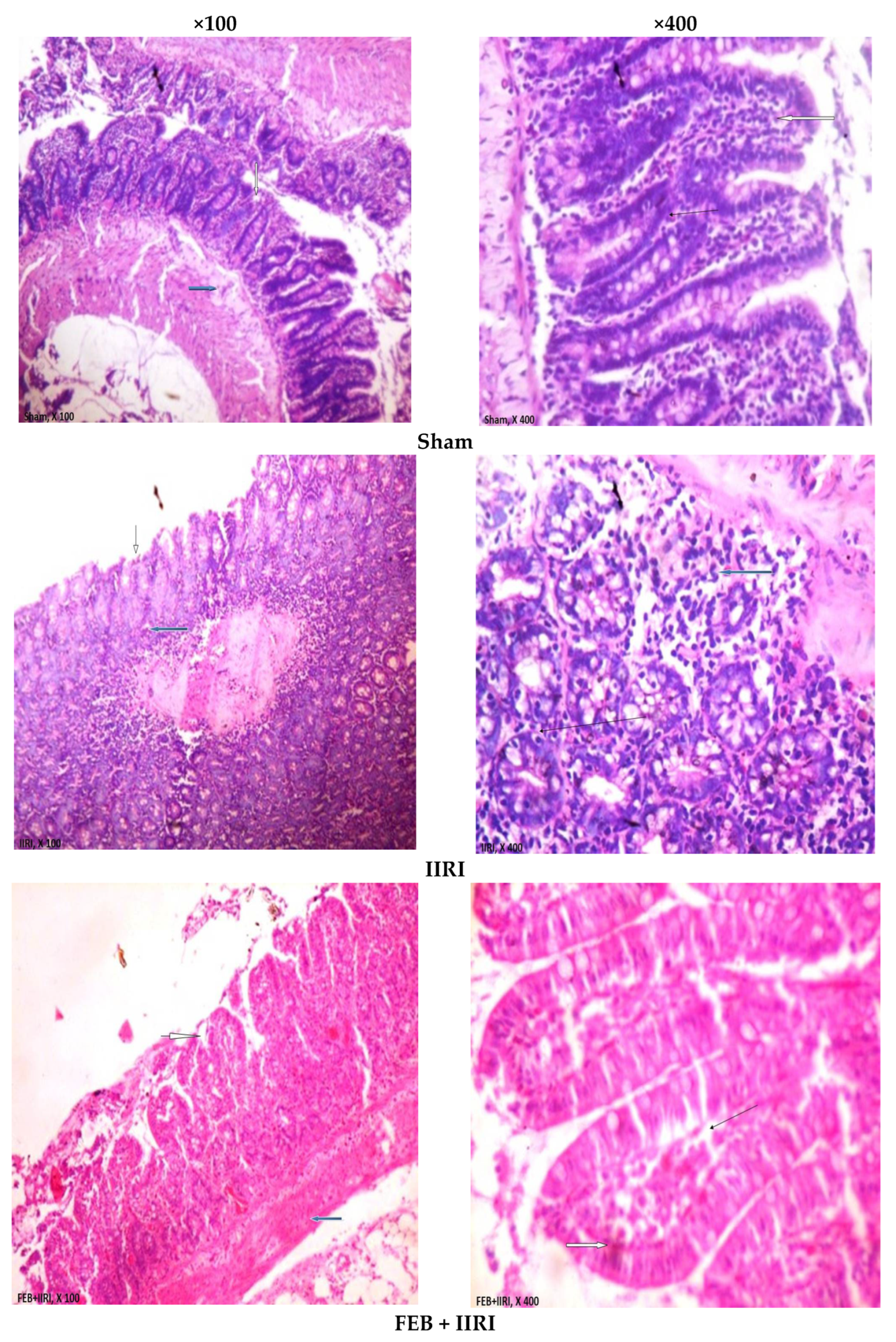
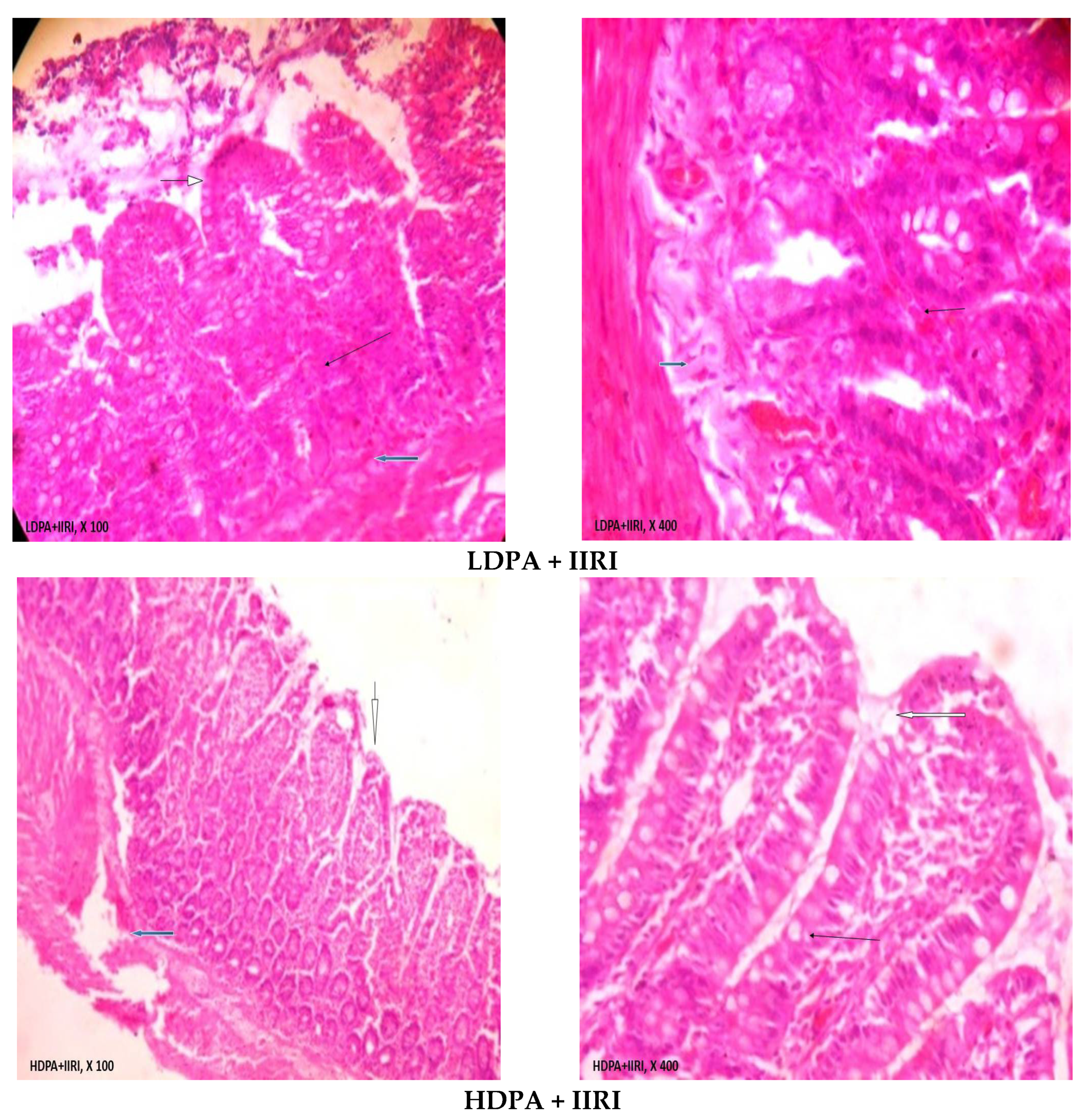
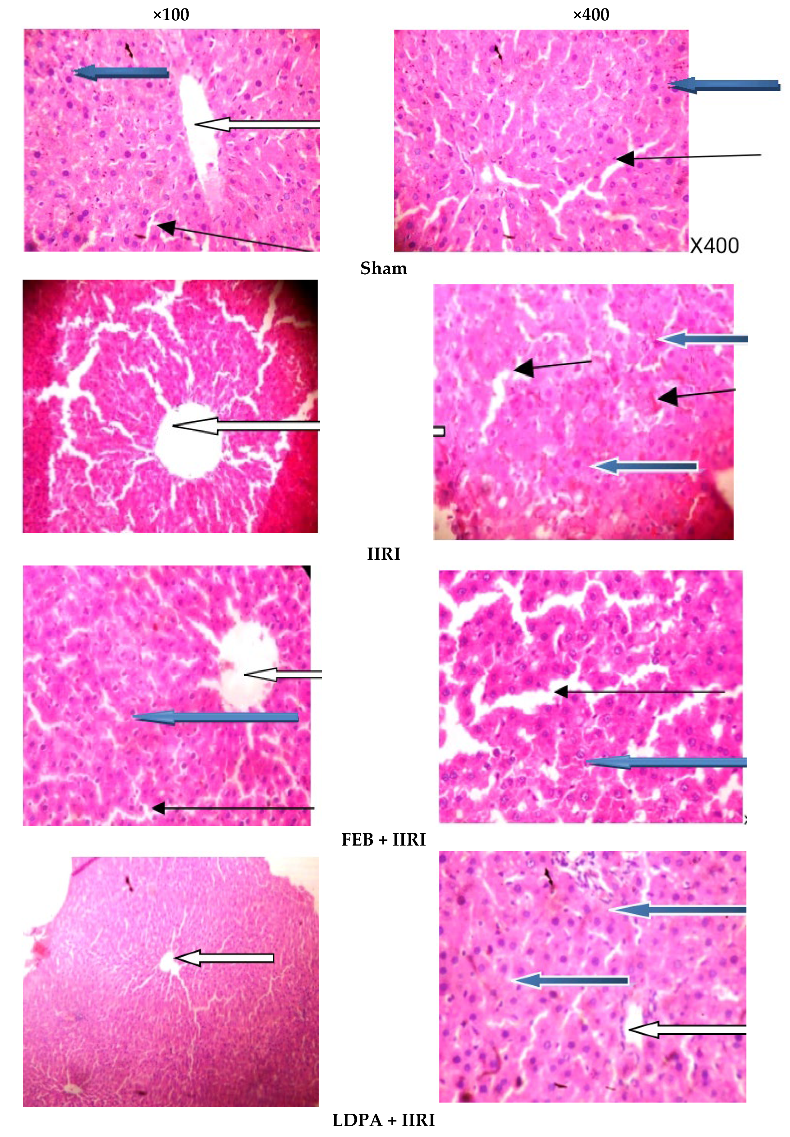
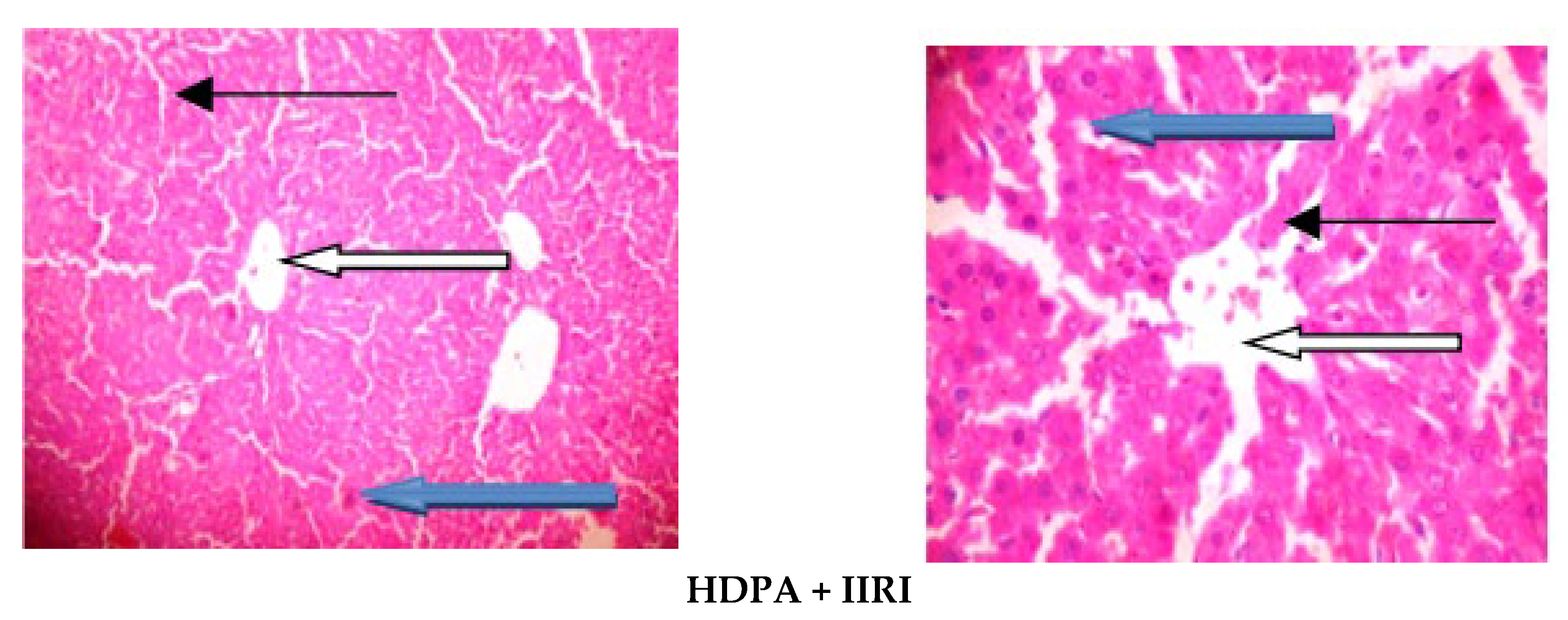
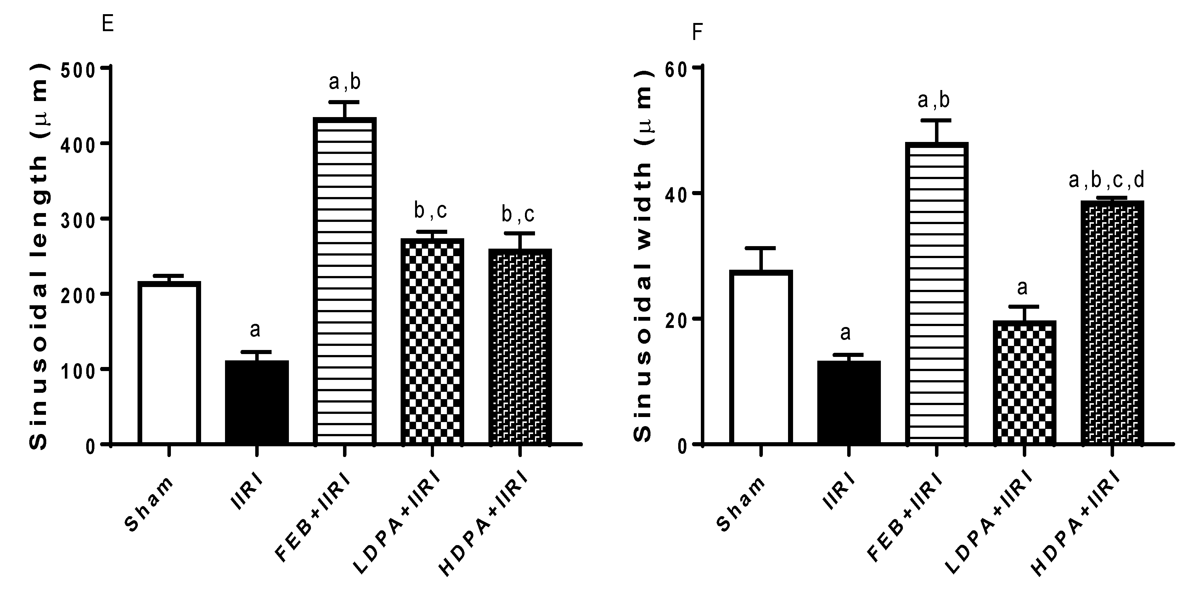
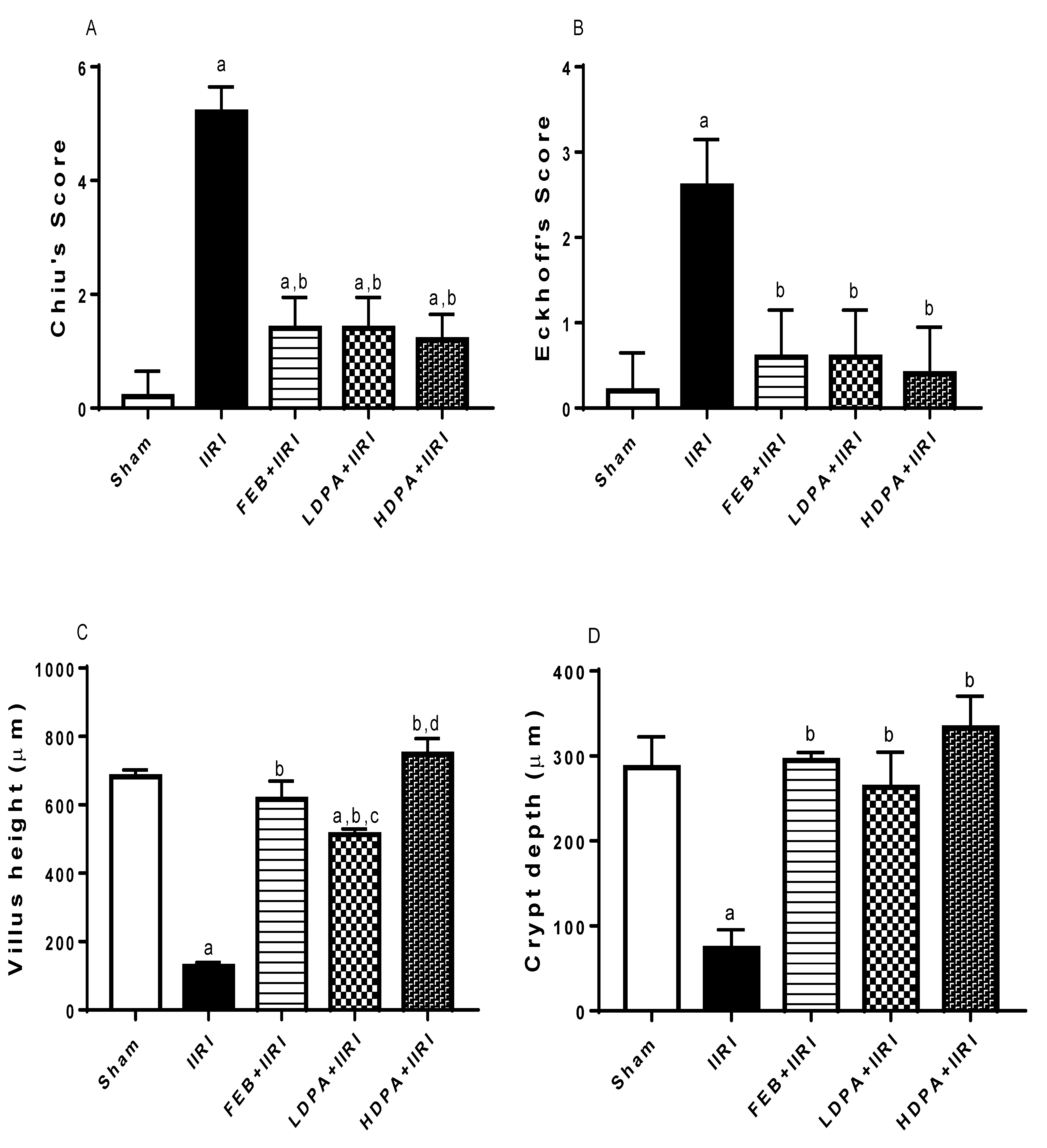
3.5. Intestinal and Hepatic Histoarchitecture

As shown in Figure 5, intestinal I/R disrupted intestinal histoarchitecture. The sham-operated animals showed normal villi from the mucosal layer, and the lumen was moderately infiltrated with lymphocytes. The propria and submucosal layer showed moderate infiltration of inflammatory cells. The animals in the IIRI group exhibited a chronically inflamed mucosal layer with sloughed and inflamed villi. There was infiltration of the lumen by lymphocytes, polymorphs, and plasma cells involving the propria and glands. The submucosal layer was infiltrated severely by inflammatory cells. Animals who received the FEB treatment showed moderately preserved villi from the mucosal layer. The lumen showed severe lymphocyte infiltration and the propria with severe inter-glandular infiltration of inflammatory cells. The submucosal layer appeared normal. Unlike the animals in the LDPA-treated group, which showed moderately preserved villi from the mucosal layer, those in the HDPA-treated group showed normal villi from the mucosal layer. In addition, the animals in both the LDPA- and HDPA-treated groups showed lumen and propria with mild lymphocyte infiltrations and inter-glandular infiltration of inflammatory cells, respectively. The submucosal layer appeared normal in the LDPA- and HDPA-treated animals.
As shown in Figure 6, the hepatic histoarchitecture was distorted following intestinal I/R compared with the animals in the sham group. Animals in the IIRI group showed mild congestion of the portal vein, some degenerated hepatocytes, and necrotic hepatocytes. At the same time, all these histopathological lesions were prevented by the FEB, LDPA, and HDPA treatments.
Figure 7 shows the histomorphological changes in the intestinal and hepatic tissues using Chiu’s and Eckhoff’s scores, respectively. When compared with the sham-operated rats, I/R caused a significant increase in Chiu’s and Eckhoff’s scores in the IIRI group. Treatments with FEB, LDPA, and HDPA significantly abolished I/R-induced alterations in intestinal and hepatic histomorphology. IIRI also significantly reduced villi length and crypt depth compared with the sham-operated rats. The observed IIRI-induced alterations in villi length and crypt depth were significantly ameliorated by FEB, LDPA, and HDPA treatments. Although the effects of LDPA and HDPA were similar on crypt depth, the effect on villi length was dose-dependent. Additionally, IIRI significantly reduced hepatic sinusoidal length and width. The noted IIRI-led reduction in sinusoidal length, but not sinusoidal width, was significantly alleviated by the FEB, LDPA, and HDPA treatments. Only FEB and HDPA significantly abrogated IIRI-driven sinusoidal width reduction.

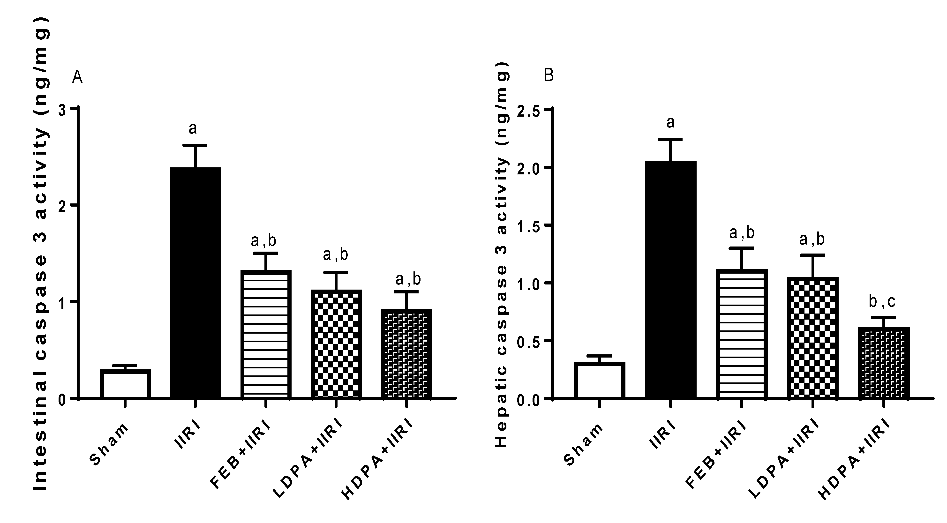
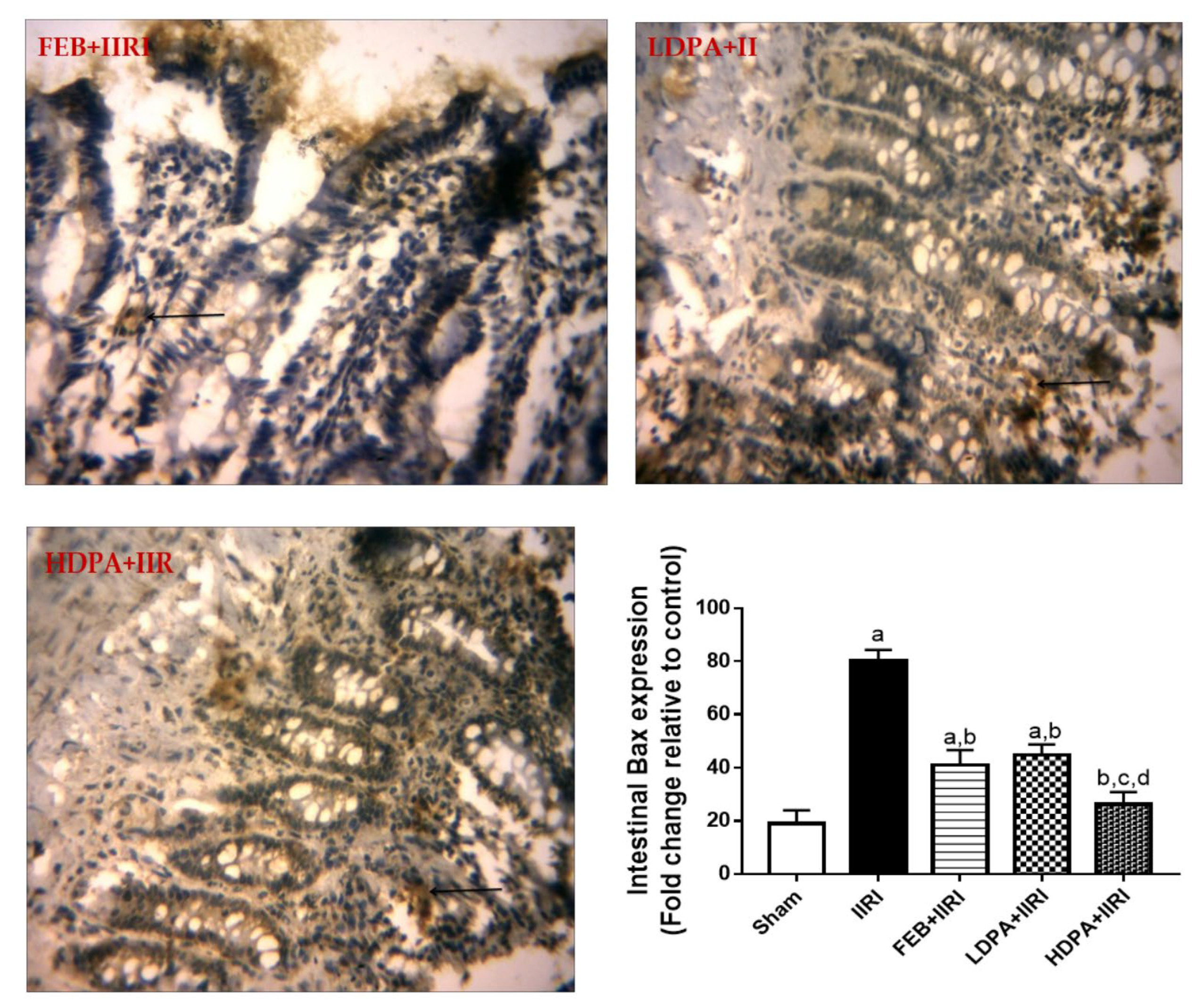
3.6. Apoptotic Markers
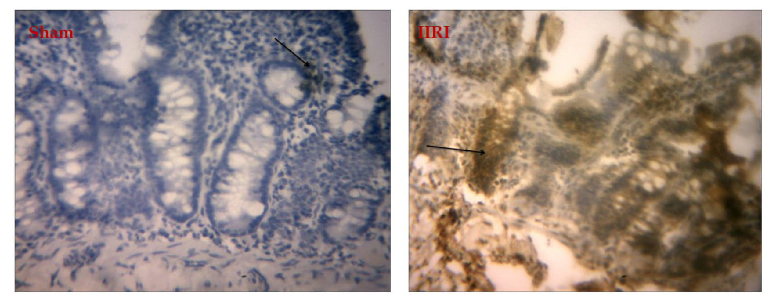
Animals in the IIRI group showed a marked rise in intestinal and hepatic caspase 3 activities compared with the sham group. The I/R-driven rise in caspase 3 activities was significantly alleviated by the FEB, LDPA, and HDPA treatments (Figure 8).

3.7. Bax Expression

When compared with the sham-operated group, the IIRI group showed a marked increase in Bax expression in the intestinal tissues. Treatments with FEB, LDPA, and HDPA significantly inhibited I/R-induced upregulation of Bax expression. Additionally, HDPA treatment significantly reduced intestinal Bax expression when compared with other treatments (Figure 9).

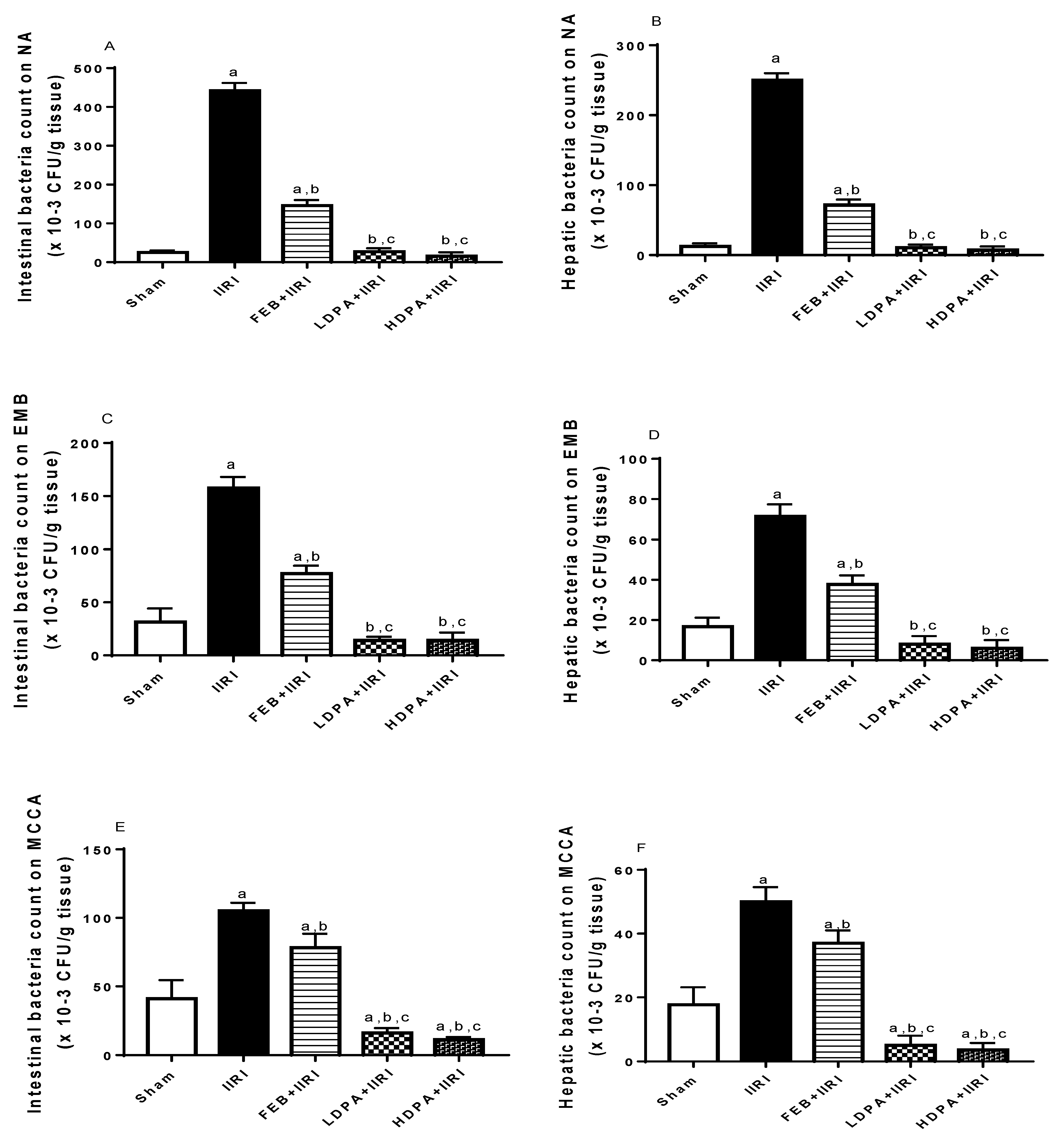
3.8. Bacterial Translocation

Using nutrient agar, eosin-methylene blue, and Mac Conkey agar as culture media, I/R significantly increased the bacteria count in intestinal and hepatic tissues in the IIRI group compared with the sham-operated group. FEB treatment significantly reduced the I/R-led rise in bacteria count. Interestingly, when compared with the IIRI and FEB + IIRI groups, the LDPA and HDPA groups showed significantly reduced bacteria counts (Figure 10).
IR promotes reactive oxygen species (ROS) and pro-inflammatory cytokine generation, which in turn translocate Bax from the cytosol into the mitochondrial outer membrane, where it becomes activated by binding with BH3-only proteins. Activated Bax oligomerizes to form pores in the mitochondrial outer membrane and triggers the release of cytochrome c into the cytosol, which in turn activates caspase 3, which induces apoptosis. Administration of PA inhibits IR-induced upregulation of Bax/caspase-3-mediated apoptosis via suppression of ROS and cytokine generation.

3.9. Correlation Study
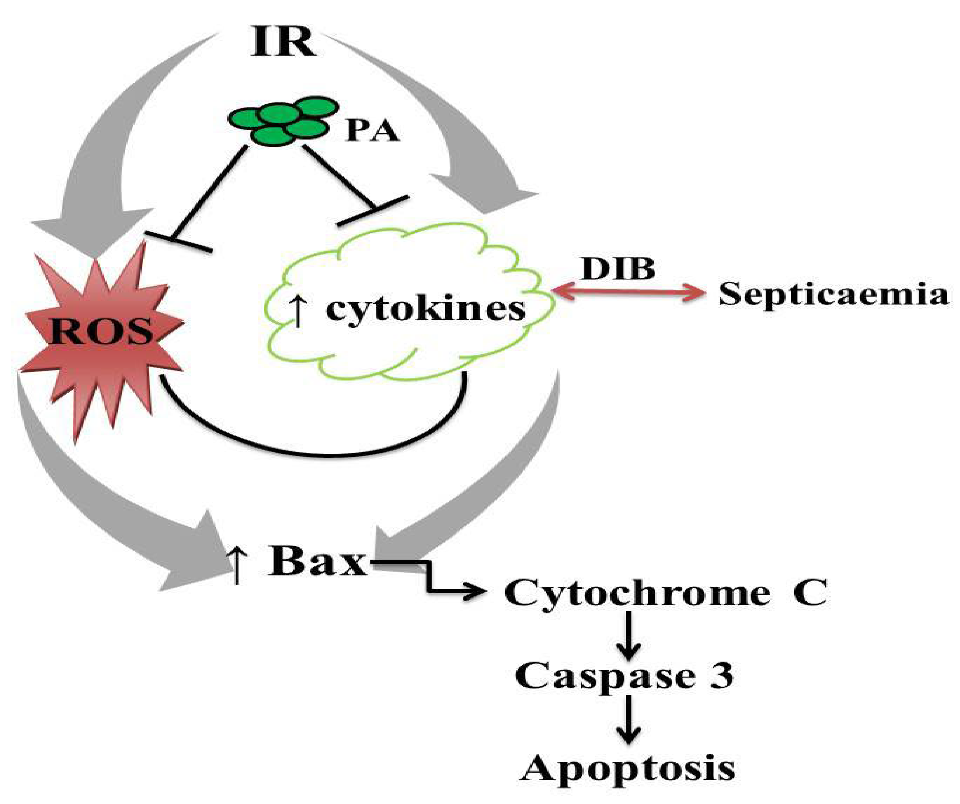
Using Pearson’s correlation analysis, it was observed that Chiu’s score of intestinal histomorphological changes was significantly positively correlated with hepatic Eckhoff’s histomorphological score, AST, and ALT, but not GGT. Furthermore, intestinal damage (using Chiu’s score) accounted for 73.12% of hepatic Eckhoff’s score, 87.04% of AST, and 77.84% of ALT (Table 5).

4. Discussion

IIR is a surgical emergency with high morbidity and mortality [51]. Although the pathological events involved in I/R are complex, the primary cause of organ injury has been established to be ischemia, which is superimposed on secondary tissue damage induced by reperfusion. The crosstalk between the intestine and other organs increases the susceptibility of vital organs to I/R injury in IIRI. Hence, the development of a safe and effective management of IIRI is pertinent in improving patients’ outcomes [52]. In the current study, we provided the seemingly first evidence that Bax/caspase 3 pathway activation can result in IIRI and hepatic injury via an oxidative-stress-sensitive signaling. Our present findings also shine a light on the protective mechanism of methanolic P. amarus leaf extract in I/R-induced intestinal–hepatic damage.
Bax proteins are nuclear-encoded proteins that are present in higher eukaryotes and are capable of piercing the outer membrane of the mitochondria to induce cell death by apoptosis [53]. Bax adopts two stable conformational states: the native Bax, which is in the cytosol, and the fully activated Bax, which is in the mitochondria [54]. In a healthy state, Bax is primarily a cytosolic protein with just a fraction in the mitochondria [55,56] and endoplasmic reticulum [57,58] via constant retranslocation from the mitochondria to the cytosol. However, during cellular stress, induced by oxidative stress or any form of stress, Bax is translocated from the cytosol to insert into the mitochondrial outer membrane and become activated by binding with BH3-only proteins [53]. BH3-only protein may also bind Bcl-2-like proteins to indirectly activate Bax [53]. Once activated, Bax oligomerizes to form pores in the mitochondrial outer membrane to trigger the release of cytochrome c into the cytosol and activates caspase 3 with consequent apoptosis [53]. Thus, IIRI-driven upregulation of caspase 3 activity observed in this study may be a consequence of IIRI-led upregulation of Bax expression. It is noteworthy that methanolic P. amarus leaf extract inhibited IIRI-led upregulation of Bax/caspase 3 signaling. Although there is no available study in the literature that documents the effect of P. amarus on IIRI, the present findings provide an extension of the reports of Guha and his colleagues [59] on the anti-apoptotic effect of P. amarus. It is likely that this botanical herb inhibits the translocation of Bax into the mitochondrial outer membrane and suppresses cytochrome c release into the cytosol, thus stultifying Bax/caspase-3-mediated apoptosis [59].
The observed I/R-induced oxidative stress evidenced by elevated intestinal and hepatic MDA concentrations and reduced GSH and thiol and non-thiol protein levels that are accompanied by the suppressed activities of enzymatic antioxidants is a possible trigger for I/R-driven activation of Bax/caspase 3 signaling [53]. The observed oxidative stress following I/R is in consonance with previous studies [5,51,60]. The observed elevated MDA is a reflection of I/R-led accumulation of reactive oxygen species (ROS), especially superoxide and hydrogen peroxide. Superoxide radicals are rapidly dismutated to hydrogen peroxide [61], which readily diffuses through the mitochondrial membrane to elicit lipid peroxidation, protein denaturation, and oxidative damage to the nucleic acids [62]. Hence, the activity of methanolic P. amarus leaf extract against MDA accumulation may be a likely mechanism for alleviating I/R-induced Bax/caspase 3 activation.
It is an established fact that the generated free radicals are decimated by the glutathione system, catalase, and the non-thiol and thiol proteins, including peroxiredoxins and GPx [63], which makes them the target for ROS. Available data in the literature have shown that upregulation of these enzymatic and non-enzymatic antioxidants protect cells from ROS-induced apoptosis [5,45,64]. The findings of this study revealed that the glutathione system and catalase, as well as the thiol and non-thiol proteins were depleted in the intestinal and hepatic tissues following IIRI, indicating a consumptive effect of I/R-led ROS generation on the antioxidant system.
Furthermore, I/R-induced intestinal and hepatic damage evidenced by distorted histoarchitecture of the intestine and liver, as well as elevated injury markers (AST, ALT, ALP, GGT, and LDH) may be due to ROS-led enhancement of MPO activity, which promotes infiltration of polymorphonuclear leucocytes, increased NO generation, and accumulation of pro-inflammatory cytokines (TNF-α and IL-6) [21]. NO plays a central role in the maintenance of tissue integrity [65]. Although endothelial nitric-oxide-synthase (eNOS)-induced NO is protective at the onset of injury, it is dysfunctional during oxidative stress [66]. Superoxide radicals react with NO to generate peroxynitrite, which in turn elicits oxido-inflammatory damage [67]. Pro-inflammatory cytokines have also been shown to trigger caspase-3-dependent apoptosis directly or indirectly by promoting ROS generation [61]. The upregulation of MDA and inflammatory markers and downregulation of antioxidants in the intestinal and hepatic tissues confirm the synchronous intestinal–hepatic damage in intestinal I/R-induced injury. The positive correlation between hepatic injury markers and the distortion of intestinal histomorphology also underscores the intestinal–hepatic crosstalk in intestinal I/R injury. The rescue effect of methanolic P. amarus leaf extract against I/R-induced oxidative stress and inflammation may be ascribed to the effective upregulation of the antioxidant system, suppression of MPO activity, and accumulation of NO and pro-inflammatory cytokines by the herbal nutraceutical.
Among all organs and tissues that are susceptible to IRI, the intestines are unique in being capable of bacterial translocation [48,68]. This unique ability may be a major contributor to the high mortality of IIRI. The P. amarus extract reduced bacterial translocation to the liver in a dose-dependent manner, with the high dose almost totally preventing translocation into the liver. Besides its antioxidant, anti-inflammatory, and antiapoptotic functions, the well-documented antibacterial activity could have contributed greatly to the near total stultification of bacterial translocation observed in this study.
The biological activities of methanolic P. amarus leaf extract against I/R-induced intestinal/hepatic damage may be attributed to its bioactive constituent compounds. Tannins, the most abundant phytochemicals in the botanical, have been reported to possess antioxidant and anti-apoptotic properties, as well as antimicrobial activities via the reduction of cytochrome c release [69,70,71]. Anthocyanins have also been shown to exert antioxidant and anti-inflammatory effect by scavenging superoxide radicals and peroxynitrites, as well as inducing enzymatic antioxidants [72,73]. The antioxidant and antibacterial activities of alkaloids and phenols have also been well documented [74,75,76,77]. Additionally, butanoic acid is a short-chain fatty acid that serves as an energy substrate and regulator of colonocytes’ reproduction [78]. It exerts anti-inflammatory and antibacterial activities by downregulating the expression of pro-inflammatory cytokines, adhesion molecules, and nuclear factor kappa-B (NF-kB), as well as the lipopolysaccharide-induced rise in IL-8 production [78]. Methenamine has also been established to possess antibacterial activities and has been demonstrated to be useful in multi-drug resistance [79]. Recent evaluations of biological activity in a series of thiazole-based agents showed that the introduction of a cyclobutane fragment into the thiazole ring led to significantly improved antifungal and antibacterial activities in the compound [80]. Octanoids and oleic acids have been demonstrated to exert anti-inflammatory activity by inhibiting NF-kB signaling and upregulating peroxisome-proliferator-activated receptors (PPARγ) [81,82,83,84]. Citric acid has also been shown to be a potent antioxidant and anti-inflammatory molecule by its ability to enhance the glutathione system and inhibit eNOS expression and accumulation of cytokines [85]. Acetic acid has been revealed to dampen inflammation and exert a bactericidal effect [86]. Furthermore, methyl stearate has been shown to possess antioxidant properties [87].

5. Conclusions

This study showed that intestinal I/R injury is mediated by the activation of Bax/caspase 3 signaling. Administration of methanolic P. amarus leaf extract could prevent intestinal and hepatic injuries associated with intestinal I/R by suppressing oxidative stress-mediated activation of Bax/caspase 3 signaling (Figure 11). This provides a novel mechanistic understanding of the pathogenesis of intestinal–hepatic crosstalk in intestinal I/R and would be beneficial in the design of a specific inhibitor of Bax/caspase 3 signaling that is safe and effective.

Supplementary Materials

The following supporting information can be downloaded at: https://www.mdpi.com/article/10.3390/molecules27165073/s1, Figure S1: Chemical structures of the Organic constituents of methanolic P. amarus leaf extract; Table S1: Intestinal histomorphometric analysis using Chiu’s grading; Table S2: Hepatic histomorphometric analysis using Eckhoff’s grading.

Author Contributions

A.O.A., T.M.A. and R.E.A. conceived of and designed the study. A.O.A. and R.E.A. supervised and managed the project. A.O.A., T.M.A., A.F.O., D.C.A., M.A.H. and R.E.A. performed the experiments, analyzed the data, and made all the figures. T.M.A. and R.E.A. wrote the first draft of the manuscript. A.O.A., T.M.A., A.F.O., D.C.A., M.A.H. and R.E.A. revised the manuscript for intellectual content. All authors have read and agreed to the published version of the manuscript.

Funding

The study was funded by the authors’ financial contributions.

Institutional Review Board Statement

The animal study protocol was approved by the Ethics Committee of The Ministry of Health, Oyo State, Nigeria (Approval Number AD13/479/44406) for studies involving animals.

Informed Consent Statement

Not applicable.

Data Availability Statement

The data that support the findings of this study are available on request from the corresponding author. The data are not publicly available due to privacy or ethical restrictions.

Conflicts of Interest

The authors declare that no competing interest exists.

Abbreviations

IRischemia/reperfusion
IIRIintestinal ischemia/reperfusion injury
MODSmultiple-organ dysfunction syndrome
MOFmultiple-organ failure
BaxBcl-2-associated X protein
Caspase 3cysteine-aspartic protease 3
GC-MSgas chromatography–mass spectrophotometric
ASTaspartate transaminase
ALTalanine transferase
GGTgamma-glutamyl transferase
LDHlactate dehydrogenase
MDAmalondialdehyde
GSHreduced glutathione
SODsuperoxide dismutase
GPxglutathione peroxidase
NOnitric oxide
MPOmyeloperoxidase
TNF-αtumor necrotic factor-α
IL-6interleukin-6
NAnutrient agar
EMBeosin-methylene blue
MCCAMac Conkey agar
Enosendothelial nitric oxide synthase
ROSreactive oxygen species
PPARγperoxisome-proliferator-activated receptors

References

  1. Bertoni, S.; Ballabeni, V.; Barocelli, E.; Tognolini, M. Mesenteric ischemia-reperfusion: An overview of preclinical drug strategies. Drug Discov. Today 2018, 23, 1416–1425. [Google Scholar] [CrossRef] [PubMed]
  2. Chen, R.; Zhang, Y.-Y.; Lan, J.-N.; Liu, H.-M.; Li, W.; Wu, Y.; Leng, Y.; Tang, L.-H.; Hou, J.-B.; Sun, Q.; et al. Ischemic postconditioning alleviates intestinal ischemia-reperfusion injury by enhancing autophagy and suppressing oxidative stress through the Akt/GSK-3β/Nrf2 pathway in mice. Oxidative Med. Cell. Longev. 2020, 2020, 6954764. [Google Scholar] [CrossRef] [PubMed] [Green Version]
  3. Liu, J.; Chen, T.; Lei, P.; Tang, X.; Huang, P. Exosomes released by bone marrow mesenchymal stem cells attenuate lung injury induced by intestinal ischemia reperfusion via the TLR4/NF-κB pathway. Int. J. Med. Sci. 2019, 16, 1238. [Google Scholar] [CrossRef] [PubMed] [Green Version]
  4. Du, J.; Fan, X.; Yang, B.; Chen, Y.; Liu, K.-X.; Zhou, J. Irisin pretreatment ameliorates intestinal ischemia/reperfusion injury in mice through activation of the Nrf2 pathway. Int. Immunopharmacol. 2019, 73, 225–235. [Google Scholar] [CrossRef] [PubMed]
  5. Akhigbe, R.E.; Hamed, M.A.; Odetayo, A.F.; Akhigbe, T.M.; Ajayi, A.F.; Ajibogun, F.A.H. Omega-3 fatty acid rescues ischaemia/perfusion-induced testicular and sperm damage via modulation of lactate transport and xanthine oxidase/uric acid signaling. Biomed. Pharmacother. 2021, 142, 111975. [Google Scholar] [CrossRef]
  6. Nadatani, Y.; Watanabe, T.; Shimada, S.; Otani, K.; Tanigawa, T.; Fujiwara, Y. Microbiome and intestinal ischemia/reperfusion injury. J. Clin. Biochem. Nutr. 2018, 63, 26–32. [Google Scholar] [CrossRef] [Green Version]
  7. Doudakmanis, C.; Bouliaris, K.; Kolla, C.; Efthimiou, M.; Koukoulis, G.D. Bacterial translocation in patients undergoing major gastrointestinal surgery and its role in postoperative sepsis. World J. Gastrointest. Pathophysiol. 2021, 12, 106. [Google Scholar] [CrossRef]
  8. Assimakopoulos, S.F.; Triantos, C.; Thomopoulos, K.; Fligou, F.; Maroulis, I.; Marangos, M.; Gogos, C.A. Gut-origin sepsis in the critically ill patient: Pathophysiology and treatment. Infection 2018, 46, 751–760. [Google Scholar] [CrossRef] [PubMed]
  9. Liu, L.; Yao, J.; Li, Z.; Zu, G.; Feng, D.; Li, Y.; Qasim, W.; Zhang, S.; Li, T.; Zeng, H.; et al. miR-381-3p knockdown improves intestinal epithelial proliferation and barrier function after intestinal ischemia/reperfusion injury by targeting nurr1. Cell Death Dis. 2018, 9, 411. [Google Scholar] [CrossRef] [Green Version]
  10. Magalhães, M.A.; Petroianu, A.; Barbosa, A.J.; Figueiredo, J.A.; Alberti, L.R.; Ribas Filho, J.M. Effects of different periods of gastric ischemia on liver as a remote organ. Acta Cir. Bras. 2018, 33, 964–974. [Google Scholar] [CrossRef]
  11. Ito, H.; Kimura, H.; Karasawa, T.; Hisata, S.; Sadatomo, A.; Inoue, Y.; Yamada, N.; Aizawa, E.; Hishida, E.; Kamata, R.; et al. NLRP3 inflammasome activation in lung vascular endothelial cells contributes to intestinal ischemia/reperfusion-induced acute lung injury. J. Immunol. 2020, 205, 1393–1405. [Google Scholar] [CrossRef] [PubMed]
  12. Douzinas, E.E.; Apeiranthitis, A. Myocardial injury secondary to intestinal ischemia/reperfusion or microbiota disturbance: Preventive and therapeutic concerns. In Modulation of Oxidative Stress in Heart Disease; Springer: Singapore, 2019; pp. 555–564. [Google Scholar]
  13. Gorecki, A.M.; Dunlop, S.A.; Rodger, J.; Anderton, R.S. The gut-brain axis and gut inflammation in Parkinson’s disease: Stopping neurodegeneration at the toll gate. Expert Opin. Ther. Targets 2020, 24, 601–604. [Google Scholar] [CrossRef] [PubMed]
  14. Su, L.-J.; Zhang, J.-H.; Gomez, H.; Murugan, R.; Hong, X.; Xu, D.; Jiang, F.; Peng, Z.-Y. Reactive oxygen species-induced lipid peroxidation in apoptosis, autophagy, and ferroptosis. Oxidative Med. Cell. Longev. 2019, 2019, 5080843. [Google Scholar] [CrossRef] [PubMed] [Green Version]
  15. Sies, H.; Jones, D.P. Reactive oxygen species (ROS) as pleiotropic physiological signalling agents. Nat. Rev. Mol. Cell Biol. 2020, 21, 363–383. [Google Scholar] [CrossRef]
  16. Ren, T.; Zhu, H.; Tian, L.; Yu, Q.; Li, M. Candida albicans infection disturbs the redox homeostasis system and induces reactive oxygen species accumulation for epithelial cell death. FEMS Yeast Res. 2020, 20, foz081. [Google Scholar] [CrossRef]
  17. Hamed, M.; Aremu, A.; Akhigbe, R. Concomitant administration of HAART aggravates anti-Koch-induced oxidative hepatorenal damage via dysregulation of glutathione and elevation of uric acid production. Biomed. Pharmacother. 2021, 137, 111309. [Google Scholar] [CrossRef]
  18. Liu, K.Y.; Nakatsu, C.H.; Jones-Hall, Y.; Kozik, A.; Jiang, Q. Vitamin E alpha-and gamma-tocopherol mitigate colitis, protect intestinal barrier function and modulate the gut microbiota in mice. Free Radic. Biol. Med. 2021, 163, 180–189. [Google Scholar] [CrossRef]
  19. Rudel, T. Caspase inhibitors in prevention of apoptosis. Herz 1999, 24, 236–241. [Google Scholar] [CrossRef]
  20. Krajewska, M.; Wang, H.G.; Krajewski, S.; Zapata, J.M.; Shabaik, A.; Gascoyne, R.; Reed, J.C. Immunohistochemical analysis of in vivo patterns of expression of CPP32(caspase-3), a cell death protease. Cancer Res. 1997, 57, 1605–1613. [Google Scholar]
  21. Akhigbe, R.E.; Ajayi, L.O.; Adelakun, A.A.; Olorunnisola, O.S.; Ajayi, A.F. Codeine-induced hepatic injury is via oxido-inflammatory damage and caspase-3-mediated apoptosis. Mol. Biol. Rep. 2020, 47, 9521–9530. [Google Scholar] [CrossRef]
  22. Adeneye, A.A. Hypoglycemic and hypochloesterolemic activities of the leaf and seed extract of Phyllanthus amarus in mice. Fitoterapia 2006, 77, 511–514. [Google Scholar] [CrossRef] [PubMed]
  23. Odukoya, O.A.; Inya-Agha, S.I.; Ilori, O.O. Immune boosting herbs: Lipid per-oxidation in liver homogenate as index of activity. J. Pharm. Toxicol. 2007, 2, 190–195. [Google Scholar] [CrossRef]
  24. Oluwafemi, F.; Debiri, F. Antimicrobial effect of Phyllanthus amarus and paraquetina nigrescens on Salamonella typhi. Afr. J. Biomed. Res. 2008, 11, 215–219. [Google Scholar]
  25. Idowu, A.T.; Bola, O.O.; Peniel, N.F. Haematologcial properties of aqueous extracts of Phyllanthus amarus (Schum and Thonn) and Xylopia anethiopica (Dunal). Ethnomed 2009, 3, 99–103. [Google Scholar]
  26. Saka, W.A.; Akhigbe, R.E.; Ajayi, A.F.; Ajayi, L.O.; Nwabuzor, O.E. Anti-diabetic and antioxidant potentials of aqueous extract of Eucalyptus glubulus in experimentally-induced diabetic rats. Afr. J. Tradit. Complement. Altern. Med. 2017, 14, 20–26. [Google Scholar]
  27. Ogundola, A.F.; Akhigbe, R.E.; Saka, W.A.; Adeniyi, A.O.; Adeshina, O.S.; Babalola, D.O.; Akhigbe, T.M. Contraceptive potential of Andrographis paniculata is via androgen suppression and not induction of oxidative stress in male Wistar rats. Tissue Cell 2021, 73, 101632. [Google Scholar] [CrossRef]
  28. Sofowora, A. Medicinal Plant and Traditional Medcine in Africa; Chichester John Willey and Sons: New York, NY, USA, 1993; p. 256. [Google Scholar]
  29. Trease, G.E.; Evans, W.C. A Textbook of Pharmacognosy; Bailliere Tindall Ltd.: London, UK, 1989; p. 53. [Google Scholar]
  30. Saka, W.A.; Ayoade, T.E.; Akhigbe, T.M.; Akhigbe, R.E. Moringa oleifera seed oil partially abrogates 2,3-dichlorovinyl dimethyl phosphate (Dichlorvos)-induced cardiac injury in rats: Evidence for the role of oxidative stress. J. Basic Clin. Physiol. Pharmacol. 2020, 32, 237–246. [Google Scholar] [CrossRef]
  31. Richardson, P.M.; Harborne, J.B. Phytochemical methods. Brittonia 1985, 37, 309. [Google Scholar] [CrossRef]
  32. Obadoni, B.O.; Ochuko, P.O. Phytochemical studies and comparative efficacy of the crude extracts of some haemostatic plants in edo and delta states of nigeria. Glob. J. Pure Appl. Sci. 2002, 8, 203–208. [Google Scholar] [CrossRef]
  33. Okwu, D.E. Phytochemical and vitamin content of indigenous spices of South Eastern Nigeria. J. Sustain. Agric. Environ. 2004, 6, 30–34. [Google Scholar]
  34. Van Buren, J.P.; Robinson, W.B. Formation of complexes between protein and tannic acid. J. Agric. Food Chem. 1969, 17, 772–777. [Google Scholar] [CrossRef]
  35. Mamza, U.T.; Sodipo, O.A.; Khan, I.Z. Gas chromatography-mass spectrometry (gc-ms) analysis of bioactive components of Phyllanthus amarus leaves. Int. Res. J. Plant Sci. 2012, 3, 208–215. [Google Scholar]
  36. Bonnie, T.C. The wistar rat as a right choice: Establishing mammalian standards and the ideal of a standard mammal. J. Hist. Biol. 1993, 26, 329–349. [Google Scholar]
  37. Tsuda, H.; Kawada, N.; Kaimori, J.-Y.; Kitamura, H.; Moriyama, T.; Rakugi, H.; Takahara, S.; Isaka, Y. Febuxostat suppressed renal ischemia-reperfusion injury via reduced oxidative stress. Biochem. Biophys. Res. Commun. 2012, 427, 266–272. [Google Scholar] [CrossRef] [PubMed]
  38. Saban-Ruiz, J.; Alonso-Pacho, A.; Fabregate-Fuente, M.; Gonzalez-Quevedo, C.D. Xanthine oxidase inhibitor febuxostat as a novel agent postulated to actagainst vascular inflammation. Anti-Inflamm. Anti-Allergy Agents Med. Chem. 2013, 12, 94–99. [Google Scholar] [CrossRef]
  39. Khames, A.; Khalaf, M.M.; Gad, A.M.; Abd El-Raouf, O.M. Ameliorative effects of Sildenafil and/or Febuxostat on Doxorubin-induced nephrotoxicity in rats. Eur. J. Pharmacol. 2017, 805, 118–124. [Google Scholar] [CrossRef] [PubMed]
  40. Kassuya, C.A.; Leite, D.F.; de Melo, L.V.; Rehder, V.L.; Calixto, J.B. Antiinflammatory properties of extracts, fractions and lignans isolated from Phyllanthus amarus. Planta Med. 2005, 71, 721–726. [Google Scholar] [CrossRef] [Green Version]
  41. Putakala, M.; Gujjala, S.; Nukala, S.; Desireddy, S. Beneficial effects of Phyllanthus amarus against high fructose diet induced insulin resistance and hepatic oxidative stress in male Wistar rats. Appl. Biochem. Biotechnol. 2017, 183, 744–764. [Google Scholar] [CrossRef]
  42. Yildiz, F.; Terzi, A.; Coban, S.; Çelik, H.; Aksoy, N.; Bitiren, M.; Ozdogan, M.K. Protective effects of resveratrol on small intestines against intestinal ischemia-reperfusion injury in rats. J. Gastroenterol. Hepatol. 2009, 24, 1781–1785. [Google Scholar] [CrossRef]
  43. Ajayi, A.F.; Akhigbe, R.E. In vivo exposure to codeine induces reproductive toxicity: Role of HER2 and p53/Bcl-2 signaling pathway. Heliyon 2020, 6, e05589. [Google Scholar] [CrossRef]
  44. Saka, W.A.; Akhigbe, R.E.; Ishola, O.S.; Ashamu, E.A.; Olayemi, O.T.; Adeleke, G.E. Hepatotherapeutic effect of Aloe vera in alcohol-induced hepatic damage. Pak. J. Biol. Sci. 2011, 14, 742–746. [Google Scholar] [CrossRef] [PubMed] [Green Version]
  45. Hamed, M.; Akhigbe, T.; Akhigbe, R.; Aremu, A.; Oyedokun, P.; Gbadamosi, J.; Anifowose, P.; Adewole, M.; Aboyeji, O.; Yisau, H.; et al. Glutamine restores testicular glutathione-dependent antioxidant defense and upregulates NO/cGMP signaling in sleep deprivation-induced reproductive dysfunction in rats. Biomed. Pharmacother. 2022, 148, 112765. [Google Scholar] [CrossRef] [PubMed]
  46. Jocelyn, P.C. Spectrophotometric assay of thiols. Methods Enzymol. 1987, 143, 44–67. [Google Scholar] [PubMed]
  47. Hu, M. Measurement of protein thiol groups and glutathione in plasma. Methods Enzymol. 1994, 233, 380–385. [Google Scholar]
  48. Ozkan, O.V.; Yuzbasioglu, M.F.; Ciralik, H.; Kurutas, E.B.; Yonden, Z.; Aydin, M.; Bulbuloglu, E.; Semerci, E.; Goksu, M.; Atli, Y.; et al. Resveratrol, a natural antioxidant, attenuates intestinal ischemia/reperfusion injury in rats. Tohoku J. Exp. Med. 2009, 218, 251–258. [Google Scholar] [CrossRef] [Green Version]
  49. Chiu, C.-J.; McArdle, A.H.; Brown, R.; Scott, H.J.; Gurd, F.N. Intestinal mucosal lesion in low-flow states. I. A morphological, hemodynamic, and metabolic reappraisal. Arch. Surg. 1970, 101, 478–483. [Google Scholar] [CrossRef]
  50. Eckhoff, D.E.; Bilbao, G.; Frenette, L.; Thompson, J.A.; Contreras, J.L. 17-Beta-estradiol protects the liver against warm ischemia/reperfusion injury and is associated with increased serum nitric oxide and decreased tumor necrosis factor-alpha. Surgery 2002, 132, 302–309. [Google Scholar] [CrossRef]
  51. Wang, Z.; Sun, R.; Wang, G.; Chen, Z.; Li, Y.; Zhao, Y.; Liu, D.; Zhao, H.; Zhang, F.; Yao, J.; et al. X.SIRT3-mediated deacetylation of PRDX3 alleviates mitochondrial oxidative damage and apoptosis induced by intestinal ischemia/reperfusion injury. Redox Biol. 2020, 28, 101343. [Google Scholar] [CrossRef]
  52. Grootjans, J.; Lenaerts, K.; Buurman, W.A.; Dejong, C.H.; Derikx, J.P. Life and death at the mucosal-luminal interface: New perspectives on human intestinal ischemia-reperfusion. World J. Gastroenterol. 2016, 22, 2760–2770. [Google Scholar] [CrossRef]
  53. Westphal, D.; Dewson, G.; Czabotar, P.E.; Kluck, R.M. Molecular biology of Bax and Bak activation and action. Biochim. Biophys. Acta BBA-Mol. Cell Res. 2011, 1813, 521–531. [Google Scholar] [CrossRef] [Green Version]
  54. Lalier, L.; Cartron, P.-F.; Juin, P.; Nedelkina, S.; Manon, S.; Bechinger, B.; Vallette, F. Bax activation and mitochondrial insertion during apoptosis. Apoptosis 2007, 12, 887–896. [Google Scholar] [CrossRef] [PubMed] [Green Version]
  55. Hsu, Y.-T.; Wolter, K.G.; Youle, R.J. Cytosol-to-membrane redistribution of Bax and Bcl-X(L) during apoptosis. Proc. Natl. Acad. Sci. USA 1997, 94, 3668–3672. [Google Scholar] [CrossRef] [PubMed] [Green Version]
  56. Goping, I.S.; Gross, A.; Lavoie, J.N.; Nguyen, M.; Jemmerson, R.; Roth, K.; Korsmeyer, S.J.; Shore, G.C. Regulated targeting of BAX to mitochondria. J. Cell Biol. 1998, 143, 207–215. [Google Scholar] [CrossRef] [PubMed]
  57. Suzuki, M.; Youle, R.J.; Tjandra, N. Structure of Bax: Coregulation of dimer formation and intracellular localization. Cell 2000, 103, 645–654. [Google Scholar] [CrossRef] [Green Version]
  58. Zong, W.-X.; Li, C.; Hatzivassiliou, G.; Lindsten, T.; Yu, Q.-C.; Yuan, J.; Thompson, C.B. Bax and Bak can localize to the endoplasmic reticulum to initiate apoptosis. J. Cell Biol. 2003, 162, 59–69. [Google Scholar] [CrossRef] [Green Version]
  59. Guha, G.; Mandal, T.; Rajkumar, V.; Kumar, R.A. Antimycin A-induced mitochondrial apoptotic cascade is mitigated by phenolic constituents of Phyllanthus amarus aqueous extract in Hep3B cells. Food Chem. Toxicol. 2010, 48, 3449–3457. [Google Scholar] [CrossRef]
  60. Wu, J.; Liu, Q.; Zhang, X.; Wu, X.; Zhao, Y.; Ren, J. STING-dependent induction of lipid peroxidation mediates intestinal ischemia-reperfusion injury. Free Radic. Biol. Med. 2021, 163, 135–140. [Google Scholar] [CrossRef]
  61. Akhigbe, R.; Ajayi, A. Testicular toxicity following chronic codeine administration is via oxidative DNA damage and up-regulation of NO/TNF-α and caspase 3 activities. PLoS ONE 2020, 15, e0224052. [Google Scholar] [CrossRef] [Green Version]
  62. Ide, T.; Tsutsui, H.; Hayashidani, S.; Kang, D.; Suematsu, N.; Nakamura, K.-I.; Utsumi, H.; Hamasaki, N.; Takeshita, A. Mitochondrial DNA damage and dysfunction associated with oxidative stress in failing hearts after myocardial infarction. Circ. Res. 2001, 88, 529–535. [Google Scholar] [CrossRef] [Green Version]
  63. Cappellini, M.D.; Fiorelli, G. Glucose-6-phosphate dehydrogenase deficiency. Lancet 2008, 371, 64–74. [Google Scholar] [CrossRef]
  64. Dvorakova, K.; Waltmire, C.N.; Payne, C.M.; Tome, M.E.; Briehl, M.M.; Dorr, R.T. Induction of mitochondrial changes in myeloma cells by imexon. Blood 2001, 97, 3544–3551. [Google Scholar] [CrossRef] [PubMed] [Green Version]
  65. Karabulut, A.B.; Kirimlioglu, V.; Yilmaz, S.; Isik, B.; Isikgil, O.; Kirimlioglu, H. Protective effects of resveratrol on spleen and ileum in rats subjected to ischemia-reperfusion. Transplant. Proc. 2006, 38, 375–377. [Google Scholar] [CrossRef] [PubMed]
  66. Roviezzo, F.; Cuzzocrea, S.; Di Lorenzo, A.; Brancaleone, V.; Mazzon, E.; Di Paola, R.; Bucci, M.; Cirino, G. Protective role of PI3-kinase-Akt-eNOS signaling pathway in intestinal injury associated with splanchnic artery occlusion shock. Br. J. Pharmacol. 2007, 151, 377–383. [Google Scholar] [CrossRef] [PubMed] [Green Version]
  67. Akhigbe, R.; Ajayi, A. The impact of reactive oxygen species in the development of cardiometabolic disorders: A review. Lipids Health Dis. 2021, 20, 23. [Google Scholar] [CrossRef] [PubMed]
  68. Oztürk, C.; Avlan, D.; Cinel, I.; Cinel, L.; Unlü, A.; Camdeviren, H.; Atik, U.; Oral, U. Selenium pretreatment prevents bacterial translocation in rat intestinal ischemia/reperfusion model. Pharmacol. Res. 2002, 46, 171–177. [Google Scholar] [CrossRef]
  69. Ho, K.Y.; Huang, J.S.; Tsai, C.C.; Lin, T.C.; Hsu, Y.F.; Lin, C.C. Antioxidant activity of tannin components from Vaccinium vitis-idaea L. J. Pharm. Pharmacol. 1999, 51, 1075–1078. [Google Scholar] [CrossRef] [PubMed]
  70. Tunon, H.; Olavsdotter, C.; Bohlin, L. Evaluation of anti-inflammatory activity of some Swedish medicinal plants. Inhibition of prostaglandin biosynthesis and PAF-induced exocytosis. J. Ethnopharmacol. 1995, 48, 61–76. [Google Scholar] [CrossRef]
  71. Maisetta, G.; Batoni, G.; Caboni, P.; Esin, S.; Rinaldi, A.C.; Zucca, P. Tannin profile, antioxidant properties, and antimicrobial activity of extracts from two Mediterranean species of parasitic plant Cytinus. BMC Complement. Altern. Med. 2019, 19, 82. [Google Scholar] [CrossRef] [Green Version]
  72. Muselík, J.; García-Alonso, M.; Martín-López, M.P.; Žemlička, M.; Rivas-Gonzalo, J.C. Measurement of antioxidant activity of wine catechins, procyanidins, anthocyanins and pyranoanthocyanins. Int. J. Mol. Sci. 2007, 8, 797–809. [Google Scholar] [CrossRef] [Green Version]
  73. Turner, M.K. Anthocyanins Increase Antioxidant Enzyme Activity in HT-29 Adenocarcinoma Cells. Master’s Thesis, University of Georgia, Athens, GA, USA, 2009. [Google Scholar]
  74. Yin, T.-P.; Cai, L.; Xing, Y.; Yu, J.; Li, X.-J.; Mei, R.-F.; Ding, Z.-T. Alkaloids with antioxidant activities from Aconitum handelianum. J. Asian Nat. Prod. Res. 2016, 18, 603–610. [Google Scholar] [CrossRef]
  75. Vattem, D.A.; Ghaedian, R.; Shetty, K. Enhancing health benefits of berries through phenolic antioxidant enrichment: Focus on cranberry. Asia Pac. J. Clin. Nutr. 2005, 14, 120–130. [Google Scholar] [PubMed]
  76. Baydar, N.G.; Özkan, G.; Sagdic, O. Total phenolic contents and antibacterial activities of grape (Vitis vinifera L.) extracts. Food Control 2004, 15, 335–339. [Google Scholar] [CrossRef]
  77. Vaquero, M.R.; Alberto, M.; de Nadra, M.M. Antibacterial effect of phenolic compounds from different wines. Food Control 2007, 18, 93–101. [Google Scholar] [CrossRef]
  78. Spina, L.; Cavallaro, F.; Fardowza, N.; Lagoussis, P.; Bona, D.; Ciscato, C.; Rigante, A.; Vecchi, M. Butyric acid: Pharmacological aspects and routes of administration. Dig. Liver Dis. Suppl. 2007, 1, 7–11. [Google Scholar] [CrossRef]
  79. Lo, T.S.; Hammer, K.D.; Zegarra, M.; Cho, W.C. Methenamine: A forgotten drug for preventing recurrent urinary tract infection in a multidrug resistance era. Expert Rev. Anti-Infect. Ther. 2014, 12, 549–554. [Google Scholar] [CrossRef] [PubMed]
  80. Antonsen, S.; Østby, R.B.; Stenstrøm, Y. Naturally occurring cyclobutanes: Their biological significance and synthesis. Stud. Nat. Prod. Chem. 2018, 57, 1–40. [Google Scholar] [CrossRef]
  81. Huang, W.-C.; Tsai, T.-H.; Chuang, L.-T.; Li, Y.-Y.; Zouboulis, C.C.; Tsai, P.-J. Anti-bacterial and anti-inflammatory properties of capric acid against Propionibacterium acnes: A comparative study with lauric acid. J. Dermatol. Sci. 2013, 73, 232–240. [Google Scholar] [CrossRef]
  82. Malapaka, R.R.V.; Khoo, S.; Zhang, J.; Choi, J.H.; Zhou, X.E.; Xu, Y.; Gong, Y.; Li, J.; Yong, E.L.; Chalmers, M.J.; et al. Identification and mechanism of 10-carbon fatty acid as modulating ligand of peroxisome proliferator-activated receptors. J. Biol. Chem. 2012, 287, 183–195. [Google Scholar] [CrossRef] [Green Version]
  83. Cui, T.; Schopfer, F.J.; Zhang, J.; Chen, K.; Ichikawa, T.; Paul, R.B.; Batthyany, C.; Chacko, B.K.; Feng, X.; Patel, R.P.; et al. Nitrated fatty acids: Endogenous anti-inflammatory signaling mediators. J. Biol. Chem. 2006, 281, 35686–35698. [Google Scholar] [CrossRef] [Green Version]
  84. Dubuquoy, L.; Rousseaux, C.; Thuru, X.; Peyrin-Biroulet, L.; Romano, O.; Chavatte, P.; Chamaillard, M.; Desreumaux, P. PPARgamma as a new therapeutic target in inflammatory bowel diseases. Gut 2006, 55, 1341–1349. [Google Scholar] [CrossRef] [Green Version]
  85. Abdel-Salam, O.M.; Youness, E.; Mohammed, N.A.; Morsy, S.; Omara, E.; Sleem, A. Citric acid effects on brain and liver oxidative stress in lipopolysaccharide-treated mice. J. Med. Food 2014, 17, 588–598. [Google Scholar] [CrossRef] [PubMed] [Green Version]
  86. Valdes, D.S.; So, D.; Gill, P.A.; Kellow, N.J. Effect of dietary acetic acid supplementation on plasma glucose, lipid profiles, and body mass index in human adults: A systematic review and meta-analysis. J. Acad. Nutr. Diet. 2021, 121, 895–914. [Google Scholar] [CrossRef] [PubMed]
  87. Pinto, M.E.A.; Araujo, S.G.; Morais, M.I.; Sa, N.P.; Lima, C.M.; Rosa, C.A.; Siqueira, E.P.; Johann, S.; Lima, L. Antifungal and antioxidant activity of fatty acid methy esters from vegetable oils. An. Acad. Bras. Ciênc. 2017, 89, 1671–1681. [Google Scholar] [CrossRef] [PubMed] [Green Version]
Figure 1. GC-MS analysis of methanolic P. amarus leaf extract. 1: Butanoic acid, 2: methenamine, 3: cyclobutane, 4: octanoic acid, 5: thiazole acid, 6: oleic acid, 7: citric acid, 8: acetic acid, 9: hexadecanoic acid, 10: methyl ester.
Molecules 27 05073 g001
Figure 2. Effect of intestinal ischemia/reperfusion (I/R) and methanolic Phyllanthus amarus leaf extract on intestinal and hepatic weight. FEB, LDPA, and HDPA significantly reduced IIRI-induced increase in absolute intestinal weight (A) and absolute hepatic weight (B). IIRI: intestinal ischemia/reperfusion injury, FEB: febuxostat, LDPA: low-dose Phyllanthus amarus, HDPA: high-dose Phyllanthus amarus, a p < 0.05 versus sham, b p < 0.05 versus IIRI. Values represent the mean for ten replicates ± the standard deviation.
Molecules 27 05073 g002
Figure 3. Effect of intestinal ischemia/reperfusion (I/R) and methanolic Phyllanthus amarus leaf extract on intestinal and hepatic lactate dehydrogenase (LDH) activities. FEB, LDPA, and HDPA significantly reduced IIRI-induced rise in intestinal LDH (A) and hepatic LDH (B). IIRI: intestinal ischemia/reperfusion injury, FEB: febuxostat, LDPA: low-dose Phyllanthus amarus, HDPA: high-dose Phyllanthus amarus, a p < 0.05 versus sham, b p < 0.05 versus IIRI, c p < 0.05 versus FEB + IIRI. Values represent the mean for ten replicates ± the standard deviation.
Molecules 27 05073 g003
Figure 4. Effect of intestinal ischemia/reperfusion (I/R) and methanolic Phyllanthus amarus leaf extract on markers of inflammation in the intestinal and hepatic tissues. FEB, LDPA, and HDPA significantly reduced IIRI-induced increase in intestinal NO (A) and hepatic NO (B), intestinal MPO (C) and hepatic MPO activity (D), intestinal TNF-α (E), hepatic TNF-α (F), intestinal IL-6 (G) and hepatic IL-6 (H) levels. IIRI: intestinal ischemia/reperfusion injury, FEB: febuxostat, LDPA: low-dose Phyllanthus amarus, HDPA: high-dose Phyllanthus amarus, a p < 0.05 versus sham, b p < 0.05 versus IIRI, c p < 0.05 versus FEB + IIRI, d p < 0.05 versus LDPA + IIRI. Values represent the mean for ten replicates ± the standard deviation.
Molecules 27 05073 g004aMolecules 27 05073 g004b
Figure 5. Effect of intestinal ischemia/reperfusion (I/R) and methanolic Phyllanthus amarus leaf extract on intestinal histoarchitecture. IIRI: intestinal ischemia/reperfusion injury, FEB: febuxostat, LDPA: low-dose Phyllanthus amarus, HDPA: high-dose Phyllanthus amarus. The sham-operated animals showed normal villi from the mucosal layer (white arrow). The lumen showed moderate lymphocyte infiltration, while the propria showed moderate inter-glandular infiltration of inflammatory cells (slender arrow), and the submucosal layer was moderately infiltrated by inflammatory cells (blue arrow). Animals in the IIRI group showed a chronically inflamed mucosal layer with sloughed and inflamed villi (white arrow). The lumen showed infiltration of lymphocytes, polymorphs, and plasma cells involving the propria and glands (slender arrow), while the submucosal layer was severely infiltrated by inflammatory cells (blue arrow). Rats in the FEB + IIRI group showed moderately preserved villi from the mucosal layer (white arrow). The lumen showed severe lymphocyte infiltration; the propria showed severe inter-glandular infiltration of inflammatory cells (slender arrow); the submucosal layer appeared normal (blue arrow). Rats in the LDPA + IIRI showed moderately preserved villi from the mucosal layer (white arrow). The lumen showed mild lymphocyte infiltration; the propria showed mild inter-glandular infiltration of inflammatory cells (slender arrow); the submucosal layer appeared normal (blue arrow). The animals in the HDPA + IIRI showed normal villi from the mucosal layer (white arrow). The lumen showed mild lymphocyte infiltration; the propria showed mild inter-glandular infiltration of inflammatory cells (slender arrow); the submucosal layer appeared normal (blue arrow).
Molecules 27 05073 g005aMolecules 27 05073 g005b
Figure 6. Effect of intestinal ischemia/reperfusion (I/R) and methanolic Phyllanthus amarus leaf extract on hepatic histoarchitecture. IIRI: intestinal ischemia/reperfusion injury, FEB: febuxostat, LDPA: low-dose Phyllanthus amarus, HDPA: high-dose Phyllanthus amarus. The sham-operated animals showed normal central venules (white arrow) and normal hepatocytes (blue arrow), and the sinusoids also appeared normal (slender arrow). No pathological lesions were seen. Animals in the IIRI group showed normal central venules (white arrow), mild congestion of the portal vein (black arrow), some degenerated hepatocytes (blue arrow), and some necrotic hepatocytes (red row). The sinusoids appeared normal (slender arrow). The rats in the FEB + IIRI group showed normal central venules without congestion (white arrow). The hepatocytes appeared normal (blue arrow), and the sinusoids appeared normal without being infiltrated by inflammatory cells (slender arrow). Animals in the LDPA + IIRI group showed normal central venules (white arrow), and the hepatocytes (blue arrow) and sinusoids (slender arrow) appeared normal. The rats in the HDPA + IIRI group showed normal central venules (white arrow). The hepatocytes appeared normal (blue arrow), and the sinusoids also appeared normal and not infiltrated (slender arrow).
Molecules 27 05073 g006aMolecules 27 05073 g006b
Figure 7. Effect of intestinal ischemia/reperfusion (I/R) and methanolic Phyllanthus amarus leaf extract on intestinal and hepatic histopathological changes using Chiu’s and Eckhoff’s score, respectively, intestinal villi height and crypt depth, and hepatic sinusoidal length and width. FEB, LDPA, and HDPA significantly reduced IIRI-induced rise in Chiu’s score (A) and Eckhoff’s score (B), as well as increase increased IIRI-induced reduction in villus height (C), crypt depth (D), sinusoidal length (E) and sinusoidal width (F). IIRI: intestinal ischemia/reperfusion injury, FEB: febuxostat, LDPA: low-dose Phyllanthus amarus, HDPA: high-dose Phyllanthus amarus, a p < 0.05 versus sham, b p < 0.05 versus IIRI, c p < 0.05 versus FEB+IIRI, d p < 0.05 versus LDPA+IIRI. Values represent the mean for five replicates ± the standard deviation.
Molecules 27 05073 g007aMolecules 27 05073 g007b
Figure 8. Effect of intestinal ischemia/reperfusion (I/R) and methanolic Phyllanthus amarus leaf extract on intestinal and hepatic caspase 3 activities. FEB, LDPA, and HDPA significantly reduced IIRI-induced rise in intestinal (A) and hepatic (B) caspase 3 activities. IIRI: intestinal ischemia/reperfusion injury, FEB: febuxostat, LDPA: low-dose Phyllanthus amarus, HDPA: high-dose Phyllanthus amarus, a p < 0.05 versus sham, b p < 0.05 versus IIRI, c p < 0.05 versus FEB + IIRI. Values represent the mean for ten replicates ± the standard deviation.
Molecules 27 05073 g008
Figure 9. Effect of intestinal ischemia/reperfusion (I/R) and methanolic Phyllanthus amarus leaf extract on intestinal Bax expression. IIRI: intestinal ischemia/reperfusion injury, FEB: febuxostat, LDPA: low-dose Phyllanthus amarus, HDPA: high-dose Phyllanthus amarus, a p < 0.05 versus sham, b p < 0.05 versus IIRI, c p < 0.05 versus FEB + IIRI, d p < 0.05 versus LDPA + IIRI. Values represent the mean for five replicates ± the standard deviation.
Molecules 27 05073 g009aMolecules 27 05073 g009b
Figure 10. Effect of intestinal ischemia/reperfusion (I/R) and methanolic Phyllanthus amarus leaf extract on bacteria translocation. FEB, LDPA, and HDPA significantly reduced IIRI-induced increase in intestinal and hepatic bacteria count using nutrient agar (NA), (A,B), Eosin-Methylene Blue (EMB) (C,D), and Mac Conkey agar (MCCA) (E,F) media for colony counts. IIRI: intestinal ischemia/reperfusion injury, FEB: febuxostat, LDPA: low-dose Phyllanthus amarus, HDPA: high-dose Phyllanthus amarus, a p < 0.05 versus sham, b p < 0.05 versus IIRI, c p < 0.05 versus FEB + IIRI. Values represent the mean for ten replicates ± the standard deviation.
Molecules 27 05073 g010
Figure 11. Schematic illustration of the effect of ischemia/reperfusion (IR) and methanolic Phyllanthus amarus leaf extract (PA) on Bax/caspase 3 signaling.
Molecules 27 05073 g011
Table 1. Phytochemical constituents of methanolic P. amarus leaf extract.
PhytochemicalsValues
Saponins0.747 ± 0.017
Tannins1.850 ± 0.030
Phenolics1.130 ± 0.014
Anthocyanin1.820 ± 0.020
Alkaloids1.607 ± 0.013
Triterpenoids0.140 ± 0.010
Glycosides0.863 ± 0.015
Values represent the mean for three replicates ± standard deviation.
Table 2. Organic constituents of methanolic P. amarus leaf extract revealed by GC-MS.
S/NCompoundMolecular Weight (g/mol)Molecular FormulaArea (%)Retention TimeQuality
1Butanoic acid88.11CH4H8O210.753.5749
2Methenamine140.186C6H12N47.583.7975
3Cyclobutane56.107C4H80.047.4769
4Octanoic acid144.211C8H16O20.179.82889
5Thiazole85.12C3H3NS0.4310.74628
6Oleic acid282.468C18H24O20.1411.35730
7Citric acid192.124C6H8O71.5312.12925
8Acetic acid128.942C2H2CL2O23.0414.5789
9Hexadeconoic acid,
methyl ester
270.45C17H34O26.0015.09498
10Methyl stearate298.5C19H38O22.4116.65595
Table 3. Effect of intestinal ischemia/reperfusion (I/R) and methanolic Phyllanthus amarus leaf extract on hepatic injury markers.
ShamIIRIFEB + IIRILDPA + IIRIHDPA + IIRI
AST (U/g tissue)35.40 ± 4.2784.80 ± 4.55 a46.40 ± 1.52 a,b40.51 ± 4.89 b37.40 ± 1.67 b,c
ALT (U/g tissue)54.67 ± 9.8687.67 ± 9.29 a58.33 ± 2.88 b47.67 ± 4.50 b56.33 ± 3.51 b
ALP (U/g tissue)50.80 ± 2.5886.34 ± 8.33 a59.60 ± 7.89 b57.52 ± 8.42 b54.01 ± 4.06 b
GGT (U/g tissue)123.51 ± 5.86178.30 ± 7.70 a142.12 ± 4.40 a,b147.35 ± 7.59 a,b137.41 ± 9.10 b
IIRI: intestinal ischemia/reperfusion injury, FEB: febuxostat, LDPA: low-dose Phyllanthus amarus, HDPA: high-dose Phyllanthus amarus, a p < 0.05 versus sham, b p < 0.05 versus IIRI, c p < 0.05 versus FEB + IIRI. Values represent the mean for ten replicates ± the standard deviation.
Table 4. Effect of intestinal ischemia/reperfusion (I/R) and methanolic Phyllanthus amarus leaf extract on oxidative stress markers and antioxidants.
ShamIIRIFEB + IIRILDPA + IIRIHDPA + IIRI
MDA (mM)
Intestinal0.683 ± 0.0561.257 ± 0.051 a0.790 ± 0.036 b0.513 ± 0.032 a,b,c0.510 ± 0.037 a,b,c
Hepatic1.967 ± 0.3055.567 ± 0.473 a1.967 ± 0.513 b2.400 ± 0.4359 b2.167 ± 0.305 b
GSH (nmoL/g tissue)
Intestinal57.470 ± 3.52232.430 ± 3.017 a58.800 ± 3.297 b78.600 ± 3.396 a,b,c78.570 ± 3.247 a,b,c
Hepatic63.130 ± 2.25933.100 ± 3.751 a63.470 ± 2.721 b83.600 ± 3.396 a,b,c83.230 ± 2.673 a,b,c
SOD (U/g)
Intestinal17.330 ± 0.49312.500 ± 0.458 a24.470 ± 0.566 a,b16.130 ± 0.611 b,c18.130 ± 0.611 b,c,d
Hepatic8.333 ± 0.2513.900 ± 0.435 a7.533 ± 0.404 b7.433 ± 0.321 b8.167 ± 0.305 b
Catalase (mU/mg)
Intestinal37.000 ± 2.40023.000 ± 2.000 a36.000 ± 2.210 b34.330 ± 2.517 b43.670 ± 2.517 a,b,c,d
Hepatic33.000 ± 2.00020.000 ± 2.646 a29.670 ± 2.082 b30.000 ± 2.646 b32.670 ± 2.082 b
GPx (nm/min/mg protein)
Intestinal161.900 ± 11.36092.310 ± 5.473 a153.900 ± 12.590 b144.800 ± 7.333 b144.800 ± 8.951 b
Hepatic185.600 ± 19.240108.900 ± 7.001 a158.400 ± 8.445 a,b148.300 ± 11.960 a,b157.000 ± 9.123 a,b
Thiol protein (U/mg protein)
Intestinal16.430 ± 0.5037.500 ± 0.360 a15.500 ± 0.400 b16.400 ± 0.625 b15.570 ± 0.416 b
Hepatic18.430 ± 0.5036.500 ± 0.361 a17.830 ± 0.945 b18.400 ± 0.625 b17.800 ± 0.360 b
Non-thiol protein (U/mg protein)
Intestinal28.370 ± 1.09718.030 ± 0.650 a32.230 ± 0.776 a,b22.070 ± 0.802 a,b,c24.240 ± 0.686 a,b,c
Hepatic30.700 ± 1.08218.370 ± 1.350 a34.230 ± 0.776 a,b25.070 ± 0.802 a,b,c27.240 ± 0.686 a,b,c
IIRI: intestinal ischemia/reperfusion injury, FEB: febuxostat, LDPA: low-dose Phyllanthus amarus, HDPA: high-dose Phyllanthus amarus, MDA: malondialdehyde, GSH: reduced glutathione, SOD: superoxide dismutase, GPx: glutathione peroxidase, a p < 0.05 versus sham, b p < 0.05 versus IIRI, c p < 0.05 versus FEB + IIRI, d p < 0.05 versus LDPA + IIRI. Values represent the mean for ten replicates ± the standard deviation.
Table 5. Pearson’s correlation between hepatic injury markers and intestinal histomorphological distortion.
Eckhoff’s ScoreASTALTGGT
Chiu’s scorePearson correlation0.85510.9330.88220.2406
R20.73120.87040.77840.0578
Sig. (2-tailed)<0.0001 *<0.001 *<0.001 *0.388
N50505050
* p < 0.05.
Publisher’s Note: MDPI stays neutral with regard to jurisdictional claims in published maps and institutional affiliations.

