
Non-Invasive and Confirmatory Differentiation of Hermaphrodite from Both Male and Female Cannabis Plants Using a Hand-Held Raman Spectrometer

 ,
, 
Abstract
:1. Introduction
2. Materials and Methods
3. Results and Discussion
4. Conclusions
Author Contributions
Funding
Institutional Review Board Statement
Informed Consent Statement
Data Availability Statement
Conflicts of Interest
References
- Adar, F. Carotenoids-Their Resonance Raman Spectra and How They Can Be Helpful in Characterizing a Number of Biological Systems. Spectroscopy 2017, 32, 12–20. [Google Scholar]
- Agarwal, U.P. Raman imaging to investigate ultrastructure and composition of plant cell walls: Distribution of lignin and cellulose in black spruce wood (Picea mariana). Planta 2006, 224, 1141–1153. [Google Scholar] [CrossRef] [PubMed]
- Ainsworth, C. Boys and girls come out to play: The molecular biology of dioecious plants. Ann. Bot 2000, 86, 211–221. [Google Scholar] [CrossRef]
- Andre, C.M.; Hausman, J.-F.; Guerriero, G. Cannabis sativa: The plant of the thousand and one molecules. Front. Plant Sci. 2016, 7, 19. [Google Scholar] [CrossRef] [Green Version]
- Appendino, G.; Gibbons, S.; Giana, A.; Pagani, A.; Grassi, G.; Stavri, M.; Smith, E.; Rahman, M.M. Antibacterial cannabinoids from Cannabis sativa: A structure-activity study. J. Nat. Prod. 2008, 71, 1427–1430. [Google Scholar] [CrossRef]
- Aryal, R.; Ming, R. Sex determination in flowering plants: Papaya as a model system. Plant. Sci. 2014, 21, 56–62. [Google Scholar] [CrossRef]
- Bai, Q.; Ma, Z.; Zhang, Y.; Su, S.; Leng, P. The sex expression and sex determining mechanism in Pistacia species. Breed. Sci. 2019, 69, 205–214. [Google Scholar] [CrossRef] [Green Version]
- Borrelli, F.; Fasolino, I.; Romano, B.; Capasso, R.; Maiello, F.; Coppola, D.; Orlando, P.; Battista, G.; Pagano, E.; Di Marzo, V.; et al. Beneficial effect of the non-psychotropic plant cannabinoid cannabigerol on experimental inflammatory bowel disease. Biochem. Pharmacol. 2013, 85, 1306–1316. [Google Scholar] [CrossRef]
- Devitt, G.; Howard, K.; Mudher, A.; Mahajan, S. Raman Spectroscopy: An Emerging Tool in Neurodegenerative Disease Research and Diagnosis. ACS Chem. Neurosci. 2018, 21, 404–420. [Google Scholar] [CrossRef]
- Dou, T.; Sanchez, L.; Irigoyen, S.; Goff, N.; Niraula, P.; Mandadi, K.; Kurouski, D. Biochemical Origin of Raman-Based Diagnostics of Huanglongbing in Grapefruit Trees. Front. Plant Sci. 2021, 12, 680991. [Google Scholar] [CrossRef]
- Edwards, H.G.; Farwell, D.W.; Webster, D. FT Raman microscopy of untreated natural plant fibres. Spectrochim. Acta A 1997, 53, 2383–2392. [Google Scholar] [CrossRef]
- Egging, V.; Nguyen, J.; Kurouski, D. Detection and Identification of Fungal Infections in Intact Wheat and Sorghum Grain Using a Hand-Held Raman Spectrometer. Anal. Chem. 2018, 90, 8616–8621. [Google Scholar] [CrossRef]
- Farber, C.; Bennett, J.S.; Dou, T.; Abugalyon, Y.; Humpal, D.; Sanchez, L.; Toomey, K.; Kolomiets, M.; Kurouski, D. Raman-Based Diagnostics of Stalk Rot Disease of Maize Caused by Colletotrichum graminicola. Front. Plant Sci. 2021, 12, 722898. [Google Scholar] [CrossRef] [PubMed]
- Farber, C.; Bryan, R.; Paetzold, L.; Rush, C.; Kurouski, D. Non-Invasive Characterization of Single-, Double- and Triple-Viral Diseases of Wheat with a Hand-Held Raman Spectrometer. Front. Plant Sci. 2020, 11, 01300. [Google Scholar] [CrossRef] [PubMed]
- Farber, C.; Mahnke, M.; Sanchez, L.; Kurouski, D. Advanced Spectroscopic Techniques for Plant Disease Diagnostics. A Review. Trends Anal. Chem. 2019, 118, 43–49. [Google Scholar] [CrossRef]
- Farber, C.; Sanchez, L.; Kurouski, D. Confirmatory Non-Invasive and Non-Destructive Identification of Poison Ivy Using a Hand-Held Raman Spectrometer. RCS Adv. 2020, 10, 21530–21534. [Google Scholar] [CrossRef]
- Farber, C.; Sanchez, L.; Pant, S.; Scheuring, D.C.; Vales, M.I.; Mandadi, K.; Kurouski, D. Potential of Spatially Offset Raman Spectroscopy for Detection of Zebra Chip and Potato Virus Y Diseases of Potatoes (Solanum tuberosum). ACS Agric. Sci. Technol. 2021, 1, 211–221. [Google Scholar] [CrossRef]
- Farber, C.; Sanchez, L.; Rizevsky, S.; Ermolenkov, A.; McCutchen, B.; Cason, J.; Simpson, C.; Burow, M.; Kurouski, D. Raman Spectroscopy Enables Non-Invasive Identification of Peanut Genotypes and Value-Added Traits. Sci. Rep. 2020, 10, 7730. [Google Scholar] [CrossRef]
- Farber, C.; Kurouski, D. Raman spectroscopy and machine learning for agricultural applications: Chemometric Assessment of Spectroscopic Signatures of Plants as The Essential Step Towards Digital Farming. Front. Plant Sci. 2022, 13, 887511. [Google Scholar] [CrossRef]
- Farber, C.; Shires, M.; Ong, K.; Byrne, D.; Kurouski, D. Raman spectroscopy as an early detection tool for rose rosette infection. Planta 2019, 250, 1247–1254. [Google Scholar] [CrossRef]
- Gupta, S.; Huang, C.H.; Singh, G.P.; Park, B.S.; Chua, N.-H.; Ram, R.J. Portable Raman leaf-clip sensor for rapid detection of plant stress. Sci. Rep. 2020, 10, 20206. [Google Scholar] [CrossRef] [PubMed]
- Hartsel, J.A.; Eades, J.; Hickory, B.; Makriyannis, A. Cannabis Sativa and Hemp. In Nutraceuticals; Academic Press: Cambridge, MA, USA, 2016; pp. 735–754. [Google Scholar]
- Heikrujam, M.; Sharma, K.; Prasad, M.; Agrawal, V. Review on different mechanisms of sex determination and sex-linked molecular markers in dioecious crops: A current update. Euphytica 2015, 201, 161–194. [Google Scholar] [CrossRef]
- Higgins, S.; Jessup, R.; Kurouski, D. Raman spectroscopy enables highly accurate differentiation between young male and female hemp plants. Planta 2022, 255, 13. [Google Scholar] [CrossRef] [PubMed]
- Kang, L.; Wang, K.; Li, X.; Zou, B. High pressure structural investigation of benzoic acid: Raman spectroscopy and x-ray diffraction. J. Phys. Chem. C 2016, 120, 14758–14766. [Google Scholar] [CrossRef]
- Kurouski, D.; Van Duyne, R.P. In Situ Detection and Identification of Hair Dyes Using Surface-Enhanced Raman Spectroscopy (SERS). Anal. Chem. 2015, 87, 2901–2906. [Google Scholar] [CrossRef]
- Lew, T.T.S.; Sarojam, R.; Jang, I.C.; Park, B.S.; Naqvi, N.I.; Wong, M.H.; Singh, G.P.; Ram, R.J.; Shoseyov, O.; Saito, K.; et al. Species-independent analytical tools for next-generation agriculture. Nat. Plants 2020, 6, 1408–1417. [Google Scholar] [CrossRef]
- Moliterni, V.M.C.; Cattivelli, L.; Ranalli, P.; Mandalino, G. The sexual differentiation of Cannabis sativa L.: A morphological and molecular study. Euphytica 2004, 140, 95–106. [Google Scholar] [CrossRef]
- Payne, W.Z.; Kurouski, D. Raman-based diagnostics of biotic and abiotic stresses in plants. A review. Front. Plant Sci. 2021, 11, 616672. [Google Scholar] [CrossRef]
- Punja, Z.K.; Holmes, J.E. Hermaphroditism in Marijuana (Cannabis sativa L.) Inflorescences-Impact on Floral Morphology, Seed Formation, Progeny Sex Ratios, and Genetic Variation. Front. Plant Sci. 2020, 11, 718. [Google Scholar] [CrossRef]
- Ram, H.Y.M.; Sett, R. Modification of growth and sex expression in Cannabis sativa by aminoethoxyvinylglycine and ethephon. Z. Pflanzenphysiol. 1981, 105, 165–172. [Google Scholar]
- Renner, S.S.; Ricklefs, R.E. Dioecy and its correlates in the flowering plants. Am. J. Bot. 1995, 82, 596–606. [Google Scholar] [CrossRef] [Green Version]
- Sanchez, L.; Farber, C.; Lei, J.; Zhu-Salzman, K.; Kurouski, D. Noninvasive and Nondestructive Detection of Cowpea Bruchid within Cowpea Seeds with a Hand-Held Raman Spectrometer. Anal. Chem. 2019, 5, 1733–1737. [Google Scholar] [CrossRef] [PubMed]
- Sanchez, L.; Pant, S.; Irey, M.S.; Mandadi, K.; Kurouski, D. Detection and Identification of Canker and Blight on Orange Trees Using a Hand-Held Raman Spectrometer. J. Raman. Spectrosc. 2019, 50, 1875–1880. [Google Scholar] [CrossRef]
- Sanchez, L.; Pant, S.; Mandadi, K.; Kurouski, D. Raman Spectroscopy vs Quantitative Polymerase Chain Reaction in Early Stage Huanglongbing Diagnostics. Sci. Rep. 2020, 10, 10101. [Google Scholar] [CrossRef] [PubMed]
- Sanchez, L.; Pant, S.; Xing, Z.; Mandadi, K.; Kurouski, D. Rapid and noninvasive diagnostics of Huanglongbing and nutrient deficits on citrus trees with a handheld Raman spectrometer. Anal. Bioanal. Chem. 2020, 10, 7730. [Google Scholar] [CrossRef]
- Schulz, H.; Baranska, M.; Baranski, R. Potential of NIR-FT-Raman spectroscopy in natural carotenoid analysis. Biopolymers 2005, 77, 212–221. [Google Scholar] [CrossRef]
- Small, E.; Antle, A. A preliminary study of pollen dispersal in Cannabis sativa in relation to wind direction. J. Ind. Hemp. 2003, 8, 37–50. [Google Scholar] [CrossRef]
- Synytsya, A.; Čopíková, J.; Matějka, P.; Machovič, V. Fourier transform Raman and infrared spectroscopy of pectins. Carbohydr. Polym. 2003, 54, 97–106. [Google Scholar] [CrossRef]
- Yu, M.M.; Schulze, H.G.; Jetter, R.; Blades, M.W.; Turner, R.F. Raman microspectroscopic analysis of triterpenoids found in plant cuticles. Appl. Spectrosc. 2007, 61, 32–37. [Google Scholar] [CrossRef]


| Band | Vibrational Mode | Assignment |
|---|---|---|
| 746 | γ(C–O-H) of COOH | Pectin [31] |
| 796 | δ ring vibration | Terpenes [32] |
| 843 | ν(C-O-C) | Cellulose [33] |
| 917 | ν(C-O-C) In plane, symmetric | Cellulose, lignin [33] |
| 1002 | -C=C- (in plane) | Carotenoids [32] |
| 1047–1068 | ν(C-O) + ν(C-C) + δ(C-O-H) | Cellulose, lignin [33] |
| 1115 | -C=C- (in plane) | Carotenoids [33] |
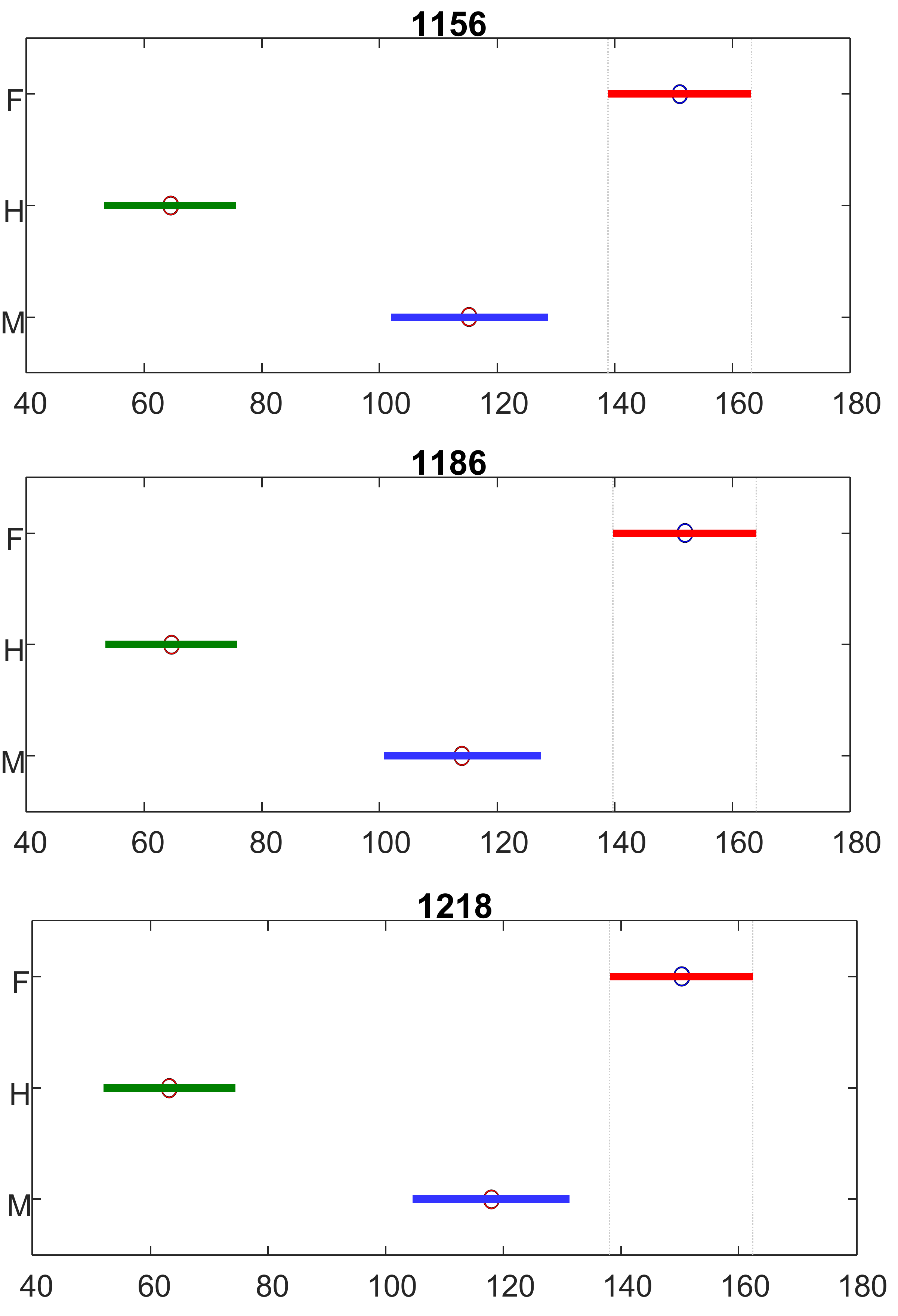
| 1156 | -C=C- (in plane) | Carotenoids [32] |
| 1186 | ν(C-O-H) Next to aromatic ring + σ(CH) | Carotenoids [15] |
| 1218 | δ(C-C-H) | Carotenoids [15] |
| 1267–1288 | δ(C-C-H) | Aliphatics [34] |
| 1326 | δCH2 Bending | Aliphatics, cellulose, lignin [33] |
| 1388 | δCH2 Bending | Aliphatics [34] |
| 1439 | δ(CH2) + δ(CH3) | Aliphatics [34] |
| 1525 | -C=C- (in plane) | Carotenoids [35,36] |
| 1609 | ν(C-C) Aromatic ring + σ(CH) | Lignin [37,38] |
| 1650–1680 | Amide I | Proteins [18] |
| Number of Spectra | TPR | Predicted as Female | Predicted as Male | Predicted as Hermaphrodite | |
|---|---|---|---|---|---|
| Female | 57 | 100% | 57 | 0 | 0 |
| Male | 50 | 100% | 0 | 50 | 0 |
| Hermaphrodite | 77 | 98.7% | 0 | 1 | 76 |
Publisher’s Note: MDPI stays neutral with regard to jurisdictional claims in published maps and institutional affiliations. |
© 2022 by the authors. Licensee MDPI, Basel, Switzerland. This article is an open access article distributed under the terms and conditions of the Creative Commons Attribution (CC BY) license (https://creativecommons.org/licenses/by/4.0/).
Share and Cite
Goff, N.K.; Guenther, J.F.; Roberts, J.K., III; Adler, M.; Molle, M.D.; Mathews, G.; Kurouski, D. Non-Invasive and Confirmatory Differentiation of Hermaphrodite from Both Male and Female Cannabis Plants Using a Hand-Held Raman Spectrometer. Molecules 2022, 27, 4978. https://doi.org/10.3390/molecules27154978
Goff NK, Guenther JF, Roberts JK III, Adler M, Molle MD, Mathews G, Kurouski D. Non-Invasive and Confirmatory Differentiation of Hermaphrodite from Both Male and Female Cannabis Plants Using a Hand-Held Raman Spectrometer. Molecules. 2022; 27(15):4978. https://doi.org/10.3390/molecules27154978
Chicago/Turabian StyleGoff, Nicolas K., James F. Guenther, John K. Roberts, III, Mickal Adler, Michael Dalle Molle, Greg Mathews, and Dmitry Kurouski. 2022. "Non-Invasive and Confirmatory Differentiation of Hermaphrodite from Both Male and Female Cannabis Plants Using a Hand-Held Raman Spectrometer" Molecules 27, no. 15: 4978. https://doi.org/10.3390/molecules27154978
APA StyleGoff, N. K., Guenther, J. F., Roberts, J. K., III, Adler, M., Molle, M. D., Mathews, G., & Kurouski, D. (2022). Non-Invasive and Confirmatory Differentiation of Hermaphrodite from Both Male and Female Cannabis Plants Using a Hand-Held Raman Spectrometer. Molecules, 27(15), 4978. https://doi.org/10.3390/molecules27154978