
Hybrid Nanobioengineered Nanomaterial-Based Electrochemical Biosensors


Abstract
:1. Introduction
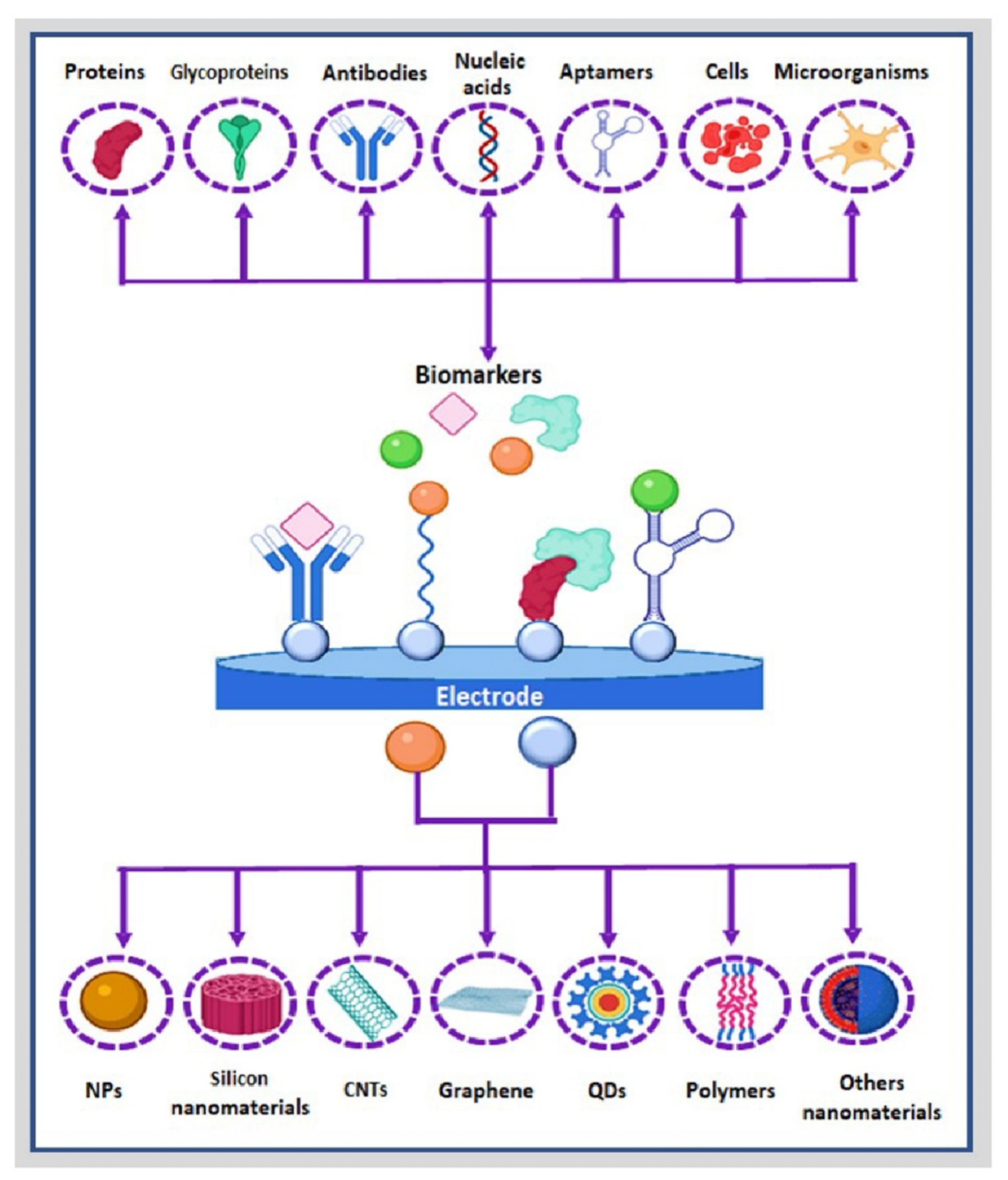
2. Nanohybrids and Nanocomposites
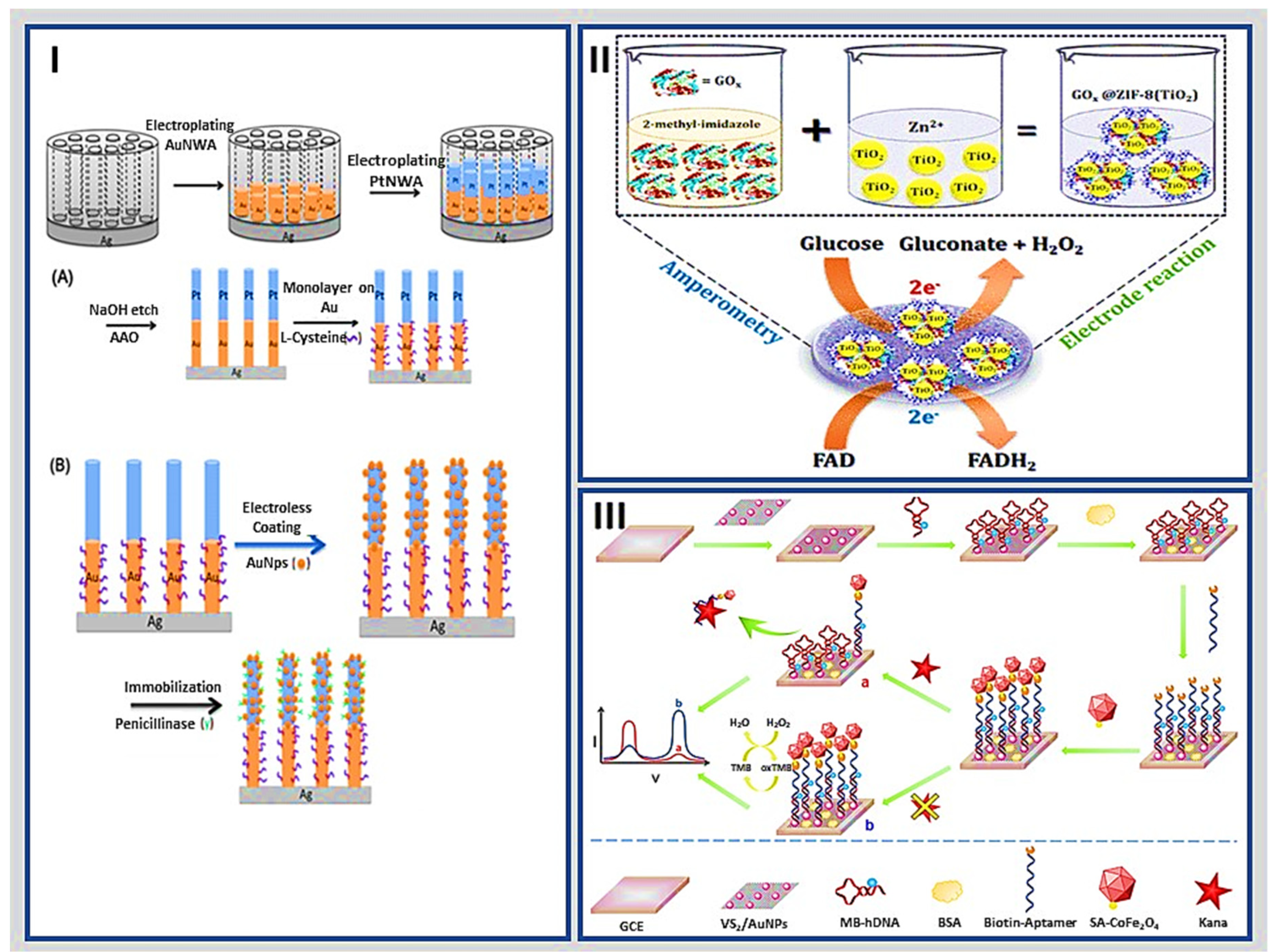
2.1. Metallic Nanostructures
2.2. Silicon Nanomaterials
2.3. Carbon Nanostructures
2.4. Polymers
2.5. Emerging Nanostructured Nanomaterial for Sensitive Biosensing

| Nanomaterial | Hybrid a | Target b | Analytical Characteristics | Comments | References | |
|---|---|---|---|---|---|---|
| Linear Range | LOD | |||||
| Metallic nanostructures | 3D hybrid graphene–GNR. | H2O2 | 0 to 50 mM | 2.9 µM | Metallic nanostructures have high catalytic activity, easy preparation, and relatively low cost. However, this kind of nanomaterial can change its oxidation state due to variations in conditions of the medium, such as pH, ionic strength, and temperature upon time. | [48] |
| TiO2 nanoparticles encapsulated ZIF-8 | Glucose | 2 to 10 mM | 80 nM | [51] | ||
| Nanohybrid of VS2/AuNP and CoFe2O4 nanozyme | Kana | 1 pM to 1 μM | 0.5 pM | [52] | ||
| Ag and hybrid Ag–Fe3O4 metallic nanoparticles. | AA | 0.2–60 μM | 74 nM | [49] | ||
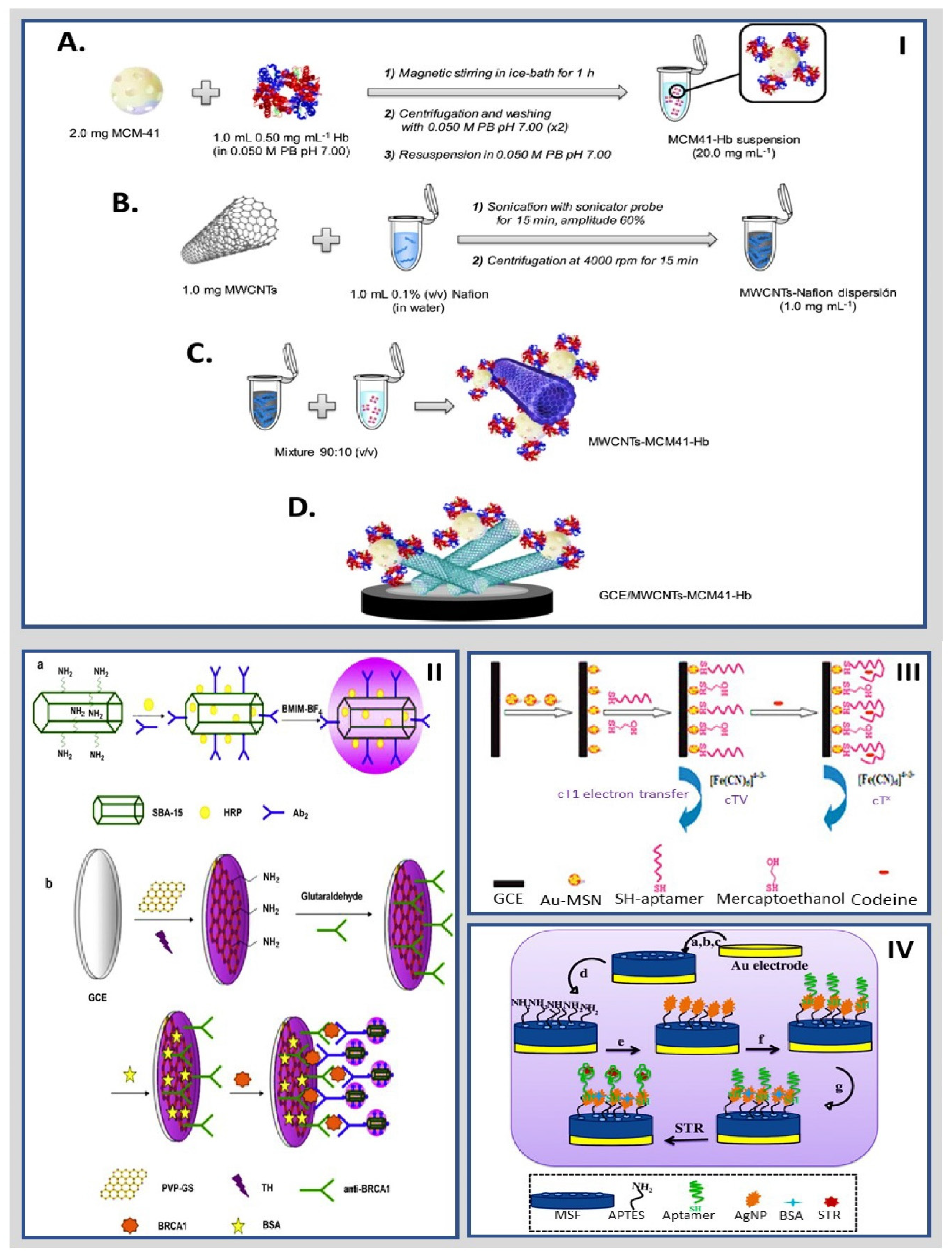
| Silicon nanomaterials | mSiO2@MWCNT. | Thrombin | 0.0001 nM and 80 nM | 50 fM | These nanomaterials have high mechanical resistance, thermal stability, long functional life, and versatility; nonetheless, they require long synthetic processes, and their application is limited to certain analytes. | [31] |
| MSF/APTES/AgNP | STR | 1 to 6.2 ng/mL | 0.33 fg/mL | [66] | ||
| Ap–GA–NH2MCM-41–GCE | hemin and Hb | 1.0 × 10⁻¹⁹ to 1.0 × 10⁻⁶ M | 7.5 × 10⁻²⁰ M and 6.5 × 10⁻²⁰ M | [63] | ||
| AuNPs loaded in functionalized MSNPs | CEA | 1.0 × 10⁻³ to 100 ng/mL | 9.8 × 10⁻⁴ ng/mL | [67] | ||
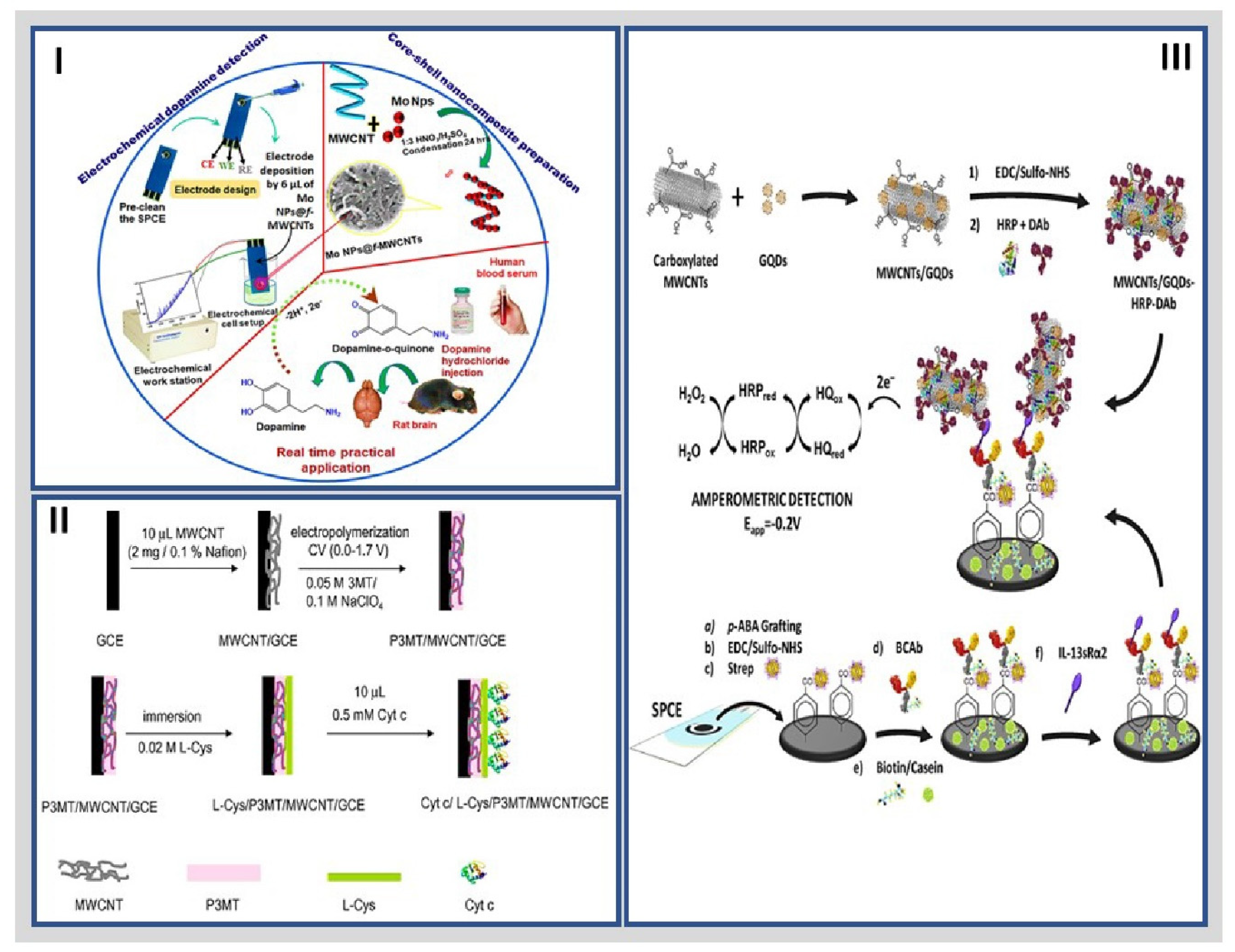
| Carbon nanostructures | MWCNTs and GQDs. | IL-13Rα2 | 2.7 to 100 ng/mL | 0.8 ng/mL | These nanomaterials enjoy thermal stability, large surface area, and a wide range of nanostructures and functional groups. They are the main nanomaterials used in the preparation of electrochemical biosensors. | [96] |
| GQDs/AuNPs. | P53 | 0.000592–1.296 pM | 0.065 fM | [97] | ||
| CQDs/AuNps | Glucose | 0.05 mM to 2.85 mM | 17 μM | [98] | ||
| CoCu-ZIF@CDs | B16-F10 cells | 1 × 10² to 1 × 10⁵ cells/mL | 33 cells/mL | [99] | ||
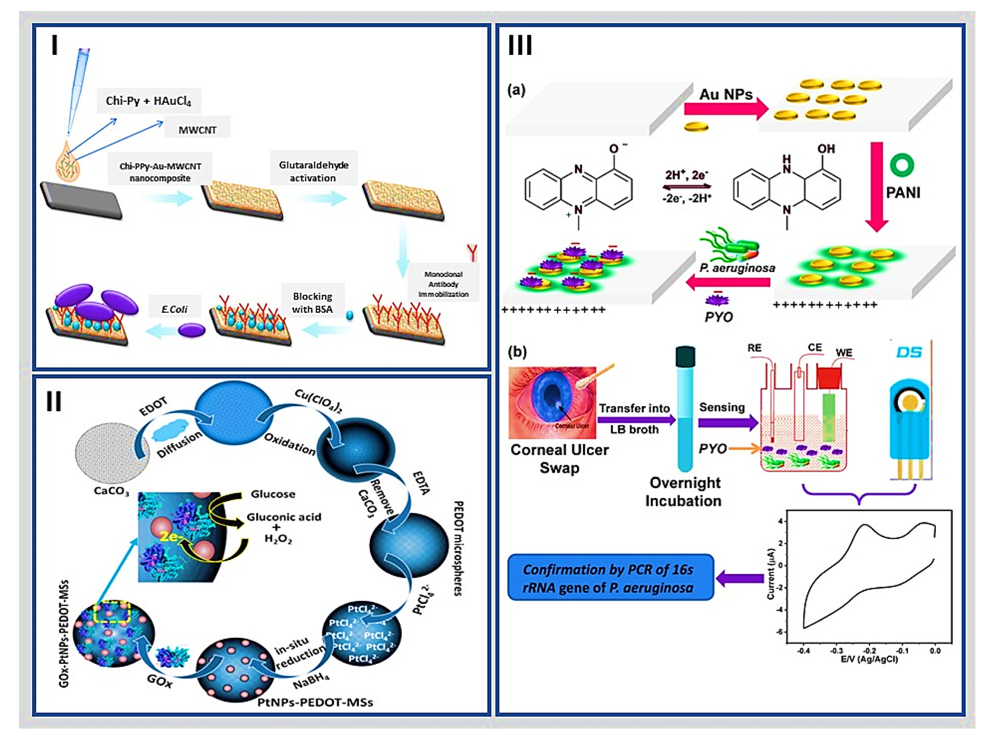
| Polymers | (Chi-Py) mixture, AuNPs, and MWCNT | Escherichia coli | 3 × 101 to 3 × 10⁷ cfu/mL | ~30 CFU/mL | These have high biocompatibility, high affinity, strong adsorption ability, low molecular permeability, physical rigidity, and chemical inertness in biological processes. However, functionalizing their surface is necessary for the anchorage of bioreceptors, and some polymers oxidize due to changes in medium conditions. | [105] |
| PANI/active carbon and n-TiO2 | Glucose | 0.02 mM to 6.0 mM | 18 μM | [106] | ||
| PEG/AuNPs/PANI | alpha-fetoprotein | 10−14 to 10−6 mg/mL | 0.007 pg/mL | [110] | ||
| Other nanostructured nanomaterials | WSe2 and AuNPs | Thrombin | 0–1 ng/mL | 190 fg/mL | Other hybrid nanostructures have a large specific surface area, excellent electrical conductivity, and electrocatalytic properties. | [112] |
| MoS2/Ti3C2 nanohybrids | miRNA | 1 fM to 0.1 nM | 0.43 fM | [113] | ||
| AuNPs/Ti3C2 MXene 3D | miRNA155 | 1.0 fM to 10 nM | 0.35 fM | [114] | ||
3. Conjugation of Nanohybrid Materials with Biomolecules
3.1. Bioreceptors
3.1.1. Protein Bioreceptors and Peptides
3.1.2. Nucleic Acids, Aptamers, Cells, and Other Bioreceptors
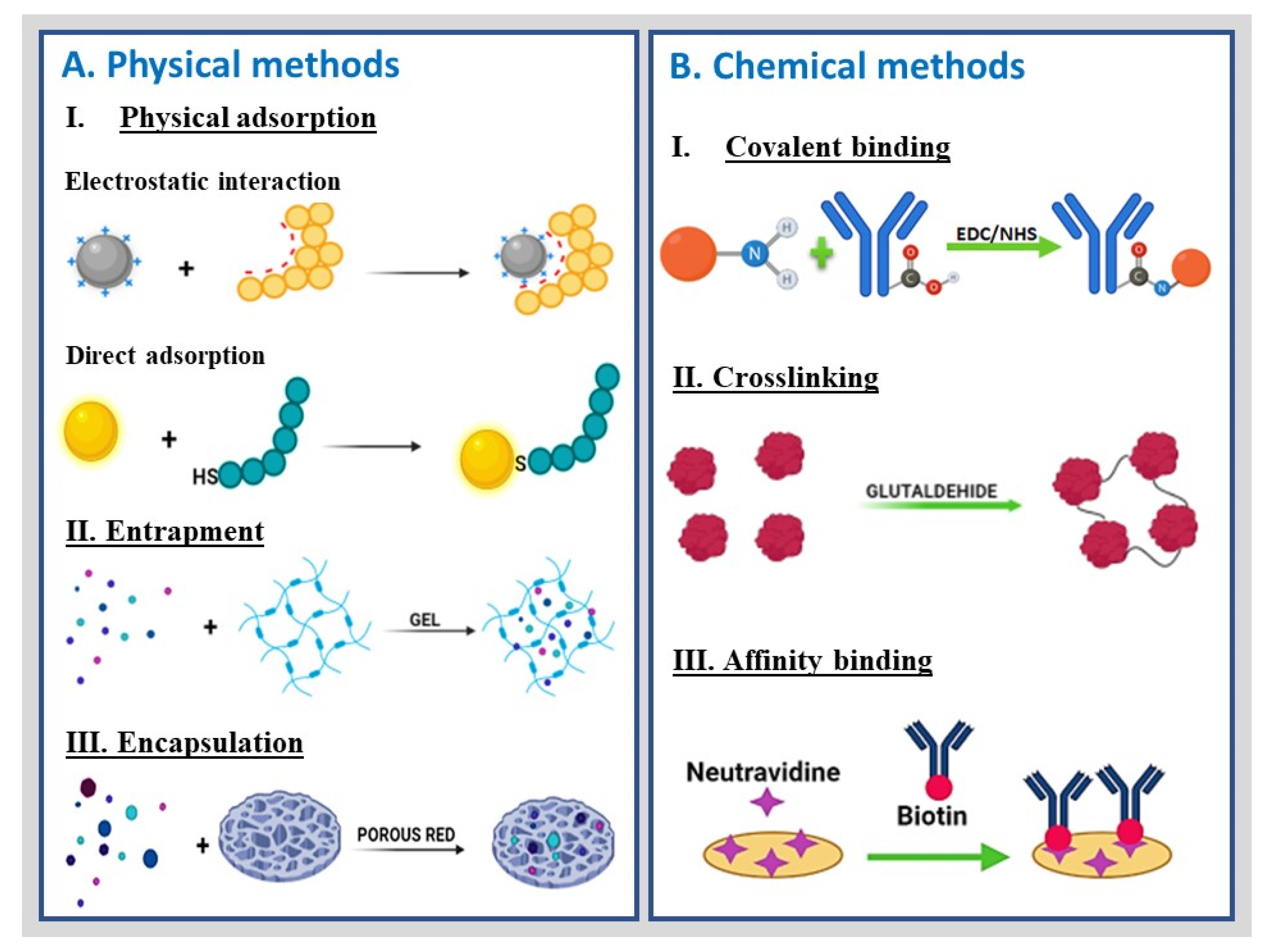
4. Functional Groups and Conjugation Chemistry
5. Characterization of Nanobioengineered Platforms
6. Examples of Nanobioengineered Platforms for Electrochemical Biosensing in the Last Five Years
| Biosensor | Application a | Nanobiohybrid: Nanomaterial and Biomolecules b | Characterization c | Analytical Performance (Linear Range and LOD) d | Reference |
|---|---|---|---|---|---|
| Immunosensor | PSA | Antibody/HP5@AuNPs@g-C3N4 bioconjugated with PSA-Ab2 | CV, EIS, and DPV | 0.0005 to 0.00 ng/mL with LOD of 0.12 pg/mL | [117] |
| HER2 | Ab/g-C3N4/AuNPs/Cu-MOF | CV and EIS | 1.00 to 100.00 ng/mL with LOD of 3.00 fg/mL | [129] | |
| AXL | Ab/fGQDs | XRD, FTIR, UV-Vis, TEM, EIS, DPV | 1.7 to 1000 pg/mL with LOD of 0.5 pg/mL | [130] | |
| CEA | CdSe-QD-melamine and Ab1-TiO2-AuNP-ITO | DPV | 0.005–1000 ng/mL with a LOD of 5 pg/mL | [155] | |
| CA19-9 | CeO2/FeOx@mC | XPS, TEM, EIS, CV | 0.1 mU/mL to 10 U/mL with a LOD of 10 μU/mL | [160] | |
| NMP-22 | Co-MOFs/CuAu NWs/Ab | SEM, XPS, CV, and chronoamperometry | 0.1 pg/mL to 1 ng/mL with a LOD of 33 fg/mL | [163] | |
| Genosensor | Zika | Anti-Dig-HRP | Chronoamperometry, CV, EIS | 5 to 300 pmol/L with LOD of 0.7 pM | [132] |
| Zika genes | AuNPs/ssDNA | SEM, CV, DPV, and chronoamperometry | 10 to 600 fM with LOD of 0.2 fM | [133] | |
| CaMV35S gen | Fe3O4-Au@Ag-sDNA on MWCNT/AuNPs/SH-sDNA | TEM, XRD, UV-Vis, CV, and DPV | 1 × 10⁻¹⁶ M to 1 × 10⁻¹⁰ M with LOD of 1.26 × 10⁻¹⁷ M | [134] | |
| mi-R21 | 3-(trimethoxysilyl)propyl methacrylate/ITO/PET/Fc-hybrid DNA hydrogel | DPV | 10 nM to 50 μM with a LOD of 5 nM | [157] | |
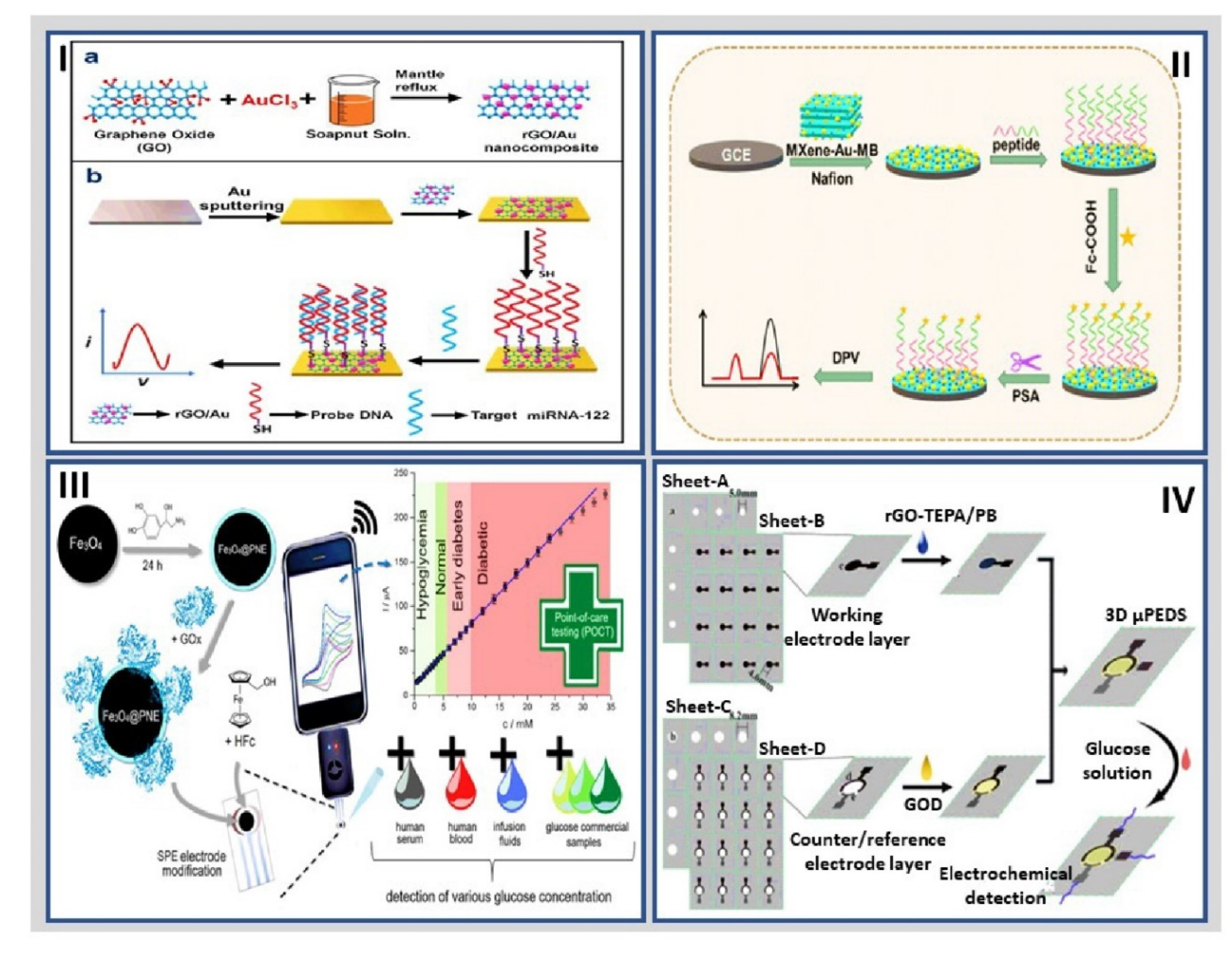
| miRNA-122 | rGO/Au/DNA | XRD, TEM, Raman, XPS, CV, and DPV | 10 μM to 10 pM with a LOD of 1.73 pM | [166] | |
| OVA | SiO2@Au/dsDNA/CeO2 | DPV | 1 pg/mL to 1000 ng/mL with a LOD of 0.87 pg/mL | [174] | |
| Enzymatic | Glucose | GOx/n-TiO2/PANI | CV and chronoamperometry | 0.02 to 6.0 mM with LOD of 18 μM | [106] |
| Glucose | Cu-nanoflowers-Gox-HRP/AuNPs-GO-PVA nanofibers | UV–Vis, SEM, TEM, XDR, CV, and chronoamperometry | 0.001 to 0.1 mM with a LOD of 0.018 μM | [158] | |
| Organophosphate pesticides | acetylcholinesterase/chitosan-transition metals/graphene/GCE | SEM, TEM, XPS, XRD, CV, DPV and EIS | 11.31 μM to 22.6 nM with LOD of 14.45 nM | [165] | |
| β-hydroxybutyric acid | Ti3C2Tx nanosheets conjugated with β-hydroxybutyrate dehydrogenase | SEM, CV, and chronoamperometry | 0.36 to 17.9 mM with a LOD of 45 μM | [168] | |
| Based on peptides | norovirus | Cys/peptide/gold layer | CV and EIS | The LOD was 99.8 nM and 7.8 copies/mL for rP2 and human norovirus, respectively. | [122] |
| PSA | MXene-Au-MB nanohybrid/peptide | DPV | 5 pg/mL to 10 ng/mL with a LOD of 0.83 pg/mL | [169] | |
| PKA and CK2 | Peptide/MSF/ITO | Chronoamperometry | The LODs were 0.083 and 0.095 U/mL, for PKA and CK2, respectively | [170] | |
| NHE | Cys-PEG-QRRMIEEPA-MB | DPV and SWV | 10 and 150 nM with a LOD of 250 pM | [177] | |
| Based on glycoproteins | Toxoplasma gondii | Ab glycosylphosphatidylinositol/SPAuE | CV, EIS | 1.0 to 10.0 IU/mL, with a LOD of 0.31 IU/mL | [140] |
| MIPs/glycoproteins | Fc/MPBA/AuNPs-SiO2 nanobioconjugate | FTIR, CV, EIS, DPV, and chronoamperometry | 1 pg/mL to 100 ng/mL and reached a LOD of 0.57 pg/mL | [142] | |
| Based on aptamers | tumor exosomes extracted from lymph node carcinoma of a prostate cells line | MNPs/aptamer-DNA/double-stranded DNA/GCE | DPV | The LOD was 70 particles/μL | [141] |
| miRNA | DSN/AuNPS/HRP | CV, EIS and chronoamperometry | The LOD was 43.3 aM | [156] | |
| CA125 and living MCF-7 cells | Tb-MOF-on-Fe-MOF | SEM, TEM, XPS, CV, and EIS | 100 μU/mL to 200 U/mL with a LOD of 58 μU/mL towards CA125. Moreover, biosensor detecting MCF-7 cells with a LOD of 19 cells/mL | [161] | |
| CEA and NSE | Paper-electrode functionalized with amino-modified graphene-Thi-AuNPs and PB-PEDOT | DPV | 0.01 to 500 ng/mL for CEA and 0.05–500 ng/mL for NSE with a LOD of 2 pg/mL for CEA and 10 pg/mL for NSE, respectively | [175] | |
| Other types of biosensors (based on cells or mimicking biosensors) | Impedimetric biosensor/Escherichia coli B. | CNT/PEI-T2 virus/GCE | EIS | 103 to 107 CFU/mL with LOD of 1.5 × 10³ CFU/mL | [135] |
| Nonenzymatic biosensor/glucose | GS/GNR/Ni | Chronoamperometry | 5 nM to 5 mM with a LOD of 2.5 nM. | [159] | |
| Mimicking biosensor/H2O2 released from H9C2 cardiac cells | AuNFs/Fe3O4@ZIF-8-MoS2 | SEM, fluorescence, CV, EIS, and chronoamperometry | 5 μM–120 mM and a LOD of 0.9 μM | [162] | |
| Electrochemical/glucose | CuOx@Co3O4 core-shell nanowires/ZIF-67 | SEM, TEM, XRD, XPS, CV, and chronoamperometry | 0.1 to 1300.0 μM with a LOD of 36 nM | [164] | |
| Mimicking/L-tyrosinase | UT-g-C3N4/Ag hybrids | TEM, XPS, XRD, AFM, EIS, CV, and DPV | 1.00 × 10⁻⁶ to 1.50 × 10⁻⁴ mol/L with a LOD of 1.40 × 10⁻⁷ mol/L | [167] | |
| Biomimetic biosensor/glucose | Fe3O4@PNE-GOx | Chronoamperometry | 0.24 to 24 mM with a LOD of 6.1 µM | [171] | |
| PAD/creatinine | CuO/IL/ERGO/SPCE | Chronoamperometry | 0.01 to 2.0 mM and a LOD of 0.22 μM | [173] | |
| 3D paper-based microfluidic electrochemical biosensor/glucose | rGO-TEPA/PB | SEM, Raman, CV, and chronoamperometry | 0.1 mM–25 mM with a LOD of 25 μM | [176] |
7. Limitations, Opportunities, and Concluding Remarks
Author Contributions
Funding
Institutional Review Board Statement
Informed Consent Statement
Data Availability Statement
Acknowledgments
Conflicts of Interest
Sample Availability
References
- Cajigas, S.; Orozco, J. Nanobioconjugates for Signal Amplification in Electrochemical Biosensing. Molecules 2020, 25, 3542. [Google Scholar] [CrossRef] [PubMed]
- Kumar, A.; Purohit, B.; Maurya, P.K.; Pandey, L.M.; Chandra, P. Engineered nanomaterial assisted signal-amplification strategies for enhancing analytical performance of electrochemical biosensors. Electroanalysis 2019, 31, 1615–1629. [Google Scholar] [CrossRef]
- Low, S.S.; Ji, D.; Chai, W.S.; Liu, J.; Khoo, K.S.; Salmanpour, S.; Karimi, F.; Deepanraj, B.; Show, P.L. Recent progress in nanomaterials modified electrochemical biosensors for the detection of MicroRNA. Micromachines 2021, 12, 1409. [Google Scholar] [CrossRef] [PubMed]
- Jones, R.G.; Kahovec, J.; Stepto, R.; Wilks, E.S.; Hess, M.; Kitayama, T.; Metanomski, W.V. Definitions of terms relating to the structure and processing of sols, gels, networks, and inorganic-organic hybrid materials (IUPAC Recommendations 2007). Pure Appl. Chem. 2007, 79, 1801–1829. [Google Scholar]
- Suni, I.I. Substrate materials for biomolecular immobilization within electrochemical biosensors. Biosensors 2021, 11, 239. [Google Scholar] [CrossRef]
- Singh, A.; Sharma, A.; Ahmed, A.; Sundramoorthy, A.K.; Furukawa, H.; Arya, S.; Khosla, A. Recent advances in electrochemical biosensors: Applications, challenges, and future scope. Biosensors 2021, 11, 336. [Google Scholar] [CrossRef]
- Yüce, M.; Kurt, H. How to make nanobiosensors: Surface modification and characterisation of nanomaterials for biosensing applications. RSC Adv. 2017, 7, 49386–49403. [Google Scholar] [CrossRef] [Green Version]
- Rocchitta, G.; Spanu, A.; Babudieri, S.; Latte, G.; Madeddu, G.; Galleri, G.; Nuvoli, S.; Bagella, P.; Demartis, M.; Fiore, V.; et al. Enzyme biosensors for biomedical applications: Strategies for safeguarding analytical performances in biological fluids. Sensors 2016, 16, 780. [Google Scholar] [CrossRef] [Green Version]
- Taylor-Pashow, K.M.L.; Della Rocca, J.; Huxford, R.C.; Lin, W. Hybrid nanomaterials for biomedical applications. Chem. Commun. 2010, 46, 5832–5849. [Google Scholar] [CrossRef]
- Hu, Y.; Huang, Y.; Tan, C.; Zhang, X.; Lu, Q.; Sindoro, M.; Huang, X.; Huang, W.; Wang, L.; Zhang, H. Two-dimensional transition metal dichalcogenide nanomaterials for biosensing applications. Mater. Chem. Front. 2016, 1, 24–36. [Google Scholar] [CrossRef] [Green Version]
- Toyos-Rodríguez, C.; García-Alonso, F.J.; Escosura-Muñiz, A. de la Electrochemical biosensors based on nanomaterials for early detection of Alzheimer’s disease. Sensors 2020, 20, 4748. [Google Scholar] [CrossRef] [PubMed]
- Holzinger, M.; Le Goff, A.; Cosnier, S. Synergetic effects of combined nanomaterials for biosensing applications. Sensors 2017, 17, 1010. [Google Scholar] [CrossRef] [PubMed] [Green Version]
- Holzinger, M.; Le Goff, A.; Cosnier, S. Nanomaterials for biosensing applications: A review. Front. Chem. 2014, 2, 1–10. [Google Scholar] [CrossRef] [PubMed] [Green Version]
- Aziz, M.A.; Oyama, M. Nanomaterials in Electrochemical Biosensor. Adv. Mater. Res. 2014, 995, 125–143. [Google Scholar] [CrossRef]
- Saxena, U.; Das, A.B. Nanomaterials towards fabrication of cholesterol biosensors: Key roles and design approaches. Biosens. Bioelectron. 2016, 75, 196–205. [Google Scholar] [CrossRef]
- Zhang, S.; Geryak, R.; Geldmeier, J.; Kim, S.; Tsukruk, V.V. Synthesis, assembly, and applications of hybrid nanostructures for biosensing. Chem. Rev. 2017, 117, 12942–13038. [Google Scholar] [CrossRef]
- Soto, D.; Alzate, M.; Gallego, J.; Orozco, J. Hybrid nanomaterial/catalase-modified electrode for hydrogen peroxide sensing. J. Electroanal. Chem. 2020, 880, 114826. [Google Scholar] [CrossRef]
- Liu, H.; Fu, Z.-e; Song, F.; Liu, Q.; Chen, L. The controllable construction and properties characterization of organic-inorganic hybrid materials based on benzoxazine-bridged polysilsesquioxanes. RSC Adv. 2017, 7, 3136–3144. [Google Scholar] [CrossRef] [Green Version]
- Zhao, X.; Zhang, P.; Chen, Y.; Su, Z.; Wei, G. Recent advances in the fabrication and structure-specific applications of graphene-based inorganic hybrid membranes. Nanoscale 2015, 7, 5080–5093. [Google Scholar] [CrossRef]
- Gong, Y.; Chen, X.; Lu, Y.; Yang, W. Self-assembled dipeptide–gold nanoparticle hybrid spheres for highly sensitive amperometric hydrogen peroxide biosensors. Biosens. Bioelectron. 2015, 66, 392–398. [Google Scholar] [CrossRef]
- Yin, P.T.; Shah, S.; Chhowalla, M.; Lee, K.-B. Design, synthesis, and characterization of graphene-nanoparticle hybrid materials for bioapplications. Chem. Rev. 2015, 115, 2483–2531. [Google Scholar] [CrossRef] [PubMed] [Green Version]
- Wang, Z.; Dai, Z. Carbon nanomaterial-based electrochemical biosensors: An overview. Nanoscale 2015, 7, 6420–6431. [Google Scholar] [CrossRef] [PubMed]
- Fernández-Sánchez, C.; Pellicer, E.; Orozco, J.; Jiménez-Jorquera, C.; Lechuga, L.M.; Mendoza, E. Plasma-activated multi-walled carbon nanotube–polystyrene composite substrates for biosensing. Nanotechnology 2009, 20, 335501. [Google Scholar] [CrossRef] [PubMed]
- Mendoza, E.; Orozco, J.; Jiménez-Jorquera, C.; González-Guerrero, A.B.; Calle, A.; Lechuga, L.M.; Fernández-Sánchez, C. Scalable fabrication of immunosensors based on carbon nanotube polymer composites. Nanotechnology 2008, 19, 075102. [Google Scholar] [CrossRef] [PubMed] [Green Version]
- Ashby, M.; Cope, E.; Cebon, D. Materials selection for engineering design. In Informatics for Materials Science and Engineering; Elsevier: Amsterdam, The Netherland, 2013; pp. 219–244. [Google Scholar]
- Gu, H.; Liu, C.; Zhu, J.; Gu, J.; Wujcik, E.K.; Shao, L.; Wang, N.; Wei, H.; Scaffaro, R.; Zhang, J.; et al. Introducing advanced composites and hybrid materials. Adv. Compos. Hybrid Mater. 2018, 1, 1–5. [Google Scholar] [CrossRef] [Green Version]
- Arduini, F.; Micheli, L.; Moscone, D.; Palleschi, G.; Piermarini, S.; Ricci, F.; Volpe, G. Electrochemical biosensors based on nanomodified screen-printed electrodes: Recent applications in clinical analysis. TrAC Trends Anal. Chem. 2016, 79, 114–126. [Google Scholar] [CrossRef] [Green Version]
- Orozco, J.; Villa, E.; Manes, C.L.; Medlin, L.K.; Guillebault, D. Electrochemical RNA genosensors for toxic algal species: Enhancing selectivity and sensitivity. Talanta 2016, 161, 560–566. [Google Scholar] [CrossRef]
- You, M.; Yang, S.; An, Y.; Zhang, F.; He, P. A novel electrochemical biosensor with molecularly imprinted polymers and aptamer-based sandwich assay for determining amyloid-β oligomer. J. Electroanal. Chem. 2020, 862, 114017. [Google Scholar] [CrossRef]
- Ahmed, J.; Rashed, M.A.; Faisal, M.; Harraz, F.A.; Jalalah, M.; Alsareii, S.A. Novel SWCNTs-mesoporous silicon nanocomposite as efficient non-nzymatic glucose biosensor. Appl. Surf. Sci. 2021, 552, 149477. [Google Scholar] [CrossRef]
- Zhang, J.; Chai, Y.; Yuan, R.; Yuan, Y.; Bai, L.; Xie, S. A highly sensitive electrochemical aptasensor for thrombin detection using functionalized mesoporous silica@multiwalled carbon nanotubes as signal tags and DNAzyme signal amplification. Analyst 2013, 138, 6938–6945. [Google Scholar] [CrossRef]
- Hasanzadeh, M.; Shadjou, N.; de la Guardia, M.; Eskandani, M.; Sheikhzadeh, P. Mesoporous silica-based materials for use in biosensors. TrAC Trends Anal. Chem. 2012, 33, 117–129. [Google Scholar] [CrossRef]
- Hasanzadeh, M.; Tagi, S.; Solhi, E.; Mokhtarzadeh, A.; Shadjou, N.; Eftekhari, A.; Mahboob, S. An innovative immunosensor for ultrasensitive detection of breast cancer specific carbohydrate (CA 15-3) in unprocessed human plasma and MCF-7 breast cancer cell lysates using gold nanospear electrochemically assembled onto thiolated graphene quantum dots. Int. J. Biol. Macromol. 2018, 114, 1008–1017. [Google Scholar] [CrossRef] [PubMed]
- Henry, P.A.; Raut, A.S.; Ubnoske, S.M.; Parker, C.B.; Glass, J.T. Enhanced electron transfer kinetics through hybrid graphene-carbon nanotube films. Electrochem. Commun. 2014, 48, 103–106. [Google Scholar] [CrossRef] [PubMed] [Green Version]
- Promphet, N.; Rattanarat, P.; Rangkupan, R.; Chailapakul, O.; Rodthongkum, N. An electrochemical sensor based on graphene/polyaniline/polystyrene nanoporous fibers modified electrode for simultaneous determination of lead and cadmium. Sens. Actuators B Chem. 2015, 207, 526–534. [Google Scholar] [CrossRef]
- Barsan, M.M.; Ghica, M.E.; Brett, C.M.A. Electrochemical sensors and biosensors based on redox polymer/carbon nanotube modified electrodes: A review. Anal. Chim. Acta 2015, 881, 1–23. [Google Scholar] [CrossRef]
- Duan, T.; Chen, Y.; Wen, Q.; Yin, J.; Wang, Y. Three-dimensional macroporous CNT–SnO2 composite monolith for electricity generation and energy storage in microbial fuel cells. RSC Adv. 2016, 6, 59610–59618. [Google Scholar] [CrossRef]
- Noreña-Caro, D.; Álvarez-Láinez, M. Functionalization of polyacrylonitrile nanofibers with β-cyclodextrin for the capture of formaldehyde. Mater. Des. 2016, 95, 632–640. [Google Scholar] [CrossRef]
- Yuan, L.; Jiang, L.; Hui, T.; Jie, L.; Bingbin, X.; Feng, Y.; Yingchun, L. Fabrication of highly sensitive and selective electrochemical sensor by using optimized molecularly imprinted polymers on multi-walled carbon nanotubes for metronidazole measurement. Sens. Actuators B Chem. 2015, 206, 647–652. [Google Scholar] [CrossRef]
- Liu, J.; Bo, X.; Yang, J.; Yin, D.; Guo, L. One-step synthesis of porphyrinic iron-based metal-organic framework/ordered mesoporous carbon for electrochemical detection of hydrogen peroxide in living cells. Sens. Actuators B Chem. 2017, 248, 207–213. [Google Scholar] [CrossRef]
- Azzouzi, S.; Rotariu, L.; Benito, A.M.; Maser, W.K.; Ben Ali, M.; Bala, C. A novel amperometric biosensor based on gold nanoparticles anchored on reduced graphene oxide for sensitive detection of l-lactate tumor biomarker. Biosens. Bioelectron. 2015, 69, 280–286. [Google Scholar] [CrossRef] [Green Version]
- Lawal, A.T. Synthesis and utilization of carbon nanotubes for fabrication of electrochemical biosensors. Mater. Res. Bull. 2016, 73, 308–350. [Google Scholar] [CrossRef]
- Mehmood, S.; Ciancio, R.; Carlino, E.; Bhatti, A.S. Role of Au(NPs) in the enhanced response of Au(NPs)-decorated MWCNT electrochemical biosensor. Int. J. Nanomed. 2018, 13, 2093. [Google Scholar] [CrossRef] [PubMed] [Green Version]
- Park, W.; Shin, H.; Choi, B.; Rhim, W.K.; Na, K.; Keun Han, D. Advanced hybrid nanomaterials for biomedical applications. Prog. Mater. Sci. 2020, 114, 100686. [Google Scholar] [CrossRef]
- Zeynaloo, E.; Yang, Y.P.; Dikici, E.; Landgraf, R.; Bachas, L.G.; Daunert, S. Design of a mediator-free, non-enzymatic electrochemical biosensor for glutamate detection. Nanomed. Nanotechnol. Biol. Med. 2021, 31, 102305. [Google Scholar] [CrossRef] [PubMed]
- Wang, J. Nanomaterial-based electrochemical biosensors. Analyst 2005, 130, 421–426. [Google Scholar] [CrossRef]
- Gallego, J.; Tapia, J.; Vargas, M.; Santamaria, A.; Orozco, J.; Lopez, D. Synthesis of graphene-coated carbon nanotubes-supported metal nanoparticles as multifunctional hybrid materials. Carbon N. Y. 2017, 111, 393–401. [Google Scholar] [CrossRef]
- Xue, C.; Kung, C.C.; Gao, M.; Liu, C.C.; Dai, L.; Urbas, A.; Li, Q. Facile fabrication of 3D layer-by-layer graphene-gold nanorod hybrid architecture for hydrogen peroxide based electrochemical biosensor. Sens. Bio-Sens. Res. 2015, 3, 7–11. [Google Scholar] [CrossRef] [Green Version]
- Hashemi, S.A.; Mousavi, S.M.; Bahrani, S.; Ramakrishna, S.; Babapoor, A.; Chiang, W.H. Coupled graphene oxide with hybrid metallic nanoparticles as potential electrochemical biosensors for precise detection of ascorbic acid within blood. Anal. Chim. Acta 2020, 1107, 183–192. [Google Scholar] [CrossRef]
- Li, Z.; Liu, C.; Sarpong, V.; Gu, Z. Multisegment nanowire/nanoparticle hybrid arrays as electrochemical biosensors for simultaneous detection of antibiotics. Biosens. Bioelectron. 2019, 126, 632–639. [Google Scholar] [CrossRef]
- Paul, A.; Srivastava, D.N. Amperometric glucose sensing at nanomolar level using MOF-encapsulated TiO2 platform. ACS Omega 2018, 3, 14634–14640. [Google Scholar] [CrossRef] [Green Version]
- Tian, L.; Zhang, Y.; Wang, L.; Geng, Q.; Liu, D.; Duan, L.; Wang, Y.; Cui, J. Ratiometric dual signal-enhancing-based electrochemical biosensor for ultrasensitive Kanamycin detection. ACS Appl. Mater. Interfaces 2020, 12, 52713–52720. [Google Scholar] [CrossRef] [PubMed]
- Kadian, S.; Arya, B.D.; Kumar, S.; Sharma, S.N.; Chauhan, R.P.; Srivastava, A.; Chandra, P.; Singh, S.P. Synthesis and application of PHT-TiO2 nanohybrid for amperometric glucose detection in human saliva sample. Electroanalysis 2018, 30, 2793–2802. [Google Scholar] [CrossRef]
- Beck, J.S.; Vartuli, J.C.; Roth, W.J.; Leonowicz, M.E.; Kresge, C.T.; Schmitt, K.D.; Chu, C.T.W.; Olson, D.H.; Sheppard, E.W.; McCullen, S.B.; et al. A new family of mesoporous molecular sieves prepared with liquid crystal templates. J. Am. Chem. Soc. 1992, 114, 10834–10843. [Google Scholar] [CrossRef]
- Shen, T.; Yao, Z.; Xia, X.; Wang, X.; Gu, C.; Tu, J. Rationally designed silicon nanostructures as anode material for Lithium-ion batteries. Adv. Eng. Mater. 2018, 20, 1700591. [Google Scholar] [CrossRef] [Green Version]
- Wongkaew, N.; Simsek, M.; Griesche, C.; Baeumner, A.J. Functional nanomaterials and nanostructures enhancing electrochemical biosensors and Lab-on-a-Chip performances: Recent progress, applications, and future perspective. Chem. Rev. 2018, 119, 120–194. [Google Scholar] [CrossRef] [PubMed]
- Parlett, C.M.A.; Wilson, K.; Lee, A.F. Hierarchical porous materials: Catalytic applications. Chem. Soc. Rev. 2013, 42, 3876–3893. [Google Scholar] [CrossRef] [PubMed]
- Triantafillidis, C.; Elsaesser, M.S.; Hüsing, N. Chemical phase separation strategies towards silica monoliths with hierarchical porosity. Chem. Soc. Rev. 2013, 42, 3833–3846. [Google Scholar] [CrossRef]
- Shi, H.; Yang, J.; Li, Z.; He, C. Introduction of organosilicon materials. In Silicon Containing Hybrid Copolymers; John Wiley & Sons, Ltd.: Hoboken, NJ, USA, 2020; pp. 1–21. [Google Scholar]
- Sanchez, C.; Shea, K.J.; Kitagawa, S.; Mizoshita, N.; Ab, T.T.; Inagaki, S. Syntheses, properties and applications of periodic mesoporous organosilicas prepared from bridged organosilane precursors. Chem. Soc. Rev. 2011, 40, 789–800. [Google Scholar]
- Ji, X.; Wang, H.; Song, B.; Chu, B.; He, Y. Silicon nanomaterials for biosensing and bioimaging analysis. Front. Chem. 2018, 6, 38. [Google Scholar] [CrossRef] [Green Version]
- Eguílaz, M.; Villalonga, R.; Rivas, G. Electrochemical biointerfaces based on carbon nanotubes-mesoporous silica hybrid material: Bioelectrocatalysis of hemoglobin and biosensing applications. Biosens. Bioelectron. 2018, 111, 144–151. [Google Scholar] [CrossRef]
- Shekari, Z.; Zare, H.R.; Falahati, A. An ultrasensitive aptasensor for hemin and hemoglobin based on signal amplification via electrocatalytic oxygen reduction. Anal. Biochem. 2017, 518, 102–109. [Google Scholar] [CrossRef] [PubMed]
- Cai, Y.; Li, H.; Du, B.; Yang, M.; Li, Y.; Wu, D.; Zhao, Y.; Dai, Y.; Wei, Q. Ultrasensitive electrochemical immunoassay for BRCA1 using BMIM·BF4-coated SBA-15 as labels and functionalized graphene as enhancer. Biomaterials 2011, 32, 2117–2123. [Google Scholar] [CrossRef] [PubMed]
- Huang, L.; Yang, X.; Qi, C.; Niu, X.; Zhao, C.; Zhao, X.; Shangguan, D.; Yang, Y. A label-free electrochemical biosensor based on a DNA aptamer against codeine. Anal. Chim. Acta 2013, 787, 203–210. [Google Scholar] [CrossRef] [PubMed]
- Roushani, M.; Ghanbari, K. An electrochemical aptasensor for streptomycin based on covalent attachment of the aptamer onto a mesoporous silica thin film-coated gold electrode. Microchim. Acta 2019, 185, 1–9. [Google Scholar] [CrossRef]
- Shekari, Z.; Zare, H.R.; Falahati, A. Developing an impedimetric aptasensor for selective label–free detection of CEA as a cancer biomarker based on gold nanoparticles loaded in functionalized mesoporous silica films. J. Electrochem. Soc. 2017, 164, 739–745. [Google Scholar] [CrossRef]
- Soto, D.; Alzate, M.; Gallego, J.; Orozco, J. Electroanalysis of an iron@graphene-carbon nanotube hybrid material. Electroanalysis 2018, 30, 1521–1528. [Google Scholar] [CrossRef]
- Zhu, Z.; Garcia-Gancedo, L.; Flewitt, A.J.; Xie, H.; Moussy, F.; Milne, W.I. A critical review of Glucose biosensors based on carbon nanomaterials: Carbon nanotubes and graphene. Sensors 2012, 12, 5996–6022. [Google Scholar] [CrossRef] [Green Version]
- Artiles, M.S.; Rout, C.S.; Fisher, T.S. Graphene-based hybrid materials and devices for biosensing. Adv. Drug Deliv. Rev. 2011, 63, 1352–1360. [Google Scholar] [CrossRef]
- Kuila, T.; Bose, S.; Khanra, P.; Mishra, A.K.; Kim, N.H.; Lee, J.H. Recent advances in graphene-based biosensors. Biosens. Bioelectron. 2011, 26, 4637–4648. [Google Scholar] [CrossRef]
- Bobrinetskiy, I.I.; Knezevic, N. Graphene-based biosensors for on-site detection of contaminants in food. Anal. Methods 2018, 10, 5061–5070. [Google Scholar] [CrossRef]
- Bollella, P.; Fusco, G.; Tortolini, C.; Sanzò, G.; Favero, G.; Gorton, L.; Antiochia, R. Beyond graphene: Electrochemical sensors and biosensors for biomarkers detection. Biosens. Bioelectron. 2017, 89, 152–166. [Google Scholar] [CrossRef] [PubMed]
- Li, B.; Pan, G.; Avent, N.D.; Lowry, R.B.; Madgett, T.E.; Waines, P.L. Graphene electrode modified with electrochemically reduced graphene oxide for label-free DNA detection. Biosens. Bioelectron. 2015, 72, 313–319. [Google Scholar] [CrossRef] [PubMed] [Green Version]
- Bahadır, E.B.; Sezgintürk, M.K. Applications of graphene in electrochemical sensing and biosensing. TrAC Trends Anal. Chem. 2016, 76, 1–14. [Google Scholar] [CrossRef]
- Wang, Y.; Hu, A. Carbon quantum dots: Synthesis, properties and applications. J. Mater. Chem. C 2014, 2, 6921. [Google Scholar] [CrossRef] [Green Version]
- Brownson, D.A.C.; Banks, C.E. The electrochemistry of CVD graphene: Progress and prospects. Phys. Chem. Chem. Phys. 2012, 14, 8264. [Google Scholar] [CrossRef]
- Ghosal, K.; Sarkar, K. Biomedical pplications of graphene nanomaterials and beyond. ACS Biomater. Sci. Eng. 2018, 4, 2653–2703. [Google Scholar] [CrossRef]
- Reina, G.; González-Domínguez, J.M.; Criado, A.; Vázquez, E.; Bianco, A.; Prato, M. Promises, facts and challenges for graphene in biomedical applications. Chem. Soc. Rev. 2017, 46, 4400–4416. [Google Scholar] [CrossRef] [Green Version]
- Zhao, C.; Song, X.; Liu, Y.; Fu, Y.; Ye, L.; Wang, N.; Wang, F.; Li, L.; Mohammadniaei, M.; Zhang, M.; et al. Synthesis of graphene quantum dots and their applications in drug delivery. J. Nanobiotechnol. 2020, 18, 1–32. [Google Scholar] [CrossRef]
- Gu, S.; Hsieh, C.T.; Chiang, Y.M.; Tzou, D.Y.; Chen, Y.F.; Gandomi, Y.A. Optimization of graphene quantum dots by chemical exfoliation from graphite powders and carbon nanotubes. Mater. Chem. Phys. 2018, 215, 104–111. [Google Scholar] [CrossRef]
- Huang, D.; Zeng, M.; Wang, L.; Zhang, L.; Cheng, Z. Biomimetic colloidal photonic crystals by coassembly of polystyrene nanoparticles and graphene quantum dots. RSC Adv. 2018, 8, 34839–34847. [Google Scholar] [CrossRef] [Green Version]
- Zhang, S.; Wang, H.; Mulchandani, A.; Badhulika, S.; Terse-Thakoor, T.; Villarreal, C. Graphene hybrids: Synthesis strategies and applications in sensors and sensitized solar cells. Front. Chem. 2015, 1, 38. [Google Scholar]
- Li, F.; Gan, S.; Han, D.; Niu, L. Graphene-based nanohybrids for advanced electrochemical sensing. Electroanalysis 2015, 27, 2098–2115. [Google Scholar] [CrossRef]
- Mondal, S.; Khastgir, D. Elastomer reinforcement by graphene nanoplatelets and synergistic improvements of electrical and mechanical properties of composites by hybrid nano fillers of graphene-carbon black and graphene-MWCNT. In Composites Part A: Applied Science and Manufacturing; Elsevier: Amsterdam, The Netherlands, 2017; Volume 102, pp. 154–165. [Google Scholar]
- Park, S.; Vosguerichian, M.; Bao, Z. A review of fabrication and applications of carbon nanotube film-based flexible electronics. Nanoscale 2013, 5, 1727. [Google Scholar] [CrossRef] [PubMed]
- Gougis, M.; Tabet-Aoul, A.; Ma, D.; Mohamedi, M. Laser synthesis and tailor-design of nanosized gold onto carbon nanotubes for non-enzymatic electrochemical glucose sensor. Sens. Actuators B Chem. 2014, 193, 363–369. [Google Scholar] [CrossRef]
- Singal, S.; Srivastava, A.K.; Dhakate, S.; Biradar, A.M.; Rajesh, R. Electroactive graphene-multi-walled carbon nanotube hybrid supported impedimetric immunosensor for the detection of human cardiac troponin-I. RSC Adv. 2015, 5, 74994–75003. [Google Scholar] [CrossRef] [Green Version]
- Dong, X.; Ma, Y.; Zhu, G.; Huang, Y.; Wang, J.; Chan-Park, M.B.; Wang, L.; Huang, W.; Chen, P. Synthesis of graphene–carbon nanotube hybrid foam and its use as a novel three-dimensional electrode for electrochemical sensing. J. Mater. Chem. 2012, 22, 17044. [Google Scholar] [CrossRef]
- Pan, M.; Gu, Y.; Yun, Y.; Li, M.; Jin, X.; Wang, S. Nanomaterials for electrochemical immunosensing. Sensors 2017, 17, 1041. [Google Scholar] [CrossRef] [Green Version]
- Eatemadi, A.; Daraee, H.; Karimkhanloo, H.; Kouhi, M.; Zarghami, N. Carbon nanotubes: Properties, synthesis, purification, and medical applications. Nanoscale Res. Lett. 2014, 9, 1–13. [Google Scholar] [CrossRef] [Green Version]
- Adabi, M.; Saber, R.; Faridi-Majidi, R.; Faridbod, F. Performance of electrodes synthesized with polyacrylonitrile-based carbon nanofibers for application in electrochemical sensors and biosensors. Mater. Sci. Eng. C 2015, 48, 673–678. [Google Scholar] [CrossRef]
- Hossain, M.F.; Slaughter, G. PtNPs decorated chemically derived graphene and carbon nanotubes for sensitive and selective glucose biosensing. J. Electroanal. Chem. 2020, 861, 113990. [Google Scholar] [CrossRef]
- Keerthi, M.; Boopathy, G.; Chen, S.M.; Chen, T.W.; Lou, B.S. A core-shell molybdenum nanoparticles entrapped f-MWCNTs hybrid nanostructured material based non-enzymatic biosensor for electrochemical detection of dopamine neurotransmitter in biological samples. Sci. Rep. 2019, 9, 1–12. [Google Scholar] [CrossRef] [PubMed]
- Eguílaz, M.; Agüí, L.; Yáñez-Sedeño, P.; Pingarrón, J.M. A biosensor based on cytochrome c immobilization on a poly-3-methylthiophene/multi-walled carbon nanotubes hybrid-modified electrode. Application to the electrochemical determination of nitrite. J. Electroanal. Chem. 2010, 644, 30–35. [Google Scholar] [CrossRef]
- Serafín, V.; Valverde, A.; Martínez-García, G.; Martínez-Periñán, E.; Comba, F.; Garranzo-Asensio, M.; Barderas, R.; Yáñez-Sedeño, P.; Campuzano, S.; Pingarrón, J.M. Graphene quantum dots-functionalized multi-walled carbon nanotubes as nanocarriers in electrochemical immunosensing. Determination of IL-13 receptor α2 in colorectal cells and tumor tissues with different metastatic potential. Sens. Actuators B Chem. 2019, 284, 711–722. [Google Scholar] [CrossRef]
- Hasanzadeh, M.; Baghban, H.N.; Shadjou, N.; Mokhtarzadeh, A. Ultrasensitive electrochemical immunosensing of tumor suppressor protein p53 in unprocessed human plasma and cell lysates using a novel nanocomposite based on poly-cysteine/graphene quantum dots/gold nanoparticle. Int. J. Biol. Macromol. 2018, 107, 1348–1363. [Google Scholar] [CrossRef]
- Buk, V.; Pemble, M.E.; Twomey, K. Fabrication and evaluation of a carbon quantum dot/gold nanoparticle nanohybrid material integrated onto planar micro gold electrodes for potential bioelectrochemical sensing applications. Electrochim. Acta 2019, 293, 307–317. [Google Scholar] [CrossRef]
- Liu, Y.; Huang, S.; Li, J.; Wang, M.; Wang, C.; Hu, B.; Zhou, N.; Zhang, Z. 0D/2D heteronanostructure–integrated bimetallic CoCu-ZIF nanosheets and MXene-derived carbon dots for impedimetric cytosensing of melanoma B16-F10 cells. Microchim. Acta 2021, 188, 1–12. [Google Scholar] [CrossRef]
- Wang, X.; Uchiyama, S. Polymers for Biosensors Construction. In State of the Art in Biosensors-General Aspects; InTech: Rijeka, Croatia, 2012; pp. 67–82. [Google Scholar]
- Peng, H.; Zhang, L.; Soeller, C.; Travas-Sejdic, J. Conducting polymers for electrochemical DNA sensing. Biomaterials 2009, 30, 2132–2148. [Google Scholar] [CrossRef]
- Wang, J.-Y.; Su, Y.-L.; Wu, B.-H.; Cheng, S.-H. Reusable electrochemical sensor for bisphenol A based on ionic liquid functionalized conducting polymer platform. Talanta 2016, 147, 103–110. [Google Scholar] [CrossRef]
- Wang, T.; Kumar, S. Electrospinning of polyacrylonitrile nanofibers. J. Appl. Polym. 2006, 102, 1023–1029. [Google Scholar] [CrossRef]
- Huang, Z.-M.; Zhang, Y.-Z.; Kotaki, M.; Ramakrishna, S. A review on polymer nanofibers by electrospinning and their applications in nanocomposites. Compos. Sci. Technol. 2003, 63, 2223–2253. [Google Scholar] [CrossRef]
- Güner, A.; Çevik, E.; Şenel, M.; Alpsoy, L. An electrochemical immunosensor for sensitive detection of Escherichia coli O157:H7 by using chitosan, MWCNT, polypyrrole with gold nanoparticles hybrid sensing platform. Food Chem. 2017, 229, 358–365. [Google Scholar] [CrossRef] [PubMed]
- Tang, W.; Li, L.; Zeng, X. A glucose biosensor based on the synergistic action of nanometer-sized TiO2 and polyaniline. Talanta 2015, 131, 417–423. [Google Scholar] [CrossRef] [PubMed]
- Liu, Y.; Turner, A.P.F.; Zhao, M.; Mak, W.C. Processable enzyme-hybrid conductive polymer composites for electrochemical biosensing. Biosens. Bioelectron. 2018, 100, 374–381. [Google Scholar] [CrossRef] [PubMed]
- Khalifa, M.M.; Elkhawaga, A.A.; Hassan, M.A.; Zahran, A.M.; Fathalla, A.M.; El-Said, W.A.; El-Badawy, O. Highly specific Electrochemical Sensing of Pseudomonas aeruginosa in patients suffering from corneal ulcers: A comparative study. Sci. Rep. 2019, 9, 18320. [Google Scholar] [CrossRef] [Green Version]
- Chen, M.; Song, Z.; Han, R.; Li, Y.; Luo, X. Low fouling electrochemical biosensors based on designed Y-shaped peptides with antifouling and recognizing branches for the detection of IgG in human serum. Biosens. Bioelectron. 2021, 178, 113016. [Google Scholar] [CrossRef]
- Hui, N.; Sun, X.; Song, Z.; Niu, S.; Luo, X. Gold nanoparticles and polyethylene glycols functionalized conducting polyaniline nanowires for ultrasensitive and low fouling immunosensing of alpha-fetoprotein. Biosens. Bioelectron. 2016, 86, 143–149. [Google Scholar] [CrossRef]
- Dou, B.; Yang, J.; Yuan, R.; Xiang, Y. Trimetallic hybrid nanoflower-decorated MoS2 nanosheet sensor for direct in situ monitoring of H2O2 secreted from live Cancer cells. Anal. Chem. 2018, 90, 5945–5950. [Google Scholar] [CrossRef]
- Wang, Y.H.; Xia, H.; Huang, K.J.; Wu, X.; Ma, Y.Y.; Deng, R.; Lu, Y.F.; Han, Z.W. Ultrasensitive determination of thrombin by using an electrode modified with WSe2 and gold nanoparticles, aptamer-thrombin-aptamer sandwiching, redox cycling, and signal enhancement by alkaline phosphatase. Mikrochim. Acta 2018, 185, 502. [Google Scholar] [CrossRef]
- Liu, L.; Wei, Y.; Jiao, S.; Zhu, S.; Liu, X. A novel label-free strategy for the ultrasensitive miRNA-182 detection based on MoS2/Ti3C2 nanohybrids. Biosens. Bioelectron. 2019, 137, 45–51. [Google Scholar] [CrossRef]
- Yang, X.; Feng, M.; Xia, J.; Zhang, F.; Wang, Z. An electrochemical biosensor based on AuNPs/Ti3C2 MXene three-dimensional nanocomposite for microRNA-155 detection by exonuclease III-aided cascade target recycling. J. Electroanal. Chem. 2020, 878, 114669. [Google Scholar] [CrossRef]
- Chansi; Bhardwaj, R.; Rao, R.P.; Mukherjee, I.; Agrawal, P.K.; Basu, T.; Bharadwaj, L.M. Layered construction of nano immuno-hybrid embedded MOF as an electrochemical sensor for rapid quantification of total pesticides load in vegetable extract. J. Electroanal. Chem. 2020, 873, 114386. [Google Scholar] [CrossRef]
- Damborský, P.; Švitel, J.; Katrlík, J. Optical biosensors. Essays Biochem. 2016, 60, 91–100. [Google Scholar] [PubMed] [Green Version]
- Zhou, X.; Yang, L.; Tan, X.; Zhao, G.; Xie, X.; Du, G. A robust electrochemical immunosensor based on hydroxyl pillar(5)arene@AuNPs@g-C3N4 hybrid nanomaterial for ultrasensitive detection of prostate specific antigen. Biosens. Bioelectron. 2018, 112, 31–39. [Google Scholar] [CrossRef] [PubMed]
- Soto, D.; Escobar, S.; Mesa, M. Study of the physicochemical interactions between Thermomyces lanuginosus lipase and silica-based supports and their correlation with the biochemical activity of the biocatalysts. Mater. Sci. Eng. C 2017, 79, 525–532. [Google Scholar] [CrossRef]
- Newman, J.D.; Setford, S.J. Enzymatic biosensors. Mol. Biotechnol. 2006, 32, 249–268. [Google Scholar] [CrossRef]
- Lin, Y.; Lu, F.; Tu, Y.; Ren, Z. Glucose biosensors based on Carbon nanotube nanoelectrode ensembles. Nano Lett. 2004, 4, 191–195. [Google Scholar] [CrossRef]
- Sassolas, A.; Blum, L.J.; Leca-Bouvier, B.D. Immobilization strategies to develop enzymatic biosensors. Biotechnol. Adv. 2012, 30, 489–511. [Google Scholar] [CrossRef]
- Hwang, H.J.; Ryu, M.Y.; Park, C.Y.; Ahn, J.; Park, H.G.; Choi, C.; Ha, S.D.; Park, T.J.; Park, J.P. High sensitive and selective electrochemical biosensor: Label-free detection of human norovirus using affinity peptide as molecular binder. Biosens. Bioelectron. 2017, 87, 164–170. [Google Scholar] [CrossRef]
- Janeway, C.A., Jr.; Travers, P.; Walport, M.; Shlomchik, M.J. Immunobiology. In Immunobiology: The Immune System in Health and Disease; Science, N.Y.G., Ed.; Garland Science: New York, NY, USA, 2001. [Google Scholar]
- Vásquez, G.; Rey, A.; Rivera, C.; Iregui, C.; Orozco, J. Amperometric biosensor based on a single antibody of dual function for rapid detection of Streptococcus agalactiae. Biosens. Bioelectron. 2017, 87, 453–458. [Google Scholar] [CrossRef]
- Kaushik, A.; Yndart, A.; Kumar, S.; Jayant, R.D.; Vashist, A.; Brown, A.N.; Li, C.Z.; Nair, M. A sensitive electrochemical immunosensor for label-free detection of Zika-virus protein. Sci. Rep. 2018, 8, 3–7. [Google Scholar] [CrossRef] [Green Version]
- Li, J.; Liu, S.; Yu, J.; Lian, W.; Cui, M.; Xu, W.; Huang, J. Electrochemical immunosensor based on graphene–polyaniline composites and carboxylated graphene oxide for estradiol detection. Sens. Actuators B Chem. 2013, 188, 99–105. [Google Scholar] [CrossRef]
- Tang, C.K.; Vaze, A.; Rusling, J.F. Fabrication of immunosensor microwell arrays from gold compact discs for detection of cancer biomarker proteins. Lab Chip 2012, 12, 281–286. [Google Scholar] [CrossRef] [PubMed] [Green Version]
- Wan, J.; Ai, J.; Zhang, Y.; Geng, X.; Gao, Q.; Cheng, Z. Signal-off impedimetric immunosensor for the detection of Escherichia coli O157:H7. Sci. Rep. 2016, 6, 19806. [Google Scholar] [CrossRef] [PubMed]
- Yola, M.L. Sensitive sandwich-type voltammetric immunosensor for breast cancer biomarker HER2 detection based on gold nanoparticles decorated Cu-MOF and Cu2ZnSnS4 NPs/Pt/g-C3N4 composite. Microchim. Acta 2021, 188, 1–13. [Google Scholar] [CrossRef] [PubMed]
- Mollarasouli, F.; Serafín, V.; Campuzano, S.; Yáñez-Sedeño, P.; Pingarrón, J.M.; Asadpour-Zeynali, K. Ultrasensitive determination of receptor tyrosine kinase with a label-free electrochemical immunosensor using graphene quantum dots-modified screen-printed electrodes. Anal. Chim. Acta 2018, 1011, 28–34. [Google Scholar] [CrossRef] [PubMed]
- Orozco, J.; Medlin, L.K. Electrochemical performance of a DNA-based sensor device for detecting toxic algae. Sens. Actuators B Chem. 2011, 153, 71–77. [Google Scholar] [CrossRef]
- Alzate, D.; Cajigas, S.; Robledo, S.; Muskus, C.; Orozco, J. Genosensors for differential detection of Zika virus. Talanta 2020, 210, 1–8. [Google Scholar] [CrossRef]
- Cajigas, S.; Alzate, D.; Orozco, J. Gold nanoparticle/DNA-based nanobioconjugate for electrochemical detection of Zika virus. Microchim. Acta 2020, 187, 1–10. [Google Scholar] [CrossRef]
- Ye, Y.; Mao, S.; He, S.; Xu, X.; Cao, X.; Wei, Z.; Gunasekaran, S. Ultrasensitive electrochemical genosensor for detection of CaMV35S gene with Fe3O4-Au@Ag nanoprobe. Talanta 2020, 206, 120205. [Google Scholar] [CrossRef]
- Zhou, Y.; Marar, A.; Kner, P.; Ramasamy, R.P. Charge-directed immobilization of bacteriophage on nanostructured electrode for whole-cell electrochemical biosensors. Anal. Chem. 2017, 89, 5734–5741. [Google Scholar] [CrossRef]
- Cummings, R.D. Lipids | Glycan-dependent cell adhesion processes. In Encyclopedia of Biological Chemistry III; Elsevier: Amsterdam, The Netherlands, 2021; Volume 2, pp. 654–662. [Google Scholar]
- Abid, S.A.; Ahmed Muneer, A.; Al-Kadmy, I.M.S.; Sattar, A.A.; Beshbishy, A.M.; Batiha, G.E.S.; Hetta, H.F. Biosensors as a future diagnostic approach for COVID-19. Life Sci. 2021, 273, 119117. [Google Scholar] [CrossRef] [PubMed]
- Hushegyi, A.; Pihíková, D.; Bertok, T.; Adam, V.; Kizek, R.; Tkac, J. Ultrasensitive detection of influenza viruses with a glycan-based impedimetric biosensor. Biosens. Bioelectron. 2016, 79, 644–649. [Google Scholar] [CrossRef] [PubMed] [Green Version]
- Silva, M.L.S. Lectin-based biosensors as analytical tools for clinical oncology. Cancer Lett. 2018, 436, 63–74. [Google Scholar] [CrossRef] [PubMed]
- Echeverri, D.; Garg, M.; Varón Silva, D.; Orozco, J. Phosphoglycan-sensitized platform for specific detection of anti-glycan IgG and IgM antibodies in serum. Talanta 2020, 217, 121117. [Google Scholar] [CrossRef] [PubMed]
- Dong, H.; Chen, H.; Jiang, J.; Zhang, H.; Cai, C.; Shen, Q. Highly sensitive electrochemical detection of tumor exosomes based on aptamer recognition-induced multi-DNA release and cyclic enzymatic amplification. Anal. Chem. 2018, 90, 4507–4513. [Google Scholar] [CrossRef] [PubMed]
- You, M.; Yang, S.; Tang, W.; Zhang, F.; He, P.G. Ultrasensitive electrochemical detection of Ggycoprotein based on boronate affinity sandwich assay and signal amplification with functionalized SiO2@Au nanocomposites. ACS Appl. Mater. Interfaces 2017, 9, 13855–13864. [Google Scholar] [CrossRef]
- Orozco, J.; Baudart, J.; Medlin, L.K. Evaluation of probe orientation and effect of the digoxigenin-enzymatic label in a sandwich hybridization format to develop toxic algae biosensors. Harmful Algae 2011, 10, 489–494. [Google Scholar] [CrossRef]
- Datta, S.; Christena, L.R.; Rajaram, Y.R.S. Enzyme immobilization: An overview on techniques and support materials. Biotech 2013, 3, 1–9. [Google Scholar] [CrossRef] [Green Version]
- Zehani, N.; Fortgang, P.; Saddek Lachgar, M.; Baraket, A.; Arab, M.; Dzyadevych, S.V.; Kherrat, R.; Jaffrezic-Renault, N. Highly sensitive electrochemical biosensor for bisphenol A detection based on a diazonium-functionalized boron-doped diamond electrode modified with a multi-walled carbon nanotube-tyrosinase hybrid film. Biosens. Bioelectron. 2015, 74, 830–835. [Google Scholar] [CrossRef]
- Ricci, F.; Zari, N.; Caprio, F.; Recine, S.; Amine, A.; Moscone, D.; Palleschi, G.; Plaxco, K.W. Surface chemistry effects on the performance of an electrochemical DNA sensor. Bioelectrochemistry 2009, 76, 208–213. [Google Scholar] [CrossRef] [Green Version]
- Orozco, J.; Jiménez-Jorquera, C.; Fernández-Sánchez, C. Gold nanoparticle-modified ultramicroelectrode arrays for biosensing: A comparative assessment. Bioelectrochemistry 2009, 75, 176–181. [Google Scholar] [CrossRef] [PubMed]
- Jungseung, K.; Desch, R.J.; Thiel, S.W.; Guliants, V.V.; Pinto, N.G.; Kim, J.; Desch, R.J.; Thiel, S.W.; Guliants, V.V.; Pinto, N.G. Energetics of protein adsorption on amine-functionalized mesostructured cellular foam silica. J. Chromatogr. A 2011, 1218, 7796–7803. [Google Scholar]
- Zhou, Z.; Piepenbreier, F.; Marthala, V.R.R.; Karbacher, K.; Hartmann, M. Immobilization of lipase in cage-type mesoporous organosilicas via covalent bonding and crosslinking. Catal. Today 2015, 243, 173–183. [Google Scholar] [CrossRef]
- Dugas, V.; Elaissari, A.; Chevalier, Y. Surface sensitization techniques and recognition receptors immobilization on biosensors and microarrays. In Recognition Receptors in Biosensors; Springer: Berlin/Heidelberg, Germany, 2010; pp. 47–134. [Google Scholar]
- Wang, J.; Zeng, H. Recent advances in electrochemical techniques for characterizing surface properties of minerals. Adv. Colloid Interface Sci. 2021, 288, 102346. [Google Scholar] [CrossRef]
- Wang, K.; Li, H.-N.; Wu, J.; Ju, C.; Yan, J.-J.; Liu, Q.; Qiu, B. TiO2-decorated graphene nanohybrids for fabricating an amperometric acetylcholinesterase biosensor. Analyst 2011, 136, 3349–3354. [Google Scholar] [CrossRef]
- Modugno, G.; Ménard-Moyon, C.; Prato, M.; Bianco, A. Carbon nanomaterials combined with metal nanoparticles for theranostic applications. Br. J. Pharmacol. 2015, 172, 975–991. [Google Scholar] [CrossRef]
- Zheng, Z.; Du, Y.; Wang, Z.; Feng, Q.; Wang, C. Pt/graphene–CNTs nanocomposite based electrochemical sensors for the determination of endocrine disruptor bisphenol A in thermal printing papers. Analyst 2013, 138, 693–701. [Google Scholar] [CrossRef]
- Li, J.; Zhang, Y.; Kuang, X.; Wang, Z.; Wei, Q. A network signal amplification strategy of ultrasensitive photoelectrochemical immunosensing carcinoembryonic antigen based on CdSe/melamine network as label. Biosens. Bioelectron. 2016, 85, 764–770. [Google Scholar] [CrossRef]
- Zhang, H.; Fan, M.; Jiang, J.; Shen, Q.; Cai, C.; Shen, J. Sensitive electrochemical biosensor for MicroRNAs based on duplex-specific nuclease-assisted target recycling followed with gold nanoparticles and enzymatic signal amplification. Anal. Chim. Acta 2019, 1064, 33–39. [Google Scholar] [CrossRef]
- Liu, S.; Su, W.; Li, Y.; Zhang, L.; Ding, X. Manufacturing of an electrochemical biosensing platform based on hybrid DNA hydrogel: Taking lung cancer-specific miR-21 as an example. Biosens. Bioelectron. 2018, 103, 1–5. [Google Scholar] [CrossRef]
- Baek, S.H.; Roh, J.; Park, C.Y.; Kim, M.W.; Shi, R.; Kailasa, S.K.; Park, T.J. Cu-nanoflower decorated gold nanoparticles-graphene oxide nanofiber as electrochemical biosensor for glucose detection. Mater. Sci. Eng. C 2020, 107, 110273. [Google Scholar] [CrossRef] [PubMed]
- Jothi, L.; Jayakumar, N.; Jaganathan, S.K.; Nageswaran, G. Ultrasensitive and selective non-enzymatic electrochemical glucose sensor based on hybrid material of graphene nanosheets/graphene nanoribbons/nickel nanoparticle. Mater. Res. Bull. 2018, 98, 300–307. [Google Scholar] [CrossRef]
- Wang, M.; Hu, M.; Hu, B.; Guo, C.; Song, Y.; Jia, Q.; He, L.; Zhang, Z.; Fang, S. Bimetallic cerium and ferric oxides nanoparticles embedded within mesoporous carbon matrix: Electrochemical immunosensor for sensitive detection of carbohydrate antigen 19-9. Biosens. Bioelectron. 2019, 135, 22–29. [Google Scholar] [CrossRef] [PubMed]
- Wang, M.; Hu, M.; Li, Z.; He, L.; Song, Y.; Jia, Q.; Zhang, Z.; Du, M. Construction of Tb-MOF-on-Fe-MOF conjugate as a novel platform for ultrasensitive detection of carbohydrate antigen 125 and living cancer cells. Biosens. Bioelectron. 2019, 142, 111536. [Google Scholar] [CrossRef]
- Lu, J.; Hu, Y.; Wang, P.; Liu, P.; Chen, Z.; Sun, D. Electrochemical biosensor based on gold nanoflowers-encapsulated magnetic metal-organic framework nanozymes for drug evaluation with in-situ monitoring of H2O2 released from H9C2 cardiac cells. Sens. Actuators B Chem. 2020, 311, 127909. [Google Scholar] [CrossRef]
- Li, S.; Yue, S.; Yu, C.; Chen, Y.; Yuan, D.; Yu, Q. A label-free immunosensor for the detection of nuclear matrix protein-22 based on a chrysanthemum-like Co-MOFs/CuAu NWs nanocomposite. Analyst 2019, 144, 649–655. [Google Scholar] [CrossRef]
- Ding, J.; Zhong, L.; Wang, X.; Chai, L.; Wang, Y.; Jiang, M.; Li, T.T.; Hu, Y.; Qian, J.; Huang, S. General approach to MOF-derived core-shell bimetallic oxide nanowires for fast response to glucose oxidation. Sens. Actuators B Chem. 2020, 306, 127551. [Google Scholar] [CrossRef]
- Wang, B.; Li, Y.; Hu, H.; Shu, W.; Yang, L.; Zhang, J. Acetylcholinesterase electrochemical biosensors with graphene-transition metal carbides nanocomposites modified for detection of organophosphate pesticides. PLoS ONE 2020, 15, e0231981. [Google Scholar] [CrossRef]
- Kasturi, S.; Eom, Y.; Torati, S.R.; Kim, C.G. Highly sensitive electrochemical biosensor based on naturally reduced rGO/Au nanocomposite for the detection of miRNA-122 biomarker. J. Ind. Eng. Chem. 2021, 93, 186–195. [Google Scholar] [CrossRef]
- Zou, J.; Mao, D.; Wee, A.T.S.; Jiang, J. Micro/nano-structured ultrathin g-C3N4/Ag nanoparticle hybrids as efficient electrochemical biosensors for l-tyrosine. Appl. Surf. Sci. 2019, 467–468, 608–618. [Google Scholar] [CrossRef]
- Koyappayil, A.; Chavan, S.G.; Mohammadniaei, M.; Go, A.; Hwang, S.Y.; Lee, M.H. β-Hydroxybutyrate dehydrogenase decorated MXene nanosheets for the amperometric determination of β-hydroxybutyrate. Microchim. Acta 2020, 187, 1–7. [Google Scholar] [CrossRef] [PubMed]
- Xu, Y.; Wang, X.; Ding, C.; Luo, X. Ratiometric antifouling electrochemical biosensors based on multifunctional peptides and MXene loaded with Au nanoparticles and methylene blue. ACS Appl. Mater. Interfaces 2021, 13, 20388–20396. [Google Scholar] [CrossRef] [PubMed]
- Liu, J.; Cheng, H.; He, D.; He, X.; Wang, K.; Liu, Q.; Zhao, S.; Yang, X. Label-free homogeneous electrochemical sensing platform for protein Kinase assay based on Carboxypeptidase Y-assisted peptide cleavage and vertically ordered mesoporous silica films. Anal. Chem. 2017, 89, 9062–9068. [Google Scholar] [CrossRef] [PubMed]
- Jędrzak, A.; Kuznowicz, M.; Rębiś, T.; Jesionowski, T. Portable glucose biosensor based on polynorepinephrine@magnetite nanomaterial integrated with a smartphone analyzer for point-of-care application. Bioelectrochemistry 2022, 145, 108071. [Google Scholar] [CrossRef]
- Schmidt-Speicher, L.M.; Länge, K. Microfluidic integration for electrochemical biosensor applications. Curr. Opin. Electrochem. 2021, 29, 100755. [Google Scholar] [CrossRef]
- Boobphahom, S.; Ruecha, N.; Rodthongkum, N.; Chailapakul, O.; Remcho, V.T. A copper oxide-ionic liquid/reduced graphene oxide composite sensor enabled by digital dispensing: Non-enzymatic paper-based microfluidic determination of creatinine in human blood serum. Anal. Chim. Acta 2019, 1083, 110–118. [Google Scholar] [CrossRef]
- Sun, X.; Jian, Y.; Wang, H.; Ge, S.; Yan, M.; Yu, J. Ultrasensitive microfluidic paper-based electrochemical biosensor based on molecularly imprinted film and boronate affinity sandwich assay for glycoprotein detection. ACS Appl. Mater. Interfaces 2019, 11, 16198–16206. [Google Scholar] [CrossRef]
- Wang, Y.; Luo, J.; Liu, J.; Sun, S.; Xiong, Y.; Ma, Y.; Yan, S.; Yang, Y.; Yin, H.; Cai, X. Label-free microfluidic paper-based electrochemical aptasensor for ultrasensitive and simultaneous multiplexed detection of cancer biomarkers. Biosens. Bioelectron. 2019, 136, 84–90. [Google Scholar] [CrossRef]
- Cao, L.; Han, G.C.; Xiao, H.; Chen, Z.; Fang, C. A novel 3D paper-based microfluidic electrochemical glucose biosensor based on rGO-TEPA/PB sensitive film. Anal. Chim. Acta 2020, 1096, 34–43. [Google Scholar] [CrossRef]
- González-Fernández, E.; Staderini, M.; Yussof, A.; Scholefield, E.; Murray, A.F.; Mount, A.R.; Bradley, M. Electrochemical sensing of human neutrophil elastase and polymorphonuclear neutrophil activity. Biosens. Bioelectron. 2018, 119, 209–214. [Google Scholar] [CrossRef]
- Yoon, J.; Shin, M.; Lee, T.; Choi, J.W. Highly sensitive biosensors based on biomolecules and functional nanomaterials depending on the types of nanomaterials: A perspective review. Materials 2020, 13, 299. [Google Scholar] [CrossRef] [PubMed] [Green Version]
- Joe, C.; Lee, B.H.; Kim, S.H.; Ko, Y.; Gu, M.B. Aptamer duo-based portable electrochemical biosensors for early diagnosis of periodontal disease. Biosens. Bioelectron. 2022, 199, 113884. [Google Scholar] [CrossRef] [PubMed]
- Garcia, P.T.; Guimarães, L.N.; Dias, A.A.; Ulhoa, C.J.; Coltro, W.K.T. Amperometric detection of salivary α-amylase on screen-printed carbon electrodes as a simple and inexpensive alternative for point-of-care testing. Sens. Actuators B Chem. 2018, 258, 342–348. [Google Scholar] [CrossRef]
- Agrahari, S.; Kumar Gautam, R.; Kumar Singh, A.; Tiwari, I. Nanoscale materials-based hybrid frameworks modified electrochemical biosensors for early cancer diagnostics: An overview of current trends and challenges. Microchem. J. 2022, 172, 106980. [Google Scholar] [CrossRef]
- Zhang, Y.; Zhou, N. Electrochemical biosensors based on micro-fabricated devices for point-of-care testing: A review. Electroanalysis 2022, 34, 168–183. [Google Scholar] [CrossRef]
- Fahmy, H.M.; Samy, E.; Serea, A.; Reem, O.; Salah-Eldin, E.; Al-Hafiry, S.A.; Ali, K.; Shalan, A.E.; Lanceros-Méndez, S. Recent progress in graphene and related carbon nanomaterial based electrochemical biosensors for early disease detection. ACS Biomater. Sci. Eng. 2022, 8, 964–1000. [Google Scholar] [CrossRef] [PubMed]
| Techniques | Physicochemical Characteristics Analyzed |
|---|---|
| Fourier transform infrared spectroscopy (FTIR). | This technique characterizes the functional groups, surface properties, structure, and conformation of hybrid nanomaterials and nanobioconjugates. |
| Thermogravimetric analysis (TGA). | Thermogravimetric analysis of nanohybrids determines their thermal stability by estimating organic and inorganic material extent. |
| Ultraviolet spectroscopy (UV-Vis). | This technique can be used to estimate variables such as Km and Vmax in enzyme nanobioconjugates. |
| Dynamic light scattering (DLS). | This technique can estimate the hydrodynamic size distribution of nanostructures. |
| Electrophoretic light scattering. | The stability of nanomaterials is highly dependent on the surface charge, among other factors. |
| X-ray diffraction (XRD). | These techniques characterize hybrid nanomaterials’ size, shape, and crystalline structure. |
| X-ray photoelectron spectroscopy (XPS). | |
| Transmission electron microscopy (TEM). | Imaging techniques study size, size distribution, aggregation, dispersion, heterogeneity, morphological characteristics, and compositional analysis of the hybrid nanomaterials and nanobioconjugates. |
| Scanning electron microscopy (SEM). | |
| Electrochemical techniques. | Electrochemical techniques such as CV and EIS are used to evaluate electron transfer before, during, and after the bioreceptors attach to the surface of hybrid nanomaterials. They are also used to characterize the analytical properties of the resultant biosensors. |
Publisher’s Note: MDPI stays neutral with regard to jurisdictional claims in published maps and institutional affiliations. |
© 2022 by the authors. Licensee MDPI, Basel, Switzerland. This article is an open access article distributed under the terms and conditions of the Creative Commons Attribution (CC BY) license (https://creativecommons.org/licenses/by/4.0/).
Share and Cite
Soto, D.; Orozco, J. Hybrid Nanobioengineered Nanomaterial-Based Electrochemical Biosensors. Molecules 2022, 27, 3841. https://doi.org/10.3390/molecules27123841
Soto D, Orozco J. Hybrid Nanobioengineered Nanomaterial-Based Electrochemical Biosensors. Molecules. 2022; 27(12):3841. https://doi.org/10.3390/molecules27123841
Chicago/Turabian StyleSoto, Dayana, and Jahir Orozco. 2022. "Hybrid Nanobioengineered Nanomaterial-Based Electrochemical Biosensors" Molecules 27, no. 12: 3841. https://doi.org/10.3390/molecules27123841
APA StyleSoto, D., & Orozco, J. (2022). Hybrid Nanobioengineered Nanomaterial-Based Electrochemical Biosensors. Molecules, 27(12), 3841. https://doi.org/10.3390/molecules27123841