Article

Synthesis, Complexation Properties, and Evaluation of New Aminodiphosphonic Acids as Vector Molecules for 68Ga Radiopharmaceuticals

by
Alesya Ya. Maruk
1,
Valery V. Ragulin
2,
Iurii A. Mitrofanov
1,
Galina S. Tsebrikova
3,*,
Vitaly P. Solov’ev
3,
Alexandr S. Lunev
1,
Kristina A. Lunyova
1,
Olga E. Klementyeva
1,
Vladimir E. Baulin
2,
Galina E. Kodina
1 and
Aslan Yu. Tsivadse
3
1
Department of Radiation Medical Technologies, State Research Center—Burnasyan Federal Medical Biophysical Center of Federal Medical Biological Agency, Zhivopisnaya str. 46, 123182 Moscow, Russia
2
Laboratory of Organophosphorus Сompounds, Institute of Physiologically Active Compounds, Russian Academy of Sciences, Severnyi proezd 1, 142432 Chernogolovka, Russia
3
Laborotary of Novel Physicochemical Problems, Frumkin Institute of Physical Chemistry and Electrochemistry, Russian Academy of Sciences, Leninskii pr. 31/4, 119071 Moscow, Russia
*
Author to whom correspondence should be addressed.
Molecules 2021, 26(8), 2357; https://doi.org/10.3390/molecules26082357
Submission received: 28 February 2021 / Revised: 8 April 2021 / Accepted: 16 April 2021 / Published: 18 April 2021

Abstract

:
Two new aminodiphosphonic acids derived from salicylic acid and its phosphonic analogue were prepared through a simple and efficient synthesis. 2-[(2-Amino-2,2-diphosphono)ethyloxy]-benzoic acid 8 and 2-[(2-amino-2,2-diphosphono)ethyloxy]-5-ethyl-phenylphosphonic acid 9 were evaluated for their applicability as 68Ga binding bone-seeking agents. Protonation constants of 8 and 9 and stability constants of the Ga3+ complexes with 8 and 9 in water were determined. The stability constant of Ga3+ complex with fully phosphorylated acid 9 (logKGaL = 31.92 ± 0.32) significantly exceeds stability constant of Ga3+ complex with 8 (logKGaL = 26.63 ± 0.24). Ligands 8 and 9 are as effective for Ga3+ cation binding as ethylenediamine-N,N’-diacetic-N,N’-bis(methy1enephosphonic) acid and ethylenediamine-N,N,N’,N’-tetrakis(methylenephosphonic) acid, respectively. The labelling process and stability of [68Ga]Ga-8 and [68Ga]Ga-9 were studied. Both 8 and 9 readily form 68Ga-complexes stable to ten-fold dilution with saline. However, in fetal bovine serum, only [68Ga]Ga-9 was stable enough to be subject to biological evaluation. It was injected into rats with bone pathology and aseptic inflammation of soft tissues. For [68Ga]Ga-9 in animals with a bone pathology model in 60 and 120 min after injection, a slight accumulation in the pathology site, stable blood percentage level, and moderate accumulation in the liver were observed. For animals with an aseptic inflammation, the accumulation of [68Ga]Ga-9 in the pathology site was higher than that in animals with bone pathology. Moreover, the accumulation of [68Ga]Ga-9 in inflammation sites was more stable than that for [68Ga]Ga-citrate. The percentage of [68Ga]Ga-9 in the blood decreased from 3.1% ID/g (60 min) to 1.5% ID/g (120 min). Accumulation in the liver was comparable to that obtained for [68Ga]Ga-citrate.

1. Introduction

Generator-produced positron-emitting 68Ga (T1/2 = 67.71 min) is one of the most promising radionuclides [1,2]. 68Ga radiopharmaceuticals are used for diagnostics of neuroendocrine tumors and prostate cancer, or for visualization of infection and inflammation [3,4,5]. Many studies are devoted to the search of osteotropic 68Ga radiopharmaceuticals for imaging bone metastases during the early stages [6,7,8,9]. Phosphonate ligands are commonly used for this purpose as they work with other radionuclides (99mTc, 111In, 153Sm, 188Re) [10,11]. 1-Amino-1,1-diphosphonic acids having fine complexation properties are of interest as ligands for radiopharmaceuticals. These compounds are structural diphosphonic analogues of physiologically important aminocarboxylic acids and have low toxicity [12,13]. Besides, they can be easily prepared by the addition of two phosphorous acid molecules to a nitrile group.
Recently, we studied two 68Ga complexes with organic ligands containing aminodiphosphonic groups (1,7-diamino-4-oxaheptane-1,1,7,7-tetraphosphonic (1, Figure 1) and 1,7-diamino-4-hydroxycarbonylheptane-1,1,7,7-tetraphosphonic acids (2, Figure 1)) as potential bone-seeking agents [14,15]. Compounds 1 and 2 are closely related to the oxa-bys-ethylenenitrile tetra(methylene phosphonic acid) (3, Figure 1), which is a component of the widely used in Russia [153Sm]samarium oxabifore therapeutic radiopharmaceutical [16]. The osteotropic properties of the previously obtained 188Re-complex with 2,5-diamino-5,5-diphosphonovaleric acid (4, Figure 1) showed that the combination of aminodiphosphonic and α-amino acid moieties in one molecule is a new way to develop radiopharmaceuticals [17]. In this regard, it is of interest to search for new ligands for radiopharmaceuticals by combining aminodiphosphonic fragment and molecules with known complexation and medicinal properties. Salicylic acid may be a promising object for further functionalization [18]. Derivatives of (2-hydroxyphenyl)phosphonic acid are poorly studied analogues of salicylic acid in which the carboxyl group is replaced by a phosphonic moiety. This can significantly affect both the complexation and biological properties. It was found that (2-hydroxy-5-ethylphenyl)phosphonic acid is of interest as a potential non-steroidal anti-inflammatory drug [19].
In this work, two new ligands were synthesized and evaluated for their applicability as 68Ga binding bone-seeking agents: 2-[(2-amino-2,2-diphosphono)ethyloxy]-benzoic acid (8) and 2-[(2-amino-2,2-diphosphono)ethyloxy]-5-ethyl-phenylphosphonic acid (9) (Figure 2). Protonation constants of 8 and 9 and stability constants of the Ga3+ complexes with 8 and 9 in water were determined. New compounds were labelled with 68Ga. Particular aspects of the labelling process and stability of [68Ga]Ga-8 and [68Ga]Ga-9 were studied. The biodistribution of [68Ga]Ga-9 was studied in animals with bone pathology and aseptic inflammation of soft tissues.

2. Results and Discussion

2.1. Synthesis

Two new ligands known as 2-[(2-amino-2,2-diphosphono)ethyloxy]-benzoic acid (8) and 2-[(2-amino-2,2-diphosphono)ethyloxy]-5-ethyl-phenylphosphonic acid (9) were prepared using a simple and efficient synthesis.
Salicylic acid and its phosphonic analogue were functionalized by alkylation of phenolic oxygen using monochloroacetonitrile. The obtained nitrile group was easily converted to the aminodiphosphonic group by the addition of two phosphorous acid molecules, resulting in acids 8 and 9 (Figure 2). Previously, we used this reaction of phosphorous acid addition to a nitrile group in order to obtain a number of aminodiphosphonic acids, promising as physiologically active compounds [17,20]. Molecules 8 and 9 contain a fragment of salicylic acid 5a or its phosphonic analogue 5b and a structural diphosphonic isostere of serine, linked by an ether bond.
The starting compounds for the synthesis of new aminodiphosphonic acids 8 and 9 were isopropyl salicylate 5a (R = COO-iPr, X = H) and diethyl (2-hydroxy-5-ethylphenyl)phosphonate 5b (R = P(O)(OEt)2, X = Et). The alkylation of phenols 5a and 5b with monochloroacetonitrile gave nitriles 6a and 6b, respectively (Figure 2). The following addition of two molecules of phosphorous acid in dioxane in the presence of phosphorus tribromide led to aminodiphosphonic acids 7a and 7b (as monoethyl ester). The desired water-soluble form of 8 was obtained by hydrolysis of isopropyl ester 7a with 6N hydrochloric acid. An attempt to obtain phosphonic analogue 9 in a similar way led to the break of the phosphorus-carbon bond and the loss of the ortho-phosphonic fragment. Acid 9 was successfully obtained after treatment of ethylphosphonate 7b with trimethylsilyl bromide in an acetonitrile solution (Figure 2).
According to 13C and 31P NMR, molecule 7a is likely a mixture of two conformers having a different orientation (syn:anti~1:4) of the aminodiphosphonic fragment and the isopropyl radical relative to the aromatic ring. The spectral data demonstrate the magnetic equivalence of phosphorus atoms of the aminodiphosphonic fragment. According to 31P NMR, molecule 7b is a mixture of two conformers with a ratio of ~3:1 (see Materials and Methods: signals of a minor conformer are marked by an asterisk (*)). The 31P NMR spectrum of each conformer includes two signals with a ratio of 1:2 from arylphosphonic and aminodiphosphonic fragments. Thus, phosphonate 7b exists in the form of a mixture of two conformers with the syn-orientation and anti-orientation of the aminodiphosphonic fragment and the ester fragments of the phosphonic function relative to the aromatic ring, similarly to aminodiphosphonic acid 7a. The presence of the second form in the case of acid 9 is likely determined by the syn-orientation or anti-orientation of the methyl group in the ethyl radical and the aminodiphosphonic fragment. Perhaps, this explains the fact that the ratio of conformers, in this case, is ~1:10.

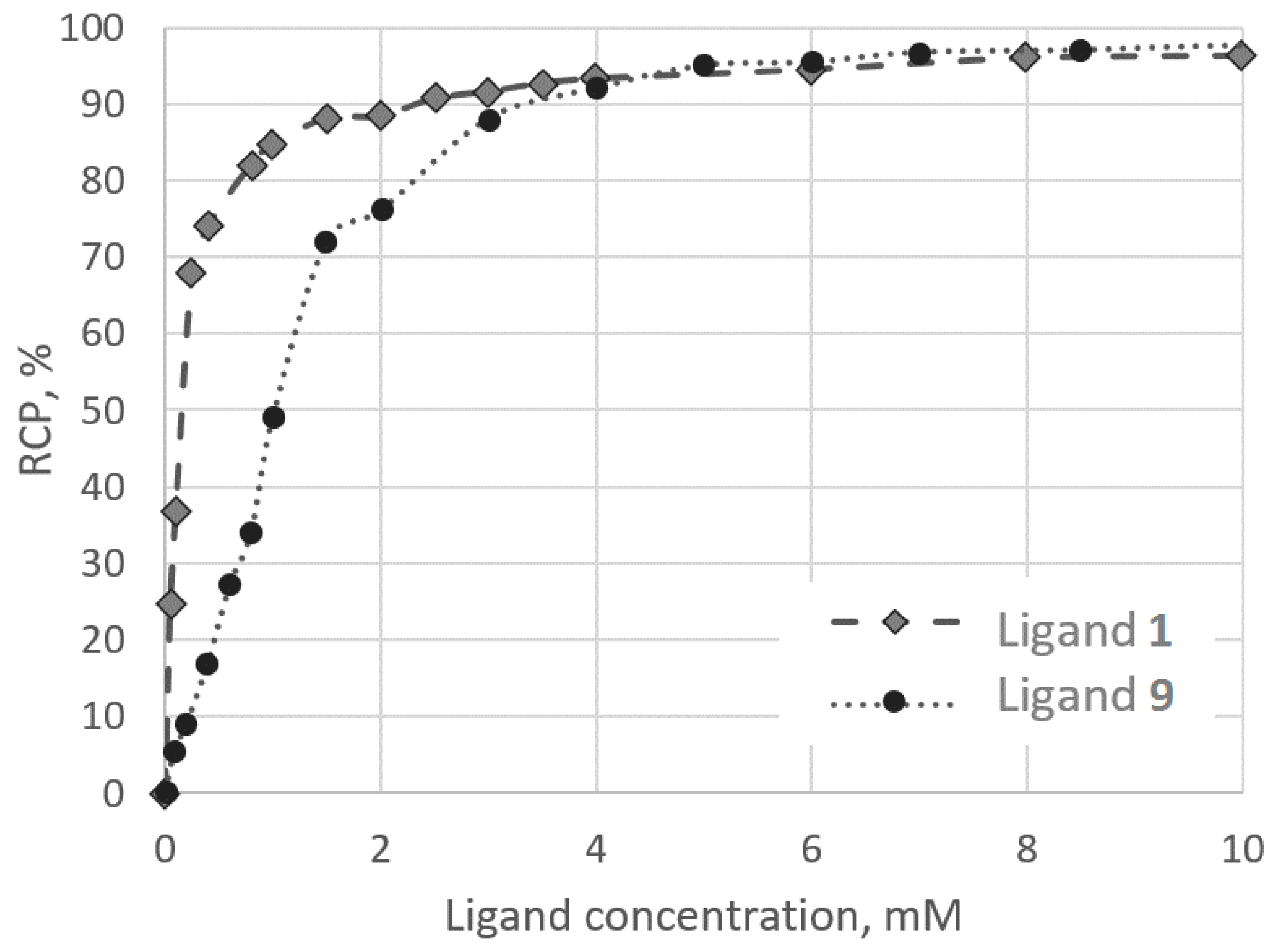
2.2. Stability Constants of Gallium(III) Complexes with 8 and 9

One of the most important criteria for the suitability of ligands as components of radiopharmaceuticals is the stability of their complexes with radionuclides [3,21,22]. For this, the protonation constants of the ligands and the stability constants of their complexes with Ga3+ were determined.
The protonation constants of 8 (H5L) and 9 (H6L) were determined at 298 К and ionic strength of I = 0.1 M KCl. Stepwise equilibrium constants of acids are given in Table 1. Full constants are given in Table S1 (see Supplementary Materials). The values of the stepwise constants of 8 and 9 are in good agreement with data for other aminodiphosphonic acids [23]. The first three protonation constants (no. 1–3, Table 1) of 8 are similar to the corresponding constants of (aminoethylene)diphosphonic acid (AEDP), which is essentially a fragment of 8.
The complexation reactions of 8 and 9 with Ga3+ in water at 298 К and ionic strength of I = 0.1 M KCl is well described by the model, which includes the complexation of Ga3+ with a deprotonated ligand and addition to Ga3+ of one to three (8) or one to four (9) protons besides the ligand (Table 2; full constants are given in Table S2, see Supplementary Materials). Stability constant of the Ga3+ complex with deprotonated ligand 9 (logKGaL = 31.92 ± 0.32) significantly exceeds the corresponding stability constant of the Ga3+ complex with 8 (logKGaL = 26.63 ± 0.24) (Table 2). This is very consistent with the fact that acid 9 has the more active P(O)(OH)2 group for the complexation instead of the COOH group of 8. This result suggests that ligand 9 is preferable over ligand 8 as a radiopharmaceutical component. The stability constants of the gallium(III)-deprotonated ligand complexes for 8 and 9 are similar to the stability of the complexes for ethylenediamine-N,N’-diacetic-N,N’-bis(methylenephosphonic) acid (EDDADPO) (logKGaL = 26.82) and for the ethylenediamine-N,N,N’,N’-tetrakis(methylenephosphonic) acid (EDTPO) (logKGaL = 31.83) [25] (Table 2 and Table S2 in Supplementary Materials). Ligands 8 and 9 are effective ligands for gallium(III) as EDDADPO and EDTPO, respectively. Since [68Ga]Ga-EDTPO showed promising results in μ-PET studies, we supposed that a study of osteotropic properties of 8 and 9 is of interest.

2.3. Radiolabelling and Stability

Both 8 and 9 ligands readily form complexes with 68Ga at room temperature when using ligand concentrations >5 mM and pH 5 ± 2 as confirmed by radio-TLC. Both [68Ga]Ga-8 and [68Ga]Ga-9 obtained in this way were stable for ten-fold dilution with saline. However, when ten-fold diluted with fetal bovine serum, up to 90% and 50% of [68Ga]Ga-8 and [68Ga]Ga-9 correspondingly undergo decomposition. Elevation of 9 concentration to 20 mM and raising reaction temperature to 95 °C (30-min reaction time) allowed us to achieve 60–90% stability in a 2 hr interval. This approach did not work out for [68Ga]Ga-8, and regardless of the concentration of 8 in the mixture, temperature, and reaction time, the stability of [68Ga]Ga-8 was very low. These results are in good agreement with the stability constants of gallium(III) complexes with 8 and 9 in water. Thus, it is once more affirmed that phosphonic groups are significantly more favorable for M3+ radiometals binding over carboxylic groups [26]. In the frame of this study, we decided to carry out further detailed studies using [68Ga]Ga-9.
Figure 3 shows the ligand concentration—labelling reaction yield dependence for the ligand 9 and previously studied 1,7-diamino-4-oxaheptane-1,1,7,7-tetraphosphonic acid (1) [10]. The conditions of 68Ga-labelling for both compounds were the same: рН 6, acetate concentration of 0.2 M, 25 °C, and 15 min reaction time. Ligand 1 contains two amidiphosphonic groups separated by a five-atoms ether chain with a weak donor oxygen atom. Ligand 9 contains one amidiphosphonic group and one phosphonic group in orto-position to it. According to potentiometric studies, ligand 1 forms one more protonated complex than ligand 9. The labelling reaction yield at low ligand concentration for [68Ga]Ga-1 is higher than that for [68Ga]Ga-9. Clearly, the structures of the Ga3+ complexes of these ligands differ significantly. This can be attributed to the fact that amidiphosphonic groups may be more effective for 68Ga binding than phosphonic groups. Another possible explanation may be the orto-position of the phosphonic group in 9 preventing the amidiphosphonic group from realizing its full binding potential toward 68Ga.
The influence of the buffering agent type on the labelling reaction yield was analysed before for [68Ga]Ga-1 [14]. The influence of acetate concentration on the labelling reaction yield was demonstrated for DOTA-conjugated molecules [27]. Here, the effect of acetate concentration was studied using four ligand 9 concentrations at a constant pH 5.9 ± 0.4. To observe the effect of ligand 9, the following concentrations were chosen: 0.8, 1.0, 2.0, and 4.0 mM. Results are presented in Figure 4a. In the case of [68Ga]Ga-9, there is a distinct correlation: the lower the acetate concentration is, the higher is the labelling reaction yield with maximal yield achieved in the absence of acetate. This correlation becomes less significant with increasing ligand concentration and becomes statistically insignificant (p > 0.05) at 5 mM of 9. In addition to the results [14], we carried out similar experiments with [68Ga]Ga-1 and found no statistically significant correlation even at a concentration of 1 in the reaction mixture being as low as 0.2 mM. Thus, there are three patterns of acetate concentration influencing the labelling reaction yield.
  • no correlation for [68Ga]Ga-1;
  • correlation with an extremum point for [68Ga]Ga-DOTA-conjugates (0.3 M of acetate corresponds to maximum labelling reaction yield) [27];
  • continuous dependence for [68Ga]Ga-9 (with maximum labelling reaction yield corresponding to the minimal concentration of acetate).
Gallium is known to form weak acetate complexes [28,29]. With logK = 3.7 [30], acetate is not able to compete with 9 for gallium binding. There is a possibility of a ternary Ga-9-OAc complex (or complexes) formation similar to that described for copper [18]. This possibility should be a subject of a separate study. In this study, acetate ion was added to reaction mixtures exclusively in the form of sodium acetate. The additional concentration of sodium in the reaction mixtures due to using NaOH for pH adjusting was ≤ 0.003 M. This suggests that sodium concentration in studied samples is virtually equal to that of acetate. To evaluate the influence of Na+ on the labelling process, additional experiments with constant Na+ concentrations were carried out. For this purpose, calculated amounts of NaCl were added to reaction mixtures at pH 5.7 ± 0.7 and 1 mM of 9. The comparison of the effects of dynamic and constant sodium concentration on the labelling reaction yield is presented in Figure 4b. At constant Na+ concentration of 0.4 M, obtained RCP (radiochemical purity) values were consistently lower than those obtained at dynamic Na+ concentration. However, the differences were mostly statistically insignificant (p > 0.05). Still, it is reasonable to assume that the acetate ion is the component responsible for the changes demonstrated in Figure 4a. The changes of water structure induced by an Na+ presence were observed in 1 M sodium chloride and sodium acetate solutions [31]. The examination of this sodium concentration in our experiments resulted in much lower RCP values as compared with corresponding samples with dynamic Na+ concentration (p < 0.05, Figure 4b). Thus, it is clear that Na+ itself has an impact on the Ga-9 complex formation process.
Finally, taking into account previous results, the effect of pH on the labelling reaction yield was studied using reaction mixtures with 50 mM of acetate and 0.8, 1.0, and 2.0 mM of ligand 9. A maximal reaction yield was observed for the samples at pH 3–4 (Figure 5), which is consistent with the data observed for 68Ga [27,32]. According to the calculations based on the obtained stability constants, the protonated complex GaH4L+ of ligand 9 dominates in this pH range.

2.4. Biodistribution of [68Ga]Ga-9

In Table 3, the biodistribution data of [68Ga]Ga-9 and [68Ga]Ga-acetate in animals with fractures are presented. Non-target biodistribution pathways and bone pathology uptake for [68Ga]Ga-9 are comparable to those of [68Ga]Ga-1 and [68Ga]Ga-2 [14]. The pathology site/intact bone ratio for [68Ga]Ga-9 is inferior to that of [68Ga]Ga-oxa-bis-ethylenenitrile tetra(methylene phosphonic acid) (3) [33] and even to that of [68Ga]Ga-acetate studied in this experiment (Table 3). Moreover, [68Ga]Ga-3 uptake in blood, liver, intestine, and kidneys is lower than that of [68Ga]Ga-9. Likely, it depends on the different stability of 68Ga-labelled complexes in vivo and requires additional research.
Since fracture healing may be accompanied by an inflammatory process, animals with an aseptic inflammation model were also studied. In Table 4, the data on the biodistribution of [68Ga]Ga-9 in animals with aseptic inflammation are presented along with data for [68Ga]Ga-citrate, which is known to have an inflammation imaging potential [34]. During the comparison of biodistribution dynamics, it was found that [68Ga]Ga-9 can be a potential agent for aseptic inflammation imaging more promising than [68Ga]Ga-citrate. Activity in blood 120 min after injection in comparison to a 60-min time point decreases three and two times for [68Ga]Ga-9 and [68Ga]Ga-citrate, respectively. [68Ga]Ga-9 pathology site/muscular tissue ratio is almost constant during the time of observation. It will allow imaging pathology foci 1 h after i.v. injection (for [68Ga]Ga-citrate—2 h).

3. Materials and Methods

3.1. Synthesis

The progress of the reactions was monitored by 31P NMR spectroscopy. All chemicals and solvents were purchased from Acros Organics (Acrus, Moscow, Russia) and Alfa Aesar (Reakor, Moscow, Russia). The 1H, 31P, and 13C NMR spectra were recorded on a Bruker DPX-200 spectrometer (Billerica, MA, USA) at 200.13, 81.0, and 50.04 MHz, respectively. Chemical shifts δ are given in ppm and coupling constants J are given in Hz. Melting points are determined on a Boetius PHMK-05 device or in the block in an open glass capillary. Chromatographic analysis was carried out for some of the compounds on LC/MSD Agilent 1100 mass spectrometer (Santa Clara, CA, USA) with DAD, ELSD, and single quadrupole mass-selective detector with ionization by electrospray. For some of the acids, elemental analysis was also performed.
The starting isopropyl salicylate 5a (R = COO-iPr, X = H) was purchase from Aldrich. Diethyl (2-hydroxy-5-ethylphenyl)phosphonate 5b (R = P(O)(OEt)2, X = Et, Figure 1) was synthesized, according to Reference [19].
General synthesis of 6a, b. Sodium hydride (20 mmol) (55% suspension in paraffin) was added in small portions to the mixture of phenol 5a or 5b (20 mmol) in 35 mL of dry dioxane at room temperature. The mixture was stirred for 0.5 h, then chloroacetonitrile (20 mmol) was added, and the formed reaction mass was boiled by stirring for 8 h. The reaction mixture evaporated in vacuo and 50 mL of water was added to the residue. The formed solution was acidified by HCl to pH~1 and the mixture was extracted with chloroform (3 × 25 mL). The organic extract was washed with water (3 × 25 mL) and evaporated in vacuo. Nitriles 6a, b were isolated by vacuum distillation of the residue.
Isopropyl 2-(cyanomethyloxy)-benzoate 6a. Сolorless oily liquid. B.p. 117–119 °C (1.1 mm). Yield 71%. 1H NMR (200 MHz, CDCl3, δ, ppm): 1.38 d (6Н, CH(CH3)2, 3JНН 6.1 Hz), 4.86 s (2H, OCH2CN), 5.25 m (1Н, CH(CH3)2), 7.15 m (1Н, arom.), 7.54 m (2Н, arom.), 7.85 m (1Н, arom.). 13С NMR (50.3 MHz, CDCl3, δ, ppm): 21.84 (CH3), 55.84 (CH2O), 68.76 (CHO), 115.06 (CN), 116.78 (Car.), 123.10 (Car.-COO), 123.69 (Car.), 131.92 (Car.), 133.44 (Car.), 155.99 (Car.-O), 164.82 (COO).
Diethyl 2-(cyanomethyloxy)-5-ethyl-phenylphosphonate 6b. Сolorless oily liquid. B.p. 164–166 °C (0.8 mm). Yield 55%. 1H NMR (200 MHz, CDCl3, δ, ppm): 1.19 t (3H, CH3, 3JНН 7.7 Hz), 1.31 t (6H, 2CH3, 3JНН 7.2 Hz), 2.61 q (2H, CH2, 3JНН 7.7 Hz), 4.12 dq (4H, 2CH2O, 3JНН 7.2 Hz, 4J 2.1 Hz), 4.83 s (2H, CH2O), 7.00 m (1H, arom.), 7.33 m (1H, arom.), 7.67 m (1H, arom.). 13С NMR (50.3 MHz, CDCl3, δ, ppm): 15.44, 16.30 d (3JРOCC 6.2 Hz), 27.86, 54.80, 62.27 d (2JРOC 5.5 Hz), 114.23 (Car.) d (3JРC 9.9 Hz), 114.95 (CN), 118.51 (Car.) d (1JРC 187.0 Hz), 133.57 (Car.), 134.71 (Car.) d (2JРC 6.9 Hz), 139.37 (Car.) d (3JРC 13.9 Hz), 156.09 (Car.-O). 31P NMR (81.0 MHz, D2O, δ, ppm): 17.1.
General synthesis of 7a, b. Phosphorus tribromide (20 mmol) was slowly added dropwise at 5 °C to a mixture of nitrile 6a, b (10 mmol) and dry phosphorous acid (20 mmol) in 15 mL of dioxane. Then the reaction mixture was stirred at room temperature for about 10 h, evaporated in vacuo, and 10 mL of dioxane was added to the residue. After the formation of a gelatinous residue, dioxane was decanted, the procedure was repeated, and 30 mL of acetic acid was added to the residue with cooling and stirring. The resulting mixture was stirred for about an hour and co-evaporated with 30 mL of toluene. Water (10 mL) dropwise was added to the oily residue and left overnight without stirring. The resulting white powder was washed with a small amount of water (3 × 3 mL) and then with ethanol, and aminodiphosphonic acids 7a and 7b was isolated with yields 63–64%.
Isopropyl 2-[(2-amino-2,2-diphosphono)ethyloxy]-benzoate 7a. White solid. Yield 63% M.p. 223–225 °C (with decomp.). 1H NMR (200 MHz, D2О, δ, ppm): 1.31 d (6Н, 2СН3, 3JНН 6.1 Hz), 4.55 d (1Н, one of CH2O, 3J 7.9 Hz), 4.60 d (1Н, second of CH2O, 3J 8.5 Hz), 5.16 m (1H, OCH), 6.95–7.20 m (2H, arom.), 7.50–7.62 m (1H, arom.), 7.80–7.90 m (1H, arom.). 13С NMR (50.3 MHz, D2О, δ, ppm): 21.11, *23.35 (СН3), 57.47 t (1JРC 116.6 Hz) (P2СN), *69.38, 69.68 (CH2O), 70.39 (OCH), *114.46, 114.84, *119.34, 119.68, 121.27, 121.73, *131.60, 131.85, 134.95, 157.83 (C = O), *167.64 (C = O). 31P NMR (81.0 MHz, D2O, δ, ppm): 10.4, * 14.1 (4:1). (*—hereinafter minor conformers). Found, %: C 37.42, 37.35; H 5.13, 5.33. C12H19NO9P2. Calculated, %: C 37.61, H 5.00. LCMS calcd for C12H19NO9P2: 383.2. Found 384.4 (protonated form).
Monoethyl 2-(2-amino-2,2-diphosphono)ethyloxy-5-ethyl-phenylphosphonate 7b. White solid. Yield 64% M.p.: 205–208 °C (with decomp.). 1H NMR (200 MHz, DMSO-d6 + drop TFA, δ, ppm): 1.12 t (3Н, СН3, 3JНН 7.3 Hz), 1.28 t (3Н, СН3, 3JНН 6.7 Hz), 2.55 q (2Н, СН2, 3JНН 7.3 Hz), 4.08 q (2H, СН2О, 3JНН 7.3 Hz), 4.48 m (broad) (2Н, СН2СР), 6.90–7.05 m (1Н, СН arom.), 7.27–7.40 m (2Н, СН arom.). 1H NMR (200 MHz, D2О, δ, ppm): 1.10 t (3Н, СН3, 3JНН 7.3 Hz), 1.15 t (3Н, СН3, 3JНН 7.3 Hz), 2.54 q (2Н, СН2, 3JНН 7.3 Hz), 3.78 q, and 4.09*q (2H, СН2, 3JНН 7.3 Hz), 4.40 d (1Н, one of СН2СР, 2JРН 9.8 Hz), 4.46 d (1Н, the second of СН2СР, 2JРН 9.8 Hz), 6.87–7.15 m (1Н, СН arom.), 7.20–7.55 m (2H, CH arom.). 13С NMR (50.3 MHz, D2О, δ, ppm): 15.22, 15.83 d (3JРC 6.5 Hz), 27.42, 58.16 t (1JРC 120.2 Hz) (P2СN), and *58.26 t (1JРC 121.5 Hz) (P2СN), 61.93 d (2JРOC 5.0 Hz), and *64.28 d (2JРOC 5.8 Hz), 68.99 and *69.43 (OCH2CN), 112.32 d (3JРC 8.4 Hz), and *112.82 d (3JРC 8.1 Hz), 118.62, 121.86, 122.13, 127.21 d (1JРC 186.3 Hz), *131.56 (3JРC 6.5 Hz) and 132.20 (3JРC 6.9 Hz), 132.64, 137.78 d (3JРC 12.7 Hz), *156.73, and 156.90. 31P NMR (81.0 MHz, DMSO-d6, δ, ppm): (10.38 + *10.90) / (*14.83 + 16.30) = 2/1. 31P NMR (81.0 MHz, D2O, δ, ppm): (*11.65 + 13.03) / (*10.76 + 14.76) = 2/1. Found, %: P 21.73, 21.85. C12H22NO10P3. Calculated, %: P 21.45.
2-[(2-Amino-2,2-diphosphono)ethyloxy]-benzoic acid 8.
Solution of isopropyl ether 7a (1.9 g, 5 mmol) in a 10 mL of 6N HCl was refluxed for 5 h. Acid 8 was isolated after evaporation of the reaction mixture and crystallization of the residue from aqueous ethanol. White solid. Yield 1.3 g (76%). M.p.: 243–244 °C (with decomposition). 1H NMR (200 MHz, D2О + NaOD, pH~10, δ, ppm): 4.27 t (2Н, СН2О, 3JРН 12.2 Hz), 6.85–6.98 m (1Н, СН arom.), 7.08–7.20 m (1Н, СН arom.), 7.24–7.38 m (2H, 2СН arom.). 13С NMR (50.3 MHz, D2О + NaOD, pH~10, δ, ppm): 58.06 t (1JРC 120.7 Hz), 72.98, 115.30, 121.50, 128.50, 129.38, 130.90, 155.87, 177.15 (C = O). 31P NMR (81.0 MHz, D2O + NaOD, δ, ppm): 9.85 (pH 5), 19.60 (pH 10). Found, %: C 31.41, 31.35, H 4.04, 3.96, P 18.23, 18.05. C9H13NO9P2. Calculated, %: C 31.69, H 3.84, P P18.16. LCMS calcd for C9H13NO9P2: 341.2. Found 342.2 (protonated form).
2-[(2-Amino-2,2-diphosphono)ethyloxy]-5-ethyl-phenylphosphonic acid 9. To a solution of 0.73 g (1.6 mmol) monoethyl ester of phosphonic acid 7b in 7 mL of dry acetonitrile 0.21 mL (1.6 mmol) of trimethylsilyl bromide was added. The mixture was gradually heated with stirring and boiled for 5 h. The reaction mixture was evaporated in vacuo. The residue was twice evaporated with 5 mL of water and evaporated to dryness. The resulting solid was washed with water and then with ethanol. Acid 9 was isolated after crystallization of the residue from aqueous ethanol. White solid. Yield 68%. M.p.: 239–240 °C (with decomposition). 1H NMR (200 MHz, D2О, δ, ppm): 1.11 t (3Н, СН3, 3JНН 7.3 Hz), 2.55 q (2Н, СН2, 3JНН 7.3 Hz), 4.52 d (1Н, one of СН2О, 3JРН 9.8 Hz), 4.55 d (1Н, the second of СН2О, 3JРН 9.3 Hz), 6.90–7.05 m (1Н, СН arom.), 7.27–7.55 m (2Н, 2СН arom.). 13С NMR (50.3 MHz, D2О, δ, ppm): 15.30, 27.55, 58.14 t (1JРC 114.6 Hz), *68.41 and 69.37, 112.39 d (3JРC 8.1 Hz), 115.20, 123.94 d (1JРC 175.2 Hz), 129.07, 131.52 d (3JРC 6.9 Hz), 131.91, 137.69 d (3JРC 13.0 Hz), 156.84. 31P NMR (81.0 MHz, D2O, δ, ppm): (*9.34 + 10.20) / (12.31 + *14.96) = 2/1. Found, %: C 29.41, 29.35; H 4.64, 4.76; P 22.95, 23.15. C10H18NO10P3. Calculated, %: C 29.64, H 4.48, P 22.93.
NMR spectra of synthesized compounds are given in Supplementary materials.

3.2. Stability Constant Measurements and Calculations

The potentiometric titration technique using the OP-300 Radelkis potentiometer was described earlier [35]. Solutions of 8 and 9 were titrated with a standard 0.1 М NaOH solution at 298 ± 0.1 K and ionic strength of I = 0.1 M KCl. The initial volume of solutions was 160 mL. Titrations were performed in the range of pH 3.0–11.6 (8) and 3.5–11.5 (9). Experiments included from 33 to 52 (8) and from 31 to 66 (9) data points. The initial analytical concentrations were 0.35–0.92 mM (8) and 0.27–0.48 mM (9). The protonation constants were estimated from four titrations using the CHEMEQUI program [36] freely available on the server [37]. CHEMEQUI evaluates equilibrium constants using four different algorithms: EQ, SIMPLEX, MONTE-CARLO, and the genetic algorithm SDE [38]. Estimation of the constants was performed based on each titration. In the case of significant correlations between the protonation constants, resulting in a shift of the constants, the calculations were performed simultaneously based on several titrations. All the computational results were used to calculate the average values of the estimated full constants logβ and their standard deviations. The average values were determined from 17 (8) and 12 (9) calculations based on several titrations and algorithms. The errors in the stepwise logK values are evaluated using standard deviations in estimated full equilibrium constants logβ and an error propagation rule for several titrations and applied algorithm calculations.
Solutions of 8 or 9 with Ga(NO3)3 were titrated with NaOH under similar conditions. Titrations were performed in the range of pH 2.9–11.5 (8) and 3.0–12.0 (9). Experiments included from 31 to 60 (8) and from 35 to 77 (9) data points. The initial analytical concentrations were 0.36–0.71 mM (8), 0.36–0.72 mM (Ga3+ in experiments with 8), 0.30–0.38 mM (9), and 0.31–0.45 mM (Ga3+ in experiments with 9). In the calculations of stability constants of Ga3+ complexes with deprotonated forms of HnL(5−n)– (for 8, n = 0, 1,..., 5) and HnL(6−n)– (for 9, n = 0, 1,..., 6), the protonation constants of acids did not vary. They were taken as previously estimated in acid titration experiments. Gallium(III) forms highly stable hydroxides in water [39]. It was taken into account for obtaining unshifted estimations of the stability constants of Ga3+ complexes with organic ligands. The following stability constants logβn of hydroxo-complexes in water were used in the calculations: −2.85, −7.28, −11.94, and −15.66 for the equilibria Ga3+ + nH2O = Ga3+(OH)n + nH+, n = 1, 2, 3, 4, respectively [40]. Stability constants of the Ga3+ complexes with 8 and 9 were obtained as average values from three titrations and 8 calculations for 8 and from four titrations and 8 calculations for 9. All four CHEMEQUI algorithms were used for the calculation of stability constants.
The Hamilton’s R-factor (HRF) and the squared determination coefficient (R2det) were used as the agreement criteria of the proposed set of equilibrium reactions and calculated constants with the experimental data [35]. Depending on the experiment and the program algorithm, HRF was 0.30–0.96% (8), 0.46–0.92% (9) at the calculations of protonation constants, 0.99–1.50% (8 + Ga3+), and 1.41–3.18% (9 + Ga3+) at the calculations of stability constants of the Ga3+ complexes.

3.3. Radiolabelling, Stability, and Quality Control

All chemicals used for labelling were of a “reagent pure” or “extra pure” grade (Sigma-Aldrich, Panreac). The 68Ge/68Ga generators (Cyclotron Ltd., Obninsk, Russia) with activity 20 and 50 mCi were used (0–12 months after the production). The generator was eluted with 0.1 M HCl as per the manufacturer’s instruction.
Radiolabelling and evaluation of [68Ga]Ga-8 and [68Ga]Ga-9 stability were carried out in triplicate using procedures similar to those described in Reference [14]. In short, for labelling with 68Ga sodium acetate (buffering agent) solutions of various concentrations, (0.1, 0.5, or 1 M) and 68Ge/68Ga generator eluate were added to the Eppendorf tubes containing 0.1–20 μmol of 8 or 9. pH of the mixtures was then adjusted using NaOH and HCl of pre-estimated concentrations. The reaction mixtures were stirred at 25 or 95 °C for 15–30 min. To evaluate the stability of the complexes obtained, 100 μL of a sample was added to 1000 μL of saline or fetal bovine serum. The mixtures were stirred at 37 °C for 2 h.
The content of major radiochemical impurities (colloidal and free 68Ga [41,42]) was determined using two strips of a radio-TLC technique. The following chromatographic systems were used: cellulose—2.4 % HCl: acetone: acetylacetone (0.8: 7: 0.5): Rf (68Gacolloid) = 0, Rf ([68Ga]Ga-8) = 0.2 ± 0.2, Rf ([68Ga]Ga-9) = 0.4 ± 0.1, Rf (68Gafree) = 0.95 ± 0.05, cellulose—HCl (1 M): methanol (2: 1): Rf (68Gacolloid) = 0, Rf ([68Ga]Ga-8) = 0.9 ± 0.1, Rf ([68Ga]Ga-9) = 0.65 ± 0.05, Rf (68Gafree) = 0.95 ± 0.05.

3.4. Biodistribution Studies

All experiments involving animals were performed following the ethical standards, Russian animal protection laws, and guidelines for scientific animal trials [43].
Animal studies were performed using female outbred albino rats with model pathologies. Animals with fractures (active bone callus formation) [44] had been grouped (N = 3) and [68Ga]Ga-9 (100 μL per rat) was i.v. injected into the tail vein. At preselected time points (60, 120 min), animals were obtained from the experiment using partial decapitation. The organs of interest were collected, blotted dry, and weighed. Radioactivity in samples of organs/tissues was counted using a WIZARD2 automatic γ-counter (PerkinElmer). The results are expressed as the percentage of injected activity dose per gram (mean % ID or mean % ID/g ± SD) for each organ/tissue. For comparison, the mixture of 68Ge/68Ga generator eluate with sodium acetate solution (pH 6.5 ± 0.5, 0.18 M total acetate concentration) was also injected into animals with bone pathology.
In addition, animals with a model of aseptic inflammation were used. The site of aseptic soft tissue inflammation was modelled by intramuscular injection of 0.2 mL of turpentine into the rat pelvic limb. An acute inflammatory reaction was observed 7 days after administration. The inflammation foci were marked by swelling of the tissue, which is sharply painful on palpation. An autopsy revealed a burn of soft tissues with elements of necrosis, pronounced as a vascular pattern. [68Ga]Ga-9 was studied using these animals in the same way it was done for animals with fractures. For comparison, the mixture of the 68Ge/68Ga generator eluate with sodium citrate solution (pH 5.0 ± 0.5, 0.084 M total citrate concentration) was also injected into animals.

4. Conclusions

The combination of aminodiphosphonic fragment with salicylic acid or its phosphonic analogue into one molecule is a promising way to develop radiopharmaceuticals. According to this technique, two new ligands with high complexation ability to gallium(III) were synthesized. Introducing phosphoryl fragment instead of carbonyl increases stability constants of the gallium(III) complexes in water. Stability constant of the Ga3+ complex with fully phosphorylated acid 9 (logKGaL = 31.92 ± 0.32) significantly exceeds stability constant of Ga3+ complex with 8 (logKGaL = 26.63 ± 0.24). Ligands 8 and 9 are as effective for Ga3+ cation binding as ethylenediamine-N,N’-diacetic-N,N’-bis(methy1enephosphonic) acid and ethylenediamine-N,N,N’,N’-tetrakis(methylenephosphonic) acid, respectively.
Both new molecules readily form 68Ga-complexes stable by ten-fold dilution with saline. However, in fetal bovine serum only, [68Ga]Ga-9 was stable enough to be subject to biological evaluation. It was injected into rats with bone pathology and aseptic inflammation of soft tissues. In vivo studies revealed that [68Ga]Ga-9 is not suitable as a bone-seeking agent, but it can be used for inflammation imaging. To an extent, as inflammation imaging, [68Ga]Ga-9 is preferable over [68Ga]Ga-Citrate due to delayed free 68Ga release from the complex.
In addition, the 68Ga-labelling reaction with 9 was studied in detail. A correlation of acetate concentration in the reaction mixture and labelling reaction was found (up to 5 mM of 9): the lower the acetate concentration is, the higher the labelling reaction yield is.

Supplementary Materials

Supplementary materials are available online. Figure S1: 1H and 13C NMR of nitrile 6a. Figure S2: 1H, 13C and 31P NMR of nitrile 6b. Figure S3: 1H, 13C and 31P NMR of aminodiphosphonic acid 7a. Figure S4a: 1H NMR of aminodiphosphonic acid 7b. Figure S4b: 13C and 31P NMR of aminodiphosphonic acid 7b. Figure S5: 1H, 13C and 31P NMR of aminodiphosphonic acid 8. Figure S6: 1H, 13C and 31P NMR of aminodiphosphonic acid 9, Table S1: protonation constants of acids 8 and 9. Table S2: full stability constants of the Ga3+ complexes with 8 and 9.

Author Contributions

Conceptualization, A.Y.M., V.E.B., V.V.R. and A.Y.T. Methodology, A.Y.M., V.P.S. and V.V.R. Software, V.P.S., V.V.R. and G.S.T. Validation, A.Y.M., V.V.R., G.S.T. and V.P.S. Formal analysis, I.A.M., K.A.L., A.S.L., V.P.S. and V.V.R. Investigation, I.A.M., K.A.L., A.S.L., V.E.B., V.V.R., G.S.T. and V.P.S. Resources, V.E.B. and A.Y.T. Data curation, I.A.M., K.A.L., A.S.L. and V.P.S. Writing—original draft preparation, A.Y.M., V.V.R., A.S.L., G.S.T., V.E.B. and V.P.S. Writing—review and editing, A.Y.M., V.E.B., V.V.R., O.E.K., G.S.T., G.E.K. and V.P.S. Visualization, I.A.M. Supervision, V.E.B., O.E.K., G.E.K. and A.Y.T. Project administration, G.E.K., V.E.B. and A.Y.T. Funding acquisition, G.E.K. and A.Y.T. All authors have read and agreed to the published version of the manuscript.

Funding

The work was performed on the government assignments for the year 2021 of the Institute of Physical Chemistry and Electrochemistry, Russian Academy of Sciences, and the Institute of Physiologically Active Compounds, Russian Academy of Sciences (topic No. 0090-2019-0008). The work on ligands synthesis was financially supported by the Russian Foundation for Basic Research (projects No. 18-03-00959 and No. 19-03-00262). The works on stability constants calculations and biodistribution were financially supported by the Russian Scientific Foundation (project No. 19-13-00294).

Institutional Review Board Statement

The study was conducted according to the guidelines of the Declaration of Helsinki, the national state standard (GOST 33216-2014. Guidelines for Accommodation and Care of Animals. Species-Specific Provisions for Laboratory Rodents and Rabbits; Russia, 2014) and approved by the Institutional Review Board of State Research Center—Burnasyan Federal Medical Biophysical Center of Federal Medical Biological Agency, Russia.

Informed Consent Statement

Not applicable.

Data Availability Statement

The data presented in this study are available in Supplementary material.

Conflicts of Interest

The authors declare no conflict of interest.

Sample Availability

Samples of the compounds 19 are available from the authors.

References

  1. Roesch, F.; Riss, P.J. The Renaissance of the 68Ge/68Ga Radionuclide Generator Initiates New Developments in 68Ga Radiopharmaceutical Chemistry. Curr. Top. Med. Chem. 2010, 10, 1633–1668. [Google Scholar] [CrossRef]
  2. Velikyan, I. Prospective of 68Ga-Radiopharmaceutical development. Theranostics 2014, 4, 47–80. [Google Scholar] [CrossRef] [Green Version]
  3. Baum, R.P.; Rosch, F. Theranostics, Gallium-68, and Other radionuclides: A Pathway to Personalized Diagnosis and Treatment; Springer: Berlin/Heidelberg, Germany, 2013. [Google Scholar]
  4. Gourni, E.; Henriksen, G.; Gamez, P.; Caballero, A.B. Metal-based PSMA radioligands. Molecules 2017, 22, 523. [Google Scholar] [CrossRef] [Green Version]
  5. Pillai, M.R.A.; Nanabala, R.; Joy, A.; Sasikumar, A.; Knapp, F.F.R. Radiolabeled enzyme inhibitors and binding agents targeting PSMA: Effective theranostic tools for imaging and therapy of prostate cancer. Nucl. Med. Biol. 2016, 43, 692–720. [Google Scholar] [CrossRef]
  6. Zha, Z.; Wu, Z.; Choi, S.R.; Ploessl, K.; Smith, M.; Alexoff, D.; Zhu, L.; Kung, H.F. A New [68Ga]Ga-HBED-CC-Bisphosphonate as a Bone Imaging Agent. Mol. Pharm. 2020, 17, 1674–1684. [Google Scholar] [CrossRef] [PubMed]
  7. Khawar, A.; Eppard, E.; Roesch, F.; Ahmadzadehfar, H.; Kürpig, S.; Meisenheimer, M.; Gaertner, F.C.; Essler, M.; Bundschuh, R.A. Preliminary results of biodistribution and dosimetric analysis of [68 Ga]Ga-DOTA ZOL: A new zoledronate-based bisphosphonate for PET/CT diagnosis of bone diseases. Ann. Nucl. Med. 2019, 33, 404–413. [Google Scholar] [CrossRef]
  8. Meckel, M.; Kubíček, V.; Hermann, P.; Miederer, M.; Rösch, F. A DOTA based bisphosphonate with an albumin binding moiety for delayed body clearance for bone targeting. Nucl. Med. Biol. 2016, 43, 670–678. [Google Scholar] [CrossRef]
  9. Meckel, M.; Fellner, M.; Thieme, N.; Bergmann, R.; Kubicek, V.; Rösch, F. In vivo comparison of DOTA based 68Ga-labelled bisphosphonates for bone imaging in non-tumour models. Nucl. Med. Biol. 2013, 40, 823–830. [Google Scholar] [CrossRef]
  10. Kodina, G.E.; Malysheva, A.O.; Klement’eva, O.E. Osteotropic radiopharmaceuticals in Russian nuclear medicine techniques. Russ. Chem. Bull. Int. Ed. 2016, 65, 350–362. [Google Scholar] [CrossRef]
  11. Palma, E.; Correia, J.D.G.; Campello, M.P.C.; Santos, I. Bisphosphonates as radionuclide carriers for imaging or systemic therapy. Mol. Biosyst. 2011, 7, 2950–2966. [Google Scholar] [CrossRef]
  12. Romanenko, V.D.; Kukhar, V.P. 1-Amino-1,1-bisphosphonates. Fundamental syntheses and new developments. Arkivoc 2012, 2012, 127–166. [Google Scholar] [CrossRef] [Green Version]
  13. Chmielewska, E.; Kafarski, P. Synthetic procedures leading towards aminobisphosphonates. Molecules 2016, 21, 1474. [Google Scholar] [CrossRef] [Green Version]
  14. Mitrofanov, I.A.; Maruk, A.Y.; Larenkov, A.A.; Kodina, G.E.; Lunev, A.S.; Luneva, K.A.; Klementyeva, O.E.; Tsebrikova, G.S.; Baulin, V.E.; Ragulin, V.V.; et al. Evaluation of Applicability of Aminodiphosphonic Acids for the Development of Bone-Seeking 68Ga-Radiopharmaceuticals. Russ. J. Gen. Chem. 2020, 90, 390–397. [Google Scholar] [CrossRef]
  15. Tsebrikova, G.; Baulin, V.; Ragulin, V.; Solov’ev, V.; Maruk, A.; Lyamtseva, E.; Malysheva, A.; Larenkov, A.; Zhukova, M.; Lunev, A.; et al. New phosphonic acids as components of bone seeking radiopharmaceuticals. J. Label. Compd. Radiopharm. 2019, 62, S287–S289. [Google Scholar]
  16. Rasulova, N.; Sagdullaev, S.; Arybzhanov, D.; Khodjibekov, M.; Krylov, V.; Lyubshin, V. Optimal Timing of Bisphosphonate Administration in Combination with Samarium-153 Oxabifore in the Treatment of Painful Metastatic Bone Disease. World J. Nucl. Med. 2013, 12, 14–18. [Google Scholar] [CrossRef] [PubMed]
  17. Tsebrikova, G.S.; Ragulin, V.V.; Baulin, V.E.; German, K.E.; Malysheva, A.O.; Klement’eva, O.E.; Kodina, G.E.; Larenkov, A.A.; Lyamtseva, E.A.; Taratonenkova, N.A.; et al. 2,5-Diamino-5,5-diphosphonovaleric Acid as a Ligand for an Osteotropic 188Re Radiopharmaceutical. Russ. J. Gen. Chem. 2018, 88, 1780–1785. [Google Scholar] [CrossRef]
  18. Teixeira, F.; Pérez, A.; Madden, W.; Hernández, L.; del Carpio, E.; Lubes, V. Speciation of the ternary complexes formed between copper(II), salicylic acid and small blood serum bioligands. J. Mol. Liq. 2016, 224, 346–350. [Google Scholar] [CrossRef]
  19. Baulin, V.E.; Kalashnikova, I.P.; Vikharev, Y.B.; Vikhareva, E.A.; Baulin, D.V.; Tsivadze, A.Y. Phosphoryl Analogs of Salicylic Acid: Synthesis and Analgesic and Anti-Inflammatory Activity. Russ. J. Gen. Chem. 2018, 88, 1786–1791. [Google Scholar] [CrossRef]
  20. Ragulin, V.V. Phosphorus-Containing Aminocarboxylic Acids: XV. α,ω-Diamino-ω,ω-diphosphonoalkylcarboxylic Acids. Russ. J. Gen. Chem. 2018, 88, 1045–1048. [Google Scholar] [CrossRef]
  21. Price, E.W.; Orvig, C. Matching chelators to radiometals for radiopharmaceuticals. Chem. Soc. Rev. 2014, 43, 260–290. [Google Scholar] [CrossRef] [PubMed]
  22. Delgado, R.; Félix, V.; Lima, L.M.P.; Price, D.W. Metal complexes of cyclen and cyclam derivatives useful for medical applications: A discussion based on thermodynamic stability constants and structural data. Dalt. Trans. 2007, 2734–2745. [Google Scholar] [CrossRef]
  23. Popov, K.; Rönkkömäki, H.; Lajunen, L.H.J. Critical evaluation of stability constants of phosphonic acids (IUPAC Technical Report). Pure Appl. Chem. 2001, 73, 1641–1677. [Google Scholar] [CrossRef]
  24. Bollinger, J.E.; Roundhill, D.M. Complexation of Indium(III), Gallium(III), Iron(III), Gadolinium(III), and Neodymium(III) Ions with Amino Diphosphonic Acids in Aqueous Solution. Inorg. Chem. 1993, 32, 2821–2826. [Google Scholar] [CrossRef]
  25. Motekaitis, R.J.; Martell, A.E. Gallium Complexes of Multidentate Ligands in Aqueous Solution. Inorg. Chem. 1980, 19, 1646–1651. [Google Scholar] [CrossRef]
  26. Stein, B.W.; Morgenstern, A.; Batista, E.R.; Birnbaum, E.R.; Bone, S.E.; Cary, S.K.; Ferrier, M.G.; John, K.D.; Pacheco, J.L.; Kozimor, S.A.; et al. Advancing Chelation Chemistry for Actinium and Other +3 f-Elements, Am, Cm, and la. J. Am. Chem. Soc. 2019, 141, 19404–19414. [Google Scholar] [CrossRef] [PubMed]
  27. Arefyeva, E.; Larenkov, A.; Maruk, A.; Kodina, G. Effects of Buffering Agents on Gallium-68 Speciation in Radiopharmaceutical Related Preparations. J. Label. Compd. Radiopharm. 2021. In Press. [Google Scholar]
  28. Hacht, B. Gallium (III)-acetate speciation studies under physiological conditions. Open Sci. J. 2016, 1, 1–13. [Google Scholar] [CrossRef] [Green Version]
  29. Clausén, M.; Öhman, L.O.; Kubicki, J.D.; Persson, P. Characterisation of gallium(III)-acetate complexes in aqueous solution: A potentiometric, EXAFS, IR and molecular orbital modelling study. J. Chem. Soc. Dalt. Trans. 2002, 2559–2564. [Google Scholar] [CrossRef]
  30. Skorik, N.A.; Artish, A.S. Stability of complexes of scandium, gallium, indium and thorium with anions of certain organic acids. Zhurnal Neorg. Khim. 1985, 30, 1994–1997. [Google Scholar]
  31. Petit, T.; Lange, K.M.; Conrad, G.; Yamamoto, K.; Schwanke, C.; Hodeck, K.F.; Dantz, M.; Brandenburg, T.; Suljoti, E.; Aziz, E.F. Probing ion-specific effects on aqueous acetate solutions: Ion pairing versus water structure modifications. Struct. Dyn. 2014, 1, 034901. [Google Scholar] [CrossRef] [PubMed]
  32. Bubenshchikov, V.B.; Maruk, A.Y.; Bruskin, A.B.; Kodina, G.E. Preparation and properties of 68Ga complexes with RGD peptide derivatives. Radiochemistry 2016, 58, 506–512. [Google Scholar] [CrossRef]
  33. Lunyov, A.S.; Clement’eva, О.Е.; Lunyova, К.А.; Zhukova, M.V.; Malysheva, А.О. Qualitative and quantitative comparisons of bone PET-imaging using 68Ga-oxabiphor and Na18F. Saratov J. Med. Sci. Res. 2017, 13, 886–891. [Google Scholar]
  34. Lunev, A.S.; Larenkov, A.A.; Petrosova, K.A.; Klementyeva, O.E.; Kodina, G.E. Fast PET imaging of inflammation using 68Ga-citrate with Fe-containing salts of hydroxy acids. EJNMMI Radiopharm. Chem. 2016, 1 (Suppl. S1), 10. [Google Scholar]
  35. Tsebrikova, G.S.; Barsamian, R.T.; Solov, V.P.; Kudryashova, Z.A.; Baulin, V.E.; Wang, Y.J.; Tsivadze, A.Y. Complexation of gallium(III) nitrate with 1,4,7,10-tetraazacyclododecane-1,4,7,10-tetrakis(methylenephosphonic acid). Russ. Chem. Bull. 2018, 67, 2184–2187. [Google Scholar] [CrossRef]
  36. Solov’ev, V.P.; Tsivadze, A.Y. Supramolecular complexes: Determination of stability constants on the basis of various experimental methods. Prot. Met. Phys. Chem. Surfaces 2015, 51, 1–35. [Google Scholar] [CrossRef]
  37. Solov’ev, V.P. The CHEMEQUI Program for Computations of Equilibrium Constants and Related Quantities from Experimental Results of UV–Vis, IR and NMR Spectroscopy, Calorimetry, Potentiometry and Conductometry. Available online: http://vpsolovev.ru/programs/chemequi/ (accessed on 8 December 2020).
  38. Ali, M.; Pant, M.; Abraham, A. A simplex differential evolution algorithm: Development and applications. Trans. Inst. Meas. Control 2012, 34, 691–704. [Google Scholar] [CrossRef]
  39. Wood, S.A.; Samson, I.M. The aqueous geochemistry of gallium, germanium, indium and scandium. Ore Geol. Rev. 2006, 28, 57–102. [Google Scholar] [CrossRef]
  40. Benézéth, P.; Diakonov, I.I.; Pokrovski, G.S.; Dandurand, J.L.; Schott, J.; Khodakovsky, I.L. Gallium speciation in aqueous solution. Experimental study and modelling: Part 2. Solubility of α-GaOOH in acidic solutions from 150 to 250°C and hydrolysis constants of gallium (III) to 300 °C. Geochim. Cosmochim. Acta 1997, 61, 1345–1357. [Google Scholar] [CrossRef]
  41. Larenkov, A.A.; Maruk, A.Y.; Kodina, G.E. Intricacies of the Determination of the Radiochemical Purity of 68Ga Preparations: Possibility of Sorption of Ionic 68Ga Species on Reversed-Phase Columns. Radiochemistry 2018, 60, 625–633. [Google Scholar] [CrossRef]
  42. Maruk, A.Y.; Larenkov, A.A. Determination of ionic 68Ga impurity in radiopharmaceuticals: Major revision of radio-HPLC methods. J. Radioanal. Nucl. Chem. 2020, 323, 189–195. [Google Scholar] [CrossRef]
  43. GOST 33216-2014. Guidelines for Accommodation and Care of Animals. Species-Specific Provisions for Laboratory Rodents and Rabbits; Standardinform: Moscow, Russia, 2014. [Google Scholar]
  44. Moiseenko, V.M.; Blinov, N.N. Sovremennaya Taktika Lecheniya Bol’nykh so Zlokachestvennymi Novoobrazovaniyami s Metastazami v Kosti: Posobie Dlya Vrachei (Modern Tactics of Treating Patients with Malignant Neoplasms with Bone Metastases: A Manual for Doctors, in Russian); Ministry of Healthcare of the Russian Federation, Petrov Research Institute of Oncology: St. Petersburg, Russia, 1996. [Google Scholar]
Figure 1. Structures of phosphonic acids mentioned in this work.
Molecules 26 02357 g001
Figure 2. General procedure for the synthesis of 2-[(2-amino-2,2-diphosphono)ethyloxy]-benzoic acid (8) and 2-[(2-amino-2,2-diphosphono)ethyloxy]-5-ethyl-phenylphosphonic acid (9).
Molecules 26 02357 g002
Figure 3. Ligand concentration—labelling reaction yield dependencies for [68Ga]Ga-1 and [68Ga]Ga-9.
Molecules 26 02357 g003
Figure 4. The effect of buffer component concentrations on the labelling reaction yield for [68Ga]Ga-9: (a) at Na+ concentration being equal to that of acetate ion and at various concentrations of ligand 9, (b) comparison of the effects of dynamic and constant Na+ concentration at the same 1 mM concentration of 9.
Molecules 26 02357 g004
Figure 5. The effect of pH on the labelling reaction yield for [68Ga]Ga-9.
Molecules 26 02357 g005
Table 1. Stepwise protonation constants of acids 8 (H5L) and 9 (H6L) in water at 298 К and ionic strength of I = 0.1 M KCl a.
No.EquilibriumlogK b
89AEDP c
1H + L = HL10.88 ± 0.1911.82 ± 0.2911.5
2HL + H = H2L9.11 ± 0.2610.65 ± 0.438.58
3H2L + H = H3L5.65 ± 0.289.76 ± 0.455.37
4H3L + H = H4L3.47 ± 0.268.50 ± 0.451.50
5H4L + H = H5L2.99 ± 0.325.05 ± 0.46
6H5L + H = H6L 2.75 ± 0.48
a For simplicity, the charges of chemical forms in the equilibria are not indicated. b The errors in the logK values are evaluated using standard deviations in estimated full equilibrium constants logβ and error propagation rule for the several titrations and applied algorithm calculations (see Experimental section). c Protonation constants of (aminoethylene)diphosphonic acid (AEDP) at 297 K and I = 0.2 M [24].
Table 2. Stepwise stability constants of the Ga3+ complexes with 8 (H5L) and 9 (H6L) in water at 298 К and ionic strength of I = 0.1 M KCl a.
No.EquilibriumlogK
89EDDADPO bEDTPO b
1Ga + L = GaL26.63 ± 0.2431.92 ± 0.3226.8231.83
2GaL + H = GaHL8.28 ± 0.408.90 ± 0.615.106.65
3GaHL + H = GaH2L4.60 ± 0.438.28 ± 0.682.245.10
4GaH2L + H = GaH3L3.25 ± 0.438.03 ± 0.65 3.29
5GaH3L + H = GaH4L ≈4.9 2.46
a See notes for Table 2. b Stepwise stability constants of the Ga3+ complexes with ethylenediamine-N,N’-diacetic-N,N’-bis(methy1enephosphonic) acid (EDDADPO) and ethylenediamine-N,N,-N’,N’-tetrakis(methylenephosphonic) acid (EDTPO) at 298 K and I = 0.1 M KNO3 [25].
Table 3. Dynamics of distribution of [68Ga]Ga-9 and [68Ga]Ga-acetate in rats with fractures (active bone callus formation) (mean ± SD).
Probe[68Ga]Ga-Acetate[68Ga]Ga-9
Organ or TissueTime after Injection
60 min120 min60 min120 min
Blood a3.0 ± 0.44.5 ± 0.42.6 ± 0.32.5 ± 0.0
Lung b3.3 ± 0.36.0 ± 0.33.0 ± 1.04.1 ± 1.4
Liver b6.8 ± 0.911.2 ± 0.74.9 ± 0.76.5 ± 0.5
Kidney b1.5 ± 0.12.6 ± 0.41.6 ± 0.21.9 ± 0.3
Intestine b4.9 ± 0.77.7 ± 0.56.4 ± 1.44.6 ± 1.3
Muscular tissue a0.2 ± 0.00.5 ± 0.00.3 ± 0.10.2 ± 0.1
Hip normal a0.8 ± 0.31.7 ± 0.30.5 ± 0.10.5 ± 0.1
Fraction site a1.9 ± 0.53.9 ± 0.90.6 ± 0.10.9 ± 0.1
Fraction site/intact bone2.42.31.21.8
a Specific activity accumulation was measured as a fraction (%) of the injected dose per gram of the considered organ or tissue (%ID/g). b Activity accumulation was measured as a fraction (%) of the injected dose per the considered organ (%ID/organ).
Table 4. Dynamics of distribution of [68Ga]Ga-9 and [68Ga]Ga-citrate in rats with aseptic inflammation (mean ± SD).
Probe[68Ga]Ga-9[68Ga]Ga-Citrate
Organ or TissueTime after Injection
60 min120 min60 min120 min
Blood a5.4 ± 1.71.9 ± 1.03.1 ± 0.21.5 ± 0.4
Lung b2.9 ± 0.93.5 ± 1.22.6 ± 1.34.9 ± 2.2
Liver b7.1 ± 0.87.2 ± 0.96.9 ± 1.26.1 ± 0.8
Kidney b1.8 ± 0.21.7 ± 0.31.5 ± 0.31.5 ± 0.3
Intestines b3.8 ± 0.35.9 ± 0.95.9 ± 0.78.0 ± 1.5
Muscular tissue a0.3 ± 0.10.2 ± 0.10.4 ± 0.030.1 ± 0.1
Pathology site a1.6 ± 0.91.1 ± 0.51.1 ± 0.20.8 ± 0.02
Pathology site/muscular tissue5.65.82.78.0
a Specific activity accumulation was measured as a fraction (%) of the injected dose per gram of the considered organ or tissue (%ID/g). b Activity accumulation was measured as a fraction (%) of the injected dose per the considered organ (%ID/organ).
Publisher’s Note: MDPI stays neutral with regard to jurisdictional claims in published maps and institutional affiliations.

