Article

Macrocystis pyrifera Extract Residual as Nutrient Source for the Production of Sophorolipids Compounds by Marine Yeast Rhodotorula rubra

1
Center for Biotechnology and Bioengineering (CeBiB), Center of Food Biotechnology and Bioseparations, BIOREN and Department of Chemical Engineering, Universidad de La Frontera, Francisco Salazar 01145, Temuco 4780000, Chile
2
Centro de Investigación y Desarrollo Tecnológico de Algas y otros Recursos Biológicos (CIDTA), Facultad de Ciencias Marinas, Universidad Católica del Norte, Coquimbo 17811421, Chile
3
Departamento de Biología Marina, Universidad Católica del Norte, Larrondo 1281, Coquimbo 17811421, Chile
4
Coastal Socio-Ecological Millenium Institute (SECOS), Santiago 8370459, Chile
5
Center for Biotechnology and Bioengineering (CeBiB), Department of Chemical Engineering, Biotechnology and Materials, Universidad de Chile, Beauchef 851, Santiago 8370459, Chile
*
Author to whom correspondence should be addressed.
Molecules 2021, 26(8), 2355; https://doi.org/10.3390/molecules26082355
Submission received: 30 March 2021 / Revised: 13 April 2021 / Accepted: 16 April 2021 / Published: 18 April 2021
(This article belongs to the Special Issue Marine Bioactive Compounds: Applications in Food Science)

Abstract

:
Seaweed processing generates liquid fraction residual that could be used as a low-cost nutrient source for microbial production of metabolites. The Rhodotorula strain is able to produce antimicrobial compounds known as sophorolipids. Our aim was to evaluate sophorolipid production, with antibacterial activity, by marine Rhodotorula rubra using liquid fraction residual (LFR) from the brown seaweed Macrocystis pyrifera as the nutrient source. LFR having a composition of 32% w/w carbohydrate, 1% w/w lipids, 15% w/w protein and 52% w/w ash. The best culture condition for sophorolipid production was LFR 40% v/v, without yeast extract, artificial seawater 80% v/v at 15 °C by 3 growth days, with the antibacterial activity of 24.4 ± 3.1 % on Escherichia coli and 21.1 ± 3.8 % on Staphylococcus aureus. It was possible to identify mono-acetylated acidic and methyl ester acidic sophorolipid. These compounds possess potential as pathogen controllers for application in the food industry.

1. Introduction

In their natural environment, microorganisms have to compete for space and nutrients; thus, the capability to produce antimicrobial compounds is a trait developed by selection pressure. In yeast strains, the production of sophorolipid compounds with antibacterial activity has been reported. Sophorolipids (SLs) are extracellular amphipathic glycolipids that consist of a disaccharide, sophorose (2-O-β-d-glucopyranosyl-β-d-glucopyranose), and a hydroxyl fatty acid [1,2]. These compounds act on the cell membrane, causing destabilization and increasing permeability [3]. SLs can have the presence of lactonic or acidic forms; the lactonic form of the carboxyl group of the fatty acid moiety is esterified to the sophorose, and the acidic form of the carboxyl group of the fatty acid moiety is not esterified [4]. Also, the fatty acid chain present in SLs might vary in size (mostly between C16 and C18) with the presence of unsaturation (saturated, monounsaturated, or polyunsaturated) [5]. This mix of structurally related molecules in SLs influence their properties and applications. Lactonic forms are more hydrophobic [6] and have been reported to have better biocide activities [7], anticancer [8], spermicide, cytotoxic, and proinflammatory activities [9]. On the other hand, acidic forms are better foaming agents, have higher water solubility [10], and have been reported to present better use in the food industry, bioremediation, and cosmetics [11].
The production of SLs is strongly stimulated when two sources of carbon (hydrophobic and hydrophilic) are present in the medium [4]. In the absence of a hydrophobic carbon source, low production levels had been observed [12]. The expression of these compounds can be greatly influenced by environmental manipulations as by the culture medium composition [13,14,15]. SLs are produced by several yeast species such as Torulopsis bombicola (formerly Candida bombicola), Starmerella apicola (formerly Candida apicola), Pseudohyphozyma bogoriensis (formerly Rhodotorula bogoriensis), and Wickerhamiella domercqiae [2].
Pseudohyphozyma (Rhodotorula) is a yeast aerobic capable of synthesizing numerous metabolites useful in industries, such as lipids, carotenoids, and sophorolipids. Species, such as P. bogoriensis and R. babjevae, are able to produce sophorolipid compounds with antibacterial and antifungal activity [14,16,17]. Its clear advantage is the capacity to grow and synthesize metabolites on substrates from industrial raw material wastes, which considerably elevates the economic profitability of biotechnological processes. In previous work, it was possible to isolate a marine Rhodotorula strain from brown seaweed tissue, which has the capacity of using the brown seaweed biomass as a nutrient source for growth and metabolite production [18].
The brown seaweed Macrocystis pyrifera (giant kelp) is an important economic resource from natural populations used as raw materials for the alginate industry [19]. In order to improve the sustainability in the exploitation of M. pyrifera, successful cultivation at pilot scales has been achieved in Chile [19,20,21]. M. pyrifera has been used as a chemical platform for the production of varied biocompounds via conversion of the carbohydrate fraction through microbial fermentation [22,23,24,25] or directly used in pharmaceuticals and food applications [26]. An example is the phlorotannin extraction processes from M. pyrifera that produce a liquid fraction residual (LFR), which was used as a microbial nutrient source to produce a carotenoid compound [18,27].
In the present work, we have evaluated the sophorolipid compounds production with antibacterial activity by the marine yeast Rhodotorula rubra (formerly Rhodotorula mucilaginosa) using LFR from Macrocystis pyrifera as a nutrient source. Additionally, the effect of culture conditions on the production of biomass and antibacterial compounds by marine R. rubra was performed.

2. Results

2.1. Characterization of LFR

The composition of LFR was 32% w/w of carbohydrate (hydrophilic sources of carbon), 1% w/w of lipids (hydrophobic sources of carbon), 15% w/w of protein, and 52% w/w of ash (Table 1). The ash present in the extract could be salts and minerals of the biomass algal, mostly, and/or formed during the extraction process (alkaline extraction and pH adjustment). The ash fraction did not need to be removed prior to its use as a nutrient source.

2.2. Effect of Culture Conditions on the Production of Biomass and Antibacterial Compounds by the Marine R. Rubra

The effect of the incubation time and temperature and the composition of the growth medium (artificial seawater, ASW) on the production of extracellular antibacterial sophorolipid compounds by R. rubra was evaluated. In the first set of experiments, the antibacterial activity was evaluated only on E. coli (Figure 1). The incubation temperature (15 and 25 °C) had no significant (p > 0.05) effect on the production of biomass and antibacterial compounds. Due to the similar temperature present in the natural environment where yeast was isolated, the low temperature (15 °C) was selected for yeast growth. The best incubation time to produce antibacterial compounds was 3 d (exponential growth phase), while the biomass concentration was higher after the incubation was carried out for 6 d. Because our objective was to improve the production of antibacterial compounds, the following experiments were conducted using a 3 d incubation period. Finally, for the higher production of antibacterial compounds, the ASW concentration was 80% v/v.
In the second set of experiments, the concentration of LFR and yeast extract on the production of antibacterial compounds was evaluated (Table 2).
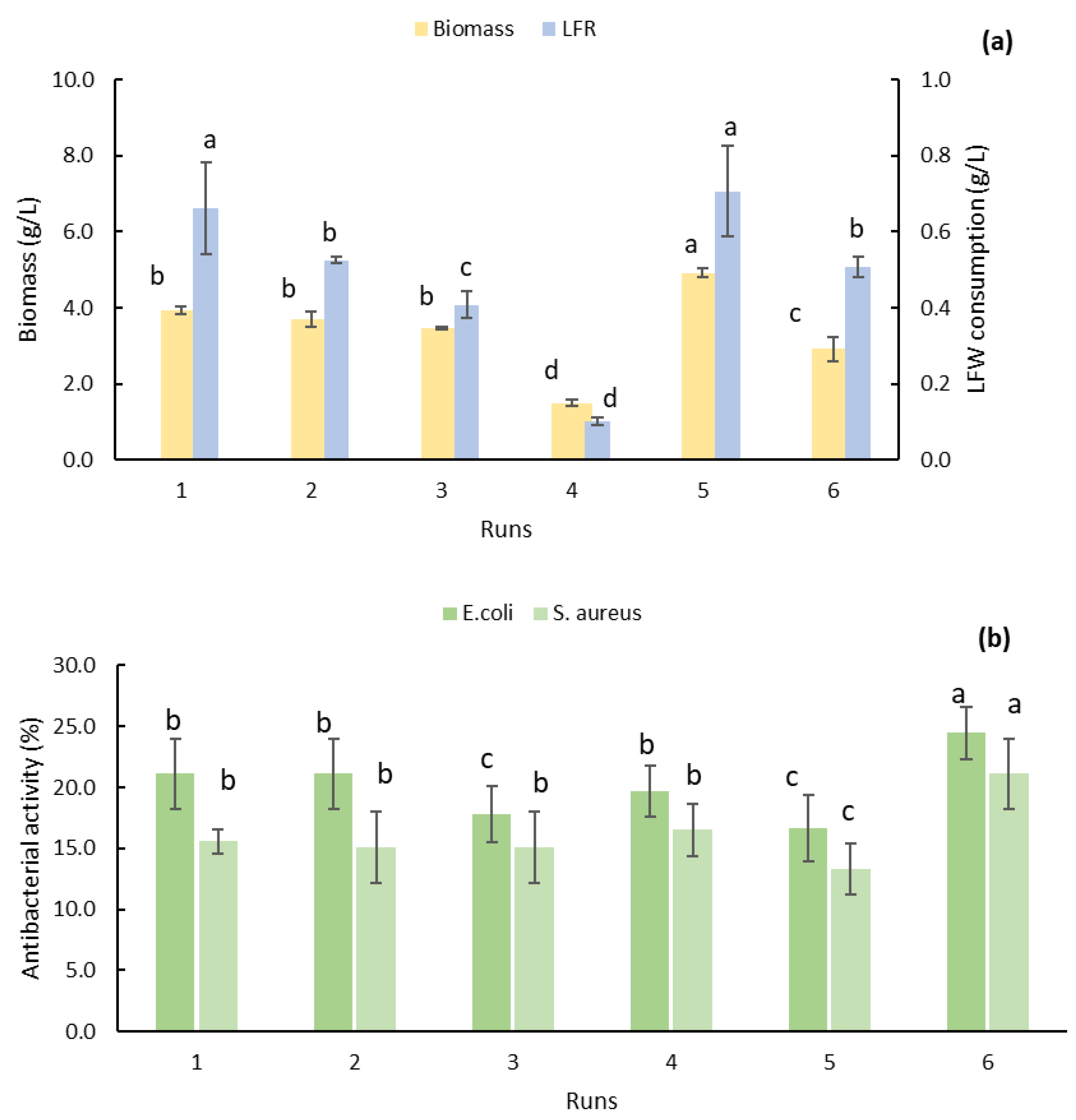
In these experiments, the antibacterial activity was tested on two pathogenic bacteria, E. coli and S. aureus (Figure 2 and Figure 3). The highest antimicrobial activity was obtained when R. rubra grew in LFR 40% v/v, without yeast extract, with the antibacterial activity of 24.4 ± 2.1 % inhibition on E. coli and 21.1 ± 2.8 % inhibition on S. aureus (Figure 2b, run 6). The lowest antibacterial activity was obtained when R. rubra was grown in LFR 40% v/v, yeast extract 6 g/L (Figure 2b, run 5), with the antibacterial activity against E. coli and S. aureus of 19.5 ± 2.1 and 10.5 ± 2.1 % inhibition, respectively. This low antibacterial activity could be related to the increase in the biomass concentration, 4.9 ± 0.1 g/L (the highest, Figure 2a), in comparison to the concentration obtained under conditions with high antimicrobial activity, 2.9 ± 0.3 g/L (Figure 2a, run 6). The same tendency for the consumption of carbohydrates in LFW was presented, 0.71 ± 0.12 g/L vs 0.51 ± 0.03 g/L (Figure 2a).
The lowest biomass concentration was obtained when R. rubra grew in LFR 30% v/v, without yeast extract (Figure 2a, run 4); conditions related with lowest LFR consumption and the absence of yeast extract in the culture medium. The lowest LFR consumption suggests a possible growth inhibition.
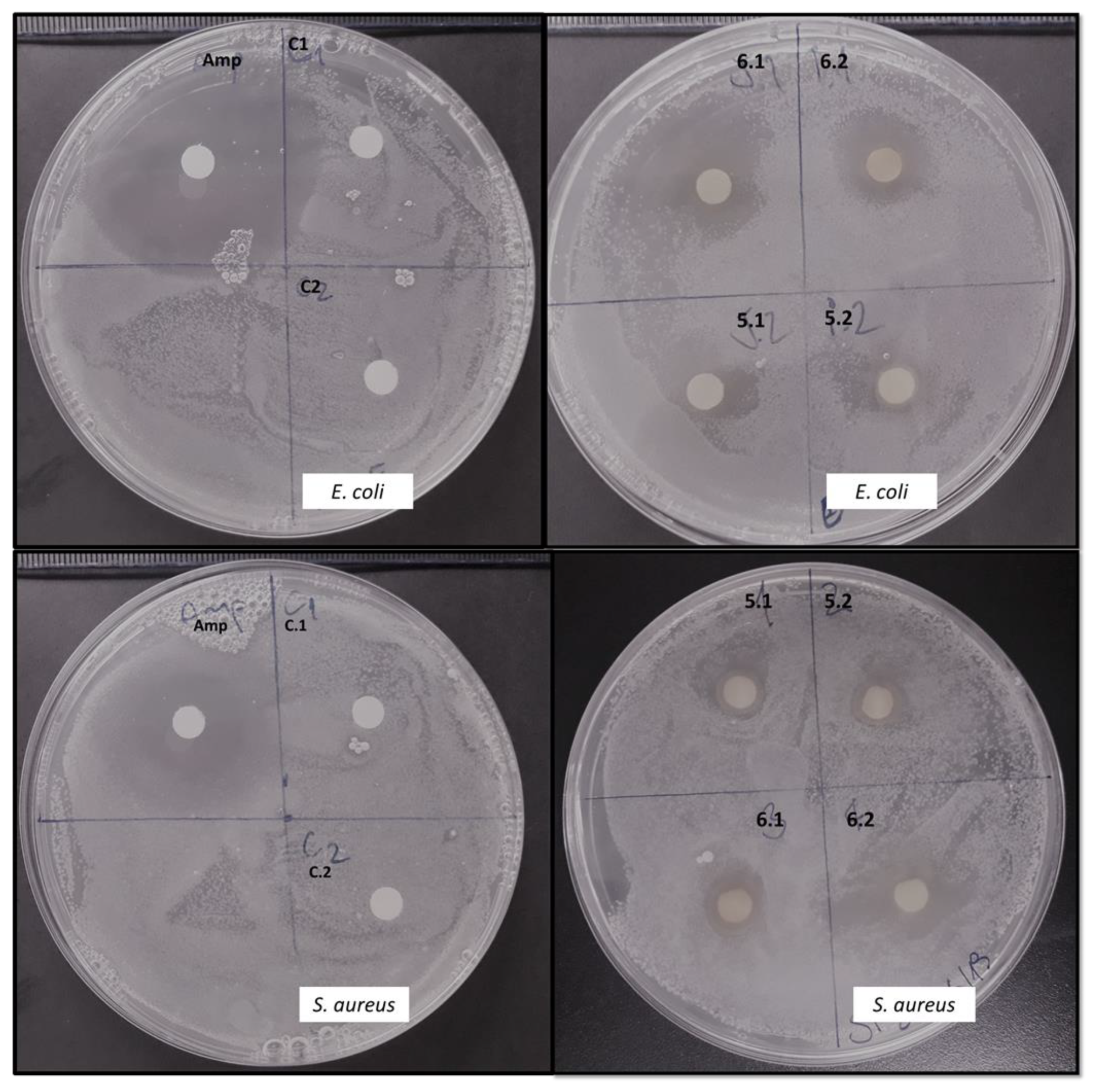
Figure 3 shows the results of the antibacterial activity of sophorolipid compounds against E. coli and S. aureus. The SLs inhibited the growth of both pathogen bacteria, resulting in a clear zone (“halo”) around the discs (Figure 3, run 5 and run 6 Table 2). In contrast, the LFR did not exhibit inhibitory activity or negative control (Figure 3, C sector).

2.3. Identification of Sophorolipid with Antibacterial Activity Produced by R. Rubra

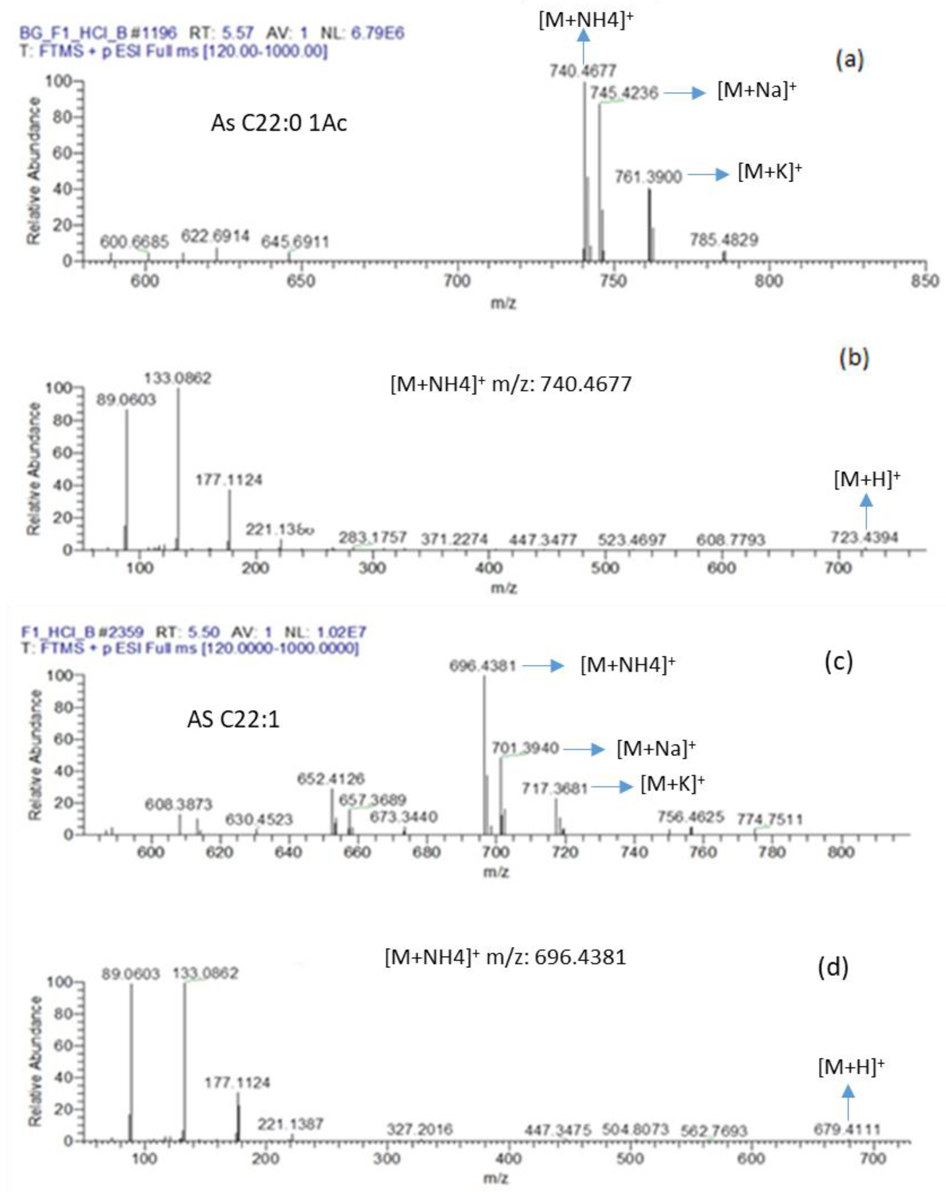
The chromatographic analysis identifies two SLs in major proportions from the extracellular extract of R. rubra—mono-acetylated (1AC) acidic sophorolipid (AS) AS C22:0-1AC and the methyl ester acidic sophorolipid AS C22:1 (Figure 4). Figure 4a shows the mass spectrum in full scan mode for AS C22:0-1AC. The m/z corresponding to the ammonium, sodium, and potassium adducts were [M + NH4] + m/z: 740.4677, [M + Na] + m/z: 745.4236, and [M + K] + m/z: 761.3900, respectively. Figure 4b shows the confirmatory mass spectrum for the fragmentation of [M + NH4] + m/z: 740.4656; therefore, the mass [M + H] + m/z: 723.4394 is observed, which corresponds to the mass of the phospholipid plus one in positive mode. The mass spectrum of Figure 4b is essential because it indicates that the mass 740.4656 corresponds to the sophorolipid with an ammonium adduct; otherwise, the mass 723.43 and its fragments would not be observed, and other masses would be seen instead.
Figure 4c shows the mass spectrum in full scan mode AS C22:1. The m/z corresponding to the ammonium, sodium, and potassium adducts were [M + NH4] + m/z: 696.4381, [M + Na] + m/z: 701.3940, and [M + K] + m/z: 717.3681, respectively. Figure 4d shows the confirmatory mass spectrum for the fragmentation of [M + NH4] + m/z: 696.4381; therefore, the mass [M + H] + m/z: 679.4111 is observed, which corresponds to the mass of the phospholipid plus one in positive mode.

3. Discussion

This study reports the antibacterial sophorolipids producing ability of a hitherto unreported marine yeast, R. rubra, using liquid fraction waste from brown seaweed M. pyrifera. The nutrient source (LFR) employed for the growth of R. rubra is a complex mix of nutrients with two sources of carbon (hydrophobic and hydrophilic), 1% w/w of lipids, and 32% w/w of carbohydrates, respectively, which would favor the SLs production by the marine yeast. In their study [28], de Oliveira et al. showed that a low N/C ratio (< 0.5) enhanced the production of SLs, while that biomass production was stimulated when the N/C ratio was near to 1 [29,30]. Our results show that the extract with the highest antibacterial activity was obtained when the marine yeast, R. rubra, was cultivated with only LFR in a high concentration (40% v/v) without yeast extract; the condition of a low N/C ratio in comparison to the culture medium supplemented with an extra nitrogen source (yeast extract).
Low-cost nutrient sources to produce SLs in the Candida strain included sugar cane molasses [31], soy molasses [32], and deproteinized whey [33], obtaining an SLs concentration of 23, 21, and 422 g/L, respectively. In this case, due to the marine origin of the yeast R. rubra, isolated from the M. pyrifera tissue [18], the use of LFR as a by-product derived to phlorotannin extraction from brown seaweed M. pyrifera is presented as a good alternative to produce sophorolipid compounds. The use of LFR as a nutrient source allowed us to obtain a low concentration of SLs, 0.1 g/L (Table 2), compared to the Candida strain. This difference could be due to the use of a culture medium supplemented with oil, in addition to the use of fed-batch culture mode in the Candida strain; therefore, there is hope that the use of this culture condition allows the increase of SLs production by R. rubra (work in progress).
SLs production also depends on the culture conditions [15]. In our study, the highest antibacterial SLs production was observed at 72 h, during the exponential phase, indicating a growth-associated antibacterial production. Similar growth-associated production of SLs was also obtained by Sen et al. [17] when they cultivated Rhodotorula babjevae YS3 in a medium containing glucose at 19 °C; a similar temperature using in this study (15 °C). Daniel et al. [33] had reported that the production of SLs with Starmerella apicola, using hydrolyzed broth from the biomass of the yeast Cutaneotrichosporon curvatum (formerly Cryptococcus curvatus) as culture medium, began in the middle of the exponential growth phase.
The production of SLs for the Rhodotorula strain has been reported by limited. Nuñez et al. [8] and Ribeiro et al. [14] reported the production of 22 carbon SLs in R. bogoriensis, with concentrations between 0.5–1 g/L. In R. rubra, the production of SLs has been reported by Chandran and Das [34] but without indicating the concentration produced. In the marine yeast strain, the SLs production has not been described. On the other hand, when comparing the production of SLs by genus, Candida vs Rhodotorula, the production of the longer carbon chain of SLs compound in the Rhodotorula strain was observed (C16–C18 in Candida vs C22–C24 in Rhodotorula), which is related to a greater antimicrobial activity [16,34].
The fatty acid profile in brown seaweed is rich in unsaturated fatty acids, such as docosenoic acid (C22:1) and saturated fatty acid such as docosanoic acid (C22:0) [35,36]. Both fatty acids could have been directed to incorporate into the structure of SLs synthetized by the marine R. rubra [1], and at the same time, the methyl esterification of the fatty acid fraction could promote the production of SLs mainly in the acidic form (AS) in comparison with the lactonic form (LS) [37]. The production of sophorolipid compounds of C22:0 and C22:1 carbon in the Rhodotorula strain had been previously reported [14,15,16].
The mass spectra analysis shows that the common structure of SLs produced by the marine R. rubra is the 13-[2-O-β-D-glucopyranosyl- β-D-glucopyranosyloxy]-docosanoic acid SLs; this structure has already been described for the Rhodotorula strain [14,16].
Finally, the antibacterial activity against Staphylococcus aureus and Escherichia coli indicates a potential application as a pathogen controller in the food industry. A similar activity was also observed by Dengle-Pulate et al. [38] against Staphylococcus aureus and Bacillus subtilis during their study involving SLs produced by Starmerella apicola using glucose as the hydrophilic source and lauryl alcohol C12–14 as the hydrophobic source.

4. Materials and Methods

4.1. Liquid Waste Fraction (LWR) from M. Pyrifera

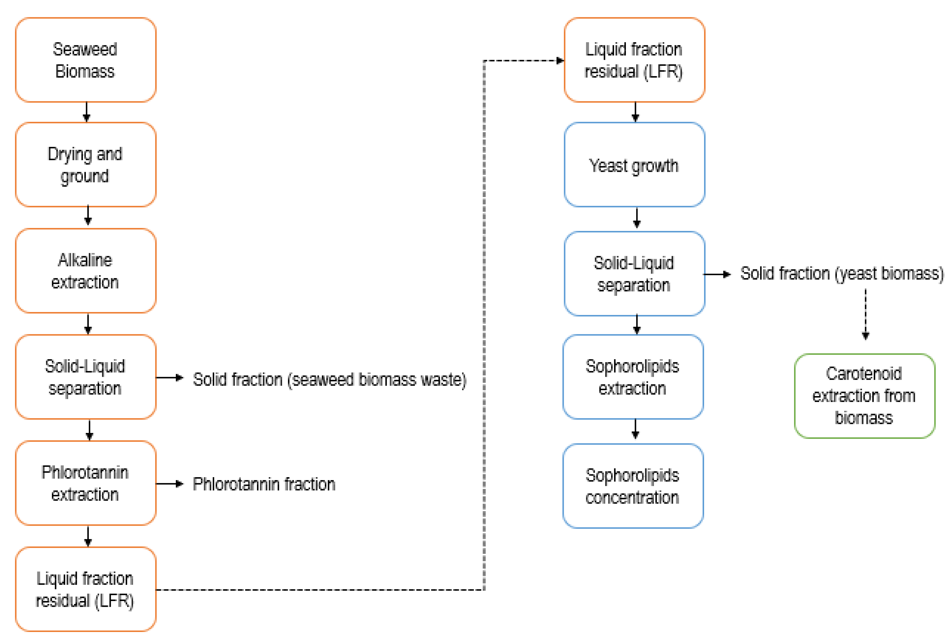
Fronds of Macrocystis pyrifera, including stipe, gas bladder, and blades, were collected by scuba diving 30 km Southwest from Puerto Montt, Chile. The samples harvested were dried at 40 °C and ground to an average size lower than 1.4 mm. The LWR was obtained from the phlorotannins extraction process, according to Leyton et al. [25], as is described in Figure 5. The phlorotannins extraction was made with NaOH 0.5 mol/L, using a seaweed mass-to-liquid ratio of 1/20 weight/volume, w/v, (180 min, 100 °C). The suspension was centrifuged (2057× g, 20 min) (Centrifuge 5804R Eppendorf AG, Hamburg, Germany), and the pH of the liquid phase was adjusted to 7.0 with HCl. Phlorotannins, which could act as cell growth inhibitors, were removed by adsorption on a macroporous resin (Amberlite XAD-16N, Sigma-Aldrich, St. Louis, MO, USA). The extract (200 mL) was incubated with the resin (40 g) under agitation (150 rpm, 25 °C) (orbital shaker Zhicheng, model ZHWY-211B, Shanghai, China) for 12 h [32]. The resin was removed by filtration and the liquid phase, LWR, was kept at −20 °C until use.

4.2. Microorganism and Inoculum Preparation

The Rhodotorula rubra strain (GenBank accession no KU167831) was isolated and identified in previous work [18]. The strain was kept at −20 °C in glycerol 50% v/v stock until use.
One mL of the yeast stock culture was seeded in 50 mL of a basal sterile medium of the following composition: yeast extract (Bacto TM) 2 g/L and glucose (Merck) 10 g/L in 60% v/v artificial seawater (ASW) [39]. The incubation was made for 3 d at 25 °C under continuous orbital shaking at 150 rpm. Then, 1 mL of the grown culture was used to inoculate a new flask with 50 mL of seaweed extract-based medium of the following composition: LFW 40% v/v, ASW 60% v/v, and yeast extract 2 g/L. The incubation was made at 25 °C, 150 rpm, for 3 d. All the culture experiments used this inoculum.

4.3. Effect of Culture Conditions on the Production of Antibacterial Compounds

The effect of incubation time (3 and 6 d), temperature (15 and 25 °C), and ASW (20%, 60%, and 80% v/v) on the antibacterial activity of the R. rubra culture extract was determined. The effect of each of the factors was evaluated changing one factor at a time, keeping the rest factors at the level specified for the basal conditions (incubation time 6 d, incubation temperature 25 °C, yeast extract concentration 6 g/L, ASW concentration 60% v/v, and LWR at 40% v/v).
The effect of LFR (20%, 30%, and 40 % v/v) and yeast extract (0 and 6 g/L) concentration was evaluated in the second set of experiments in which the incubation time and temperature, and the concentration of ASW defined in the first set of experiments were used.
In each set of experiments, two Erlenmeyer flasks were prepared with 50 mL of the sterile-specified medium and each flask was inoculated with 5 mL of the above-specified inoculum. After the incubation at the specified conditions, the total content of the flask was centrifuged (7000× g, 4 °C, 10 min) to recover the biomass; the supernatant was stored at −20 °C until use.

4.4. Separation of Antibacterial Compounds

The cell-free culture broth (50 mL) was extracted with 20 mL of ethyl acetate under agitation (150 rpm, 15 °C, 1 h). The mixture was transferred to decanting funnel to allow phases of separation at room temperature for 1 h. The organic phase was vacuum-dried at 40 °C to remove the solvent. The dry sample was dissolved in 0.5 mL of methanol and stored at −20 °C until antimicrobial assay.

4.5. Evaluation of the Antimicrobial Activity

The antimicrobial activity of the extracellular extract (EE) of R. rubra was evaluated by the disk diffusion method against one Gram-positive bacteria (Staphylococcus aureus ATCC25923) and one Gram-negative bacteria (Escherichia coli ATCC25922) in Mueller–Hinton agar medium (Sigma Aldrich). Pathogen assays were prepared by diluting the 24 h culture grown in 10 mL of sterile Luria–Bertani (LB) broth to cell density diluted of 106 UFC/mL.
Twenty µL of the bacterial sample was deposited into sterile paper discs (5 mm in diameter of Whatmann N°1) and placed on a Petri dish containing the pathogen in Mueller–Hinton agar medium. Positive control was ampicillin (100 mg/L), and negative control was LWF. Plates were kept to 4 °C for 1 h to allow the diffusion of chemicals; after, these plates were incubated at 37 °C for 12 h. Inhibition zones were measured using software ImageJ (ImageJ bundled with 64-bit Java 1.8.0_112) and expressed as a percent of inhibition, antibacterial activity.

4.6. Assay Methods

Characterization of LWR. The characterization of LWR was estimated indirectly from the chemical composition of the initial biomass less. The chemical composition of residual biomass was obtained later in the phlorotannins extraction process. The moisture content, as well as protein, lipid, ash, and fiber contents in the seaweed biomass were quantified following the official methods of the Association of Official Analytical Chemistry (AOAC): 930.04, 978.04, 991.36, 930.05, and 962.09, respectively [40]. The non-nitrogen extract was estimated as the difference between 100 and the sum of the percentages of protein, ash, lipid, and fiber content.
Biomass concentration. The biomass concentration of R. rubra was determined gravimetrically; a known culture volume (2 mL) was centrifuged (2057× g, 10 min; CT15E, Himac, Hitachi, Tokyo, Japan), and the cell pellet was washed twice with distilled water, centrifuged, and dried at 105 °C until constant weight.
Determination of the total carbohydrate concentration. Total carbohydrate concentration was measured using the phenol-sulfuric acid method [41]. Briefly, 200 µL of the sample was added to a 200 µL phenol solution (5 w/v %) and supplemented with 1 mL of concentrated sulfuric acid. The mixture was equilibrated for 20 min at room temperature. The absorbance was measured at 476 nm against a water blank. A calibration curve of glucose at different concentrations (0.02–0.1 g/L) was prepared; the results were expressed as grams of glucose per liter of LFR. The consumption of LFR by the difference between the initial and final concentration was determined.
Identification of sophorolipids compounds. UHPLC system (Dionex UltiMate 3000) coupled to mass spectrometry (Orbitrap®® Q Exactive Focus detector from Thermo Scientific was used. A Gemini®® 3 µm NX-C18 column (50 × 2 mm) and a gradient elution at 0.35 mL/min were used for the separation. The two solvents used were HPLC water (phase A) and acetonitrile (phase B); both phases contained 6.7 mM of ammonia. Conditions of the elution gradient were: 0%–10% B 1.8min, 30% B 3min, 90% B 6 min, 90% B 7.5 min; 15% B was where it remained constant for 1.5 min. Finally, phase B dropped to 15%, where it remained constant for 4.5 min. Detection was carried out in electrospray positive ion mode (ESI+). Measurements were recorded in full scan mode, scan range 150–1000 m/z, and microscan tree scan per second and then confirmed by MS/MS. The performance of fragmentation was at CE 20 eV. The ESI condition was sheath gas flow rate 35, aux gas flow rate 10, sweep gas flow rate 0, spray voltage 3.5 kV, capillary temperature 350 °C, S-lens RF level 100, and aux gas heater temperature 250 °C.

4.7. Statistical Analysis

All the culture experiments were made in duplicate. The results are presented as average ± standard deviation. The significance and relative influence of each individual factor on the production of R. rubra biomass and antibacterial compounds were determined using the variance analysis (ANOVA). The significance of the factors was determined at 5% confidence level.

5. Conclusions

Sophorolipids production with antibacterial activity on E. coli and S. aureus from marine R. rubra that was cultivated in a growth medium based on liquid fractions residual (LFR) from the brown seaweed Macrocystis pyrifera was demonstrated. Sophorolipid compounds were identified as a mono-acetylated acidic (AS C22:0-1Ac) and a methyl ester acidic (AS C22:1) sophorolipid. The synthesis of sophorolipid compounds by the marine R. rubra depends on the culture medium composition and especially on the type of carbohydrate source present.

Author Contributions

Conceptualization, C.S., M.E.L., and A.L.; methodology, A.L., L.F., and M.A.; formal analysis, M.A. and A.L.; investigation, A.L.; resources, C.S., M.E.L., F.T., and A.L.; data curation, M.A. and A.L.; writing—original draft preparation, C.S., M.E.L., F.T., and A.L.; supervision, A.L.; project administration, C.S. and A.L. All authors have read and agreed to the published version of the manuscript.

Funding

This research received no external funding.

Institutional Review Board Statement

Not applicable.

Informed Consent Statement

Not applicable.

Data Availability Statement

Not applicable.

Acknowledgments

This research was supported by the postdoctoral project Fondecyt N°3170610; the Centre for Biotechnology and Bioengineering (CeBiB) FB-000; Fondequip EQM150055 (CIDTA-UCN) and Conicyt PAI 79160069 (UCN). The authors also acknowledge the Buschmann (Universidad de Los Lagos) for providing the algal samples and Lic. Samantha Garcia for help in the LC-Ms analysis from CIDTA.

Conflicts of Interest

The authors declare no conflict of interest.

References

  1. Nuñez, A.; Ashby, R.; Foglia, T.A.; Solaiman, D.K.Y. Analysis and characterization of sophorolipids by liquid chromatography with atmospheric pressure chemical ionization. Chromatographia 2001, 53, 673–677. [Google Scholar] [CrossRef]
  2. Van Bogaert, I.N.A.; Saerens, K.; Muynck, C.; Develter, D.M.; Soetaert, W.; Vandamme, E.J. Microbial production and application of sophorolipids. Appl. Microbiol. Biotechnol. 2007, 76, 23–34. [Google Scholar] [CrossRef] [PubMed]
  3. Azim, A.; Shah, V.; Doncel, G.F.; Peterson, N.; Gao, W.; Gross, R. Amino Acid Conjugated Sophorolipids: A New Family of Biologically Active Functionalized Glycolipids. Bioconjugate Chem. 2006, 17, 1523–1529. [Google Scholar] [CrossRef] [PubMed]
  4. Asmer, H.J.; Lang, S.; Wagner, F.; Wray, V. Microbial production, structure elucidation and bioconversion of sophorose lipids. J. Acad Ind. Res. 1988, 65, 1460–1466. [Google Scholar] [CrossRef]
  5. Hu, Y.; Ju, L.-K. Sophorolipid production from different lipid precursors observed with LC-MS. Enzym. Microb. Technol. 2001, 29, 593–601. [Google Scholar] [CrossRef]
  6. Joshi-Navare, K.; Khanvilkar, P.; Prabhune, A. Jatropha oil derived sophorolipids: Production and characterization as laundry detergent additive. Biochem. Res. Int. 2013, 2013, 169797. [Google Scholar] [CrossRef] [PubMed]
  7. Ito, S.; Kinta, M.; Inoue, S. Growth of yeasts on n-alkanes: Inhibition by a lactonic sophorolipid produced by Torulopsis bombicola. Agric. Biol. Chem. 1980, 44, 2221–2223. [Google Scholar] [CrossRef]
  8. Shao, L.; Song, X.; Ma, X.; Li, H.; Qu, Y. Bioactivities of sophorolipid with different structures against human esophageal cancer cells. J. Surg. Res. 2012, 173, 286–291. [Google Scholar] [CrossRef]
  9. Shah, V.; Doncel, G.F.; Seyoum, T.; Eaton, K.M.; Zalenskaya, I.; Hagver, R.; Azim, A.; Gross, R. Sophorolipids Microbial Glycolipids with Anti-Human Immunodeficiency Virus and Sperm-Immobilizing Activities. Antimicrobial Agents Chemotherapy. 2005, 49, 4093–4100. [Google Scholar] [CrossRef] [Green Version]
  10. Hirata, Y.; Ryu, M.; Igarashi, K.; Nagatsuka, A.; Furuta, T.; Kanaya, S.; Sugiura, M. Natural synergism of acid and lactone type mixed sophorolipids in interfacial activities and cytotoxicities. J. Oleo Sci. 2009, 58, 565–572. [Google Scholar] [CrossRef] [Green Version]
  11. Ma, X.; Li, H.; Shao, L.-J.; Shen, J.; Song, X. Effects of nitrogen sources on production and composition of sophorolipids by Wickerhamiella domercqiae var. sophorolipid CGMCC 1576. Appl. Microbiol. Biotechnol. 2011, 91, 1623–1632. [Google Scholar] [CrossRef]
  12. Cooper, D.G.; Paddock, D.A. Production of a Biosurfactant from Torulopsis bombicola. Appl. Environ. Microbiol. 1984, 47, 173–176. [Google Scholar] [CrossRef] [Green Version]
  13. Sanchez, S.; Demain, A.L. Regulation of fermentation processes. Enzym. Microb. Technol. 2002, 31, 895–906. [Google Scholar] [CrossRef]
  14. Ribeiro, I.A.; Bronze, M.R.; Castro, M.F.; Ribeiro, M.H. Design of selective production of sophorolipids byRhodotorula bogoriensisthrough nutritional requirements. J. Mol. Recognit. 2012, 25, 630–640. [Google Scholar] [CrossRef]
  15. Nuñez, A.; Ashby, R.; Foglia, T.A.; Solaiman, D.K. LC/MS analysis and lipase modification of the sophorolipids produced by Rhodotorula bogoriensis**. Biotechnol. Lett. 2004, 26, 1087–1093. [Google Scholar] [CrossRef]
  16. Solaiman, D.K.; Ashby, R.D.; Crocker, N.V. High-titer production and strong antimicrobial activity of sophorolipids fromRhodotorula bogoriensis. Biotechnol. Prog. 2015, 31, 867–874. [Google Scholar] [CrossRef]
  17. Sen, S.; Borah, S.N.; Bora, A.; Deka, S. Production, characterization, and antifungal activity of a biosurfactant produced by Rhodotorula babjevae YS3. Microb. Cell Factories 2017, 16, 1–14. [Google Scholar] [CrossRef] [Green Version]
  18. Leyton, A.; Flores, L.; Mäki-Arvela, P.; Lienqueo, M.; Shene, C. Macrocystis pyriferasource of nutrients for the production of carotenoids by a marine yeastRhodotorula mucilaginosa. J. Appl. Microbiol. 2019, 127, 1069–1079. [Google Scholar] [CrossRef] [PubMed]
  19. Camus, C.; Infante, J.; Buschmann, A.H. Overview of 3 year precommercial seafarming of Macrocystis pyrifera along the Chilean coast. Rev. Aquac. 2016, 10, 543–559. [Google Scholar] [CrossRef]
  20. Macchiavello, J.; Araya, E.; Bulboa, C. Production of Macrocystis pyrifera (Laminariales; Phaeophyceae) in northern Chile on spore-based culture. Environ. Boil. Fishes 2010, 22, 691–697. [Google Scholar] [CrossRef]
  21. Westermeier, R.; Patiño, D.J.; Murúa, P.; Müller, D.G. Macrocystis mariculture in Chile: Growth performance of heterosis genotype constructs under field conditions. Environ. Boil. Fishes 2010, 23, 819–825. [Google Scholar] [CrossRef] [Green Version]
  22. Gao, K.; McKinley, K.R. Use of macroalgae for marine biomass production and CO2 remediation: A review. Environ. Boil. Fishes 1994, 6, 45–60. [Google Scholar] [CrossRef]
  23. Hafting, J.T.; Craigie, J.S.; Stengel, D.D.; Loureiro, R.R.; Buschmann, A.H.; Yarish, C.; Edwards, M.D.; Critchley, A.T. Prospects and challenges for industrial production of seaweed bioactives. J. Phycol. 2015, 51, 821–837. [Google Scholar] [CrossRef] [PubMed]
  24. Tierney, M.S.; Smyth, T.J.; Rai, D.K.; Soler-Vila, A.; Croft, A.K.; Brunton, N. Enrichment of polyphenol contents and antioxidant activities of Irish brown macroalgae using food-friendly techniques based on polarity and molecular size. Food Chem. 2013, 139, 753–761. [Google Scholar] [CrossRef] [PubMed]
  25. Leyton, A.; Lienqueo, M.E.; Shene, C. Macrocystis pyrifera: Substrate for the production of bioactive compounds. Environ. Boil. Fishes 2020, 32, 2335–2341. [Google Scholar] [CrossRef]
  26. Purcell-Meyerink, D.; Packer, M.; Wheeler, T.; Hayes, M. Aquaculture Production of the Brown Seaweeds Laminaria digitata and Macrocystis pyrifera: Applications in Food and Pharmaceuticals. Mol. 2021, 26, 1306. [Google Scholar] [CrossRef]
  27. Leyton, A.; Pezoa-Conte, R.; Barriga, A.; Buschmann, A.-H.; Mäki-Arvela, P.; Mikkola, J.-P.; Lienqueo, M.-E. Identification and efficient extraction method of phlorotannins from the brown seaweed Macrocystis pyrifera using an orthogonal experimental design. Algal Res. 2017, 16, 201–208. [Google Scholar] [CrossRef]
  28. De Oliveira, M.R.; Magri, A.; Baldo, C.; Camilios-Neto, D.; Minucelli, T.; Colabone- Celligoi, M.A.P. Review: Sophorolipids A Promising Biosurfactant and it’s Applications. Int. J. Adv. Biotechnol. Res. 2015, 6, 161–174. [Google Scholar]
  29. Casas, J.A.; De Lara, S.G.; Garcia-Ochoa, F. Optimization of a synthetic medium for Candida bombicola growth using factorial design of experiments. Enzym. Microb. Technol. 1997, 21, 221–229. [Google Scholar] [CrossRef]
  30. Casas, J.A.; García-Ochoa, F. Sophorolipid production by Candida bombicola: Medium composition and culture methods. J. Biosci. Bioeng. 1999, 88, 488–494. [Google Scholar] [CrossRef]
  31. Daverey, A.; Pakshirajan, K. Production, Characterization, and Properties of Sophorolipids from the Yeast Candida bombicola using a Low-cost Fermentative Medium. Appl. Biochem. Biotechnol. 2009, 158, 663–674. [Google Scholar] [CrossRef]
  32. Leyton, A.; Vergara-Salinas, J.; Pérez-Correa, J.; Lienqueo, M. Purification of phlorotannins from Macrocystis pyrifera using macroporous resins. Food Chem. 2017, 237, 312–319. [Google Scholar] [CrossRef]
  33. Daniel, H.J.; Reuss, M.; Syldatk, C. Production of sophorolipids in high concentration from deproteinized whey and rapeseed oil in a two stage fed batch process using Candida bombicola ATCC 22214. Biotechnol. Lett. 1998, 20, 1153–1156. [Google Scholar] [CrossRef]
  34. Chandran, P.; Das, N. Characterization of sophorolipid biosurfactant produced by yeast species grown on diesel oil. Int. J. Sci. Nat. 2011, 2, 63–71. [Google Scholar]
  35. Polat, S.; Ozogul, Y. Seasonal proximate and fatty acid variations of some seaweeds from the northeastern Mediterranean coast. Oceanol. 2013, 55, 375–391. [Google Scholar] [CrossRef] [Green Version]
  36. Silva, G.; Pereira, R.B.; Valentao, P.; Andrade, P.B.; Sousa, C. Distinct fatty acid profile of ten Brown macroalgae. Braz. J. Pharmacogn. 2013, 23, 608–613. [Google Scholar] [CrossRef] [Green Version]
  37. Shin, J.D.; Lee, J.; Kim, Y.B.; Han, I.-S.; Kim, E.-K. Production and characterization of methyl ester sophorolipids with 22-carbon-fatty acids. Bioresour. Technol. 2010, 101, 3170–3174. [Google Scholar] [CrossRef] [PubMed]
  38. Dengle-Pulate, V.; Chandorkar, P.; Bhagwat, S.S.; Prabhune, A.A. Antimicrobial and SEM Studies of Sophorolipids Synthesized Using Lauryl Alcohol. J. Surfactants Deterg. 2014, 17, 543–552. [Google Scholar] [CrossRef]
  39. Shene, C.; Leyton, A.; Rubilar, M.; Pinelo, M.; Acevedo, F.; Morales, E. Production of lipids and docosahexasaenoic acid (DHA) by a native Thraustochytrium strain. Eur. J. Lipid Sci. Technol. 2013, 115, 890–900. [Google Scholar] [CrossRef]
  40. AOAC, Association of Official Analytical Chemistry. Official Methods of Analysis of AOAC International; AOAC International: Gaithersburg, MD, USA, 2000. [Google Scholar]
  41. Dubois, M.; Gilles, K.A.; Hamilton, J.K.; Rebers, P.A.; Smith, F. Colorimetric Method for Determination of Sugars and Related Substances. Anal. Chem. 1956, 28, 350–356. [Google Scholar] [CrossRef]
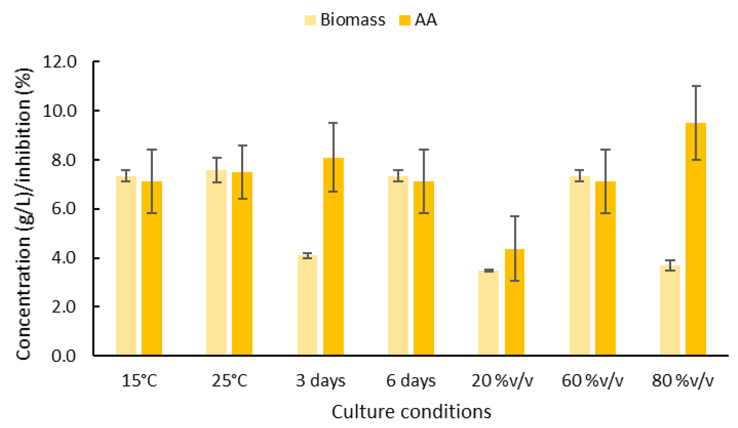
Figure 1. Effect of culture conditions: incubation temperature (15 and 25 °C), incubation time (3 and 6 days), and growth medium composition: artificial seawater concentration (20, 60, and 80 %v/v) on the biomass concentration and antibacterial activity on E. coli (AA) of the extract from cultures of R. rubra.
Molecules 26 02355 g001
Figure 2. Effect of LFR and yeast extract concentration on biomass concentration and LFR consumption (a) and antibacterial activity (b) in cultures of R. rubra. Symbols indicate statistical differences (p < 0.05).
Molecules 26 02355 g002
Figure 3. Antibacterial activity of the extract from cultures of R. rubra against Staphylococcus aureus ATCC25923 and Escherichia coli ATCC25922 in Mueller–Hinton agar medium. Amp: ampicillin (positive control); C: LFR (negative control); run 6 and run 5 extract from cultures of R. rubra, 1 and 2 are duplicates of the samples.
Molecules 26 02355 g003
Figure 4. Full scan LC-MS mass spectrum of the R. rubra extract with antibacterial activity under culture conditions LFR 40% v/v, without yeast extract, seawater 80% v/v, incubation temperature of 15 °C and incubation time of 3 days. (a) m/z for AS C22:0-1AC; (b) confirmatory mass spectrum for the fragmentation of [M + NH4] + m/z: 740.4656; (c) m/z for AS C22:1 and (d) confirmatory mass spectrum for the fragmentation of [M + NH4] + m/z: 696.4403. AS: acidic sophorolipid; AC: acetyl group.
Molecules 26 02355 g004
Figure 5. Liquid waste fraction (LFR) production and its sequential use for the production of antimicrobial compounds by the marine Rhodotorula rubra.
Molecules 26 02355 g005
Table 1. Chemical composition of LFR from M. pyrifera.
Chemical CompositionUnitSeaweed Extract
Dry Basis
Lipids%1.4 ± 0.1
Ash%51.5 ± 1.5
Protein%15 ± 0.5
Carbohydrates*%32 ± 1.0
Carbohydrate* estimated from no nitrogen extract + fiber.
Table 2. LFR and yeast extract concentration evaluated on biomass and antibacterial sophorolipids production by marine R. rubra.
RunsLFR (%)YE (g/L)SLs (g/L)
12060.12
22000.13
33060.09
43000.12
54060.07
64000.20
Publisher’s Note: MDPI stays neutral with regard to jurisdictional claims in published maps and institutional affiliations.