
Plant-Based Gums and Mucilages Applications in Pharmacology and Nanomedicine: A Review

 , ,
, ,  ,
,  and
and 
Abstract
1. Introduction
2. Methodology
3. Chemical Character of Gums and Mucilages
Classification of Gums and Mucilages
4. Plant-Based Gums and Pharmaceutical Applications
4.1. Use of Gums in Medicinal Formulations
4.2. Use of Gums to Improve Metformin Microspheres
4.3. Use of Gums as a Drug Carrier to Form Hydrogels and Improve Pharmacokinetics
4.4. Investigation of Antibacterial Properties of a Mixture of Polymers and Guar Gums
4.5. Establishing an Oral Delivery System of Protein Drugs by Gums
5. Plant-Based Mucilages and Pharmaceutical Applications
5.1. Use of Mucilages as an Adjunct and Suspending Factor in Medicinal Formulations
5.2. Use of Mucilages to Create Porous Physical Structures and Cell Scaffolds
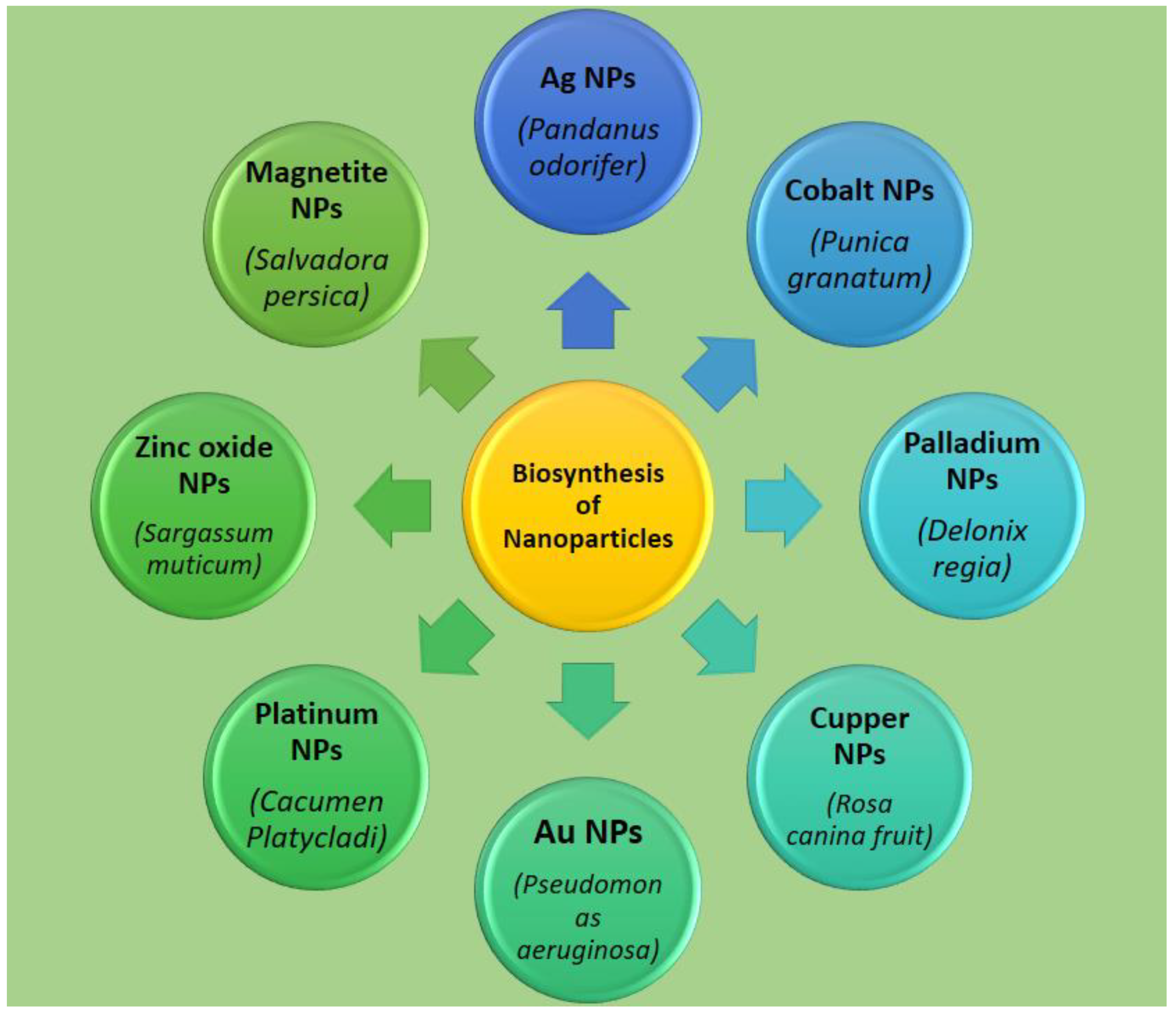
6. Importance of Plants in the Biosynthesis of Nanoparticles
7. Applications
7.1. Applications of Plant-Derived Gums in Nanomedicine
7.2. Applications of Plant-Based Mucilages in Nanomedicine
8. Challenges and Future Scope
9. Conclusions
Author Contributions
Funding
Institutional Review Board Statement:
Informed Consent Statement
Data Availability Statement
Conflicts of Interest
References
- Mishra, P.; Srivastava, A.K.; Yadav, T.C.; Pruthi, V.; Prasad, R. Pharmaceutical and Therapeutic Applications of Fenugreek Gum; Springer: Berlin/Heidelberg, Germany, 2021; pp. 379–408. [Google Scholar]
- Ulbrich, K.; Hola, K.; Subr, V.; Bakandritsos, A.; Tucek, J.; Zboril, R. Targeted drug delivery with polymers and magnetic nanoparticles: Covalent and noncovalent approaches, release control, and clinical studies. Chem. Rev. 2016, 116, 5338–5431. [Google Scholar] [CrossRef] [PubMed]
- Yazdi, M.E.T.; Amiri, M.S.; Akbari, S.; Sharifalhoseini, M.; Nourbakhsh, F.; Mashreghi, M.; Abbasi, M.R.; Modarres, M.; Es-haghi, A. Green synthesis of silver nanoparticles using helichrysum graveolens for biomedical applications and wastewater treatment. BioNanoScience 2020, 10, 1–7. [Google Scholar]
- Ashna, M.; Es-Haghi, A.; Karimi Noghondar, M.; Al Amara, D.; Yazdi, M.E.T. Greener synthesis of cerium oxide nanoemulsion using pollen grains of Brassica napus and evaluation of its antitumour and cytotoxicity properties. Mater. Technol. 2020, 1–8. [Google Scholar] [CrossRef]
- Baranei, M.; Taheri, R.A.; Tirgar, M.; Saeidi, A.; Oroojalian, F.; Uzun, L.; Asefnejad, A.; Wurm, F.R.; Goodarzi, V. Anticancer effect of green tea extract (GTE)-Loaded pH-responsive niosome Coated with PEG against different cell lines. Mater. Today Commun. 2020, 101751. [Google Scholar] [CrossRef]
- Barani, M.; Bilal, M.; Rahdar, A.; Arshad, R.; Kumar, A.; Hamishekar, H.; Kyzas, G.Z. Nanodiagnosis and nanotreatment of colorectal cancer: An overview. J. Nanoparticle Res. 2021, 23, 1–25. [Google Scholar] [CrossRef]
- Barani, M.; Bilal, M.; Sabir, F.; Rahdar, A.; Kyzas, G.Z. Nanotechnology in ovarian cancer: Diagnosis and treatment. Life Sci. 2020, 266, 118914. [Google Scholar] [CrossRef]
- Barani, M.; Mirzaei, M.; Mahani, M.T.; Nematollahi, M.H. Lawsone-loaded Niosome and its Antitumor Activity in MCF-7 Breast Cancer Cell Line: A Nano-herbal Treatment for Cancer. DARU J. Pharm. Sci. 2018, 26, 1–7. [Google Scholar] [CrossRef]
- Barani, M.; Mirzaei, M.; Torkzadeh-Mahani, M.; Adeli-sardou, M. Evaluation of Carum-loaded Niosomes on Breast Cancer Cells: Physicochemical Properties, In Vitro Cytotoxicity, Flow Cytometric, DNA Fragmentation and Cell Migration Assay. Sci. Rep. 2019, 9, 1–10. [Google Scholar] [CrossRef]
- Nair, L.S.; Laurencin, C.T. Biodegradable polymers as biomaterials. Prog. Polym. Sci. 2007, 32, 762–798. [Google Scholar] [CrossRef]
- Deogade, U.M.; Deshmukh, V.N.; Sakarkar, D.M. Natural gums and mucilage’s in NDDS: Applications and recent approaches. Int. J. PharmTech. Res. 2012, 4, 799–814. [Google Scholar]
- Darroudi, M.; Yazdi, M.E.T.; Amiri, M.S. Plant-Mediated Biosynthesis of Nanoparticles. In 21st Century Nanoscience—A Handbook; CRC Press: Boca Raton, FL, USA, 2020; pp. 1-1–1-18. [Google Scholar]
- Shamasi, Z.; Es-haghi, A.; Taghavizadeh Yazdi, M.E.; Amiri, M.S.; Homayouni-Tabrizi, M. Role of Rubia tinctorum in the synthesis of zinc oxide nanoparticles and apoptosis induction in breast cancer cell line. Nanomed. J. 2020. [Google Scholar] [CrossRef]
- Hashemzadeh, M.R.; Yazdi, M.E.T.; Amiri, M.S.; Mousavi, S.H. Stem cell therapy in the heart: Biomaterials as a key route. Tissue Cell 2021, 71, 101504. [Google Scholar] [CrossRef] [PubMed]
- Barani, M.; Mirzaei, M.; Torkzadeh-Mahani, M.; Lohrasbi-Nejad, A.; Nematollahi, M.H. A new formulation of hydrophobin-coated niosome as a drug carrier to cancer cells. Mater. Sci. Eng. C 2020, 113, 110975. [Google Scholar] [CrossRef] [PubMed]
- Barani, M.; Mukhtar, M.; Rahdar, A.; Sargazi, G.; Thysiadou, A.; Kyzas, G.Z. Progress in the application of nanoparticles and graphene as drug carriers and on the diagnosis of brain infections. Molecules 2021, 26, 186. [Google Scholar] [CrossRef] [PubMed]
- Barani, M.; Nematollahi, M.H.; Zaboli, M.; Mirzaei, M.; Torkzadeh-Mahani, M.; Pardakhty, A.; Karam, G.A. In silico and in vitro study of magnetic niosomes for gene delivery: The effect of ergosterol and cholesterol. Mater. Sci. Eng. C 2019, 94, 234–246. [Google Scholar] [CrossRef]
- Barani, M.; Sabir, F.; Rahdar, A.; Arshad, R.Z.; Kyzas, G. Nanotreatment and nanodiagnosis of prostate cancer: Recent Updates. Nanomaterials 2020, 10, 1696. [Google Scholar] [CrossRef] [PubMed]
- Es-haghi, A.; Javadi, F.; Yazdi, M.E.T.; Amiri, M.S. The expression of antioxidant genes and cytotoxicity of biosynthesized cerium oxide nanoparticles against hepatic carcinoma cell line. Avicenna J. Med. Biochem. 2019, 7, 16–20. [Google Scholar] [CrossRef]
- Mohammad Sadegh Amiri, M.E.T.Y.; Rahnama, M. Medicinal plants and phytotherapy in Iran: Glorious history, current status and future prospects. Plant Sci. Today 2021, 8, 95–111. [Google Scholar] [CrossRef]
- Bhosale, R.R.; Osmani, R.A.M.; Moin, A. Natural gums and mucilages: A review on multifaceted excipients in pharmaceutical science and research. Int. J. Pharmacogn. Phytochem. Res. 2014, 15, 901–912. [Google Scholar]
- Prajapati, V.D.; Jani, G.K.; Moradiya, N.G.; Randeria, N.P. Pharmaceutical applications of various natural gums, mucilages and their modified forms. Carbohydr. Polym. 2013, 92, 1685–1699. [Google Scholar] [CrossRef]
- Avachat, A.M.; Dash, R.R.; Shrotriya, S.N. Recent investigations of plant based natural gums, mucilages and resins in novel drug delivery systems. Ind. J. Pharm. Edu. Res. 2011, 45, 86–99. [Google Scholar]
- Anbalahan, N. Pharmacological activity of mucilage isolated from medicinal plants. Int. J. Appl. Pure Sci. Agric. 2017, 3, 98–113. [Google Scholar]
- Barani, M.; Torkzadeh-Mahani, M.; Mirzaei, M.; Nematollahi, M.H. Comprehensive evaluation of gene expression in negative and positive trigger-based targeting niosomes in HEK-293 cell line. Iran. J. Pharm. Res. IJPR 2020, 19, 166. [Google Scholar]
- Bilal, M.; Barani, M.; Sabir, F.; Rahdar, A.; Kyzas, G.Z. Nanomaterials for the treatment and diagnosis of Alzheimer’s disease: An overview. NanoImpact 2020, 20, 100251. [Google Scholar] [CrossRef]
- Das, S.S.; Bharadwaj, P.; Bilal, M.; Barani, M.; Rahdar, A.; Taboada, P.; Bungau, S.; Kyzas, G.Z. Stimuli-responsive polymeric nanocarriers for drug delivery, imaging, and theragnosis. Polymers 2020, 12, 1397. [Google Scholar] [CrossRef] [PubMed]
- Davarpanah, F.; Yazdi, A.K.; Barani, M.; Mirzaei, M.; Torkzadeh-Mahani, M. Magnetic delivery of antitumor carboplatin by using PEGylated-Niosomes. DARU J. Pharm. Sci. 2018, 26, 57–64. [Google Scholar] [CrossRef]
- Ebrahimi, A.K.; Barani, M.; Sheikhshoaie, I. Fabrication of a new superparamagnetic metal-organic framework with core-shell nanocomposite structures: Characterization, biocompatibility, and drug release study. Mater. Sci. Eng. C 2018, 92, 349–355. [Google Scholar] [CrossRef]
- Ghazy, E.; Kumar, A.; Barani, M.; Kaur, I.; Rahdar, A.; Behl, T. Scrutinizing the therapeutic and diagnostic potential of nanotechnology in thyroid cancer: Edifying drug targeting by nano-oncotherapeutics. J. Drug Deliv. Sci. Technol. 2020, 61, 102221. [Google Scholar] [CrossRef]
- Taghipour, Y.D.; Bahramsoltani, R.; Marques, A.M.; Naseri, R.; Rahimi, R.; Haratipour, P.; Panah, A.I.; Farzaei, M.H.; Abdollahi, M. A systematic review of nano formulation of natural products for the treatment of inflammatory bowel disease: Drug delivery and pharmacological targets. DARU J. Pharm. Sci. 2018, 26, 229–239. [Google Scholar] [CrossRef]
- Jani, G.K.; Shah, D.P.; Prajapati, V.D.; Jain, V.C. Gums and mucilages: Versatile excipients for pharmaceutical formulations. Asian J. Pharm. Sci. 2009, 4, 309–323. [Google Scholar]
- Mirhosseini, H.; Amid, B.T. A review study on chemical composition and molecular structure of newly plant gum exudates and seed gums. Food Res. Int. 2012, 46, 387–398. [Google Scholar] [CrossRef]
- Daas, P.J.; Schols, H.A.; de Jongh, H.H. On the galactosyl distribution of commercial galactomannans. Carbohydr. Res. 2000, 329, 609–619. [Google Scholar] [CrossRef]
- Aminabhavi, T.M.; Nadagouda, M.N.; Joshi, S.D.; More, U.A. Guar gum as platform for the oral controlled release of therapeutics. Expert Opin. Drug Deliv. 2014, 11, 753–766. [Google Scholar] [CrossRef]
- Thombare, N.; Jha, U.; Mishra, S.; Siddiqui, M. Guar gum as a promising starting material for diverse applications: A review. Int. J. Biol. Macromol. 2016, 88, 361–372. [Google Scholar] [CrossRef]
- Rani, G.U.; Konreddy, A.K.; Mishra, S.; Sen, G. Synthesis and applications of polyacrylamide grafted agar as a matrix for controlled drug release of 5-ASA. Int. J. Biol. Macromol. 2014, 65, 375–382. [Google Scholar] [CrossRef]
- Mahfoudhi, N.; Sessa, M.; Chouaibi, M.; Ferrari, G.; Donsì, F.; Hamdi, S. Assessment of emulsifying ability of almond gum in comparison with gum arabic using response surface methodology. Food Hydrocoll. 2014, 37, 49–59. [Google Scholar] [CrossRef]
- Kumar, S.; Gupta, S.K. Natural polymers, gums and mucilages as excipients in drug delivery. Polim. Med. 2012, 42, 191–197. [Google Scholar] [PubMed]
- Verma, C.; Pathania, D.; Anjum, S.; Gupta, B. Smart designing of tragacanth gum by graft functionalization for advanced materials. Macromol. Mater. Eng. 2020, 305, 1900762. [Google Scholar] [CrossRef]
- Nyandoro, V.O.; Ogaji, J.I.; Audu-Peter, J.D. Effect of particle size of okra gum as a suspending agent on some physicochemical properties of reconstituted dry paracetamol suspension. WJPR Res. 2019, 8, 129–141. [Google Scholar]
- Taghavizadeh Yazdi, M.E.; Nazarnezhad, S.; Mousavi, S.H.; Sadegh Amiri, M.; Daurroudi, M.; Baino, F.; Kargozar, S. Gum Tragacanth (GT): A versatile biocompatible material beyond borders. Molecules 2021, 26, 1510. [Google Scholar] [CrossRef]
- Nep, E.; Kaur, N.; Shaboun, S.; Adebisi, A.; Smith, A.; Conway, B.; Asare-Addo, K. Mechanical and release behaviour of theophylline from matrix tablets containing psyllium powder in combination with grewia polysaccharides. Coll. Surf. B Biointerfaces 2020, 188, 110809. [Google Scholar] [CrossRef]
- Saha, T.; Masum, Z.; Mondal, S.; Hossain, M.; Jobaer, M.; Shahin, R.; Fahad, T. Application of natural polymers as pharmaceutical excipients. Global J Life Sci. Biol. Res. 2018, 4. [Google Scholar] [CrossRef]
- Oke, E.O.; Adeyi, O.; Adeyi, A.J.; Adekunle, K.F. Modelling of Grewia mollis stem bark gum extraction yield using neuro-fuzzy technique. Proc. Int. J. Eng. Res. Afr. 2018, 34, 70–80. [Google Scholar] [CrossRef]
- Martins, E.; Christiana, I.; Olobayo, K. Effect of Grewia gum on the mechanical properties of Paracetamol tablet formulations. Afr. J. Pharm. Pharmacol. 2008, 2, 001–006. [Google Scholar]
- Nep, E.; Conway, B.R. Polysaccharide gum matrix tablets for oral controlled delivery of cimetidine. J. Pharm. Sci. Res. 2010, 2, 708–716. [Google Scholar]
- Ogaji, I.; Okafor, I.S. Potential of Grewia gum as film coating agent: Some physicochemical properties of coated praziquantel tablets. Int. J. Pharm. Res. 2011, 3, 16–19. [Google Scholar]
- Azubuike, C.P.; Alfa, M.A.; Oseni, B.A. Characterization and Evaluation of the Suspending Potentials of Corchorus Olitorius Mucilage in Pharmaceutical Suspensions; University of Lagos: Lagos, Nigeria, 2017. [Google Scholar]
- Sharma, N.; Sharma, A.; Bhatnagar, A.; Nishad, D.; Karwasra, R.; Khanna, K.; Sharma, D.; Kumar, N.; Jain, G.K. Novel gum acacia based macroparticles for colon delivery of Mesalazine: Development and gammascintigraphy study. J. Drug Deliv. Sci. Technol. 2019, 54, 101224. [Google Scholar] [CrossRef]
- Pal, K.; Bera, D. Natural polymers, gums and mucilages as efficacious green emissaries of essential therapeutics. In MOL2NET, International Conference Series on Multidisciplinary Sciences; MDPI Sciforum: Basel, Switzerland, 2020; Volume 6, ISSN 2624–5078. [Google Scholar]
- Nayak, A.K.; Hasnain, M.S. Plant polysaccharides in drug delivery applications. In Plant Polysaccharides-Based Multiple-Unit Systems for Oral Drug Delivery; Springer: Berlin/Heidelberg, Germany, 2019; pp. 19–23. [Google Scholar]
- Malik, K.; Arora, G.; Singh, I. Locust bean gum as superdisintegrant—Formulation and evaluation of nimesulide orodispersible tablets. Polim. Med. 2011, 41, 17–28. [Google Scholar]
- Jenita, J.J.L.; Vijaya, K.; Suma, R.; Raj, B.; Siddiqca, A. Formulation and evaluation of compression coated tablets of mesalazine for colon delivery. Int. J. PharmTech Res. 2010, 2, 535–541. [Google Scholar]
- Kaur, L.; Singh, I. Microwave grafted, composite and coprocessed materials: Drug delivery applications. Ther. Deliv. 2016, 7, 827–842. [Google Scholar] [CrossRef]
- Mohammadi, H.; Roshan, S.; Bhikshapathi, D. Development and evaluation of fast disintegrating tablets of lornoxicam solid dispersions. Int. J. Pharm. Sci. Nanotechnol. 2019, 12, 4585–4592. [Google Scholar]
- Kumar, S.V.; Sasmal, D.; Pal, S.C. Rheological characterization and drug release studies of gum exudates of Terminalia catappa Linn. Aaps Pharmscitech 2008, 9, 885–890. [Google Scholar] [CrossRef]
- Bai, L.; Zhu, P.; Wang, W.; Wang, M. The influence of extraction pH on the chemical compositions, macromolecular characteristics, and rheological properties of polysaccharide: The case of okra polysaccharide. Food Hydrocoll. 2020, 102, 105586. [Google Scholar] [CrossRef]
- Lett, J.A.; Sundareswari, M.; Ravichandran, K.; Sagadevan, S. The fabrication of porous hydroxyapatite scaffold using gaur gum as a natural binder. Digest J. Nanomater. Biostruct. (DJNB) 2018, 13, 235–243. [Google Scholar]
- Kawahara, R.; Watanabe, K.; Yamane, R.; Yasui, H.; Kikugawa, N.; Mori, N.; Akiyama, R.; Matsubara, T.; Harada, M.; Kaneda, S. Four-week repeated dose oral toxicity study of gum ghatti in rats. Fundam. Toxic. Sci. 2020, 7, 227–232. [Google Scholar] [CrossRef]
- Odeku, O.A.; Fell, J.T. In-vitro evaluation of khaya and albizia gums as compression coatings for drug targeting to the colon. J. Pharmacy Pharmacol. 2005, 57, 163–168. [Google Scholar] [CrossRef]
- Goswami, S.; Naik, S. Natural gums and its pharmaceutical application. J. Sci. Innov. Res. 2014, 3, 112–121. [Google Scholar]
- Kumar, R.; Patil, M.; Patil, S.R.; Paschapur, M.S. Evaluation of Anacardium occidentale gum as gelling agent in aceclofenac gel. Int. J. PharmTech Res. 2009, 1, 695–704. [Google Scholar]
- Ofori-Kwakye, K.; Asantewaa, Y.; Kipo, S.L. Physicochemical and binding properties of cashew tree gum in metronidazole tablet formulations. Int. J. Pharm. Pharm. Sci. 2010, 2, 105–109. [Google Scholar]
- Ganesh, G.; Sureshkumar, R.; Jawahar, N.; Senthil, V.; Nagasamy Venkatesh, D.; Shanmukha Srinivas, M. Preparation and evaluation of sustained release matrix tablet of diclofenac sodium using natural polymer. J. Pharm. Sci. Res. 2010, 2, 360–368. [Google Scholar]
- Shankar, N.B.; Kumar, N.U.; Balakrishna, P.K.; Kumar, R.P. Design and evaluation of controlled release bhara gum microcapsules of famotidine for oral use. Res. J. Pharm. Technol. 2008, 1, 433–437. [Google Scholar]
- Mate, C.J.; Mishra, S. Exploring the potential of moi gum for diverse applications: A Review. J. Polym. Environ. 2020, 28, 1579–1591. [Google Scholar] [CrossRef]
- Rajamma, A.; Yogesha, H.; Sateesha, S. Natural gums as sustained release carriers: Development of gastroretentive drug delivery system of ziprasidone HCl. DARU J. Pharm. Sci. 2012, 20, 1–9. [Google Scholar]
- Santos, M.B.; de Carvalho, M.G.; Garcia-Rojas, E.E. Carboxymethyl tara gum-lactoferrin complex coacervates as carriers for vitamin D3: Encapsulation and controlled release. Food Hydrocoll. 2021, 112, 106347. [Google Scholar] [CrossRef]
- Singh, A.V. Biopolymers in drug delivery: A review. Pharmacologyonline 2011, 1, 666–674. [Google Scholar]
- Ahad, H.; Kumar, C.; Kumar, B.; Reddy, B.; Shekar, A.; Sagar, N. Permeation studies of diclofenac sodium from Ficus carica fruit mucilage matrices for transdermal delivery. Int. J. ChemTech Res. 2010, 2, 937–941. [Google Scholar]
- Gangurde, A.; Malode, S.; Bhambar, R. Preliminary evaluation of neem gum as tablet binder. Indian J. Pharm. Educ. Res. 2008, 42, 344–347. [Google Scholar]
- Panda, D.S. Studies on gum of Moringa oleifera for its emulsifying properties. J. Pharm. Bioallied Sci. 2014, 6, 92. [Google Scholar] [CrossRef] [PubMed]
- Patel, M.T.; Patel, J.K.; Upadhyay, U.M. Assessment of various pharmaceutical excipient properties of natural Moringa oleifera gum [Mucoadhesion, disintegration, binder]. Int. J. Pharm. Life Sci. 2012, 3, 1833–1847. [Google Scholar]
- Mehetre, G.; Pande, V.; Kendre, P. Isolation and characterization of bionanofibers from Moringa oleifera gum as a platform for drug delivery. Nanosci. Nanotechnol. 2015, 3, 1–5. [Google Scholar]
- Aderinola, T.A.; Alashi, A.M.; Nwachukwu, I.D.; Fagbemi, T.N.; Enujiugha, V.N.; Aluko, R.E. In vitro digestibility, structural and functional properties of Moringa oleifera seed proteins. Food Hydrocoll. 2020, 101, 105574. [Google Scholar] [CrossRef]
- Krishna, R.R.; Murthy, T.E.G.K. Preparation and evaluation of mucoadhesive microcapsules of glipizide formulated with gum kondagogu: In vitro and in vivo. Acta Pharm. Sci. 2010, 52, 3. [Google Scholar]
- Thombre, N.; Aher, A.; Shimpi, P. Formulation Development and Evaluation of Gum Damar Based Sustained Release Matrix Tablet of Metoprolol Succinate. Asian J. Pharm. Res. Develop. 2020, 8, 81–86. [Google Scholar] [CrossRef]
- Alur, H.H.; Desai, R.P.; Mitra, A.K.; Johnston, T.P. Inhibition of a model protease—Pyroglutamate aminopeptidase by a natural oligosaccharide gum from Hakea gibbosa. Int. J. Pharm. 2001, 212, 171–176. [Google Scholar] [CrossRef]
- Bahadur, S.; Sahu, U.K.; Sahu, D.; Sahu, G.; Roy, A. Review on natural gums and mucilage and their application as excipient. J. Appl. Pharm. Res. 2017, 5, 13–21. [Google Scholar]
- Singh, P.; Mishra, G.; Dinda, S.C. Natural Excipients in Pharmaceutical Formulations. In Evidence Based Validation of Traditional Medicines; Springer: Berlin/Heidelberg, Germany, 2021; pp. 829–869. [Google Scholar]
- Shingala, V.K.; Singh, A.K.; Yadav, S.K.; Sivakumar, T. Design and characterization of Diclofenac sodium tablets containing Mangifera indica resin as release retardant. Int. J. PharmTech Res. 2010, 2, 2107–2111. [Google Scholar]
- Ravi, K.; Sachin, R.; Mirtyunjaya, B. Evaluation of disintegrating properties of mangifera indica. RGUHS J. Pharm. Scirnces 2011, 1, 11–20. [Google Scholar]
- Bala, R.; Rana, R.; Madaan, R. Natural gums and mucilage as matrix formers in sustained released dosage forms. Res. J. Pharm. Technol. 2019, 12, 5119–5125. [Google Scholar] [CrossRef]
- Pasha, B.; Ramarao, N. Evaluation of some natural gums as sustained release carriers in the manufacturing of tablets. Indian J. Res. Pharm. Biotechnol. 2017, 5, 224–228. [Google Scholar]
- Bamiro, O.A.; Ajala, T.O.; Adenokun, E.G. A New emulsifying agent: Cucumis sativus Linnaeus Mucilage. J. Pharm. Res. Int. 2017, 17, 1–9. [Google Scholar] [CrossRef]
- Jiang, M.; Li, H.; Shi, J.-s.; Xu, Z.-h. Depolymerized konjac glucomannan: Preparation and application in health care. J. Zhejiang Univ. Sci. B 2018, 19, 505–514. [Google Scholar] [CrossRef]
- Amiri, M.S.; Joharchi, M.R.; Nadaf, M.; Nasseh, Y. Ethnobotanical knowledge of Astragalus spp.: The world’s largest genus of vascular plants. Avicenna J. Phytomed. 2020, 10, 128. [Google Scholar]
- Seyedabadi, M.M.; Rostami, H.; Jafari, S.M.; Fathi, M. Development and characterization of chitosan-coated nanoliposomes for encapsulation of caffeine. Food Biosci. 2020, 40, 100857. [Google Scholar] [CrossRef]
- Rahim, H.; Sadiq, A.; Khan, S.; Khan, M.A.; Amin, F.; Jan, N.U.; Shahid, M.; Kifayatullah, M.; Ali, N.; Chishti, K.A. Prunus armeniaca and Prunus domestica gums: Exploring their synergistic binding potential in tablets. Lat. Am. J. Pharm. 2018, 37, 1672–1683. [Google Scholar]
- Rahim, H.; Khan, M.A.; Sadiq, A.; Khan, S.; Chishti, K.A.; Rahman, I.U. Comparative studies of binding potential of Prunus armeniaca and Primus domestica gums in tablets formulations. Pak. J. Pharm. Sci. 2015, 28, 909–914. [Google Scholar] [PubMed]
- Salarbashi, D.; Jahanbin, K.; Tafaghodi, M.; Fahmideh-Rad, E. Prunus armeniaca gum exudates: An overview on purification, structure, physicochemical properties, and applications. Food Sci. Nutr. 2021, 9, 1255. [Google Scholar] [CrossRef] [PubMed]
- Ozoude, C.H.; Azubuike, C.P.; Ologunagba, M.O.; Tonuewa, S.S.; Igwilo, C.I. Formulation and development of metformin-loaded microspheres using Khaya senegalensis (Meliaceae) gum as co-polymer. Fut. J. Pharm. Sci. 2020, 6, 1–11. [Google Scholar]
- Singh, B.; Sharma, K.; Dutt, S. Dietary fiber tragacanth gum based hydrogels for use in drug delivery applications. Bioact. Carbohydr. Diet. Fibre 2020, 21, 100208. [Google Scholar] [CrossRef]
- Sharma, S.; Virk, K.; Sharma, K.; Bose, S.K.; Kumar, V.; Sharma, V.; Focarete, M.L.; Kalia, S. Preparation of gum acacia-poly (acrylamide-IPN-acrylic acid) based nanocomposite hydrogels via polymerization methods for antimicrobial applications. J. Mol. Struct. 2020, 1215, 128298. [Google Scholar] [CrossRef]
- Iqbal, D.N.; Shafiq, S.; Khan, S.M.; Ibrahim, S.M.; Abubshait, S.A.; Nazir, A.; Abbas, M.; Iqbal, M. Novel chitosan/guar gum/PVA hydrogel: Preparation, characterization and antimicrobial activity evaluation. Int. J. Biol. Macromol. 2020, 164, 499–509. [Google Scholar] [CrossRef] [PubMed]
- Freitas, A.A.; Ribeiro, A.J.; Santos, A.C.; Veiga, F.; Nunes, L.C.; Silva, D.A.; Soares-Sobrinho, J.L.; Silva-Filho, E.C. Sterculia striata gum as a potential oral delivery system for protein drugs. Int. J. Biol. Macromol. 2020, 164, 1683–1692. [Google Scholar] [CrossRef]
- Ameri, A.; Heydarirad, G.; Mahdavi Jafari, J.; Ghobadi, A.; Rezaeizadeh, H.; Choopani, R. Medicinal plants contain mucilage used in traditional Persian medicine (TPM). Pharm. Biol. 2015, 53, 615–623. [Google Scholar] [CrossRef]
- Malviya, R. Extraction characterization and evaluation of selected mucilage as pharmaceutical excipient. Polim. Med. 2011, 41, 39–44. [Google Scholar]
- Ghazy, E.; Rahdar, A.; Barani, M.; Kyzas, G.Z. Nanomaterials for Parkinson disease: Recent progress. J. Mol. Struct. 2020, 1231, 129698. [Google Scholar] [CrossRef]
- Hajizadeh, M.R.; Maleki, H.; Barani, M.; Fahmidehkar, M.A.; Mahmoodi, M.; Torkzadeh-Mahani, M. In vitro cytotoxicity assay of D-limonene niosomes: An efficient nano-carrier for enhancing solubility of plant-extracted agents. Res. Pharm. Sci. 2019, 14, 448. [Google Scholar] [PubMed]
- Hajizadeh, M.R.; Parvaz, N.; Barani, M.; Khoshdel, A.; Fahmidehkar, M.A.; Mahmoodi, M.; Torkzadeh-Mahani, M. Diosgenin-loaded niosome as an effective phytochemical nanocarrier: Physicochemical characterization, loading efficiency, and cytotoxicity assay. DARU J. Pharm. Sci. 2019, 27, 329–339. [Google Scholar] [CrossRef] [PubMed]
- Hasanein, P.; Rahdar, A.; Barani, M.; Baino, F.; Yari, S. Oil-In-water microemulsion encapsulation of antagonist drugs prevents renal ischemia-reperfusion injury in rats. Appl. Sci. 2021, 11, 1264. [Google Scholar] [CrossRef]
- Mukhtar, M.; Bilal, M.; Rahdar, A.; Barani, M.; Arshad, R.; Behl, T.; Brisc, C.; Banica, F.; Bungau, S. Nanomaterials for diagnosis and treatment of brain cancer: Recent Updates. Chemosensors 2020, 8, 117. [Google Scholar] [CrossRef]
- Nokhodchi, A.; Nazemiyeh, H.; Khodaparast, A.; Sorkh-Shahan, T.; Valizadeh, H.; Ford, J. An in vitro evaluation of fenugreek mucilage as a potential excipient for oral controlled-release matrix tablet. Drug Dev. Ind. Pharm. 2008, 34, 323–329. [Google Scholar] [CrossRef]
- Chodavarapu, N.P.; Yendluri, R.B.; Haritha, S.; Prabhakar, R.; Pranati, C. Formulation and evaluation of Abelmoschus esculentus mucilage based metformin hydrochloride floating matrix tablets. Int. J. Pharm. Technol. 2011, 3, 2725–2745. [Google Scholar]
- Huang, X.; Kakuda, Y.; Cui, W. Hydrocolloids in emulsions: Particle size distribution and interfacial activity. Food Hydrocoll. 2001, 15, 533–542. [Google Scholar] [CrossRef]
- Bashir, S.; Erum, A.; Saghir, S.; Tulain, U.R.; Rashid, A. Physicochemical characterization and evaluation of suspending properties of arabinoxylan from Ispaghula (Plantago ovata) husk. Pak. J. Pharm. Sci. 2014, 27, 1761–1766. [Google Scholar]
- Kamel, R.; Abbas, H. Self-assembled carbohydrate hydrogels for prolonged pain management. Pharm. Develop. Technol. 2013, 18, 990–1004. [Google Scholar] [CrossRef]
- Koocheki, A.; Mortazavi, S.A.; Shahidi, F.; Razavi, S.M.A.; Taherian, A. Rheological properties of mucilage extracted from Alyssum homolocarpum seed as a new source of thickening agent. J. Food Eng. 2009, 91, 490–496. [Google Scholar] [CrossRef]
- Kamel, R.; Afifi, S.M.; Kassem, I.A.; Elkasabgy, N.A.; Farag, M.A. Arabinoxylan and rhamnogalacturonan mucilage: Outgoing and potential trends of pharmaceutical, environmental, and medicinal merits. Int. J. Biol. Macromol. 2020, 165, 2550–2564. [Google Scholar] [CrossRef]
- Haile, T.G.; Sibhat, G.G.; Molla, F. Physicochemical Characterization of grewia ferruginea hochst. ex A. Rich Mucilage for potential use as a pharmaceutical excipient. BioMed Res. Int. 2020, 2020. [Google Scholar] [CrossRef] [PubMed]
- Haile, T.G.; Sibhat, G.G.; Tadese, E.; Tesfay, D.; Molla, F. Evaluation of grewia ferruginea hochst ex A. Rich mucilage as suspending agent in metronidazole benzoate suspension. BioMed Res. Int. 2020, 2020. [Google Scholar] [CrossRef] [PubMed]
- Şimşek, E.; Karaca, B.; Arslan, Y.E. Bioengineered three-dimensional physical constructs from quince seed mucilage for human adipose-derived mesenchymal stem cells. J. Bioact. Compat. Polym. 2020, 35, 240–253. [Google Scholar] [CrossRef]
- Joseph, B.; George, J.; Mohan, J. Pharmacology and traditional uses of Mimosa pudica. Int. J. Pharm. Sci. Drug Res. 2013, 5, 41–44. [Google Scholar]
- Choudhary, P.D.; Pawar, H.A. Recently investigated natural gums and mucilages as pharmaceutical excipients: An overview. J. Pharm. 2014, 2014, 9. [Google Scholar] [CrossRef]
- Beikzadeh, S.; Khezerlou, A.; Jafari, S.M.; Pilevar, Z.; Mortazavian, A.M. Seed mucilages as the functional ingredients for biodegradable films and edible coatings in the food industry. Adv. Coll. Interf. Sci. 2020, 280, 102164. [Google Scholar] [CrossRef] [PubMed]
- Pawar, H.A.; Kamat, S.R.; Choudhary, P.D. An overview of natural polysaccharides as biological macromolecules: Their chemical modifications and pharmaceutical applications. Biol. Med. 2015, 7, 1. [Google Scholar] [CrossRef]
- Mazhar, M.; Ahmad, M.; Mumtaz, S.M.; Kumar, Y. Indian medicinal herbs-useful in diabetes. Hosp. Pharm. 2018, 13, 33. [Google Scholar]
- Kothawade, S.; Shinde, P.; Agrawal, M.; Aragade, P.; Kamble, H. Preliminary evaluation of Dendropthoe falcata mucilage as tablet binder. Int. J. PharmTech Res. 2010, 2, 1474–1476. [Google Scholar]
- Krishna, L.N.V.; Kulkarni, P.; Dixit, M.; Lavanya, D.; Raavi, P.K. Brief introduction of natural gums, mucilages and their applications in novel drug delivery systems-a review. IJDFR 2011, 2, 54–71. [Google Scholar]
- Keshani-Dokht, S.; Emam-Djomeh, Z.; Yarmand, M.-S.; Fathi, M. Extraction, chemical composition, rheological behavior, antioxidant activity and functional properties of Cordia myxa mucilage. Int. J. Biol. Macromol. 2018, 118, 485–493. [Google Scholar] [CrossRef]
- Haruna, S.; Aliyu, B.S.; Bala, A. Plant gum exudates (Karau) and mucilages, their biological sources, properties, uses and potential applications: A review. Bayero J. Pure Appl. Sci. 2016, 9, 159–165. [Google Scholar] [CrossRef]
- Thakkar, K.N.; Mhatre, S.S.; Parikh, R.Y. Biological synthesis of metallic nanoparticles. Nanomed. Nanotechnol. Biol. Med. 2010, 6, 257–262. [Google Scholar] [CrossRef]
- Modarres, M.; Yazdi, M.E.T. Elicitation improves phenolic acid content and antioxidant enzymes activity in salvia leriifolia cell cultures. Iran. J. Sci. Technol. Trans. A Sci. 2021, 18, 1–7. [Google Scholar]
- Yazdi, M.E.T.; Modarres, M.; Amiri, M.S.; Darroudi, M. Phyto-synthesis of silver nanoparticles using aerial extract of Salvia leriifolia Benth and evaluation of their antibacterial and photo-catalytic properties. Res. Chem. Interme. 2019, 45, 1105–1116. [Google Scholar] [CrossRef]
- Yazdi, M.E.T.; Darroudi, M.; Amiri, M.S.; Hosseini, H.A.; Nourbakhsh, F.; Mashreghi, M.; Farjadi, M.; Kouhi, S.M.M.; Mousavi, S.H. Anticancer, antimicrobial, and dye degradation activity of biosynthesised silver nanoparticle using Artemisia kopetdaghensis. Micro Nano Lett. 2020, 15, 1046–1050. [Google Scholar]
- Gericke, M.; Pinches, A. Biological synthesis of metal nanoparticles. Hydrometallurgy 2006, 83, 132–140. [Google Scholar] [CrossRef]
- Singh, P.; Kim, Y.-J.; Zhang, D.; Yang, D.-C. Biological synthesis of nanoparticles from plants and microorganisms. Trends Biotechnol. 2016, 34, 588–599. [Google Scholar] [CrossRef]
- Marslin, G.; Siram, K.; Maqbool, Q.; Selvakesavan, R.K.; Kruszka, D.; Kachlicki, P.; Franklin, G. Secondary metabolites in the green synthesis of metallic nanoparticles. Materials 2018, 11, 940. [Google Scholar] [CrossRef]
- Kalaiselvi, A.; Roopan, S.M.; Madhumitha, G.; Ramalingam, C.; Elango, G. Synthesis and characterization of palladium nanoparticles using Catharanthus roseus leaf extract and its application in the photo-catalytic degradation. Spectrochim. Acta Part A Mol. Biomol. Spectrosc. 2015, 135, 116–119. [Google Scholar] [CrossRef] [PubMed]
- Baranwal, A.; Mahato, K.; Srivastava, A.; Maurya, P.K.; Chandra, P. Phytofabricated metallic nanoparticles and their clinical applications. RSC Adv. 2016, 6, 105996–106010. [Google Scholar] [CrossRef]
- Yazdi, T.; Ehsan, M.; Housaindokht, M.R.; Sadeghnia, H.R.; Esmaeilzadeh Bahabadi, S.; Amiri, M.S.; Darroudi, M. Assessment of phytochemical components and antioxidant activity of Rheum turkestanicum Janisch. Stud. Med. Sci. 2020, 31, 75–81. [Google Scholar]
- Hamidi, A.; Yazdi, M.E.T.; Amiri, M.S.; Hosseini, H.A.; Darroudi, M. Biological synthesis of silver nanoparticles in Tribulus terrestris L. extract and evaluation of their photocatalyst, antibacterial, and cytotoxicity effects. Res. Chem. Intermed. 2019, 45, 2915–2925. [Google Scholar] [CrossRef]
- Vijayaraghavan, K.; Ashokkumar, T. Plant-mediated biosynthesis of metallic nanoparticles: A review of literature, factors affecting synthesis, characterization techniques and applications. J. Environ. Chem. Eng. 2017, 5, 4866–4883. [Google Scholar] [CrossRef]
- Riddin, T.; Gericke, M.; Whiteley, C. Analysis of the inter-and extracellular formation of platinum nanoparticles by Fusarium oxysporum f. sp. lycopersici using response surface methodology. Nanotechnology 2006, 17, 3482. [Google Scholar] [CrossRef]
- Lade, B.D.; Shanware, A.S. Phytonanofabrication: Methodology and factors affecting biosynthesis of nanoparticles. In Smart Nanosystems for Biomedicine, Optoelectronics and Catalysis; IntechOpen: London, UK, 2020. [Google Scholar]
- Samari, F.; Salehipoor, H.; Eftekhar, E.; Yousefinejad, S. Low-temperature biosynthesis of silver nanoparticles using mango leaf extract: Catalytic effect, antioxidant properties, anticancer activity and application for colorimetric sensing. N. J. Chem. 2018, 42, 15905–15916. [Google Scholar] [CrossRef]
- Tsuji, M.; Miyamae, N.; Lim, S.; Kimura, K.; Zhang, X.; Hikino, S.; Nishio, M. Crystal structures and growth mechanisms of Au@ Ag core—shell nanoparticles prepared by the microwave—polyol method. Cryst. Growth Des. 2006, 6, 1801–1807. [Google Scholar] [CrossRef]
- Pantidos, N.; Horsfall, L.E. Biological synthesis of metallic nanoparticles by bacteria, fungi and plants. J. Nanomed. Nanotechnol. 2014, 5, 1. [Google Scholar] [CrossRef]
- Akhtar, M.S.; Panwar, J.; Yun, Y.-S. Biogenic synthesis of metallic nanoparticles by plant extracts. ACS Sustain. Chem. Eng. 2013, 1, 591–602. [Google Scholar] [CrossRef]
- Kalu, V.; Odeniyi, M.; Jaiyeoba, K. Matrix properties of a new plant gum in controlled drug delivery. Arch. Pharm. Res. 2007, 30, 884–889. [Google Scholar] [CrossRef] [PubMed]
- Luo, Y.; Wang, Q. Recent development of chitosan-based polyelectrolyte complexes with natural polysaccharides for drug delivery. Int. J. Biol. Macromol. 2014, 64, 353–367. [Google Scholar] [CrossRef]
- Beneke, C.E.; Viljoen, A.M.; Hamman, J.H. Polymeric plant-derived excipients in drug delivery. Molecules 2009, 14, 2602–2620. [Google Scholar] [CrossRef]
- Bhardwaj, T.R.; Kanwar, M.; Lal, R.; Gupta, A. Natural gums and modified natural gums as sustained-release carriers. Drug Develop. Ind. Pharm. 2000, 26, 1025–1038. [Google Scholar] [CrossRef]
- Nikazar, S.; Barani, M.; Rahdar, A.; Zoghi, M.; Kyzas, G.Z. Photo-and magnetothermally responsive nanomaterials for therapy, controlled drug delivery and imaging applications. Chem. Select 2020, 5, 12590–12609. [Google Scholar] [CrossRef]
- Rahdar, A.; Hajinezhad, M.R.; Nasri, S.; Beyzaei, H.; Barani, M.; Trant, J.F. The synthesis of methotrexate-loaded F127 microemulsions and their in vivo toxicity in a rat model. J. Mol. Liq. 2020, 313, 113449. [Google Scholar] [CrossRef]
- Rahdar, A.; Hajinezhad, M.R.; Sargazi, S.; Barani, M.; Bilal, M.; Kyzas, G.Z. Deferasirox-loaded pluronic nanomicelles: Synthesis, characterization, in vitro and in vivo studies. J. Mol. Liq. 2021, 323, 114605. [Google Scholar] [CrossRef]
- Rahdar, A.; Hajinezhad, M.R.; Sargazi, S.; Bilal, M.; Barani, M.; Karimi, P.; Kyzas, G.Z. Biochemical effects of deferasirox and deferasirox-loaded nanomicellesin iron-intoxicated rats. Life Sci. 2021, 119146. [Google Scholar] [CrossRef]
- Rahdar, A.; Taboada, P.; Hajinezhad, M.R.; Barani, M.; Beyzaei, H. Effect of tocopherol on the properties of Pluronic F127 microemulsions: Physico-chemical characterization and in vivo toxicity. J. Mol. Liq. 2019, 277, 624–630. [Google Scholar] [CrossRef]
- Sabir, F.; Barani, M.; Rahdar, A.; Bilal, M.; Nadeem, M. How to face skin cancer with nanomaterials: A review. Biointerface Res. Appl. Chem. 2020, 11, 11931–11955. [Google Scholar]
- Torkzadeh-Mahani, M.; Zaboli, M.; Barani, M.; Torkzadeh-Mahani, M. A combined theoretical and experimental study to improve the thermal stability of recombinant D-lactate dehydrogenase immobilized on a novel superparamagnetic Fe3O4NPs@ metal–organic framework. Appl. Organomet. Chem. 2020, 34, e5581. [Google Scholar] [CrossRef]
- Patel, J.J.; Karve, M.; Patel, N.K. Guar gum: A versatile material for pharmaceutical industries. Int. J. Pharm. Pharm. Sci. 2014, 6, 13–19. [Google Scholar]
- Rana, V.; Rai, P.; Tiwary, A.K.; Singh, R.S.; Kennedy, J.F.; Knill, C.J. Modified gums: Approaches and applications in drug delivery. Carbohydr. Polym. 2011, 83, 1031–1047. [Google Scholar] [CrossRef]
- George, B.; Suchithra, T. Plant-derived bioadhesives for wound dressing and drug delivery system. Fitoterapia 2019, 137, 104241. [Google Scholar] [CrossRef]
- Kora, A.J.; Sashidhar, R.; Arunachalam, J. Gum kondagogu (Cochlospermum gossypium): A template for the green synthesis and stabilization of silver nanoparticles with antibacterial application. Carbohydr. Polym. 2010, 82, 670–679. [Google Scholar] [CrossRef]
- Vinod, V.; Saravanan, P.; Sreedhar, B.; Devi, D.K.; Sashidhar, R. A facile synthesis and characterization of Ag, Au and Pt nanoparticles using a natural hydrocolloid gum kondagogu (Cochlospermum gossypium). Coll. Surf. B Biointerfaces 2011, 83, 291–298. [Google Scholar] [CrossRef]
- Iravani, S. Green synthesis of metal nanoparticles using plants. Green Chem. 2011, 13, 2638–2650. [Google Scholar] [CrossRef]
- Padil, V.V.T.; Černík, M. Green synthesis of copper oxide nanoparticles using gum karaya as a biotemplate and their antibacterial application. Int. J. Nanomed. 2013, 8, 889. [Google Scholar]
- Deshmukh, A.; Aminabhavi, T. Pharmaceutical applications of various natural gums. In Polysaccharides; Ramawat, K., Mérillon, J.M., Eds.; Springer: Berlin/Heidelberg, Germany, 2015. [Google Scholar]
- Juby, K.; Dwivedi, C.; Kumar, M.; Kota, S.; Misra, H.; Bajaj, P. Silver nanoparticle-loaded PVA/gum acacia hydrogel: Synthesis, characterization and antibacterial study. Carbohydr. Polym. 2012, 89, 906–913. [Google Scholar] [CrossRef]
- Pooja, D.; Panyaram, S.; Kulhari, H.; Rachamalla, S.S.; Sistla, R. Xanthan gum stabilized gold nanoparticles: Characterization, biocompatibility, stability and cytotoxicity. Carbohydr. Polym. 2014, 110, 1–9. [Google Scholar] [CrossRef]
- Goyal, R.; Tripathi, S.; Tyagi, S.; Ram, K.R.; Ansari, K.; Shukla, Y.; Chowdhuri, D.K.; Kumar, P.; Gupta, K. Gellan gum blended PEI nanocomposites as gene delivery agents: Evidences from in vitro and in vivo studies. Eur. J. Pharm. Biopharm. 2011, 79, 3–14. [Google Scholar] [CrossRef] [PubMed]
- Goyal, R.; Tripathi, S.; Tyagi, S.; Ram, K.R.; Ansari, K.; Kumar, P.; Shukla, Y.; Chowdhuri, D.K.; Gupta, K. Gellan gum-PEI nanocomposites as efficient gene delivery agents. J. Biomed. Nanotechnol. 2011, 7, 38–39. [Google Scholar] [CrossRef] [PubMed]
- Yue, Y.; Wu, C. Progress and perspectives in developing polymeric vectors for in vitro gene delivery. Biomater. Sci. 2013, 1, 152–170. [Google Scholar] [CrossRef] [PubMed]
- Lungwitz, U.; Breunig, M.; Blunk, T.; Göpferich, A. Polyethylenimine-based non-viral gene delivery systems. Eur. J. Pharm. Biopharm. 2005, 60, 247–266. [Google Scholar] [CrossRef]
- Chen, X.-A.; Zhang, L.-J.; He, Z.-J.; Wang, W.-W.; Xu, B.; Zhong, Q.; Shuai, X.-T.; Yang, L.-Q.; Deng, Y.-B. Plasmid-encapsulated polyethylene glycol-grafted polyethylenimine nanoparticles for gene delivery into rat mesenchymal stem cells. Int. J. Nanomed. 2011, 6, 843. [Google Scholar]
- Jana, P.; Sarkar, K.; Mitra, T.; Chatterjee, A.; Gnanamani, A.; Chakraborti, G.; Kundu, P. Synthesis of a carboxymethylated guar gum grafted polyethyleneimine copolymer as an efficient gene delivery vehicle. RSC Adv. 2016, 6, 13730–13741. [Google Scholar] [CrossRef]
- Aspinall, G. Gums and mucilages. Adv. Carbohydr. Chem. Biochem. 1969, 24, 333–379. [Google Scholar] [PubMed]
- Malviya, R.; Srivastava, P.; Kulkarni, G. Applications of mucilages in drug delivery-a review. Adv. Biol. Res. 2011, 5, 1–7. [Google Scholar]
- Cascone, M.G.; Sim, B.; Sandra, D. Blends of synthetic and natural polymers as drug delivery systems for growth hormone. Biomaterials 1995, 16, 569–574. [Google Scholar] [CrossRef]
- Huang, S.; Fu, X. Naturally derived materials-based cell and drug delivery systems in skin regeneration. J. Control. Release 2010, 142, 149–159. [Google Scholar] [CrossRef]
- Ghoreishi, S.; Akbari, I.; Hedayati, A. Preparation of basil seed mucilage aerogels loaded with paclitaxel nanoparticles by the combination of phase inversion technique and gas antisolvent process. Nanomed. Res. J. 2017, 2, 179–188. [Google Scholar]
- Urena-Saborio, H.; Alfaro-Viquez, E.; Esquivel-Alvarado, D.; Madrigal-Carballo, S.; Gunasekaran, S. Electrospun plant mucilage nanofibers as biocompatible scaffolds for cell proliferation. Int. J. Biol. Macromol. 2018, 115, 1218–1224. [Google Scholar] [CrossRef]
- Tamri, P.; Hemmati, A.; Boroujerdnia, M.G. Wound healing properties of quince seed mucilage: In vivo evaluation in rabbit full-thickness wound model. Int. J. Surg. 2014, 12, 843–847. [Google Scholar] [CrossRef]
- Carvalho, E.G.; Soares, C.P.; Blau, L.; Menegon, R.F.; Joaquim, W.M. Wound healing properties and mucilage content of Pereskia aculeata from different substrates. Rev. Bras. Farmacogn. 2014, 24, 677–682. [Google Scholar] [CrossRef]
- Sanzari, I.; Leone, A.; Ambrosone, A. Nanotechnology in plant science: To make a long story short. Front. Bioeng. Biotechnol. 2019, 7, 120. [Google Scholar] [CrossRef]
- Naeeji, N.; Shahbazi, Y.; Shavisi, N. In vitro antimicrobial effect of basil seed mucilage-chitosan films containing Ziziphora clinopodioides essential oil and MgO nanoparticles. Nanomed. Res. J. 2020, 5, 225–233. [Google Scholar]
- Allafchian, A.; Jalali, S.A.H.; Mousavi, S.E.; Hosseini, S.S. Preparation of cell culture scaffolds using polycaprolactone/quince seed mucilage. Int. J. Biol. Macromol. 2020, 155, 1270–1276. [Google Scholar] [CrossRef] [PubMed]
- Shahbazi, Y.; Moosavy, M.-H. Physico-mechanical and antimicrobial properties of quince seed mucilage supplemented with titanium dioxide and silicon oxide nanoparticles. Nanomed. Res. J. 2019, 4, 157–163. [Google Scholar]
- Devanesan, S.; Ponmurugan, K.; AlSalhi, M.S.; Al-Dhabi, N.A. Cytotoxic and antimicrobial efficacy of silver nanoparticles synthesized using a traditional phytoproduct, asafoetida gum. Int. J. Nanomed. 2020, 15, 4351. [Google Scholar] [CrossRef] [PubMed]
- Jalili, M.A.; Allafchian, A.; Karimzadeh, F.; Nasiri, F. Synthesis and characterization of magnetite/Alyssum homolocarpum seed gum/Ag nanocomposite and determination of its antibacterial activity. Int. J. Biol. Macromol. 2019, 139, 1263–1271. [Google Scholar] [CrossRef]
- Kanikireddy, V.; Varaprasad, K.; Rani, M.S.; Venkataswamy, P.; Reddy, B.J.M.; Vithal, M. Biosynthesis of CMC-Guar gum-Ag0 nanocomposites for inactivation of food pathogenic microbes and its effect on the shelf life of strawberries. Carbohydr. Polym. 2020, 236, 116053. [Google Scholar] [CrossRef]
- Samrot, A.V.; Angalene, J.L.A.; Roshini, S.; Stefi, S.; Preethi, R.; Raji, P. Purification, characterization and exploitation of Azadirachta indica gum for the production of drug loaded nanocarrier. Mater. Res. Express 2020, 7, 055007. [Google Scholar] [CrossRef]
- Khan, T.A.; Nazir, M.; Ali, I.; Kumar, A. Removal of chromium (VI) from aqueous solution using guar gum–nano zinc oxide biocomposite adsorbent. Arab. J. Chem. 2017, 10, S2388–S2398. [Google Scholar] [CrossRef]
- Raeisi, S.; Ojagh, S.M.; Quek, S.Y.; Pourashouri, P.; Salaün, F. Nano-encapsulation of fish oil and garlic essential oil by a novel composition of wall material: Persian gum-chitosan. LWT 2019, 116, 108494. [Google Scholar] [CrossRef]
- Saravanan, P.; Vinod, V.; Sreedhar, B.; Sashidhar, R. Gum kondagogu modified magnetic nano-adsorbent: An efficient protocol for removal of various toxic metal ions. Mater. Sci. Eng. C 2012, 32, 581–586. [Google Scholar] [CrossRef]
- El Sheikh, D.M. Rheological characteristics of Arabic gum suspension and Plantago seeds mucilage. J. Am. Sci. 2014, 10, 18–24. [Google Scholar]
- Fierascu, I.; Fierascu, I.C.; Brazdis, R.I.; Baroi, A.M.; Fistos, T.; Fierascu, R.C. Phytosynthesized metallic nanoparticles—Between nanomedicine and toxicology. A brief review of 2019’ s findings. Materials 2020, 13, 574. [Google Scholar] [CrossRef] [PubMed]


| Substance (Common Name) | Botanical Name | Family | Structure | Pharmaceutical Application | Ref |
|---|---|---|---|---|---|
| Guar gum | Cyamopsis tetragonoloba (L.) Taub. | Fabaceae | Galactose Mannose | Sustained release Controlled drug delivery Suspending agent | [34,35,36,37] |
| Almond Gum | Prunus dulcis (Mill.) D.A.Webb | Rosaceae | Aldobionic acid L-arabinose L-galactose D-mannose | Emulsifying Thickening Suspending Adhesive Stabilizing ↑Drug release Uncoated tablet dosage form | [38] |
| Karaya gum | Firmiana simplex (L.) W.Wight | Malvaceae | α-d-galacturonic acid α-l-rhamnose | In vivo → gastric retentive dosage forms ↑Dissolution rate of drug solid dispersions Suspending agent Emulsifying agent Dental adhesive Sustaining agent Mucoadhesive Buccoadhesive | [39] |
| Tragacanth gum | Astragalus brachycalyx Fisch. ex Boiss., A. gummifer Labill. | Fabaceae | Pectinaceous Arabino galactans Xylogalacturonans | Sustain release Suspending agent Emulsifying agent | [40,41,42] |
| Tamarind gum | Tamarindus indica L. | Fabaceae | Glucosyl: Xylosyl: Galactosyl 3:2:1 | Matrix tablets ↓Drug release Biodegradable carrier for colon specific release | [23] |
| Grewia gum | Grewia mollis Juss. | Malvaceae | Glucose Rhamnose Galacturonic acid | Controlled release dosage forms Suspending agent Binding property ↑Degree of packing ↑Fluidity granules In vitro drug release → control the release of cimetidine from tablets delaying the release of cimetidine from tablets Film forming property | [43,44,45,46,47,48] |
| Gum acacia | Acacia nilotica (L.) Delile | Fabaceae | 1,3-linked β-d-galactopyranosyl | Binder Suspending agent Emulsifying agent Demulcent Emollient | [49,50] |
| Khaya gum | Khaya grandifoliola C.DC. | Meliaceae | Protein Sugar Phenol 61% Galactose 14% Arabinose 7% Rhamnose, 8% Glucose 5% Glucuronic acid <2% other sugar residues | Binding agent Drug targeting Controlled release | [51,52] |
| Locust bean gum (carob gum) | Ceratonia siliqua L. | Fabaceae | D-galacto- Dmannoglycan pentane Proteins Cellulose | Super disintegrant Controlled drug delivery Drug targeting to the colon Super disintegrants Mucoadhesive | [53,54,55,56] |
| Terminalia catappa gum | Terminalia catappa L. | Combretaceae | _____ | Oral sustained release tablets | [57] |
| Okra gum | Abelmoschus esculentus (L.) Moench | Malvaceae | Galactose Galacturonic acid Rhamnose Glucose Mmannose Arabinose Xylose | Controlled release tablet Sustained-release tablets Suspending agent | [58] |
| Gum ghatti | Anogeissus latifolia (Roxb. ex DC.) Wall. ex Guillem. and Perr. | Combretaceae | β-1-3-linked D galactose units with some ß1-6- linked D-galactose units | Binder Emulsifier Suspending agent | [59,60] |
| Albizia gum | Albizia zygia (DC.) J.F.Macbr. | Fabaceae | Galactose Mannose Arabinose Glucuronic acid 4-0-α-methyl analogue | Tablet binder Emulsifier Coating materials in compression-coated tablets | [61,62] |
| Cashew gum | Anacardium occidentale L. | Anacardiaceae | Galactose Arabinose Rhamnose Glucose Glucuronic acid L-arabinose L-rhamnose D-galactose Glucuronic acid | Suspending agent ↑Disintegration time ↑Polymer ratio → ↓drug release to a greater extent | [63,64,65] |
| Bhara gum | Terminalia bellirica (Gaertn.) Roxb. | Combretaceae | ß-sitosterol Gallic acid Ellagic acid Ethyl gallate Galloyl glucose Chebulaginic acid | Sustained release Microcapsules employing bhara gum → ↓release of famotidine | [66,67] |
| Cordia gum | Cordia myxa L. | Boraginaceae | Galactose (27%) Rhamnose (21%) Mannose (17%) Xylose (11%) Glucose (10%) Arabinose (9.5%) and uronic acids (5%) | Oral sustained release matrix tablets | [62] |
| Honey Locust Gum | Gleditsia triacanthos L. | Fabaceae | Proteins Fats Carbohydrates Fibers | Matrix tablets at different concentrations (5% and 10%) | [64,68] |
| Tara Gum | Caesalpinia spinosa (Molina) Kuntze | Fabaceae | Galactomannans. ratio of mannose to galactose in tara gumis 3:1 | Controlled release carrier | [69,70] |
| Neem Gum | Azadirachta indica A.Juss. | Meliaceae | Mannose Glucosamine Arabinose Galactose Fucose Xylose Glucose | Binding property Sustained release ↑Matrix tablet | [71,72] |
| Moringa oleifera Gum | Moringa oleifera Lam. | Moringaceae | Arabinose Galactose Glucuronic acid in the preparation of 10:7:2 Rhamnose | Gelling property Binding property Release retardant property Disintegrating property Emulsifying property | [73,74,75,76] |
| Gum Damar | Shorea javanica Koord. and Valeton | Dipterocarpaceae | 40% a Alpha resin (resin that dissolves in alcohol) 22% Beta-resin 23% Dammarol acid 2.5% Water | Sustained release | [77,78] |
| Hakea Gum | Hakea gibbosa Cav. | Proteaceae | Glucuronic acid Galactose Arabinose Mannose Xylose which is 12: 43: 32: 5: 8. | Sustained release Binding agent | [79,80,81] |
| Mango Gum | Mangifera indica L. | Anacardiaceae | ______ | Binding agent Sustained release Disintegrating | [82,83] |
| Olibanum Gum | Boswellia serrata Roxb. ex Colebr. | Burseraceae | 5–9% Oil content 13–17% Resin acids, 20–30% Polysaccharides 40–60% boswellic acid | Sustained release Binding agent | [84,85] |
| Terminalia Gum | Terminalia randii Baker f. | Combretaceae | ______ | Binding agent ↑Strength friability ↓Friability | [86] |
| Konjac Glucomannan. | Amorphophallus konjac K.Koch | Araceae | D-glucose D-mannose in the ratio 1: 1.6 | Gelling properties | [87] |
| Substance (Common Name) | Botanical Name | Family | Structure | Pharmaceutical Application | Ref |
|---|---|---|---|---|---|
| Mimosa mucilage | Mimosa pudica L. | Fabaceae | D-xylose, D-glucuronic acid | ↓Release of drug from tablets In vitro release→↑mucilage ↓Release of drug ↑Mucilage in tablets→ ↑Percent swelling ↓Percent erosion of tablets | [115] |
| Hibiscus rosa-sinensis | Hibiscus rosa-sinensis L. | Malvaceae | L-rhamnose, D-galactose, D-galacturonic acid, D-glucuronic acid | Sustained release Binding agent Release-retarding agent | [116] |
| Asario Mucilage | Lepidium sativum L. | Brassicaceae | _______ | Suspending agent Emulsifying agent | [21] |
| Fenugreek Mucilage | Trigonella foenum-graecum L. | Fabaceae | Mannose, Galactose, Xylose | Better release retardant | [117] |
| Aloe Mucilage | Aloe vera (L.) Burm.f. | Xanthorrhoeaceae | Arabinan, Arabinorhamnogalactan, Galactan, Galactogalacturan, Glucogalactomannan, Galactoglucoarabinomannan, Glucuronic acid, Polysaccharides | A controlled delivery system | [118] |
| Phoenix Mucilage | Phoenix dactylifera L. | Arecaceae | Carbohydrates 44–88%, Fructose, Sucrose, Mannose, Glucose, Maltose, Pectin (0.5–3.9%), Starch, Cellulose | Binding properties | [116] |
| Cassia tora Mucilage | Senna tora (L.) Roxb. | Fabaceae | Cinnamaldehyde, Tannins, Mannitol, Coumarins, Essential oils, (aldehydes, eugenol, pinene), Sugars, Resins | Binding Property ↑Hardness ↓Disintegration Suspending agent | [119] |
| Dendrophthoe Mucilage | Dendrophthoe falcata (L.f.) Ettingsh. | Loranthaceae | Binder | [120] | |
| Cocculus Mucilage | Cocculus hirsutus (L.) W.Theob. | Menispermaceae | Polysaccharides, Gelatinous type of material | Gelling property Anti-inflammatory | [121] |
| Cordia Mucilage | Cordia dichotoma G.Forst. | Boraginaceae | ______ | Binding agent Emulsifying | [122] |
| Ocimum Mucilage | Ocimum americanum L. | Lamiaceae | Xylose, Arabinose, Rhamnose, Galacturonic acids | Disintegrating property | [123] |
| No | Genus and Used Form | Application | Results | Reference |
|---|---|---|---|---|
| 1 | Basil seed mucilage | Antimicrobial | basil seed mucilage–chitosan films containing Ziziphora clinopodioides essential oil and MgO nanoparticles can be used for increasing shelf-life of stored food commodities | [178] |
| 2 | Quince seed mucilage | cell culture scaffolds | the electrospun quince seed mucilage, in combination with polycaprolactone based scaffolds with 3D structures and 75–150 nm mean fiber diameters, are able to maximize adhesion and growth of epithelial Vero cells. | [179] |
| 3 | Quince seed mucilage | Structural improvement and antibacterial | quince seed mucilage supplemented with titanium dioxide (TiO2) and silicon oxide (SiO2) nanoparticles greatly improved the antibacterial and physico-mechanical properties of the prepared films. | [180] |
| 4 | Asafoetida gum | Cell toxicity and antimicrobial | Synthesized silver nanoparticles using Asafoetida were found to be effective in inhibiting the multiplication of cancer cells (MCF-7). They also exhibited significant antibacterial and antifungal activity. | [181] |
| 5 | Alyssum homolocarpum seed gum | Synthesis of magnetite nanoparticles and antibacterial | Magnetic nanocomposite (Fe3O4 NPs) was synthesized and coated via Alyssum homo-locarpum seed gum successfully. The fabricated nanocomposite exhibits excellent antibacterial activity against Gram-positive and Gram-negative bacteria. | [182] |
| 6 | Guar gum | Biosynthesis of nanocomposites and Agricultural industry | Novel (Carboxymethyl cellulose) CMC–guar gum silver nanocomposites (CG-Ag0NC) are fabricated. Antimicrobial results displayed greater performance of the CG-Ag0NC. Developed CG-Ag0NC enhanced the shelf life of strawberries. | [183] |
| 7 | Azadirachta indica gum | Nano-carrier | Gum purified from Azadirachta indica did not have antibacterial activity but possessed good antioxidant and anticancer activity. The extracted polysaccharide was further carboxymethylated and used for the synthesis of nanocarrier to carry anticancer drug, curcumin. The nanocarriers were found to be effective against MCF7 cancer cell line. | [184] |
| 8 | Guar gum | Water purification | Guar gum–nano zinc oxide (GG/nZnO) biocomposite was used as an adsorbent for enhanced removal of Cr(VI) from aqueous solution. | [185] |
| 9 | Persian gum | Food industry | Nano-capsules with fish oil–garlic essential oil using persian gum were successfully produced. Nano-capsules produced have good physicochemical properties indicating good stability. | [186] |
| 10 | Gum kondagogu | Removal of various toxic metal ions | Gum kondagogu (GK) modified magnetic iron oxide nanoparticles (MNP). The removal efficiencies for a variety of metal cations by the GK–MNP were determined quantitatively in the order: Cd2+ > Cu2+ > Pb2+ > Ni2+ > Zn2+ > Hg2+ | [187] |
Publisher’s Note: MDPI stays neutral with regard to jurisdictional claims in published maps and institutional affiliations. |
© 2021 by the authors. Licensee MDPI, Basel, Switzerland. This article is an open access article distributed under the terms and conditions of the Creative Commons Attribution (CC BY) license (http://creativecommons.org/licenses/by/4.0/).
Share and Cite
Amiri, M.S.; Mohammadzadeh, V.; Yazdi, M.E.T.; Barani, M.; Rahdar, A.; Kyzas, G.Z. Plant-Based Gums and Mucilages Applications in Pharmacology and Nanomedicine: A Review. Molecules 2021, 26, 1770. https://doi.org/10.3390/molecules26061770
Amiri MS, Mohammadzadeh V, Yazdi MET, Barani M, Rahdar A, Kyzas GZ. Plant-Based Gums and Mucilages Applications in Pharmacology and Nanomedicine: A Review. Molecules. 2021; 26(6):1770. https://doi.org/10.3390/molecules26061770
Chicago/Turabian StyleAmiri, Mohammad Sadegh, Vahideh Mohammadzadeh, Mohammad Ehsan Taghavizadeh Yazdi, Mahmood Barani, Abbas Rahdar, and George Z. Kyzas. 2021. "Plant-Based Gums and Mucilages Applications in Pharmacology and Nanomedicine: A Review" Molecules 26, no. 6: 1770. https://doi.org/10.3390/molecules26061770
APA StyleAmiri, M. S., Mohammadzadeh, V., Yazdi, M. E. T., Barani, M., Rahdar, A., & Kyzas, G. Z. (2021). Plant-Based Gums and Mucilages Applications in Pharmacology and Nanomedicine: A Review. Molecules, 26(6), 1770. https://doi.org/10.3390/molecules26061770