Article

Low-Dimensional Architectures in Isomeric cis-PtCl2{Ph2PCH2N(Ar)CH2PPh2} Complexes Using Regioselective-N(Aryl)-Group Manipulation

by
Peter De’Ath
,
Mark R. J. Elsegood
,
Noelia M. Sanchez-Ballester
and
Martin B. Smith
*
Department of Chemistry, Loughborough University, Loughborough LE11 3TU, UK
*
Author to whom correspondence should be addressed.
Molecules 2021, 26(22), 6809; https://doi.org/10.3390/molecules26226809
Submission received: 28 September 2021 / Revised: 5 November 2021 / Accepted: 7 November 2021 / Published: 11 November 2021

Abstract

:
The solid-state behaviour of two series of isomeric, phenol-substituted, aminomethylphosphines, as the free ligands and bound to PtII, have been extensively studied using single crystal X-ray crystallography. In the first library, isomeric diphosphines of the type Ph2PCH2N(Ar)CH2PPh2 [1ae; Ar = C6H3(Me)(OH)] and, in the second library, amide-functionalised, isomeric ligands Ph2PCH2N{CH2C(O)NH(Ar)}CH2PPh2 [2ae; Ar = C6H3(Me)(OH)], were synthesised by reaction of Ph2PCH2OH and the appropriate amine in CH3OH, and isolated as colourless solids or oils in good yield. The non-methyl, substituted diphosphines Ph2PCH2N{CH2C(O)NH(Ar)}CH2PPh2 [2f, Ar = 3-C6H4(OH); 2g, Ar = 4-C6H4(OH)] and Ph2PCH2N(Ar)CH2PPh2 [3, Ar = 3-C6H4(OH)] were also prepared for comparative purposes. Reactions of 1ae, 2ag, or 3 with PtCl24-cod) afforded the corresponding square-planar complexes 4ae, 5ag, and 6 in good to high isolated yields. All new compounds were characterised using a range of spectroscopic (1H, 31P{1H}, FT–IR) and analytical techniques. Single crystal X-ray structures have been determined for 1a, 1b∙CH3OH, 2f∙CH3OH, 2g, 3, 4b∙(CH3)2SO, 4c∙CHCl3, 4d∙½Et2O, 4e∙½CHCl3∙½CH3OH, 5a∙½Et2O, 5b, 5c∙¼H2O, 5d∙Et2O, and 6∙(CH3)2SO. The free phenolic group in 1b∙CH3OH, 2f∙CH3OH, 2g, 4b∙(CH3)2SO, 5a∙½Et2O, 5c∙¼H2O, and 6∙(CH3)2SO exhibits various intra- or intermolecular O–H∙∙∙X (X = O, N, P, Cl) hydrogen contacts leading to different packing arrangements.

Graphical Abstract

1. Introduction

Tertiary phosphines, and their phosphine oxides, have played an important role in the study of supramolecular and self-assembly processes [1,2,3]. Their synthetic versatility, coupled with ease of substituent modification, has no doubt played a significant contribution over the years. Hydrogen bonding interactions are routinely encountered in supramolecular ligand systems as illustrated by the elegant studies from Breit [4], Reek [5], and others [6,7]. More recently, amongst other common types of non-covalent interactions, those based on halogen bonding [8,9] and Hδ+∙∙∙Hδ− have been reported [10].
For a number of years, we [11,12,13,14,15,16], and others [17,18,19,20,21,22], have been interested in aminomethylphosphines, readily amenable by Mannich condensation reactions. Such interest stems from the relative ease of accessing P-monodentate ligands based on a P–C–N linker [11,15,16,19,20,22] or P/P-bidentate derivatives bearing a P–C–N–C–P backbone [12,13,14,17,18,19,21]. Previously, we have shown that the N-arene group can be easily tuned with, for example, various H-bonding donor/acceptor sites based on –CO2H/OH groups [12,13,14,15,16]. In continuation of these studies, we report here our work on the regioselective positioning of amide/hydroxy and methyl groups within a series of aminomethylphosphines, both as the free ligands and when coordinated to a square-planar Pt(II) metal centre. Our rationale for introducing an –C(O)NH– group is based on the known use of this functionality in supramolecular chemistry [23] and, furthermore, the recent interest in amide-modified phosphines for their variable coordination chemistry [24,25,26], binding nitroaromatics [27], and relevance to catalysis based on Pd [28]. Our choice of metal fragment in this work, “cis-PtCl2”, is based on its capability to support a relatively small bite angle diphosphine ligand in a cis, six-membered ring conformation, and to provide up to two “acceptor” sites for potential H-bonding [29]. For this purpose, we elected to pursue a double Mannich condensation reaction of Ph2PCH2OH with a series of isomeric primary amines bearing either OH/CH3 groups and/or an amide spacer between the arene and P–C–N–C–P backbone (Chart 1).

2. Results and Discussion

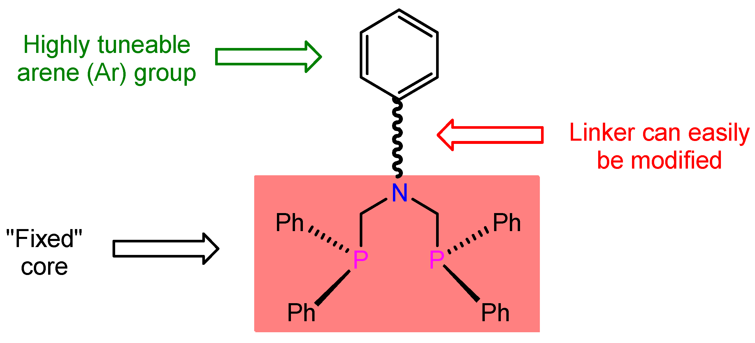
2.1. Ligand Synthesis

We [11,12,13,14,15,16,29], and others [17,19,20,21,22], have previously used Mannich condensations as a versatile method for the synthesis of aminomethylphosphines. Accordingly, two equivalents of Ph2PCH2OH were reacted with one equivalent of the amine, for 24 h at r.t. under N2, yielding the desired phenol-substituted ditertiary phosphines 1ae and 3 (Scheme 1).
For 1ae, colourless solids were isolated in 38–97% yields and found to be air stable in the solid state, but oxidise rapidly in solution. Compounds 1ae and 3 exhibit single resonances in their 31P{1H} NMR spectra (in d6-dmso) around δ(P) −26 ppm [12,13,14,15,29], indicating the presence of only one PIII environment. The ligands were also characterised by 1H NMR, FT–IR, and elemental analysis (Table 1). In particular, the absence of an NH resonance, in the 1H NMR spectra, confirmed that double condensation had occurred.
The synthesis of ditertiary phosphines, containing a flexible backbone presenting extra donor/acceptor sites with additional H-bonding capability, is described here with the opportunity to enhance solid-state packing behaviour. The precursors for the synthesis of the desired functionalised ditertiary phosphines 2ag were prepared using, in step (i), 1 equiv. of primary amine, N-carbobenzyloxyglycine (1 equiv.) and dicyclohexylcarbodiimide (DCC, 1 equiv.) in THF affording the corresponding carbamates followed by, in step (ii), treatment with Pd/C and cyclohexene in C2H5OH, to give the desired primary alkylamines in moderate to good yields [30,31]. Using a similar procedure to that described for 1ae, the amide-functionalised diphosphines 2ae were prepared in 65–89% yields by condensation using 1 equiv. of primary amine and two equiv. of Ph2PCH2OH at r.t. in CH3OH (Scheme 1). Furthermore, the phenol-substituted phosphines 2f and 2g were synthesised to investigate what effect, if any, an absent methyl group on the N-arene ring displays. In the case of 2dg, the diphosphines were obtained as solids whereas 2ac were obtained as yellow oils that were sufficiently pure to be used in complexation studies. All compounds displayed a single 31P NMR resonance around δ(P) −26 ppm [12,13,14,15,29] indicating the inclusion of an amide spacer has negligible effect on the 31P chemical shift. Other spectroscopic and analytical data are given in Table 1.

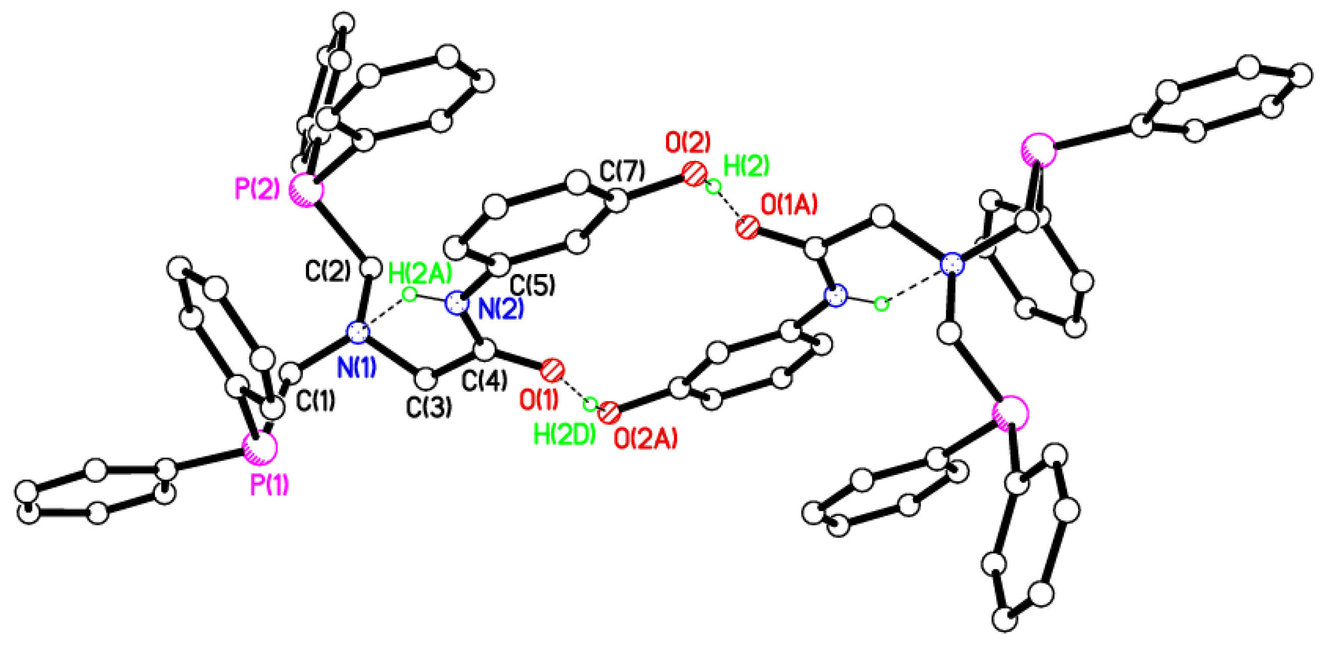
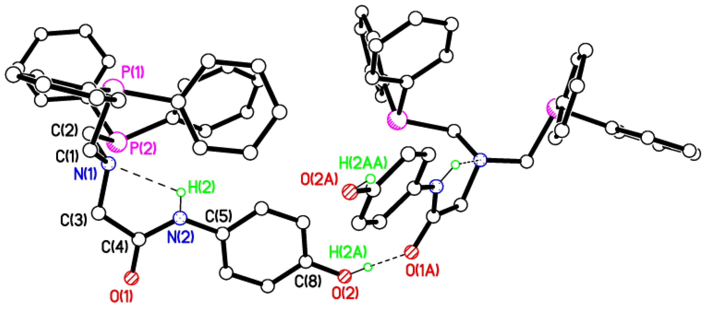
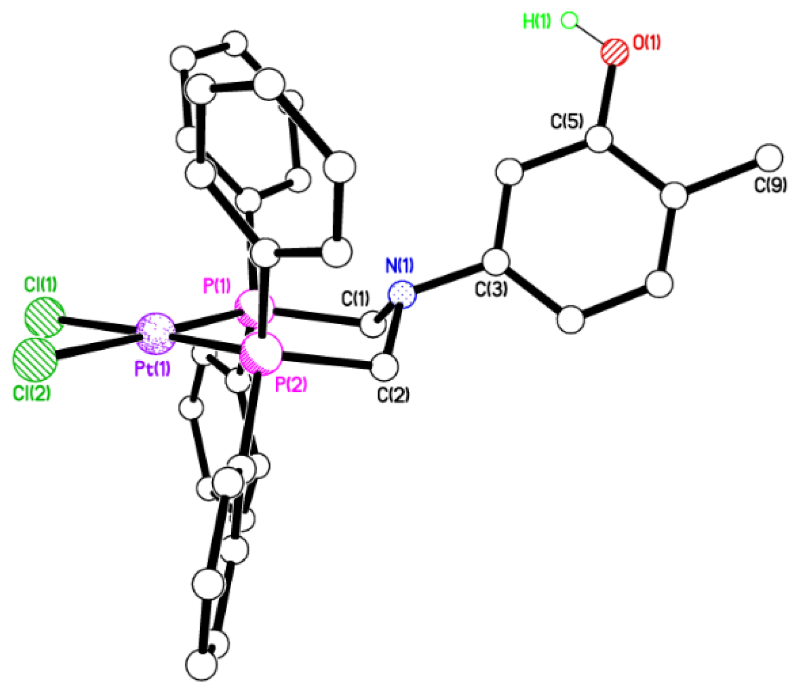
2.2. Single Crystal X-ray Studies of 1a, 1b∙CH3OH, 2f∙CH3OH, 2g, and 3

X-ray quality crystals of 1a, 1b∙CH3OH, 2f∙CH3OH, 2g, and 3 were obtained by slow evaporation of a methanol solution, while for 2g diethyl ether was diffused into a deuterochloroform/methanol solution (Table 2).
The geometry around each phosphorus atom is essentially pyramidal as would be anticipated (Figure 1, Figure 2, Figure 3, Figure 4 and Figure 5). The PIII atoms are in an anti conformation, presumably to minimise steric repulsions between the phenyl groups. The geometry about the N(1) centre is approx. pyramidal [Σ(C–N(1)–C) angles: 337.0(3)° for 1a; 335(2)° for 1b∙CH3OH; 335.2(2)/336.6(2)° for 2f∙CH3OH; 333.7(2)° for 2g] and approximately trigonal planar for 3 [Σ(C–N–C) = 359.05(11)°]. In 1a and 1b∙CH3OH, the N-arene ring [C(3) > C(8)] is twisted by ca. 88° (1a) and 86° (1b∙CH3OH) [12,32] such that it is almost perpendicular to the C(1)–N(1)–C(2) plane, whereas for 3, the twist of the C(1)–N(1)–C(2) fragment is around 9° from co-planarity with the N-arene group, apparently as a result of the intermolecular H-bonding requirements (vide infra).

2.3. Secondary Interactions in 1a, 1b∙CH3OH, 2f∙CH3OH, 2g, and 3

The synthons observed in the solid state for these highly modular ligands may be dictated by various factors including the nature of the ligand, the flexibility of the P–C–N–C–P backbone, the predisposition of the OH/CH3 groups about the N-arene ring, and the solvent used in the crystallisation. In order to probe the OH/CH3 interplay of groups, the crystal structure of 1a, with the –OH group in the ortho position with respect to the N(1) atom, is described first. Ligand 1a crystallises with an intramolecular S(5) [33,34,35] H-bonded ring with d = 2.26(5) Å [denoting the hydrogen (H) to acceptor (A) distance in an H-bond D–H···A] [36] for the O–H···N interaction (Figure 1). The intramolecular H-bonding in 1a limits the dimensionality of the packing of the diphosphine ligand. Therefore, the structure of 1a is essentially zero-dimensional (Table 3).
Compound 3, where the −OH functional group is in the meta position with respect to the tertiary N(1) atom, aggregates in the solid state in such a way that fairly weak hydrogen bonds, O−H···P [d = 2.60(2) Å], form between symmetry-related molecules, creating dimers in which two ligands are held in an R22(16) H-bonding motif (Figure 2). The distance between symmetry-related nitrogen atoms is 8.257 Å. The structure of 3 shows a 0D arrangement.
Compound 1b∙CH3OH, which contains the −OH group in a para position with respect to the N-arene, displays a similar structure to 3 with intramolecular O–H···P interactions at d = 2.60 Å. However, instead of forming dimers, there are 1D zig-zag chains in the c direction (Figure 3). The para hydroxyl oxygen acts as an acceptor for an O–H···O intermolecular H-bond from approximately alternate CH3OH molecules of crystallisation with d = 2.05 Å. These CH3OH molecules are 50/50 disordered with the second component H-bonding to its neighbour with d = 1.95 Å. Selected hydrogen parameters for 1b∙CH3OH are listed in Table 3.
Compound 2f∙CH3OH crystallises with two, similarly behaved, molecules in the asymmetric unit. A pair of H-bonded molecules, related by inversion symmetry, and with d = 1.81(3) Å for the intermolecular O–H···O interaction [1.78(3) Å for molecule 2] affords R22(16) ring motifs (Figure 4). The intramolecular N–H∙∙∙N S(5) H-bond motif with d = 2.25(3) Å [2.26(3) Å for molecule 2] results in an intermediate twist angle of 64.23(13)° [but a rather more perpendicular 78.70(8)° for molecule 2] between planes C(1)/N(1)/C(2) and ring C(5) > C(10) [plane C(35)/N(4)/C(36) and ring C(39) > C(44) for molecule 2]. The meta hydroxy group in 2f facilitates 0D dimer formation, as opposed to the chains observed in 2g (vida infra).
For 2g, molecules form H-bonded, 1D, zig-zag chains in the c direction via strong O–H∙∙∙O interactions with d = 1.83(5) Å (Figure 5). The intramolecular N–H∙∙∙N S(5), H-bond motif with d = 2.29(3) Å again results in an almost perpendicular twist angle of 82.09(15)° between planes C(1)/N(1)/C(2) and arene ring C(5) > C(10). The para hydroxy group promotes chain formation.

2.4. Dichloroplatinum(II) Complexes of 1ae, 2ag, and 3

The synthesis of P,P-chelate complexes cis-PtCl2(1ae) [4ae], cis-PtCl2(2ag) [5ag], and cis-PtCl2(3) [6] (Chart 2) was achieved by stirring the ligands and PtCl24-cod) (1:1 ratio) in CH2Cl2 for 1.5 h with displacement of the cod ligand. The products were isolated in good yields as colourless solids. Downfield shifts of the 31P NMR resonances were observed for all complexes, with 1JPtP coupling constants of approx. 3400 Hz, indicative of a cis conformation [29]. This was further supported by two characteristic νPtCl IR vibrations in the range of 279–316 cm−1 (Table 4). Furthermore, compounds 4ae, 5ag, and 6 present ν(NH) and ν(OH) IR absorptions in the range 3050–3465 cm–1 and also a strong band in the region of 1653–1675 cm–1, indicative of ν(C=O amide).

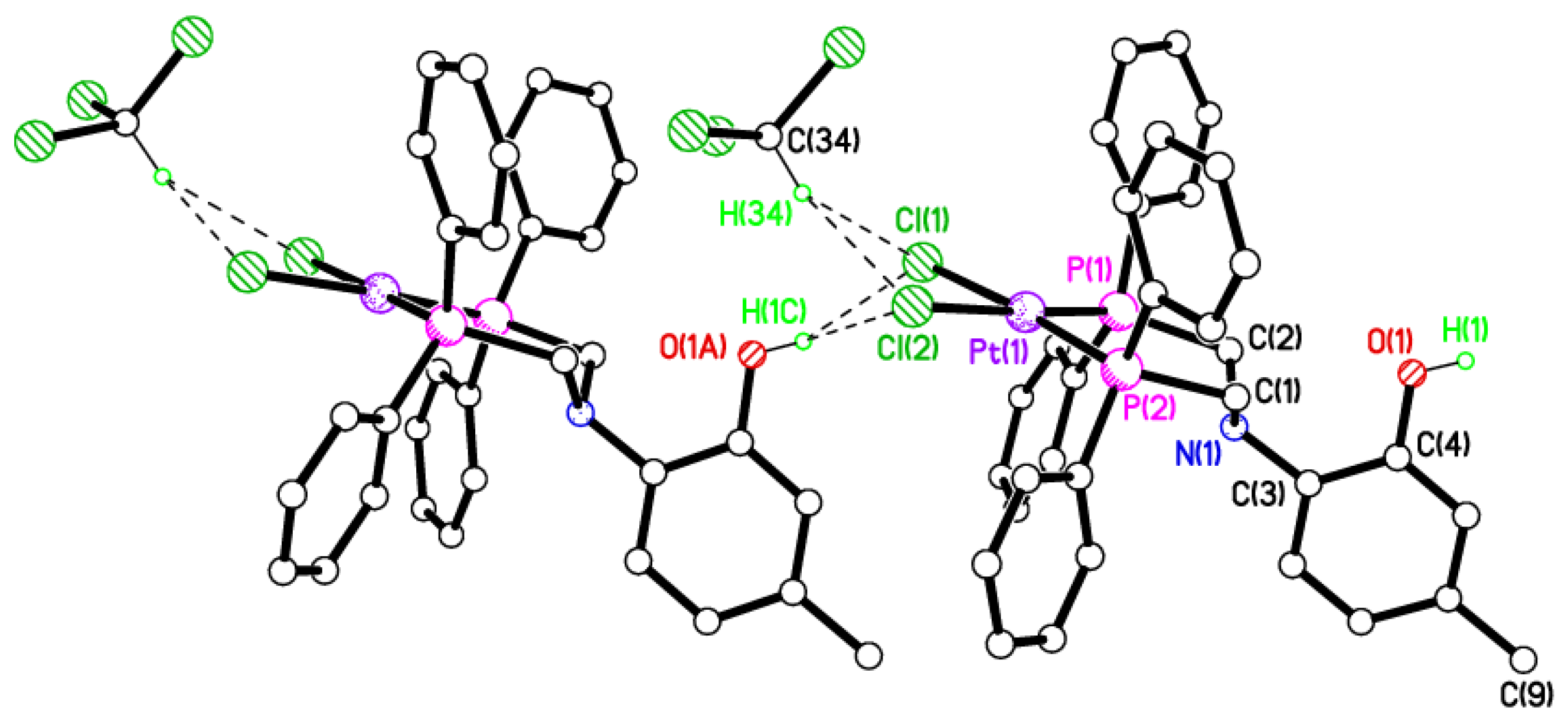
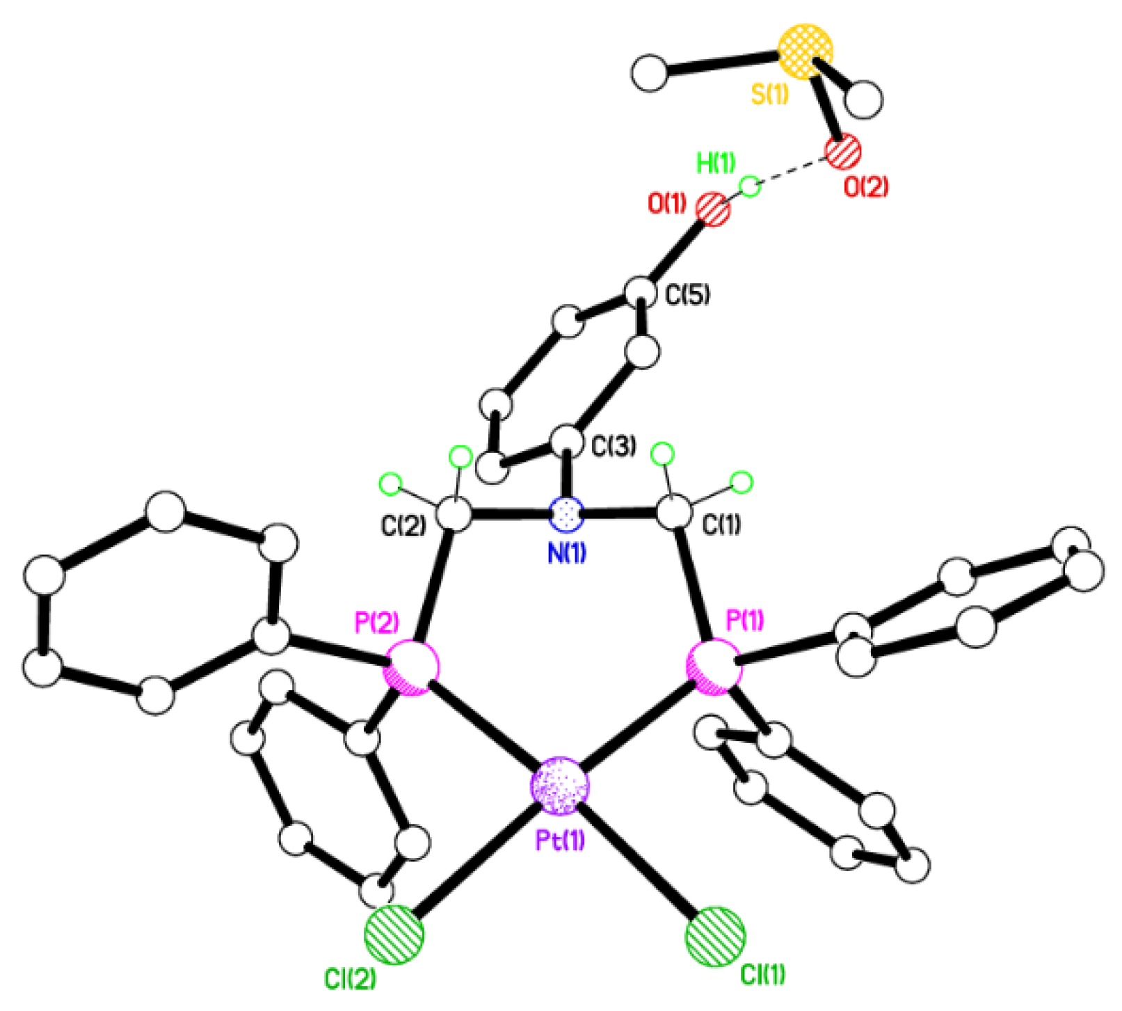
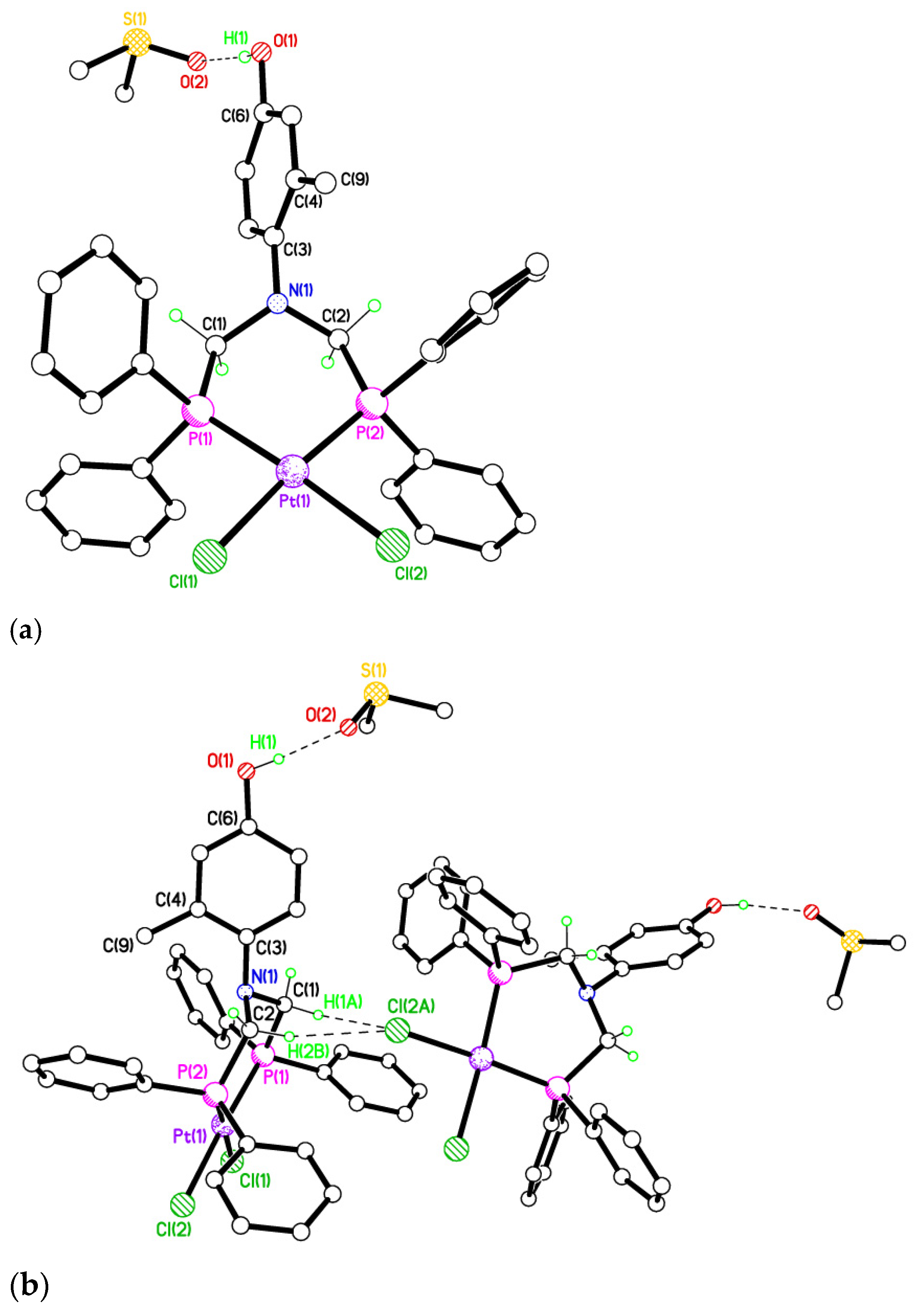
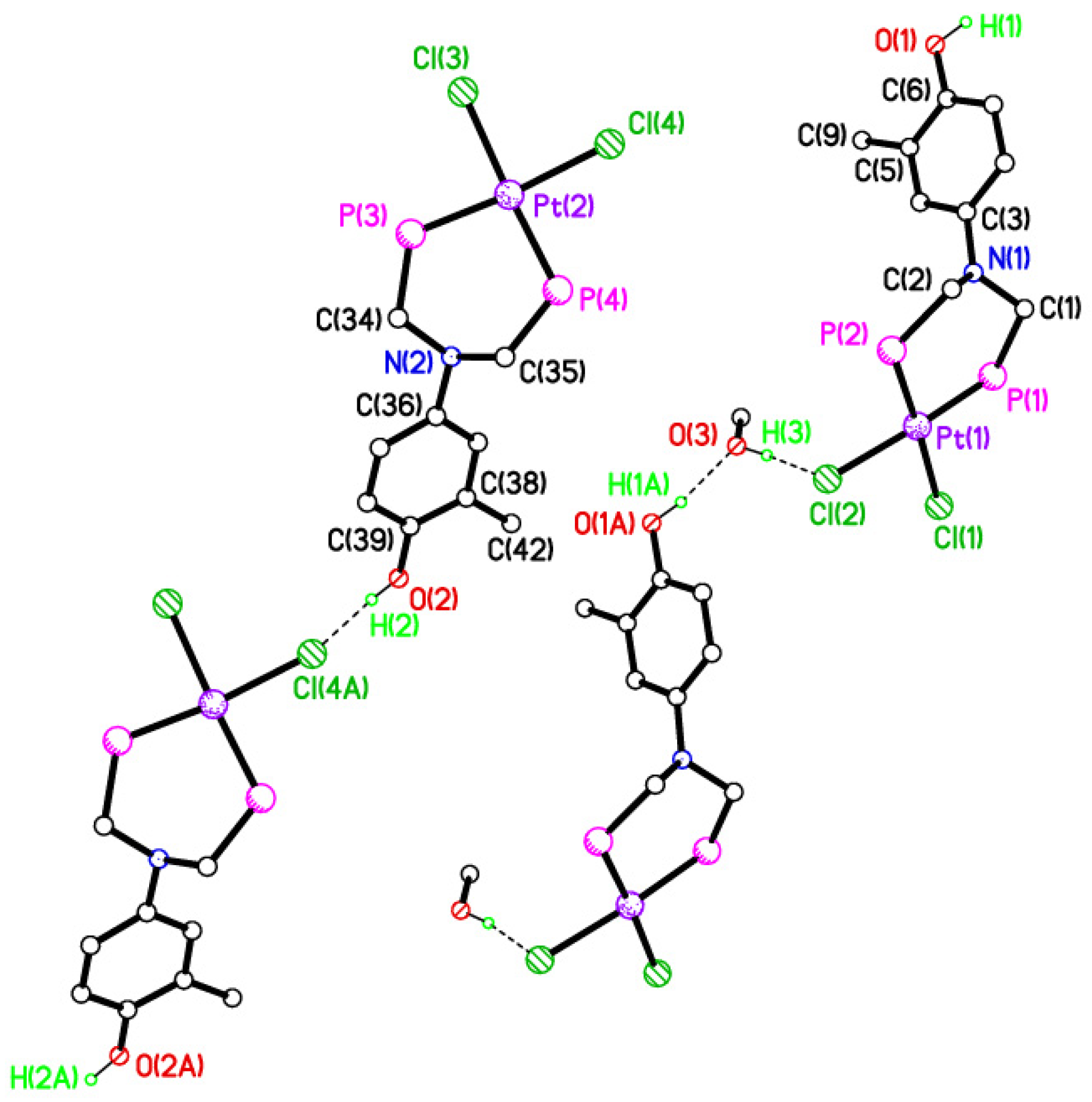
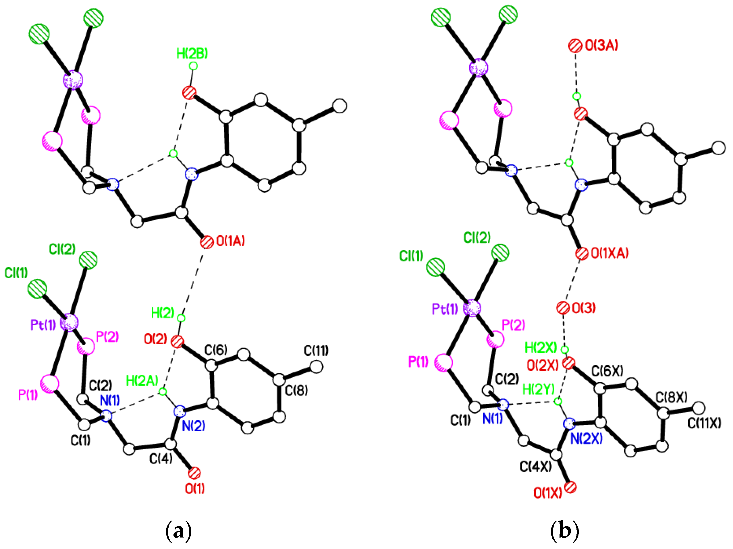
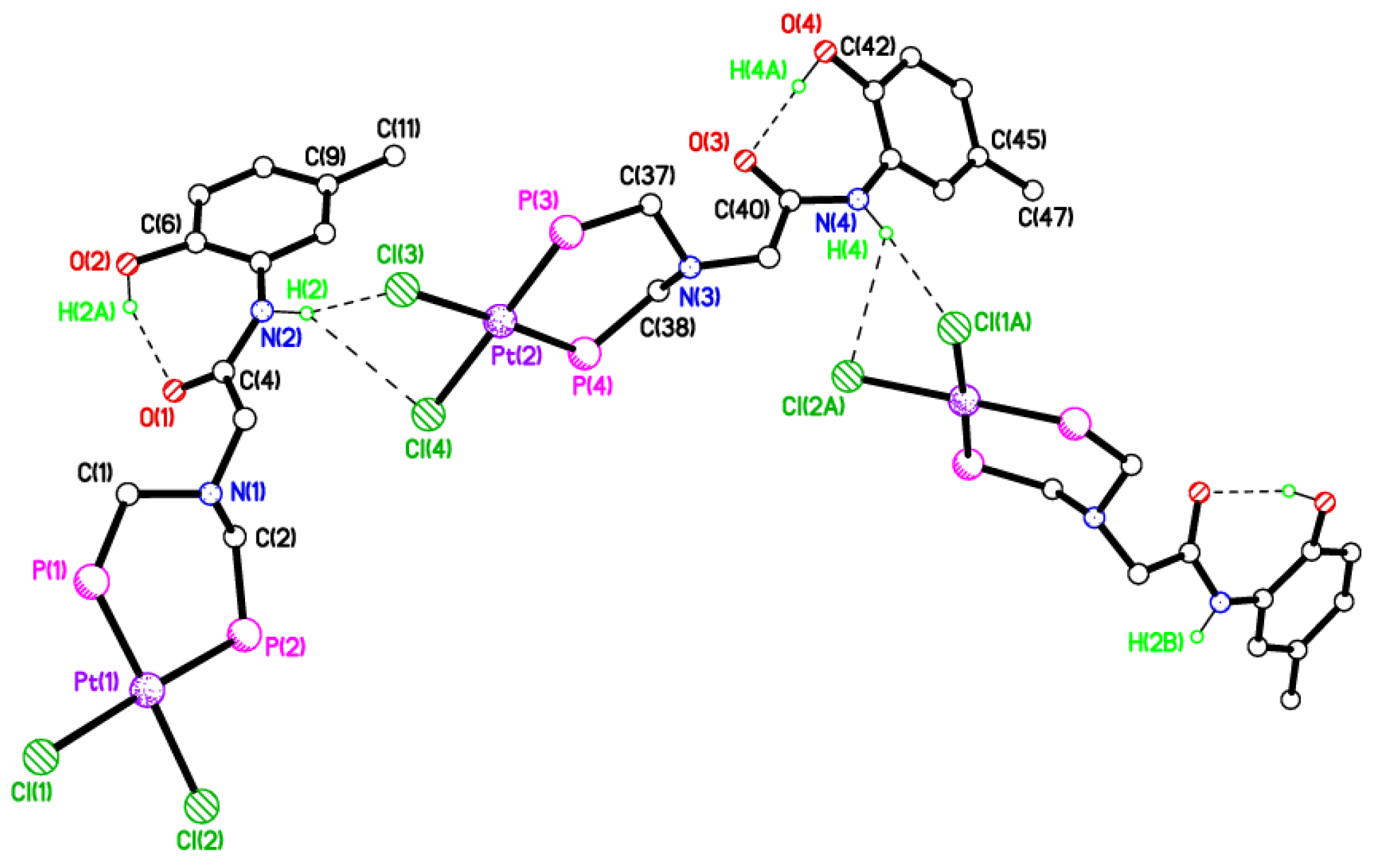
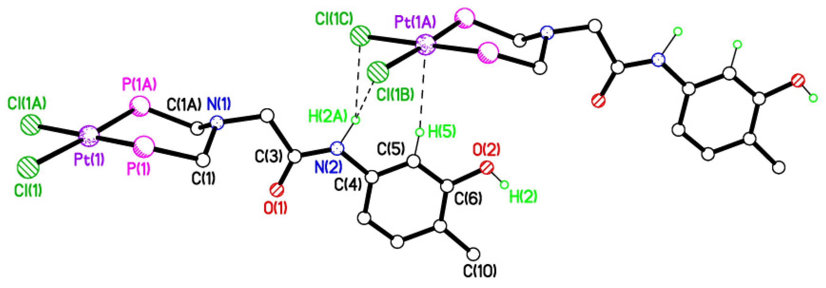
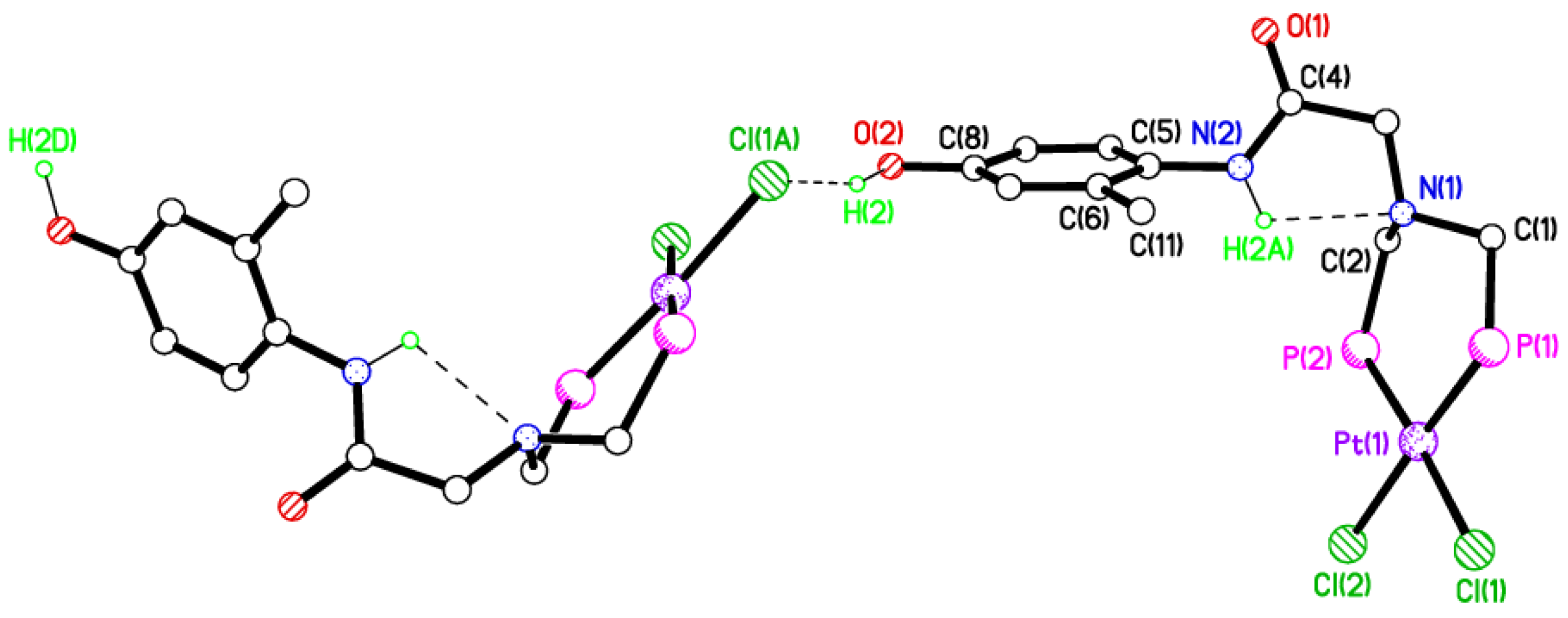
2.5. Single Crystal X-ray Studies of Complexes 4b∙(CH3)2SO, 4c∙CHCl3, 4d∙½Et2O, 4e∙½CHCl3∙½CH3OH, 5a∙½Et2O, 5b, 5c∙¼H2O, 5d∙Et2O, and 6∙(CH3)2SO

Detailed single crystal X-ray analysis (Table 5 and Table 6) of complexes 4b∙(CH3)2SO, 4c∙CHCl3, 4d∙½Et2O, 4e∙½CHCl3∙½CH3OH, 5a∙½Et2O, 5b, 5c∙¼H2O, 5d∙Et2O, and 6∙(CH3)2SO shows that the geometry about each Pt(II) centre is approximately square planar [P–Pt–P range 90.23(9)–96.52(3)°] (Table 7 and Table 8). The Pt–Cl and Pt–P bond distances are consistent with literature values [29] and the conformation of the Pt–P–C–N–C–P six-membered ring in each complex is best described as a boat. The dihedral angle measured between the P2C2 plane and N-arene ring least-squares planes varies between 50.98(12)° [in 6∙(CH3)2SO] and 90° (in 5d∙Et2O), the difference of ca. 39° may tentatively be explained by the predisposition of the –OH group about the N-arene group and subsequent H-bonding requirements. Upon metal chelation, a degree of freedom, compared with the free ligands 1a, 1b∙CH3OH, 2f∙CH3OH, 2g, and 3 has been removed, as the P–C–N–C–P backbone is locked into a specific conformation. Unfortunately, we were unable to obtain suitable X-ray quality crystals of compounds 4a and 5eg.
Despite the ortho position of the hydroxy group in 4c∙CHCl3, molecules do not form an intramolecular S(5) O–H∙∙∙N interaction as seen in 1a (Figure 1), instead forming a bifurcated H-bond with the two coordinated chloride ligands of an adjacent molecule (Figure 6). This generates a 1D chain, and also attracts a bifurcated H-bonded chloroform molecule. There are somewhat asymmetric distances d for H(1C) to Cl(1) and Cl(2) are 2.45(4) and 2.76(4) Å, while those from H(34) to Cl(1) and Cl(2) are 2.66 and 2.86 Å, so are also asymmetric. The twist angle between planes P(1)/P(2)/C(1)/C(2) and ring C(3) > C(8) is 84.83(8)°, so is almost perpendicular. Atoms N(1) and Pt(1) lie 0.795(4) and 0.024(2) Å away from the P(1)/P(2)/C(1)/C(2) plane, respectively. The hinge angle across the P(1)–P(2) vector is 2.51(5)°. Selected hydrogen bonding geometric parameters for 4c∙CHCl3 are shown in Table 9.
Compound 6∙(CH3)2SO, in which the –OH group is meta to the N-arene group H-bonds to the DMSO molecule of crystallisation resulting in a 0D structure (Figure 7). The distance d for this H-bond is 1.79(2) Å. The twist angle between plane P(1)/P(2)/C(1)/C(2) and ring C(3) > C(8) is 50.98(12)°. Atoms N(1) and Pt(1) lie 0.758(4) and 0.404(2) Å away from the P(1)/P(2)/C(1)/C(2) plane, respectively, so is more chair-shaped than some of the other platinum(II) complexes reported here. The hinge angle across the P(1)–P(2) vector is 11.87(13)°.
For 4d∙½Et2O (Figure 8) a molecule of badly disordered diethyl ether, modelled by the Platon Squeeze procedure, is not shown, but is in the vicinity of the hydroxy group and may H-bond to it resulting in a 0D structure. The twist angle between plane P(1)/P(2)/C(1)/C(2) and ring C(3) > C(8) is 67.82(7)°. Atoms N(1) and Pt(1) lie 0.797(3) and 0.2378(16) Å away from the P(1)/P(2)/C(1)/C(2) plane, respectively. The hinge angle across the P(1)–P(2) vector is 9.20(9)°.
The crystal structure of 4b∙(CH3)2SO shows the hydroxy group H-bonding to the DMSO molecule of crystallisation (Figure 9a). The distance d for this H-bond is 1.89 Å. The twist angle between plane P(1)/P(2)/C(1)/C(2) and ring C(3) > C(8) is 72.2(4)°. Atoms N(1) and Pt(1) lie 0.781(17) and 0.180(10) Å away from the P(1)/P(2)/C(1)/C(2) plane, respectively. The hinge angle across the P(1)–P(2) vector is 8.7(6)°. Molecules form 1D, weakly H-bonded, undulating chains in the c direction via the methylene H atoms on C(1) and C(2) to a single, coordinated chloride ligand in an adjacent molecule (Figure 9b). Selected hydrogen bonding parameters for 4b∙(CH3)2SO are shown in Table 9.
For compound 4e∙½CHCl3∙½CH3OH there are two independent Pt complexes, one CH3OH, and one CHCl3 in the asymmetric unit. Both Pt complexes form 1D chains aligned parallel to b, but these chains are different (Figure 10). The chain involving Pt(2) forms simple O–H∙∙∙Cl H-bonds with the adjacent molecules via the para hydroxy group with d = 2.39(4) Å. For the chain involving the Pt(1)-containing molecules, the intermolecular H-bond has an inserted methanol molecule. The distances, d, are 2.32(5) and 1.82 Å for H(3)∙∙∙Cl(2) and H(1A)∙∙∙O(3), respectively. Atoms N(1)/N(2) and Pt(1)/Pt(2) lie 0.765(9)/0.798(9) and 0.424(5)/0.364(5) Å away from the P(1)/P(2)/C(1)/C(2) or P(3)/P(4)/C(34)/C(35) planes, respectively. So, as in 6∙(CH3)2SO, the core 6-membered Pt–P–C–N–C–P rings adopt more chair-shaped conformations. The hinge angles across the P(1)–P(2)/P(3)–P(4) vectors are 13.44(16)/12.47(16)°. The twist angles between planes P(1)/P(2)/C(1)/C(2) or P(3)/P(4)/C(34)/C(35) and rings C(3) > C(8) or C(36) > C(41) are 88.17(19)/54.62(15)°. So, while the other geometric parameters are similar between the two molecules, this twist angle is significantly different.
In 5c, the amide and ring atoms from C(4) > C(11) are disordered over two sets of almost equally occupied positions. The disorder highlights two or more chain-forming possibilities for this structure, analogous to that observed in in 4e∙½CHCl3∙½CH3OH, with one possibility being simple (hydroxyl)O–H∙∙∙O(amide) links (Figure 11a), while the other, shown in Figure 11b, shows an alternative, water-inserted linkage. There is also likely to be some alternation of these motifs, given the random disorder and approx. 25% occupancy observed for water atom O(3). Unlike almost all of the other structures herein, the core 6-membered Pt–P–C–N–C–P ring adopts a conformation with atoms Pt(1)/P(1)/P(1)/C(2) being in a plane and atoms C(1) and N(2) being 1.021(6) and 1.237(6) Å, respectively, away from that plane. There is no C=O∙∙∙HN intermolecular H-bonding observed between molecules. Instead, the amide NH forms a bifurcated H-bond with the two neighbouring acceptor atoms N(1) and the ortho hydroxyl O(2) with d = 2.37 and 2.28 Å, respectively, while d = 2.89 Å for H(2)∙∙∙O(1A).
In the second motif, adjacent molecules have an inserted water molecule in the H-bond pattern (Figure 11b). The amide NH again forms a bifurcated H-bond with the two neighbouring acceptor atoms N(1) and O(2X) with d = 2.14 and 2.25 Å, respectively, while d = 2.89 Å for H(2X)∙∙∙O(3), which is a little long, and d for O(3)∙∙∙O(1XA) = 2.21(3) Å, which is rather short. The distance d from water oxygen O(3) to O(1A), however, is entirely reasonable for an H-bond at 2.74 Å, suggesting a predominantly alternating pattern between the two disorder options is most likely.
Complex 5a∙½Et2O was crystallised from a diethyl ether solution, including half a solvent molecule per complex molecule in the crystal lattice. There are two Pt complexes and two, half-occupied, Et2O solvent molecules of crystallisation in the asymmetric unit. The packing adopted by this second complex with an ortho hydroxyl group is very different to 5c (Figure 12). Here there is no intramolecular N–H∙∙∙N H-bond, instead the ortho hydroxyl forms an intramolecular H-bond with the amide oxygen with d = 1.80 and 1.77(4) Å in the molecules containing Pt(1) and Pt(2), respectively. This does leave the two unique amide NH atoms free to form intermolecular interactions, which they do via highly asymmetric, bifurcated H-bonds with the coordinated chloride ligands on adjacent Pt complexes. From H(2) d = 2.60(11) and 2.95(13) Å to Cl(3) and Cl(4), respectively, while d = 2.52(7) and 3.12(15) Å from H(4) to Cl(1A) and Cl(2A), respectively. N(1)/N(3) and Pt(1)/Pt(2) lie 0.771(13)/0.781(14) and 0.349(8)/0.346(8) Å out of the planes P(1)/P(2)/C(1)/C(2) and P(3)/P(4)/C(37)/C(38), respectively. The twist angle between planes P(1)/P(2)/C(1)/C(2) and P(3)/P(4)/C(37)/C(38) relative to the rings C(5) > C(10) and C(41) > C(46) are 51.3(5) and 51.71(4)°, respectively. Hinge angles across P(1)–P(2) and P(3)–P(4) are 12.3(5) and 12.0(4)°, respectively. Differences between the two systems involving ortho hydroxyl groups are the position of the methyl ring substituent in the meta or para position, and the co-crystallised solvent being a small amount of water or Et2O. Either, or both of these differences might account for the different intra- and intermolecular packing motifs observed. Selected hydrogen bonding parameters for 5a∙½Et2O are shown in Table 9.
Molecules of 5d∙Et2O lie on a mirror plane, passing through Pt(1), between pairs of P and Cl atoms, and including the atoms from N(1) to the terminal hydroxy-substituted ring. Again, here the amide NH is involved in the 1D chain propagation (Figure 13), forming a symmetrical bifurcated H-bond with the two coordinated chloride ligands on the adjacent molecule with d = 2.66(15) Å. Supporting this is an additional (Ar)C–H(5)∙∙∙Pt(1) interaction at 2.78 Å. The twist angle between the P(1)/P(1A)/C(1)/C(1A) plane and the ring C(4) > C(9) = 90° due to the imposed crystallographic symmetry. The hinge angle at P(1)–P(1A) = 29.5(5)°. Atoms N(1) and Pt(1) lie 0.79(2) and 0.782(14) Å away from the P(1)/P(2)/C(1)/C(2) plane, respectively. So, this is the most chair shaped core Pt–P–C–N–C–P 6-membered ring. The meta hydroxyl group is not involved in the chain propagating intermolecular interactions and points into a cleft between a pair of Ph rings. It does not make an H-bond with the solvent of crystallisation.
For compound 5b, the para position of the hydroxyl group facilitates 1D chain formation, forming an H-bond with one of the chloride ligands on an adjacent molecule with d = 2.09(6) Å (Figure 14). The amide NH here forms the familiar, but not universal, H-bond with the amine N(1) with d = 2.29(5) Å. The twist angle between the P(1)/P(2)/C(1)/C(2) plane and the ring C(5) > C(10) = 68.39(12)°. The hinge angle at P(1)–P(1A) = 4.95(10)°. Atoms N(1) and Pt(1) lie 0.810(4) and 0.164(3) Å away from the P(1)/P(2)/C(1)/C(2) plane, respectively.

3. Conclusions

In summary, we have shown that the position of the OH/CH3 groups with respect to the N-arene, the inclusion of an amide spacer, and the solvent used in the crystallisation can dictate the solid-state packing behaviour of both non coordinated and cis-PtCl2 bound diphosphine ligands. Unsurprisingly, the use of highly polar solvents (DMSO, CH3OH) in this study has been shown to play an important role in disrupting packing behaviour. Our work reinforces the importance of substituent effects, not only those commonly associated with −PR2 groups which may be alkyl or aryl based [37,38], but also those functional moieties positioned on the arene group of the central tertiary amine.

4. Materials and Methods

4.1. General Procedures

The synthesis of ligands 1ae, 2ag, and 3 were undertaken using standard Schlenk-line techniques and an inert nitrogen atmosphere. Ph2PCH2OH was prepared according to a known procedure [39]. All coordination reactions were carried out in air, using reagent grade quality solvents. The compound PtCl24-cod) (cod = cycloocta-1,5-diene) was prepared according to a known procedure [40]. All other chemicals were obtained from commercial sources and used directly without further purification

4.2. Instrumentation

Infrared spectra were recorded as KBr pellets on a Perkin-Elmer Spectrum 100S (4000–250 cm−1 range) Fourier-Transform spectrometer. 1H NMR spectra (400 MHz) were recorded on a Bruker DPX-400 spectrometer with chemical shifts (δ) in ppm to high frequency of Si(CH3)4 and coupling constants (J) in Hz. 31P{1H} NMR (162 MHz) spectra were recorded on a Bruker DPX-400 spectrometer with chemical shifts (δ) in ppm to high frequency of 85% H3PO4. NMR spectra were measured in CDCl3 or (CD3)2SO at 298 K. Elemental analyses (Perkin-Elmer 2400 CHN Elemental Analyser) were performed by the Loughborough University Analytical Service within the Department of Chemistry.

4.3. Preparation of Ligands 1ae, 2ag, and 3

The following general procedure was used for the synthesis of 1ae, 2ag, and 3. A mixture of Ph2PCH2OH (2 equiv.) and the appropriate amine (1 equiv.) in CH3OH (20 mL) was stirred under N2 for 24 h. The volume of the solution was evaporated to ca. 2–3 mL, under reduced pressure, to afford the desired ligands which were collected by suction filtration (except 2ac) and dried in vacuo. Isolated yields in range 38–97%. Characterising details are given in Table 1.

4.4. Preparation of cis-Dichloroplatinum(II) Phosphine Complexes 4ae, 5ag, and 6

The following general procedure was used for the synthesis of 4ae, 5ag, and 6. To a solution of PtCl24-cod) (1 equiv.) in CH2Cl2 (5 mL) was added a solution of the appropriate ligand (1 equiv.) in CH2Cl2 (5 mL). The colourless (or pale yellow) solution was stirred for 30 min at r.t., evaporated to ca. 2–3 mL under reduced pressure, and diethyl ether (10 mL) added. The solids were collected by suction filtration and dried in vacuo. Isolated yields in range 73–99%. Characterising details are given in Table 4.

4.5. Single Crystal X-ray Crystallography

Suitable crystals of 1a, 1b∙CH3OH, 2f·CH3OH, and 3 were obtained by slow evaporation of a CH3OH solution whereas 2g was obtained by vapour diffusion of Et2O into a CDCl3/CH3OH solution. Crystals of 4b∙(CH3)2SO, 5a∙½Et2O, 5b, and 5c∙¼H2O were obtained by slow diffusion of Et2O into a CDCl3/(CH3)2SO/CH3OH solution. Slow diffusion of hexanes [for 6∙(CH3)2SO] into a CDCl3/(CH3)2SO solution or vapour diffusion of Et2O into a CHCl3/(CH3)2SO/CH3OH [for 4c∙CHCl3, 4e∙½CHCl3∙½CH3OH) or CH2Cl2/CH3OH (for 5d∙Et2O)]. Slow evaporation of a CH2Cl2/Et2O/hexanes solution gave suitable crystals of 4d∙½Et2O. Table 2, Table 5 and Table 6 summarise the key data collection and structure refinement parameters. Diffraction data for compounds 1a, 1b∙CH3OH, 2f∙CH3OH 3, 4b∙(CH3)2SO, 4c∙CHCl3, 4d 4e∙½CHCl3∙½CH3OH, 5d∙Et2O, and 6∙(CH3)2SO, were collected using a Bruker or Bruker-Nonius APEX 2 CCD diffractometer using graphite-monochromated Mo-Kα radiation. Data for compounds 5b and 5c∙¼H2O, were collected using a Bruker APEX 2 CCD diffractometer using synchrotron radiation at Daresbury SRS Station 9.8 or 16.2 SMX for 5a·½Et2O. Data for compound 2g was collected using a Bruker SMART 1000 CCD diffractometer using graphite-monochromated Mo-Kα radiation. All structures were solved by direct methods [except structures 4b∙(CH3)2SO, 5a∙½Et2O, and 5b which were solved using Patterson synthesis] and refined by full-matrix least-squares methods on F2. All CH atoms were placed in geometrically calculated positions and were refined using a riding model (aryl C–H 0.95 Å, methyl C–H 0.98 Å, methylene C–H 0.99 Å. Where data quality allowed, OH and NH atom coordinates and Uiso were freely refined, or refined with mild geometrical restraints; otherwise, they were placed geometrically with O/N–H = 0.84 Å. Uiso(H) values were set to be 1.2 times Ueq of the carrier atom for aryl CH and NH, and 1.5 times Ueq of the carrier atom for OH and CH3. Throughout the text and tabulated data, where H atom geometry does not include a SU, the coordinates were constrained. Unless stated, all structural determinations proceeded without the need for restraints or disorder modelling. Where disorder was modelled it was supported with appropriate geometrical and U value restraints. In 1b∙CH3OH, the methanol was modelled as disordered over two equally occupied sets of positions. In 2f·CH3OH the methanol was modelled using the Platon Squeeze procedure [41]. Compound 3 was found to contain a disordered methanol and was modelled over two sets of positions, each at half weight. In 4d·½Et2O, atoms C(1) > C(7) and N(1) were modelled with U value restraints. The Et2O was modelled using Platon Squeeze due to significant disorder. In 4e∙½CHCl3∙½CH3OH the chloroform molecule was modelled over two sets of positions with major occupancy 57.1(7)% Restraints were applied to that molecule and also ring C(55) > C(60). In 5a·½Et2O three Ph rings were modelled as disordered over two sets of positions with occupancies close to 50%. Restraints were applied to these rings and also the two half-occupancy Et2O solvent molecules of crystallisation. In 5c∙¼H2O, atoms Cl(1) and C(3) > C(11), O(1), O(2) and N(1) were modelled as split over two sets of positions with major occupancy 56(4) and 50.9(6)%, respectively and restraints were applied. In 5d∙Et2O the Et2O was modelled as a diffuse area of electron density by the Platon Squeeze procedure and restraints were applied to atoms C(1) > C(10), C(11) > C(22) and N(2) O(2). In 6∙(CH3)2SO the DMSO was modelled with restraints as disordered over two sets of positions with major component 71.0(5)% and with C(33) coincident for both components Programs used during data collection, refinement and production of graphics were Bruker SMART, Bruker APEX 2, SAINT, SHELXTL, COLLECT, DENZO and local programs [41,42,43,44,45,46,47,48,49,50,51]. CCDC 2101643-2101656 contain the supplementary crystallographic data for this paper. These data can be obtained free of charge from The Cambridge Crystallographic Data Centre via www.ccdc.cam.ac.uk/structures (accessed on 3 November 2021).

Author Contributions

Conceptualisation, M.B.S.; synthesis and characterisation of the compounds, N.M.S.-B., P.D.; single crystal X-ray crystallography, N.M.S.-B., M.R.J.E.; writing-original draft preparation, M.B.S.; writing-review and editing, M.R.J.E., M.B.S. All authors have read and agreed to the published version of the manuscript.

Funding

This research received no external funding.

Institutional Review Board Statement

Not applicable.

Informed Consent Statement

Not applicable.

Data Availability Statement

Not applicable.

Acknowledgments

We thank the EPSRC Centre for Doctoral Training in Embedded Intelligence under grant reference EP/L014998/1 for financial support (PD). Johnson Matthey are acknowledged for their kind donation of precious metals and the UK National Crystallography Service at the University of Southampton for three of the data collections. The STFC is thanked for the allocation of beam time at Daresbury Laboratory.

Conflicts of Interest

The authors declare no conflict of interest.

Sample Availability

Samples of the compounds in this article are not available from the authors.

References

  1. Lehn, J.-M. Supramolecular Chemistry-Scope and Perspectives. Molecules, Supermolecules, and Molecular Devices (Nobel Lecture). Angew. Chem. Int. Ed. Engl. 1988, 27, 89–112. [Google Scholar] [CrossRef]
  2. Jongkind, L.J.; Caumes, X.; Hartendorp, A.P.T.; Reek, J.N.H. Ligand Template Strategies for Catalyst Encapsulation. Acc. Chem. Res. 2018, 51, 2115–2128. [Google Scholar] [CrossRef] [PubMed]
  3. James, S.L. Phosphines as building blocks in coordination-based self-assembly. Chem. Soc. Rev. 2009, 38, 1744–1758. [Google Scholar] [CrossRef]
  4. Breit, B. Supramolecular Approaches to Generate Libraries of Chelating Bidentate Ligands for Homogeneous Catalysis. Angew. Chem. Int. Ed. 2005, 44, 6816–6825. [Google Scholar] [CrossRef] [PubMed]
  5. Daubignard, J.; Detz, R.J.; de Bruin, B.; Reek, J.N.H. Phosphine Oxide Based Supramolecular Ligands in the Rhodium-Catalysed Asymmetric Hydrogenation. Organometallics 2019, 38, 3961–3969. [Google Scholar] [CrossRef]
  6. Koshti, V.S.; Sen, A.; Shinde, D.; Chikkali, S.H. Self-assembly of P-chiral supramolecular phosphines on rhodium and direct evidence for Rh-catalyst-substrate interactions. Dalton Trans. 2017, 46, 13966–13973. [Google Scholar] [CrossRef]
  7. Vasseur, A.; Membrat, R.; Palpacelli, D.; Giorgi, M.; Nuel, D.; Giordano, L.; Martinez, A. Synthesis of chiral supramolecular bisphosphinite palladcycles through hydrogen transfer-promoted self-assembly process. Chem. Commun. 2018, 54, 10132–10135. [Google Scholar] [CrossRef] [PubMed] [Green Version]
  8. Romero-Nieto, C.; de Cózar, A.; Regulska, E.; Mullenix, J.B.; Rominger, F.; Hindenberg, P. Controlling the molecular arrangement of racemates through weak interactions: The synergy between p-interactions and halogen bonds. Chem. Commun. 2021, 57, 7366–7369. [Google Scholar] [CrossRef] [PubMed]
  9. Carreras, L.; Serrano-Torné, M.; van Leeuwen, P.W.N.M.; Vidal-Ferran, A. XBphos-Rh: A halogen-bond assembled supramolecular catalyst. Chem. Sci. 2018, 9, 3644–3648. [Google Scholar] [CrossRef] [Green Version]
  10. García-Márquez, A.; Frontera, A.; Roisnel, T.; Gramage-Doria, R. Ultrashort Hd+…Hd- intermolecular distance in a supramolecular system in the solid state. Chem. Commun. 2021, 57, 7112–7115. [Google Scholar] [CrossRef]
  11. Blann, K.; Bollmann, A.; Brown, G.M.; Dixon, J.T.; Elsegood, M.R.J.; Raw, C.R.; Smith, M.B.; Tenza, K.; Willemse, A.; Zweni, P. Ethylene oligomerisation chromium catalysts with unsymmetrical PCNP ligands. Dalton Trans. 2021, 50, 4345–4354. [Google Scholar] [CrossRef] [PubMed]
  12. De’Ath, P.; Elsegood, M.R.J.; Halliwell, C.A.G.; Smith, M.B. Mild intramolecular P-C(sp3) bond cleavage in bridging diphosphine complexes of RuII, RhIII, and IrIII. J. Organomet. Chem. 2021, 937, 121704. [Google Scholar] [CrossRef]
  13. Smith, M.B.; Dale, S.H.; Coles, S.J.; Gelbrich, T.; Hursthouse, M.B.; Light, M.E.; Horton, P.N. Hydrogen bonded supramolecular assemblies based on neutral square-planar palladium(II) complexes. CrystEngCommun 2007, 9, 165–175. [Google Scholar] [CrossRef]
  14. Smith, M.B.; Dale, S.H.; Coles, S.J.; Gelbrich, T.; Hursthouse, M.B.; Light, M.E. Isomeric dinuclear gold(I) complexes with highly functionalised ditertiary phosphines: Self-assembly of dimers, rings and 1-D polymeric chains. CrystEngCommun 2006, 8, 140–149. [Google Scholar] [CrossRef]
  15. Dann, S.E.; Durran, S.E.; Elsegood, M.R.J.; Smith, M.B.; Staniland, P.M.; Talib, S.; Dale, S.H. Supramolecular chemistry of half-sandwich organometallic building blocks based on RuCl2(p-cymene)Ph2PCH2Y. J. Organomet. Chem. 2006, 691, 4829–4842. [Google Scholar] [CrossRef]
  16. Durran, S.E.; Smith, M.B.; Slawin, A.M.Z.; Gelbrich, T.; Hursthouse, M.B.; Light, M.E. Synthesis and coordination studies of new aminoalcohol functionalised tertiary phosphines. Can. J. Chem. 2001, 79, 780–791. [Google Scholar] [CrossRef]
  17. Jiang, M.-S.; Tao, Y.-H.; Wang, Y.-W.; Lu, C.; Young, D.J.; Lang, J.-P.; Ren, Z.-G. Reversible Solid-State Phase Transitions between Au-P Complexes Accompanied by Switchable Fluorescence. Inorg. Chem. 2020, 59, 3072–3078. [Google Scholar] [CrossRef]
  18. Pandey, M.K.; Kunchur, H.S.; Mondal, D.; Radhakrishna, L.; Kote, B.S.; Balakrishna, M.S. Rare AuH Interactions in Gold(I) Complexes of Bulky Phosphines Derived from 2,6-Dibenzhydryl-4-methylphenyl Core. Inorg. Chem. 2020, 59, 3642–3658. [Google Scholar] [CrossRef] [PubMed]
  19. Bálint, E.; Tajti, A.; Tripolszky, A.; Keglevich, G. Synthesis of platinum, palladium and rhodium complexes of α-aminophosphine ligands. Dalton Trans. 2018, 47, 4755–4778. [Google Scholar] [CrossRef] [PubMed] [Green Version]
  20. Zhang, Y.-P.; Zhang, M.; Chen, X.-R.; Lu, C.; Young, D.J.; Ren, Z.-G.; Lang, J.-P. Cobalt(I) and Nickel(II) Complexes of a PNN Type Ligand as Photoenhanced Electrocatalysts for the Hydrogen Evolution Reaction. Inorg. Chem. 2020, 59, 1038–1045. [Google Scholar] [CrossRef]
  21. Hou, R.; Huang, T.-H.; Wang, X.-J.; Jiang, X.-F.; Ni, Q.-L.; Gui, L.-C.; Fan, Y.-J.; Tan, Y.-L. Synthesis, structural characterisation and luminescent properties of a series of Cu(I) complexes based on polyphosphine ligands. Dalton Trans. 2011, 40, 7551–7558. [Google Scholar] [CrossRef] [Green Version]
  22. Wang, X.-J.; Gui, L.-C.; Ni, Q.-L.; Liao, Y.-F.; Jiang, X.-F.; Tang, L.-H.; Zhang, Z.; Wu, Q. p-Stacking induced complexes with Z-shape motifs featuring a complimentary approach between electron-rich arene diamines and electron-deficient aromatic N-heterocycles. CrystEngComm 2008, 10, 1003–1010. [Google Scholar] [CrossRef]
  23. Au, R.H.W.; Jennings, M.C.; Puddephatt, R.J. Supramolecular Organoplatinum(IV) Chemistry: Sequential Introduction of Amide Hydrogen Bonding Groups. Organometallics 2009, 28, 3754–3762. [Google Scholar] [CrossRef]
  24. Coles, N.T.; Gasperini, D.; Provis-Evans, C.B.; Mahon, M.F.; Webster, R.L. Heterobimetallic Complexes of 1,1-Diphosphineamide Ligands. Organometallics 2021, 40, 148–155. [Google Scholar] [CrossRef]
  25. Navrátil, M.; Císařová, I.; Alemayehu, A.; Škoch, K.; Štěpnička, P. Synthesis and Structural Characterisation of an N-Phosphanyl Ferrocene Carboxamide and its Ruthenium, Rhodium and Palladium Complexes. ChemPlusChem 2020, 85, 1325–1338. [Google Scholar] [CrossRef]
  26. Navrátil, M.; Císařová, I.; Štěpnička, P. Intermolecular interactions in the crystal structures of chlorogold(I) complexes with N-phosphinoamide ligands. Inorg. Chim. Acta 2021, 516, 120138. [Google Scholar] [CrossRef]
  27. Pachisia, S.; Kishan, R.; Yadav, S.; Gupta, R. Half-Sandwich Ruthenium Complexes of Amide-Phosphine Based Ligands: H-Bonding Cavity Assisted Binding and Reduction of Nitro-substrates. Inorg. Chem. 2021, 60, 2009–2022. [Google Scholar] [CrossRef] [PubMed]
  28. Nasser, N.; Eisler, D.J.; Puddephatt, R.J. A chiral diphosphine as trans-chelate ligand and its relevance to catalysis. Chem. Commun. 2010, 46, 1953–1955. [Google Scholar] [CrossRef] [PubMed]
  29. Elsegood, M.R.J.; Lake, A.J.; Smith, M.B.; Weaver, G.W. Ditertiary phosphines bearing a -N-C-C(O)-N(H)- linker and their corresponding dichloroplatinum(II) complexes. Phosphorus Sulfur Silicon Relat. Elem. 2019, 194, 540–544. [Google Scholar] [CrossRef]
  30. Hoyos, O.L.; Bermejo, M.R.; Fondo, M.; García-Deibe, A.; González, A.M.; Maneiro, M.; Pedrido, R. Mn(III) complexes with asymmetrical N2O3Schiff bases. The unusual crystal structure of [Mn(phenglydisal-3-Br,5-Cl)(dmso)] (H3phenglydisal = 3-aza-N-{2-[1-aza-2-(2-hydroxyphenyl)vinyl]phenyl}-4-(2-hydroxyphenyl)but-3-enamide), a mononuclear single-stranded helical manganese(III) complex. J. Chem. Soc. Dalton Trans. 2000, 3122–3127. [Google Scholar] [CrossRef]
  31. Bermejo, M.R.; González, A.M.; Fondo, M.; García-Deibe, A.; Maneiro, M.; Sanmartín, J.; Hoyos, O.L.; Watkinson, M. A direct route to obtain manganese(III) complexes with a new class of asymmetrical Schiff base ligands. New J. Chem. 2000, 24, 235–241. [Google Scholar] [CrossRef]
  32. Elsegood, M.R.J.; Smith, M.B.; Staniland, P.M. Neutral Molecular Pd6 Hexagons Using k3-P2O Terdentate Ligands. Inorg. Chem. 2006, 45, 6761–6770. [Google Scholar] [CrossRef] [PubMed]
  33. Etter, M.C.; MacDonald, J.C.; Bernstein, J. Graph-Set Analysis of Hydrogen-Bond Patterns in Organic Crystals. Acta Crystallogr. 1990, B46, 256–262. [Google Scholar] [CrossRef]
  34. Etter, M.C. Encoding and Decoding Hydrogen-Bond Patterns of Organic Compounds. Acc. Chem. Res. 1990, 23, 120–126. [Google Scholar] [CrossRef]
  35. Bernstein, J.; Davis, R.E.; Shimoni, L.; Chang, N.-L. Patterns in Hydrogen Bonding: Functionality and Graph Set Analysis in Crystals. Angew. Chem. Int. Ed. Engl. 1995, 34, 1555–1573. [Google Scholar] [CrossRef]
  36. Desiraju, G.; Steiner, T. The Weak Hydrogen Bond; Oxford University Press: Oxford, UK, 2001. [Google Scholar]
  37. Klemps, C.; Payet, E.; Magna, L.; Saussine, L.; Le Goff, X.F.; Le Floch, P. PCNCP Ligands in the Chromium-Catalysed Oligomerisation of Ethylene: Tri-versus Tetramerization. Chem. Eur. J. 2009, 15, 8259–8268. [Google Scholar] [CrossRef]
  38. Walsh, A.P.; Laureanti, J.A.; Katipamula, S.; Chambers, G.M.; Priyadarshani, N.; Lense, S.; Bays, J.T.; Linehan, J.C.; Shaw, W.J. Evaluating the impacts of amino acids in the second and outer coordination spheres of Rh-bis(diphosphine) complexes for CO2 hydrogenation. Faraday Discuss. 2019, 215, 123–140. [Google Scholar] [CrossRef] [PubMed]
  39. Hellman, H.; Bader, J.; Birkner, H.; Schumacher, O. Hydroxymethyl-phosphine, Hydroxymethyl-phosphoniumsalze und Chlormethyl-phosphoniumsalze. Justus Liebigs Ann. Chem. 1962, 659, 49–56. [Google Scholar] [CrossRef]
  40. McDermott, J.X.; White, J.F.; Whitesides, G.M. Thermal Decomposition of Bis(phosphine)platinum(II) Metallocycles. J. Am. Chem. Soc. 1976, 98, 6521–6528. [Google Scholar] [CrossRef]
  41. Spek, A.L. PLATON SQUEEZE: A tool for the calculation of the disordered solvent contribution to the calculated structure factors. Acta Crystallogr. Sect. C-Struct. Chem. 2015, 71, 9–18. [Google Scholar] [CrossRef] [Green Version]
  42. Sluis, P.v.d.; Spek, A.L. BYPASS: An effective method for the refinement of crystal structures containing disordered solvent regions. Acta Crystallogr. 1990, A46, 194–201. [Google Scholar] [CrossRef]
  43. Bruker SMART Version 5.611; Bruker AXS Inc.: Fitchburg, WI, USA, 2001.
  44. Area-Detector Integration Software, APEX-II, Version V1; Bruker-Nonius: Madison, WI, USA, 2004.
  45. Denzo, Z.; Otwinowski, W. Processing of X-ray diffraction data in oscillation mode, Methods in Enzymology. In Macromolecular Crystallography; Carter, C.W., Jr., Sweet, R.M., Eds.; Academic Press: Cambridge, MA, USA, 1997; Volume 276, pp. 307–326. [Google Scholar]
  46. Hooft, R.W.W. COLLECT: Data Collection Software; Nonius B.V.: Delft, The Netherlands, 1998. [Google Scholar]
  47. SAINT Software for CCD Diffractometers; Bruker AXS Inc.: Madison, WI, USA, 2004.
  48. Krause, L.; Herbst-Irmer, R.; Sheldrick, G.M.; Stalke, D.J. SADABS software. J. Appl. Cryst. 2015, 48, 3–10. [Google Scholar] [CrossRef] [PubMed] [Green Version]
  49. Sheldrick, G.M. Crystal structure refinement with SHELXL. Acta Crystallogr. Sect. 2015, A71, 3–8. [Google Scholar] [CrossRef] [Green Version]
  50. Sheldrick, G.M. A short history of SHELX. Acta Crystallogr. Sect. 2008, A64, 112–122. [Google Scholar] [CrossRef] [PubMed] [Green Version]
  51. Sheldrick, G.M. SHELXTL User Manual, Version 6.12; Bruker AXS Inc.: Madison, WI, USA, 2001. [Google Scholar]
Chart 1. Potential modification sites of a Ph2P–C–N(Ar)–C–PPh2 backbone.
Molecules 26 06809 ch001
Scheme 1. Synthesis of 1ae, 2ag, and 3.
Molecules 26 06809 sch001
Figure 1. Molecular structure of 1a. All hydrogens, except on C(1), C(2) and O(1), have been omitted for clarity.
Molecules 26 06809 g001
Figure 2. Molecular structure of 3 showing a dimer pair. All hydrogens, except on C(1), C(2) and O(1), have been omitted for clarity. Symmetry code: A = 1 − x, 1 − y, 1 − z.
Molecules 26 06809 g002
Figure 3. Crystal structure packing plot for 1b∙CH3OH. Most H atoms, two Ph groups per P atom have been omitted for clarity. Symmetry code: A = x, −y + ½, z + ½.
Molecules 26 06809 g003
Figure 4. Dimers of 2f forming R22(16) graph set motifs. Most H atoms omitted for clarity. The second unique molecule which adopts a similar, centrosymmetric motif, is not shown.
Molecules 26 06809 g004
Figure 5. Intra- and intermolecular interactions in the crystal structure of 2g. Most H atoms omitted for clarity. Symmetry operator A = x, −y + 3/2, z − ½.
Molecules 26 06809 g005
Chart 2. Structures of compounds 4ae, 5ag, and 6.
Molecules 26 06809 ch002
Figure 6. H-bonded packing arrangement in the crystal structure of 4c∙CHCl3. Most H atoms omitted for clarity. Symmetry operator A = x+½, –y+½, z+½.
Molecules 26 06809 g006
Figure 7. Crystal structure of 6∙(CH3)2SO showing the hydroxyl group H-bonding to the (CH3)2SO molecule of crystallisation. Most H-atoms omitted for clarity.
Molecules 26 06809 g007
Figure 8. Crystal structure of 4d∙½Et2O. Most H atoms and the disordered OEt2 molecule omitted for clarity.
Molecules 26 06809 g008
Figure 9. (a) Crystal structure of 4b∙(CH3)2SO showing the hydroxyl group H-bonding to the DMSO molecule of crystallisation. Most H-atoms removed for clarity. (b) Packing interactions in the crystal structure of 4b∙(CH3)2SO. Most H atoms omitted for clarity. Symmetry operator A = y − 1, 1 − x, ¼ + z.
Molecules 26 06809 g009
Figure 10. H-bonded packing motifs in the crystal structure of 4e∙½CHCl3∙½CH3OH. Most H atoms, two Ph groups per P atom, and the disordered chloroform of crystallisation which is not involved in any significant intermolecular interactions, are omitted for clarity. Symmetry operators are x, y − 1, z and x, y + 1, z.
Molecules 26 06809 g010
Figure 11. Most H atoms and 2 Ph groups per P atom have been omitted for clarity. (a) Packing motif 1 in the crystal structure of 5c. Symmetry operator A = x + 1, y, z. (b) Packing motif 2 in the crystal structure of 5c. The true structure is most likely an alternation of motifs 1 and 2. Symmetry operator A = x + 1, y, z.
Molecules 26 06809 g011
Figure 12. Packing motif in the crystal structure of 5a∙½Et2O. Most H atoms, two Ph groups per P atom and the two, half-occupied, Et2O molecules have been omitted for clarity.
Molecules 26 06809 g012
Figure 13. Packing plot of 5d∙Et2O. Most H atoms, two Ph groups per P atom, and a diordered Et2O molecule modelled by the Platon Squeeze procedure, are omitted for clarity. Symmetry operators: (i) for the mirror x, y, −z + ½, (ii) for the chain direction x + 1, y, z.
Molecules 26 06809 g013
Figure 14. Packing plot in the crystal structure of 5b. Phenyl groups and hydrogen atoms not involved in hydrogen bonding have been omitted for clarity.
Molecules 26 06809 g014
Table 1. Selected spectroscopic and analytical data for compounds 1a3 a.
Com-pound aδ(P) bδ(H)
/OH
(NH)
δ(H)
/arom. H.
δ(H)
/CH2
δ(H)
/CH2 d
δ(H)
/CH3
νOH
NH) e
Microanalysis
(CHN)
1a (79)−27.58.627.33–7.23,
6.76, 6.69–6.57
4.15 (2.4) 2.103398Calc. for C33H31NOP2,
C, 76.29; H, 6.01; N, 2.70
Found, C, 76.07; H, 6.13; N, 2.78
1b (56)−27.39.067.36–7.26,
7.15, 6.50, 6.44
3.96 (5.6)1.743282Calc. for C33H31NOP2.2MeOH,
C, 72.03; H, 6.74; N, 2.40
Found, C, 72.45; H, 6.04; N, 2.58
1c (97)−27.58.777.44–7.22,
6.86, 6.54, 6.48
4.09 (3.4)2.123389Calc. for C33H31NOP2,
C, 76.29; H, 6.01; N, 2.70
Found, C, 75.99; H, 6.00; N, 2.76
1d (38)−26.78.637.40–7.30,
6.55
4.02 (3.2)1.963432Calc. for C33H31NOP2,
C, 76.29; H, 6.01; N, 2.70
Found, C, 75.53; H, 6.05; N, 2.74
1e (96)−26.49.067.49–7.33,
6.85, 6.50, 6.27
3.88 (3.6)2.083387Calc. for C33H31NOP2.MeOH,
C, 74.03; H, 6.40; N, 2.54
Found, C, 74.81; H, 5.93; N, 2.61
2a (81)−26.08.157.77–7.195.063.62 (8.0)1.19--
2b (89)−26.07.837.60–7.215.073.69 (3.6)1.63--
2c (88)−26.5 c9.34
(8.17)
7.71–7.195.273.61 (4.8)1.63--
2d (65)−27.19.05
(8.68)
7.55–7.32, 6.95, 6.61, 6.413.693.81 (4.8) 2.043047
(3228)
Calc. for C35H34N2O2P2,
C, 72.91; H, 5.94; N, 4.86
Found, C, 72.72; H, 5.95; N, 4.88
2e (80)−27.19.29 (9.08)7.46–7.35, 7.29, 6.86, 6.153.733.82 (4.4)2.083178
(3317)
Calc. for C35H34N2O2P2,
C, 72.91; H, 5.94; N, 4.86
Found, C, 72.71; H, 5.94; N, 4.82
2f (70)−27.59.31
(9.07)
7.41–7.03, 6.94, 6.40, 6.303.693.77 (4.4) 3163
(3283)
Calc. for C34H32N2O2P2,
C, 72.59; H, 5.73; N, 4.98
Found, C, 72.10; H, 5.80; N, 4.95
2g (85)−26.89.09 (8.78)7.36–7.25, 6.83, 6.533.613.72 (4.4) 3300 (3257)Calc. for C34H32N2O2P2,
C, 72.59; H, 5.73; N, 4.98
Found, C, 72.15; H, 5.72; N, 4.95
3 (53)−27.69.127.38–7.31, 6.92, 6.30, 6.13 3.85 3376Calc. for C32H29NOP2,
C, 76.03; H, 5.78; N, 2.77
Found, C, 75.67; H, 5.71; N, 2.74
a Isolated yields in parentheses. b Recorded in (CD3)2SO unless otherwise stated. c Recorded in CDCl3. d 2J(PH) coupling in brackets. e Recorded as KBr discs.
Table 2. Details of the X-ray data collections and refinements for compounds 1a, 1b·CH3OH, 2f·CH3OH, 2g, and 3.
Compound1a1b·CH3OH2f·CH3OH2g3
FormulaC33H31NOP2C34H35NO2P2C35H36N2O3P2C34H32N2O2P2C32H29NOP2
M519.53551.57594.60562.55505.50
Crystal dimensions0.42 × 0.15 × 0.030.13 × 0.12 × 0.020.24 × 0.18 × 0.160.25 × 0.18 × 0.150.31 × 0.28 × 0.03
Crystal morphology and colourPlate,
colourless
Block,
colourless
Block,
colourless
Block,
colourless
Plate,
colourless
Crystal systemMonoclinicMonoclinicTriclinicMonoclinicTriclinic
Space groupP21/nP21/cP 1 ¯ IaP 1 ¯
a/Å17.367(5)10.3050(3)12.6198(3)11.6234(10)10.5860(4)
b/Å8.522(2)32.8017(10)16.2027(4)21.7359(19)10.7397(4)
c/Å20.382(6)8.5189(2)17.8529(4)11.6340(10)13.4172(6)
α/° 64.0678(10) 73.1667(6)
β/°114.673(4)92.7318(16)76.7403(14)93.8717(14)80.4518(7)
γ/° 75.5070(14) 63.1422(6)
V32741.2(13)2876.30(14)3148.15(13)2932.6(4)1301.45(9)
Z44442
λ/Å0.710730.710730.710730.710730.71073
Τ150(2)120(2)120(2)150(2)150(2)
Density (calcd.)/Mg/m31.2591.2741.2551.2741.290
μ/mm−10.1850.1830.1760.1820.193
θ range/°2.02–26.603.03–27.533.24–25.001.87–28.821.59–30.62
Measured reflections23,52527,24761,33012,57715,576
Independent reflections5708654511,04765867814
Observed reflections
(F2 > 2σ(F2))
31155019755952936116
Rint0.1240.0580.0950.0390.027
R[F2 > 2σ(F2)] a0.07430.07990.05170.03890.0441
wR2 [all data] b0.22050.16500.12200.08610.1248
Largest difference map features/eÅ−31.40, −0.490.46, −0.520.38, −0.300.29, −0.160.51, −0.21
a R = ∑||Fo| − |Fc||/∑|Fo|. b wR2 = [∑[w(Fo2Fc2)2]/∑[w(Fo2)2]]1/2.
Table 3. Selected data (D···A/Å, ∠D–H···A/°) for key inter- and intramolecular contacts for compounds 1a, 1b·CH3OH, 2f·CH3OH, 2g, and 3.
1a1b·CH3OH2f·CH3OH a2g3
O–H···Nintra2.745(4), 119(4)
O–HMeOH···Ointer
O–HMeOH···OMeOH
2.844(8), 157
2.781(11), 172
O–H···Pinter 3.432(3), 173 3.4400(12), 167(2)
O–H···(O)Cinter 2.671(3), 171(3)
[2.659(3), 165(3)]
2.706(4), 169(4)
N–H···Nintra 2.695(3), 114(2)
[2.714(3), 117(2)]
2.748(4), 114(3)
a Values in parentheses are for the second independent molecule.
Table 4. Selected spectroscopic and analytical data for compounds 4a6 a.
Com-pound aδ(P) bδ(H)
/OH
(NH)
δ(H)
/arom. H.
δ(H)
/CH2
δ(H)
/CH2
δ(H)
/CH3
νOH
NH) f
νPtClMicroanalysis
(CHN)
4a (98)−9.4 d
(3424)
9.257.89–7.80, 7.64–7.46, 6.68, 5.90 4.211.933314316, 289Calc. for C33H31Cl2NOP2Pt.CH2Cl2,
C, 46.91; H, 3.82; N, 1.61
Found, C, 47.07; H, 3.77; N, 1.69
4b (89)−4.9 d
(3426)
9.227.96–7.53, 6.96, 6.49, 6.33 4.191.293373315, 282Calc. for C33H31Cl2NOP2Pt,
C, 50.46; H, 3.98; N, 1.78
Found, C, 50.51; H, 4.13; N, 1.83
4c (78)−8.6 d
(3436)
9.427.89–7.84, 7.56–7.43, 6.59, 6.30, 6.05 4.162.063433309, 290Calc. for C33H31Cl2NOP2Pt.0.5CH2Cl2,
C, 48.96; H, 3.87; N, 1.68
Found, C, 49.42; H, 3.96; N, 1.73
4d (98)−11.7 d (3410)8.447.94–7.87, 7.78–7.62, 6.86, 6.47 4.432.023421314, 290Calc. for C33H31Cl2NOP2Pt,
C, 50.46; H, 3.98; N, 1.78
Found, C, 50.24; H, 3.98; N, 1.85
4e (81)−7.8 d
(3421)
9.017.96–7.85, 7.59–7.45, 6.75, 6.27, 6.03 4.332.093416316, 284Calc. for C33H31Cl2NOP2Pt,
C, 50.46; H, 3.98; N, 1.78
Found, C, 50.66; H, 4.61; N, 1.70
5a (89)−9.8 d,e
(3398)
9.45
(8.91)
7.84–7.80, 7.53–7.44, 6.693.494.052.223051
(3249)
305, 283Calc. for C35H34Cl2N2O2P2Pt.0.5CH2Cl2,
C, 48.63; H, 3.74; N, 3.15
Found, C, 49.00; H, 4.07; N, 3.13
5b (65)−11.0 d
(3397)
9.16
(8.61)
7.83–7.80, 7.57–7.41, 7.05, 6.484.034.031.803050
(3350)
316, 283Calc. for C35H34Cl2N2O2P2Pt,
C, 49.89; H, 4.07; N, 3.32
Found, C, 49.32; H, 4.17; N, 3.25
5c (73)−9.9 d
(3405)
9.56
(8.94)
7.85–7.77, 7.59–7.38, 6.63, 6.513.174.052.173075
(3347)
315, 290Calc. for C35H34Cl2N2O2P2Pt,
C, 49.89; H, 4.07; N, 3.32
Found, C, 49.28; H, 4.05; N, 2.91
5d (99)−9.8 c,d
(3406)
9.17
(8.90)
7.98–7.50, 6.97–6.84, 6.68, 6.733.204.662.133323
(3465)
309, 283Calc. for C35H34Cl2N2O2P2Pt.0.5C4H10O,
C, 50.52; H, 4.47; N, 3.19
Found, C, 50.91; H, 4.53; N, 3.61
5e (90)−9.7 c,d
(3406)
9.46
(9.21)
7.94–7.78, 7.54–7.42, 7.09, 6.87, 6.693.434.122.023287
(3439)
312, 286Calc. for C35H34Cl2N2O2P2Pt,
C, 49.89; H, 4.07; N, 3.32
Found, C, 49.77; H, 3.95; N, 3.38
5f (85)−9.5 c,d
(3425)
9.62
(9.36)
7.91–7.86, 7.60–7.42, 7.05, 6.83, 6.453.474.18 3053
(3312)
304, 279Calc. for C34H32Cl2N2O2P2Pt,
C, 49.29; H, 3.89; N, 3.38
Found, C, 48.98; H, 3.38; N, 3.37
5g (84)−9.5 c,d
(3405)
9.52
(9.31)
8.01–7.97, 7.70–7.61, 7.34, 6.783.494.26 3054
(3325)
311, 287Calc. for C34H32Cl2N2O2P2Pt,
C, 49.29; H, 3.89; N, 3.38
Found, C, 48.72; H, 3.66; N, 3.33
6 (89)−4.0 d
(3436)
8.457.45–7.05, 6.89–6.76, 6.31, 4.31 3356311, 289Calc. for C32H29Cl2NOP2Pt,
C, 49.82; H, 3.79; N, 1.82
Found, C, 49.31; H, 3.58; N, 1.79
a Isolated yields in parentheses. b Recorded in (CD3)2SO unless otherwise stated. c Recorded in CDCl3.d 1J(PtP) coupling in parentheses. e Recorded in CDCl3/CD3OD. f Recorded as KBr discs.
Table 5. Details of the X-ray data collections and refinements for compounds 4b·(CH3)2SO, 4c·CHCl3, 4d·½OEt2, and 4e·½CHCl3·½CH3OH.
Compound4b·(CH3)2SO4c·CHCl34d·½OEt24e·½CHCl3·½CH3OH
FormulaC35H37Cl2NO2P2PtSC34H32Cl5NOP2PtC35H36Cl2NO1.5P2PtC34H33.5Cl3.5NO1.5P2Pt
M863.64904.88822.58861.22
Crystal dimensions0.19 × 0.02 × 0.010.30 × 0.18 × 0.040.13 × 0.06 × 0.030.15 × 0.04 × 0.02
Crystal morphology and colourNeedle,
colourless
Plate,
colourless
Lath,
colourless
Needle,
colourless
Crystal systemTetragonalMonoclinicMonoclinicMonoclinic
Space groupP43P21/nP21/cP21/n
a/Å11.373(3)11.6938(4)15.7344(6)21.4521(4)
b/Å 16.7052(6)17.0714(6)12.5164(2)
c/Å26.773(6)18.2242(7)13.9632(5)24.5837(4)
α/°
β/° 99.7066(6)92.0800(4)92.2343(5)
γ/°
V33463(2)3509.1(2)3748.2(2)6595.78(19)
Z4448
λ/Å0.710730.710730.67100.71073
Τ150(2)150(2)150(2)120(2)
Density (calcd.)/Mg/m31.6571.7131.4581.735
μ/mm−14.3914.5003.4254.666
θ range/°1.79–26.091.67–31.091.78–31.102.94–27.49
Measured reflections29,84832,60048,26884,353
Independent reflections685210,99713,23915,063
Observed reflections
(F2 > 2σ(F2))
5560892610,91812,905
Rint0.1100.0430.0390.049
R[F2 > 2σ(F2)] a0.04730.03030.02660.0561
wR2 [all data] b0.10150.06600.06460.1202
Largest difference map features/eÅ−31.43, −0.911.29, −1.080.84, −0.671.64, −1.48
a R = ∑||Fo| − |Fc||/∑|Fo|. b wR2 = [∑[w(Fo2Fc2)2]/∑[w(Fo2)2]]1/2.
Table 6. Details of the X-ray data collections and refinements for compounds 5a ½OEt2, 5b, 5c·¼H2O, 5d OEt2, and 6 (CH3)2SO.
Compound5a·½OEt25b5c·¼H2O5d·OEt26·(CH3)2SO
FormulaC37H39Cl2N2O2.50P2PtC35H34Cl2N2O2P2PtC35H34.5Cl2N2O2.25P2PtC39H44Cl2N2O3P2PtC34H35Cl2NO2P2PtS
M879.63842.57842.57916.69849.62
Crystal dimensions0.12 × 0.06 × 0.050.05 × 0.02 × 0.010.09 × 0.05 × 0.020.13 × 0.12 × 0.020.32 × 0.11 × 0.02
Crystal morphology and colourBlock,
colourless
Plate,
colourless
Plate,
colourless
Plate,
colourless
Needle,
colourless
Crystal systemTrigonalMonoclinicTriclinicOrthorhombicMonoclinic
Space groupP32P21/nP 1 ¯ PbcmP21/n
a/Å24.3688(7)18.2384(7)8.4021(6)10.125(6)9.7763(4)
b/Å 8.1955(3)10.3896(7)19.790(11)13.0930(5)
c/Å10.6567(6)23.5809(10)21.8810(15)18.407(10)25.8715(10)
α/° 92.8380(10)
β/° 111.4543(5)97.9841(9) 95.1690(6)
γ/° 106.6253(8)
V35480.5(4)3280.5(2)1804.5(2)3688(4)3298.1(2)
Z64244
λ/Å0.78480.69100.67100.710730.71073
Τ150(2)120(2)150(2)150(2)150(2)
Density (calcd.)/Mg/m31.5991.7061.5591.6511.711
μ/mm−15.2664.2253.5614.0774.609
θ range/°3.69–33.171.19–31.012.71–30.942.01–25.001.58–30.64
Measured
reflections
48,82237,55322,91025,09238,887
Independent
reflections
19,29810,64212,184335710,104
Observed
reflections
(F2 > 2σ(F2))
17,145828310,10420517753
Rint0.0710.0630.0530.15040.0572
R[F2 > 2σ(F2)] a0.05420.03630.05070.07460.0356
wR2 [all data] b0.15510.08420.13410.20650.0816
Largest difference map features/eÅ−31.59, −1.711.40, −1.472.34, −3.462.77, −1.911.97, −1.50
a R = ∑||Fo| − |Fc||/∑|Fo|. b wR2 = [∑[w(Fo2Fc2)2]/∑[w(Fo2)2]]1/2.
Table 7. Selected bond distances and angles for dichloroplatinum(II) compounds 4b·(CH3)2SO, 4c·CHCl3, 4d, and 4e·½CHCl½CH3OH.
Bond Length (Å)4b·(CH3)2SO4c·CHCl34d4e·½CHCl3·½MeOH a
Pt(1)–P(1)2.223(4) 2.2226(6)2.2257(6)2.2386(18) [2.2353(18)]
Pt(1)–P(2)2.225(4)2.2186(7)2.2146(6)2.2475(18) [2.2464(18)]
Pt(1)–Cl(1)2.358(4)2.3625(6)2.3558(6)2.3560(18) [2.3574(17)]
Pt(1)–Cl(2)2.359(4)2.3484(6)2.3553(6)2.3694(17) [2.3616(18)]
Bond angle (°)
Cl(1)–Pt(1)–P(1)87.91(14)86.12(2)87.63(2)87.30(7) [86.87(7)]
Cl(1)–Pt(1)–P(2)174.97(15)176.08(3)175.93(2)176.50(7) [176.84(7)]
Cl(1)–Pt(1)–Cl(2)88.95(13)88.68(2)90.43(2)88.13(7) [87.34(7)]
Cl(2)–Pt(1)–P(2)86.71(13)88.94(2)85.53(2)88.54(7) [90.04(7)]
Cl(2)–Pt(1)–P(1)176.37(15)174.73(2)177.81(2)169.14(7) [170.73(7)]
P(1)–Pt(1)–P(2)96.35(14)96.30(3)96.42(2)96.17(7) [95.95(7)]
a Values in parentheses are for the second independent molecule.
Table 8. Selected bond distances and angles for dichloroplatinum(II) compounds 5a·½OEt2, 5b, 5c·¼H2O, 5d·OEt2, and 6·(CH3)2SO.
Bond Length (Å)5a·½OEt2 a5b5c·¼H2O5d·OEt26·(CH3)2SO
Pt(1)–P(1)2.233(3) [2.234(3)]2.2172(9)2.2268(12)2.220(3)2.2219(9)
Pt(1)–P(2)2.230(3) [2.229(3)]2.2249(9)2.2196(12)c2.2288(9)
Pt(1)–Cl(1)2.381(3) [2.378(3)]2.3685(9)2.347(4) b2.348(3)2.3421(9)
Pt(1)–Cl(2)2.361(3) [2.365(3)2.3425(9)2.3638(12)c2.3618(10)
Bond angle (°)
Cl(1)–Pt(1)–P(1)86.31(10) [86.48(10)]85.73(3)92.55(11)89.98(12)88.81(3)
Cl(1)–Pt(1)–P(2)177.81(12) [177.51(12)]176.29(3)167.1(2)176.96(13)173.98(3)
Cl(1)–Pt(1)–Cl(2)90.20(13) [90.16(13)]89.33(3)88.17(11)87.38(17)88.78(4)
Cl(2)–Pt(1)–P(2)87.75(14) [87.51(13)]88.31(3)87.76(4)89.98(12)87.17(3)
Cl(2)–Pt(1)–P(1)175.76(12) [175.67(12)]174.72(3)178.70(5)176.96(13)174.52(4)
P(1)–Pt(1)–P(2)90.20(13) [95.81(11)]96.52(3)91.30(4)92.62(17)94.83(3)
a Values in parentheses are for the second independent molecule. b 2-fold disorder. c Molecule lies on a mirror plane.
Table 9. Selected data (D···A/Å, ∠D–H···A/°) for key inter- and intramolecular contacts for compounds 4b·(CH3)2SO, 4c·CHCl3, 4e·½CHCl½CH3OH, 5a·½OEt2, 5b, 5c·¼H2O, 5d·OEt2, and 6·(CH3)2SO.
4b·(CH3)2SO4c·CHCl34e·½CHCl3
·½CH3OH
5a·½OEt2 a5b5c·¼H2O5d·OEt26·(CH3)2SO
O–H···(O)Cinter 3.714(14), 169
N–H···Nintra 2.711(5), 107(4)2.776(12), 108 b
O–Hinter···OMeOH 2.624(10),
160
O–Hinter···ClPt 3.145(2), 145(3)
3.361(2), 133(3)
3.197(6), 160(9) 3.065(3), 161(5)
O–Hinter···O(CH3)2SO2.716(17), 170 1.79(2),
173(5)
N–Hinter···ClPt 3.328(12), 145(16)
3.320(11), 159(16)
3.505(15), 138(6)
O–H···(O)Cintra 2.596(13),
157
2.610(13), 175(20)
a Values in parentheses are for the second independent molecule. b For the major disorder component; 2.658(12), 117 for the minor component.
Publisher’s Note: MDPI stays neutral with regard to jurisdictional claims in published maps and institutional affiliations.

