Article

Aurones and Flavonols from Coreopsis lanceolata L. Flowers and Their Anti-Oxidant, Pro-Inflammatory Inhibition Effects, and Recovery Effects on Alloxan-Induced Pancreatic Islets in Zebrafish

1
Graduate School of Biotechnology and Department of Oriental Medicinal Biotechnology, Kyung Hee University, Yongin 17104, Korea
2
Department of Food Science and Biotechnology, Kyung Hee University, Yongin 17104, Korea
3
Department of Herbal Crop Research, National Institute of Horticultural and Herbal Science, RDA, Eumseong 27709, Korea
4
Department of Food Technology and Services, Eulji University, Seongnam 13135, Korea
*
Authors to whom correspondence should be addressed.
Molecules 2021, 26(20), 6098; https://doi.org/10.3390/molecules26206098
Submission received: 13 September 2021 / Revised: 1 October 2021 / Accepted: 4 October 2021 / Published: 9 October 2021
(This article belongs to the Special Issue Antioxidant Therapeutic Agents to Prevent Inflammation)

Abstract

:
(1) Background: Many flavonoids have been reported to exhibit pharmacological activity; a preparatory study confirmed that Coreopsis lanceolata flowers (CLFs) contained high flavonoid structure content; (2) Methods: CLFs were extracted in aqueous methanol (MeOH:H2O = 4:1) and fractionated into acetic ester (EtOAc), normal butanol (n-BuOH), and H2O fractions. Repeated column chromatographies for two fractions led to the isolation of two aurones and two flavonols; (3) Results: Four flavonoids were identified based on a variety of spectroscopic data analyses to be leptosidin (1), leptosin (2), isoquercetin (3), and astragalin (4), respectively. This is the first report for isolation of 24 from CLFs. High-performance liquid chromatography (HPLC) analysis determined the content levels of compounds 14 in the MeOH extract to be 2.8 ± 0.3 mg/g (1), 17.9 ± 0.9 mg/g (2), 3.0 ± 0.2 mg/g (3), and 10.9 ± 0.9 mg/g (4), respectively. All isolated compounds showed radical scavenging activities and recovery activities in Caco-2, RAW264.7, PC-12, and HepG2 cells against reactive oxygen species. MeOH extract, EtOAc fraction, and 13 suppressed NO formation in LPS-stimulated RAW 264.7 cells and decreased iNOS and COX-2 expression. Furthermore, all compounds recovered the pancreatic islets damaged by alloxan treatment in zebrafish; (4) Conclusions: The outcome proposes 14 to serve as components of CLFs in standardizing anti-oxidant, pro-inflammatory inhibition, and potential anti-diabetic agents.

1. Introduction

Humans living in the modern era enjoy many advantages such as economical leisure, improvement of living standard, and growth of medicine. However, at the same time, these benefits have led to the birth of an aging society, increased stress, and caused many chronic diseases. In particular, human beings suffer from chronic metabolic diseases and so-called adult diseases, and diabetes is the most representative of these diseases. Diabetes mellitus is a metabolic disease caused by hyperglycemia, which affects more than 400 million people worldwide [1,2]. Diabetes is a disease with a high concentration of glucose in the blood due to a lack of insulin secretion or poor functioning [3]. These types of diabetes include type 1 and type 2 diabetes [4]. Type 1 diabetes results from the β-cell destruction leading to deficiency of insulin, and then high glucose in blood cannot get into cells, resulting in various symptom of diabetes [5]. However, type 2 diabetes is caused by insulin resistance and defects in insulin secretion. Then, blood glucose and insulin concentrations rise abnormally and persistent hyperglycemia causes complications that result in retinopathy and nephropathy [6]. Currently, there is no development of a method for the treatment of diabetes, and it is best to keep it at the normal blood glucose level through the hypoglycemic agent [6,7]. Acarbose, voglibose, and other synthetic hypoglycemic agents are commonly used [8], but they have a high risk of hypoglycemia and may have side effects [9]. Therefore, in order to overcome the side effects of synthetic hypoglycemic agents, researches on diabetes prevention and adjuvant therapy using natural materials have been actively carried out.
Many studies insisted that diabetic patients were exposed to oxidative stress due to abnormal activation of the superoxide dismutase (SOD) enzyme and reduction in the antioxidant, such as reduced glutathione [10]. Such increased oxidative stress causes dysfunction of pancreatic β-cells and results in insulin secretory disorder.
Recent studies proved that reducing the inflammatory response by macrophages reduces insulin resistance and treats type 2 diabetes [11]. Macrophages are typical immune cells that cause an inflammatory reaction. They infiltrate tissues, such as liver and muscle, to secrete cytokines and induce inflammatory responses. Therefore, materials inhibiting nitric oxide (NO) production by repressing inducible nitric oxide synthase (iNOS) and cyclooxygenase-2 (COX-2) enzymes can be used as adjuvant anti-type 2 diabetic agents.
The zebrafish model has been widely used in the search for anti-diabetic drugs because of its physiological and genetic similarities to mammalian systems [12,13]. In addition, it is easy conserve in laboratories, multitudinous progeny, pellucid embryos, and amenability to genetic and chemical screens [14,15]. In order to study diabetes, zebrafish can cause variations in the size of pancreatic islets (PI) and in glucose absorption by treating alloxan (AX) to damage the PI.
Methanolic extract and their fractions of Coreopsis lanceolata flower (CLFs) showed DPPH and ABTS radical scavenging capacities, repression in nitrile oxide (NO) production in lipopolysaccharide (LPS)-induced RAW 264.7, reduction in iNOS and COX-2 expression in RAW264.7 cells, and recovery effect on AX-induced PI in zebrafish. Thus, this paper studied to clarify the antioxidant, anti-inflammatory, and anti-diabetes constituents in C. lanceolata flowers was carried out.
C. lanceolata (Asteraceae) is a herbaceous perennial plant originated in America, South Africa, and Eastern Asia continents, and is previously reported to have antioxidant [16], antibacterial, antiallergic [17], antileukemia [18], and nematicidal [19] effects. Notwithstanding several reported pharmacological activities of C. lanceolata, there are only a few studies to isolate flavonoids in C. lanceolata flowers [20].
Accordingly, we started exploring the principal components in C. lanceolata flowers. Finally, two aurones (1 and 2) and two flavonols (3 and 4) were isolated and identified. This is the first report for isolation of 24 from C. lanceolata flowers. All of the solvent fractions and flavonoids were measured for antioxidant and anti-inflammatory effects using enteric epithelial cells (Caco-2), macrophage cells (RAW264.7), neuron cells (PC-12), and hepatic cells (HepG2) from various angles (epithelium-blood-organ). Furthermore, we not only evaluated the inhibition effects on LPS-induced NO production in RAW264.7 macrophage by repressing iNOS and COX-2 but also for protective activity against alloxan-induced damage to the pancreatic islets of zebrafish larvae. The manuscript details the strategy for isolation and identification of the compounds but also their anti-oxidant, pro-inflammatory inhibition effects, and potential anti-diabetic strength. This suggested the potentiality of C. lanceolata flowers as nutraceuticals for anti-diabetic agents.

2. Results and Discussion

2.1. Structure Elucidation

C. lanceolata flowers were extracted in aqueous methanol (MeOH:H2O = 4:1), and the condensed extracts were segmentationed into EtOAc, n-BuOH, and H2O fractions, successively. Among the fractions, the EtOAc and n-BuOH fractions were used for isolation of metabolites through column chromatography to yield two aurones (1 and 2) and two flavonols (3 and 4). All the isolated compounds 14 showed UV absorption (254 nm and 360 nm) and exhibited red (1 and 2) and yellow (3 and 4) color on the TLC plate upon spraying with 10% H2SO4 and heating, suggesting them to be flavonoids. Comparing the 1D-, 2D-NMR and FAB/MS data with reported values allowed us to identify two known flavonols, isoquercetin (3) [21] and astragalin (4) [22] (Figure 1).
Compound 1, red amorphous powder, was given a molecular weight (MW) of 300, based on the detected molecular ion peak (MIP) [M + H]+ m/z 301 in the positive FAB/MS spectrum (FAB+). FT-IR data exhibited the absorption (cm-1) of hydroxy (3366), conjugated ketone (1661), and aromatic double bond (1604). Based on gHSQC spectra, the mentioned 1H-NMR (PMR) and 13C-NMR (CMR) data indicated that 1 was an aurone. PMR displayed the signals of three olefinic methines [δH 6.84 (coupling pattern, coupling constant in Hz), d, 8.2, H-5′; δH 7.26, dd, 8.2, 1.6, H-6′; δH 7.46, d, 1.6, H-2′) derived from a 1,2,4-trisubstituted benzene moiety; two olefinic methines (δH 6.73, d, 8.4, H-6; δH 7.33, d, 8.4, H-5), owing to a 1,2,3,4-tetrasubstituted benzene moiety; one olefinic methine (δH 6.70, s, H-2); and one methoxy (δH 4.12, s, 8-OCH3) were observed. The above mentioned PMR indicated that 1 was an aurone with a methoxy group. CMR showed 16 carbon signals, confirming 1 to be composed of an aurone and one methoxy group (δC 61.7, 8-OCH3) including one conjugated ketone (δC 184.6), six oxygen substituted olefinic quaternaries (δC 133.94, 146.89, 147.62, 149.71, 159.62, 159.92), two olefinic quaternaries (δC 114.70, 125.63), and six olefinic methines (δC 114.70, 115.23, 116.78, 119.26, 120.81, 126.42). CMR indicated 1 to be an aurone with three hydroxy and one methoxy groups. gHMBC correlation of the methoxy proton (δH 4.12) with the oxygen-substituted olefinic quaternary carbon (δC 133.94, C-8) which exhibited disclosing the methoxy to be at C-8. Consequently, 1 was identified to be 8-methoxymaritimetin (leptosidin), which was confirmed by comparing literature data [23] (Figure 1). Leptosidin was first isolated from Flemengia strobilifera in 1975 [24], and was reported to exhibit antioxidant activity [23].
Compound 2, red amorphous powder, was decided for MW to be 462, based on MIP [M + H]+ m/z 463 in the FAB+. FT−IR data showed the absorption (cm−1) of hydroxy (3360), conjugated ketone (1659), and an aromatic double bond (1617). The 1D-NMR spectra of 2 were resembled nearly to those of 1 with the preclusion of one additional hexose, including a hemiacetal (δH 5.10, d, 7.0, H-1″; δC 102.54 ppm); four oxygen substituted methines [(δH 3.43, overlap, H-4″; δC 71.23 ppm), (δH 3.49, overlap, H-5″; δC 77.91 ppm), (δH 3.50, overlap, H-3″; δC 78.45 ppm), (δH 3.55, overlap, H-2″; δC 74.79 ppm)]; and one oxygen substituted methylene (δH 3.71, dd, 12.0, 5.2, H-6″b; 3.91, dd, 12.0, 1.2, H-6″a; δC 62.42 ppm) signal. The J value (7.0 Hz) of the anomeric proton proved that the anomeric hydroxy have a β-configuration. The gHMBC correlation of the anomer proton (δH 5.10) with the oxygenated olefinic quaternary carbon (δC 158.11, C-7) exhibited disclosing the presence of β-d-glucopyranose at C-7. Accordingly, 2 was identified as a leptosidin 7-O-β-d-glucopyranoside (leptosin), which was first isolated from Coreopsis grandiflora in 1943 [25] (Figure 1), and has not been reported so far any activity.

2.2. Quantitative Analysis of Compounds 14 Using HPLC

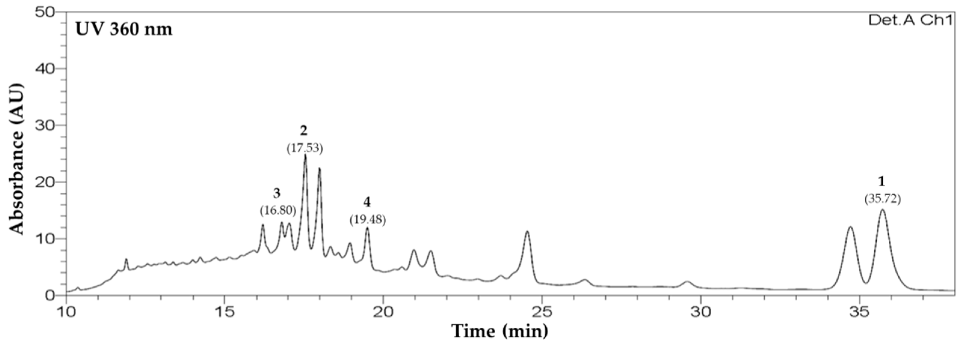
High-performance liquid chromatography (HPLC) analysis was conducted to determine the content of compounds 14 in the MeOH extract from C. lanceolata flowers. Gradient elution using H2O (+0.1% formic acid) and acetonitrile (ACN) was performed, using a XBridge C18 5 μm LC column. Peaks were detected using photo diode arrays at 360 nm. Peaks appeared at 16.80, 17.53, 19.48, and 35.72 min, and were identified as those of isoquercetin (3), leptosin (2), astragalin (4), and leptosidin (1), respectively (Figure 2). Extrapolation from the regression curve of compounds determined the content of compounds 14 in the extract from C. lanceolata flowers to be 2.8 ± 0.3 mg/g (1), 18.0 ± 0.9 mg/g (2), 3.0 ± 0.2 mg/g (3), and 10.9 ± 0.9 mg/g (4), respectively (Table 1). This analytic method was dependable for the r2 values of regression curves for all compounds were higher than 0.999. Quantitative analysis was replicated three times.

2.3. Radical Scavenging Assays for Extract, Solvent Fractions, and Flavonoids 14 Using DPPH and ABTS

The antioxidant capacities of extract (CLF), fractions (CLFE, CLFB, and CLFW), and compounds 14 of C. lanceolata flowers by the DPPH and ABTS assays are shown in Table 2. CLFE showed the highest scavenging capacities in DPPH and ABTS assay than other fractions. It was suggested that the CLFE mainly contained high amount of antioxidant flavonoids, such as leptosidin. Compounds 14 showed high radical scavenging capacities in order 1 > 3 > 2 > 4 in DPPH and ABTS radicals. A previous report showed that the radical scavenging capacities measured by DPPH and ABTS radicals depend on the numeric of hydroxy (OH) groups [26]. Leptosidin (1) showed the highest antioxidant capacity because of 3′, 4′-disubstitution of OH groups on the B-ring, forming a pyrocatechol structure, which is widely known to be the key structure with radical scavenging capacity. Compound 1 has one less sugar than others (24). The steric hindrance caused by sugars was reduced in compound 1. The aglycone of compound 3, quercetin, also has the OH group at 3′ and 4′ on the B-ring, forming a pyrocatechol structure. As shown in Table 2, the leptosidin glucoside, compound 2, and the quercetin glycoside, compound 3, showed two times higher activity than the kaempferol glycoside, compound 4.

2.4. Inhibition Effects of Compounds 14 on Intracellular Oxidative Stress in PC-12, HepG2, Caco-2, and RAW264.7 Cells

The reduction products of oxygen, i.e., reactive oxygen species (ROS), are produced by metabolic processes or external factors in normal cells in the body. Most of them have an unstable state that reaches a stable state by losing or gaining electrons. These properties are known to cause damage, various diseases, inflammation, and aging by causing oxidative stress (OS) in DNA and cell membranes in vivo [27,28]. For measuring ROS in cells, 2′,7′-dichlorofluorescein (DCFH) diacetate is used as a typical substance. It can freely cross the cell membrane. When the acetate group is removed by esterase, it is deacetylated with non-fluorescent DCFH. Deacetylated DCFH is oxidized by ROS and, as a result, becomes a strong fluorescent DCF. Compounds 14 from CLF inhibited intracellular ROS in colon epithelial (Caco-2), macrophage (RAW 264.7), and neuronal (PC-12) cells. ROS levels in all cell lines were raised by OS (200 μM H2O2), unlike their levels in control cells (Caco-2: 265.8%, RAW264.7: 188.2%, PC-12: 137.0%, and HepG2: 138.1%). After treating the cells with 10-μM isolated compounds, the results showed that all compounds significantly lowered stress induced by ROS. In particular, the decrease rates diversified by the cell line (Figure 3). In Caco-2 colon epithelial cells, aurones 1 and 2 significantly lowered oxidative stresses, compounds 13 were effectual in RAW264.7 macrophages, and all flavonoids lowered oxidative stresses in PC-12 neuronal cells. These diverse rates also showed unambiguous structure–activity relationships. In the Caco-2 cells, aurones 1 and 2 were most effectual, indicating that the two pyrocatechol structure in the A- and B-ring decreased oxidative stresses. Aurone 2, which has an additional glucose compared with aurone 1, showed a little higher capacity than aurone 1. Additionally, flavonol 3, which has an pyrocatechol structure in the B-ring compared with flavonol 4, showed a little higher capacity than flavonol 4. In the RAW264.7 cells, aurones 1, 2, and flavonol 3, i.e., more catechol structures in the B-ring, correlated with higher recovery effects. In PC-12 and HepG2 cells, all compounds recuperated intracellular ROS to the control level, and no differences caused by disparateness in the structure were observed. Structure divergence of flavonoids have dissimilar absorption and conveyance rates in cells [29,30]. Thus, flavonoids with different substituents are thought to have different levels of access and absorption in different types of cells. In addition, the flavonoids have different antioxidant capacities depending on the position or numeric of their hydroxyl (-OH) and various other structural characteristics, such as double bond, methylation (-OCH3), and number of saccharide [31,32]. In consequence, even though the aglycone compounds were the strongest radical scavenging effect, aurone 2 had a higher ability to reduce ROS in cells than 1. Thus, the ability to decrease the ROS in a particular cell depends on a combination of absorption, penetrability, and molecular antioxidant capacity [26].

2.5. Inhibition Effects of Extract, Solvent Fractions, and Flavonoids 14 on NO formation in RAW 264.7 Macrophages

2.5.1. Cytotoxicity for Extract, Fractions, and Flavonoids 14 in RAW 264.7 Macrophages

To evaluate cytotoxicity for extract, fractions, as well as isolated compounds, an MTT assay was conducted. The results showed that extract (CLF), EtOAc fraction (CLFE), n-BuOH fraction (CLFB), and H2O fraction (CLFW) had no cytotoxicity up to 100 μg/mL in RAW 264.7 cells. Furthermore, compounds 14 had no cytotoxicity up to 100 μM, either (Supplementary Materials S7).

2.5.2. Inhibition Effects of Extract, Solvent Fractions, and Flavonoids 14 on NO formation in RAW 264.7 Macrophages

Stuehr and Marletta reported that nitric oxide (NO) is produced by mouse macrophage in response to bacterial lipopolysaccharide (LPS) [33]. Although the role of NO prevents the host from the invader, such as bacteria, excessive NO production is able to cause chronic inflammation [34]. Thus, the suppression effect of extract, solvent fractions, and compounds 14 on NO formation in RAW 264.7 induced with LPS was measured. Extract and CLFE dose-dependently showed the suppression effect on NO production (Figure 4). Except for astragalin (4), all isolated compounds 13 significantly inhibited NO production. A total of 100 μM of flavonoids 14 showed the NO production by 32.9 ± 1.1%, 49.5 ± 0.7%, 62.4 ± 0.3%, and 125.7 ± 1.5%, respectively, compared with LPS-treated cells (100%) (Figure 5). A distinct structure–activity relationship was observed in this case also. Aurones 1 and 2 showed higher efficacy in inhibiting NO production than flavonols 3 and 4 because of the existence of a catechol group. In addition, flavonol 3, which has a catechol structure in the B-ring compared with flavonol 4, showed a little higher efficacy in inhibiting NO production than flavonol 4. The pyrocatechol group was previously reported to inhibit NO production through the inhibition of LPS signaling and direct scavenging of NO [35].

2.6. Effects of Ethyl Acetate Fraction (CLFE), and Flavonoids 14 on Levels of Tumor Necrosis Factor (TNF)-α, Interleukin (IL)-1β, and Interleukin (IL)-6 in LPS-Stimulated RAW264.7 Cells

Macrophages and monocytes are representative immune cells, and they are activated by components derived from invading bacteria or cytokines secreted by other immune cells in the body to induce an effective inflammatory response. Stimulation of lipopolysaccharide (LPS) secretes inflammation-inducing cytokines, such as tumor necrosis factor (TNF)-α, interleukin (IL)-6, and IL-1β from macrophages (including RAW 264.7 cells). Therefore, we assessed the inhibitory effects of CLFE (100 μg/mL) and flavonoids 14 (100 μM) on the LPS-induced production of TNF-α, IL-6, and IL-1β. RAW264.7 cells were incubated with CLFE and flavonoids 14 for 3 h and then stimulated with LPS for 24 h. Figure 6 showed that CLFE and flavonoids 14 significantly repressed TNF-α, IL-6, and IL-1β in RAW 264.7 cells.

2.7. Inhibition Effects of Extract, Solvent Fractions, and Flavonoids 14 on Expression of iNOS and COX-2 in RAW264.7 Cells

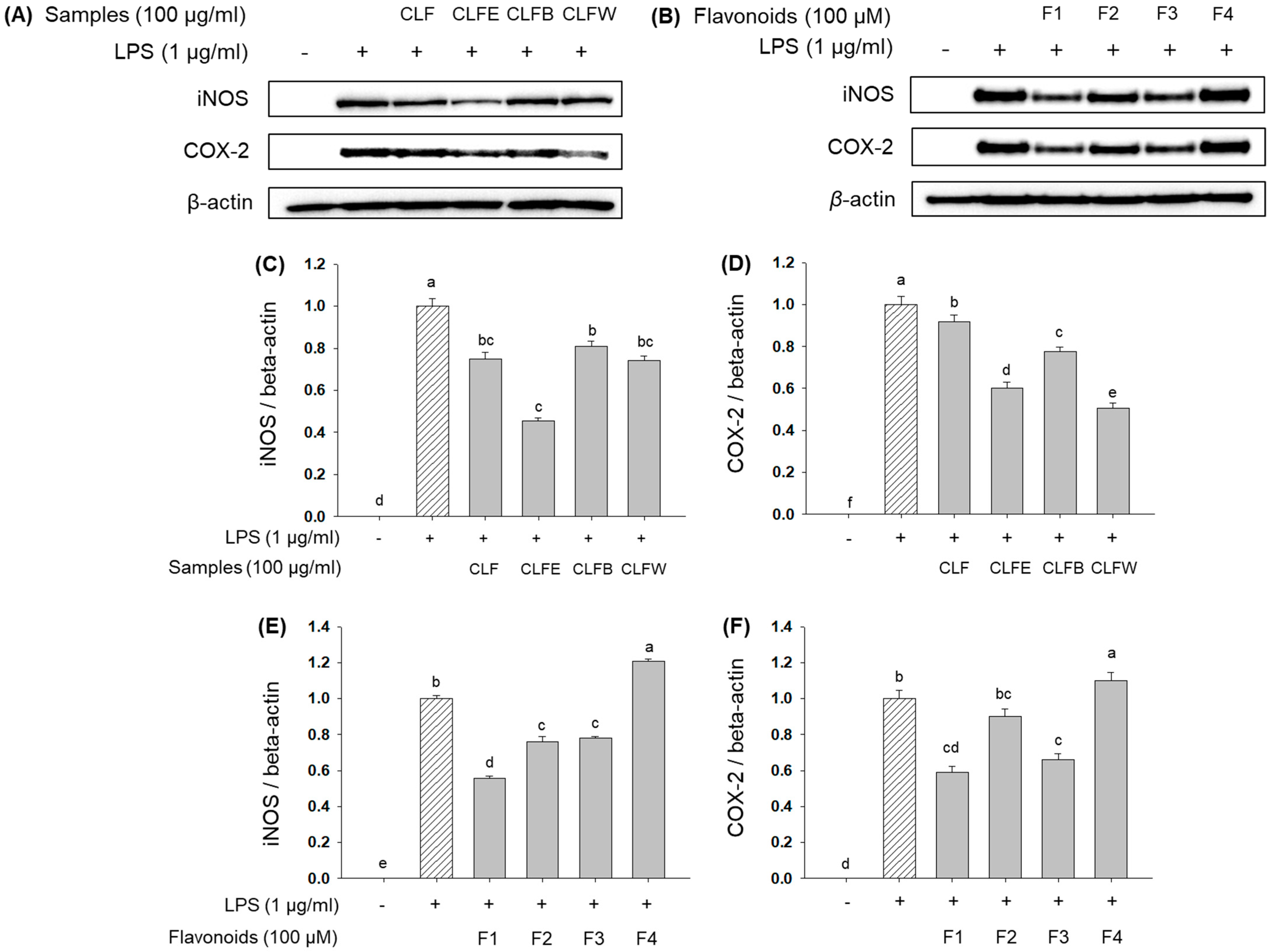
Once inflammation occurred by inflammatory stimuli, iNOS continuously produces high levels of NO [36]. Furthermore, NO activates COX-2, which induces the release of pro-inflammatory prostaglandins [37]. Because extract, CLFE, leptosidin (1), leptosin (2), and isoquercetin (3) inhibited NO production, we hypothesized that they would also reduce the production of iNOS and COX-2. As expected, they were suppressed by compounds 13. The protein levels of iNOS and COX-2 were decreased by treatments with extract (74.9 ± 3.3% and 91.6 ± 3.5%), CLFE (45.3 ± 1.7% and 60.1 ± 3.1%), CLFB (80.8 ± 2.5% and 77.5 ± 2.3%), CLFW (74.3 ± 2.1% and 50.7 ± 2.2%), leptosidin (1) (55.5 ± 1.6% and 59.1 ± 3.3%), leptosin (2) (76.2 ± 2.6% and 89.9 ± 4.4%), isoquercetin (3) (77.9 ± 1.1% and 66.2 ± 3.3%), and astragalin (4) (120.7 ± 1.3% and 110.1 ± 4.6%), compared with LPS-treated cells, respectively (Figure 7). In particular, 13 highly inhibited the expression of iNOS and COX-2 because of having catechol groups in B-ring, and compound 1 showed the highest inhibition effects because of the absence of attached sugar [38].

2.8. Protective Effects of Extract, Solvent Fractions, and Flavonoids 14 on Pancreatic Islets (PI) in Zebrafish Treated by Alloxan

Protective activity of extract, solvent fractions, and isolated flavonoids 14 was assessed against alloxan-treated PI in zebrafish. Because of their physiological resemblances to mammals, zebrafish PI was used for model type 1 and 2 diabetes with alloxan (AX), a diabetogenic material that was reported to reduce β-cell mass in PIs [39,40,41]. To assess alloxan-treated PI, the size changes of the PIs and the fluorescence intensities of NBDG-stained PIs using fluorescence microscope were analyzed. When the zebrafish larvae were exposed to alloxan, the PI size was reduced significantly by 47.8% (p = 0.0003) compared to the normal group (Figure 8A). Zebrafish larvae treated with glimepiride, a positive control, revealed recovery capacity against AX-treated PI by a 99.5% (p = 0.0047) increase compared to AX-treated group. The PI sizes in the groups treated with extract (CLF), EtOAc (CLFE), n-BuOH (CLFB), and H2O (CLFW) fractions were increased by 70.0% (p = 0.0091), 68.4% (p = 0.0065), 69.8% (p = 0.005), and 73.2% (p = 0.0083), respectively, compared to the AX-treated group (Figure 8). All of the isolated compounds also resulted in an increase in PI size with statistical significance. Flavonoids 14 (F1–F4) increased the sizes of the damaged PIs by 98.7% (p = 0.0037), 89.8% (p = 0.0012), 78.0% (p = 0.0002), and 87.9% (p = 0.0011), respectively, compared to the AX-treated group (Figure 8A). All isolated compounds in this study increased the sizes of AX-treated PIs with high levels of significance. In particular, leptosidin (1) displayed almost the same recovery effects as that of the positive control, i.e., glimepiride.

2.9. Action of Diazoxide (DZ) on Alloxan-Induced PIs in Zebrafish

In glucose-stimulated insulin secretion, metabolism of glucose in PIs is the key step [42]. Diazoxide (DZ), a KATP channel opener, was used to examine the involvement of pancreatic β-cell KATP channel stimulation activity. The PI size in the DZ-treated normal group was significantly smaller (70.4%, p = 0.0052) compared to the normal group. The AX group showed no significant differences compared to the AX + DZ-treated group. PI size in the 10-μg/mL glimepiride (GLM) + AX + DZ co-treatment groups was significantly lower (50.9%, p = 0.002) compared to the GLM + AX-treated groups. The AX + CLFW-treated groups were not significantly different after treatment with DZ, indicating that CLFW had no act as a KATP channel opener. Furthermore, groups co-treated with 1 + AX, 2 + AX, and 4 + AX were not significantly different after treatment with DZ, indicating that compounds 1, 2, and 4 did not act as a KATP channel opener. Moreover, the 3 + AX group showed significantly smaller PI sizes after treatment with DZ (61.6%, p = 0.0358) compared to the non-DZ group (Figure 8B). These results indicate that compound 3 can stimulate insulin secretion by Ca2+ influx via closure of KATP channels in β-cells.

3. Materials and Methods

3.1. Plant Materials

Coreopsis lanceolata flowers were obtained at Kyung Hee University, Yongin, Korea in 2020 and identified by Professor Dae-Keun Kim, Woosuk University, Jeonju, Korea. A voucher specimen (KHU2020-0701) was reserved at the Laboratory of Natural Products Chemistry, Kyung Hee University, Yongin, Korea.

3.2. General Experimental Procedures

The materials and equipment used for the isolation and structure determination of constituents are described in a previous study [27].

3.3. Extraciton and Isolation

Preparation procedure of MeOH extracts (CLF) and solvent fractions (Frs), ethyl acetate (CLFE 194 g), n-butanol (CLFB 254 g), and H2O (CLFW 652 g) Frs from C. lanceolata flowers and the isolation procedures of compounds 14 from CLF are presented in Figure 9 and Figure S1. The physico-chemical and spectroscopic characteristics of isolated flavonoids are presented in Table 3, Table 4 and Table 5.

3.4. Quantitative Analysis of Flavonoids 14 Using HPLC

The materials, equipment, and methods used for quantitative analysis of the isolated flavonoids from CLF are described in Supplementary Material S6. The solvent elution was graded as Table 6 and Table S6. The quantitative analysis was repeated three times.

3.5. Antioxidant Activities

3.5.1. Free Radical Scavenging Activity

The materials, equipment, and methods used for the free radical scavenging assay of extract (CLF), fractions (CLFE, CLFB, and CLFW), and flavonoids 14 from C. lanceolata flowers are described in Supplementary Material S8 and previous studies [27,43,44].

3.5.2. Cell Culture and Cytotoxicity Assessment

The materials, equipment, and methods used for the cell culture and cytotoxicity assessment of extract (CLF), fractions (CLFE, CLFB, and CLFW), and flavonoids 14 from C. lanceolata flowers are described in Supplementary Material S8 and previous studies [27,45].

3.5.3. Measurement of Intracellular Oxidative Stress

The materials, equipment, and methods used for the measurement of intracellular oxidative stress of flavonoids 14 from C. lanceolata flowers are described in Supplementary Material S8 and previous studies [27,46].

3.6. Pro-Inflammatory Inhibition Activities

3.6.1. Determination of NO Production

NO produced by RAW 264.7 cells was determined using a method described in Supplementary Material S8 and the literature [27,47].

3.6.2. Assays for IL-1β, IL-6, and TNF-α

The materials, equipment, and methods used for the assays for IL-1β, IL-6, and TNF-α of ethyl acetate fraction (CLFE), as well as the flavonoids 14 from C. lanceolata flowers, are described in Supplementary Material S8 and a previous study [47].

3.6.3. Western Blot Analysis for Protein Expression

The materials, equipment, and methods used for the western blot analysis for protein expression of extract (CLF), fractions (CLFE, CLFB, and CLFW), and flavonoids 14 from C. lanceolata flowers are described in Supplementary Material S8 and previous studies [27,47].

3.7. Antidiabetic Activity

3.7.1. Chemicals and Animals

The chemical materials used for the antidiabetic activity are described in Supplementary Material S8 and previous studies [48,49,50].

3.7.2. Animals

The animal preparation used for the antidiabetic activity are also described in Supplementary Material S8 and previous studies [48,49,50].

3.7.3. Ethics Statement

All zebrafish experimental procedures were carried out in accordance with standard zebrafish protocols and were approved by the Animal Care and Use Committee of Kyung Hee University [KHUASP(SE)-15-10].

3.7.4. Evaluation of Recovery Efficacy on Pancreatic Islet Damaged by Alloxan in Zebrafish

The materials, equipment, and methods used for evaluation of recovery efficacy of extract (CLF), fractions (CLFE, CLFB, and CLFW), and flavonoids 14 from C. lanceolata flowers on pancreatic islet damaged by alloxan in zebrafish are described in Supplementary Material S8 and a previous study [48,49,50].

3.7.5. Action of Diazoxide on Alloxan-Induced Diabetic Zebrafish

The materials, equipment, and methods used for action of diazoxide on alloxan-induced diabetic zebrafish are described in supplementary material S8 and previous studies [48,49,50].

3.8. Statistical Anlaysis

Results (mean ± SD, n = 3) were assessed using one-way analysis of variance and the Tukey–Kramer honestly significant difference (HSD) test, whereby p < 0.05 was considered to represent statistical significance. All statistical analyses were performed using SPSS 22.0 (SPSS Inc., Chicago, IL, USA).

4. Conclusions

We carried out this research to find other pharmacological active compounds from C. lanceolata flowers. Two aurones and two flavonol glucosides were isolated through repeated column chromatography using silica gel (SiO2), octadecyl SiO2 (ODS), and Sephadex LH-20 resins, and were identified by analysis of UV, IR, NMR, and MS data. Leptosidin (1) exhibited high DPPH and ABTS radical scavenging activity. All four flavonoids showed powerful antioxidation by reducing oxidative stress in intestinal epithelial cells (Caco-2), macrophage cells (RAW264.7), neuron cells (PC-12), and hepatic cells (HepG2). Extract, EtOAc fraction, and flavonoids 13 inhibited NO production and extract, EtOAc, n-BuOH, H2O fractions, and flavonoids 13 suppressed iNOS protein expression in RAW 264.7 cells treated with LPS. Additionally, EtOAc, n-BuOH, H2O fractions, and flavonoids 1 and 3 decreased COX-2 protein expression. Extract, EtOAc fraction, and flavonoids 1 and 3 were notably capable of inhibiting the pro-inflammatroy cytokines (TNF-α, IL-1β, and IL-6). Furthermore, H2O fraction and all flavonoids 14 recovered the alloxan-treated PI in zebrafish with high levels of significance. In particular, isoquercetin (3) stimulated insulin secretion by a Ca2+ influx via closure of KATP channels in pancreatic β-cells. The data indicate that the fractions and compounds from C. lanceolata flowers can activate the potential antioxidant effect, immune response by inhibiting excessive inflammation, and a recovery effect on damaged pancreatic β–cells. These results indicate that C. lanceolata flowers and its isolated flavonoids are used as potential anti-oxidant, pro-inflammatory inhibition, and anti-diabetic agents.

Supplementary Materials

The following are available online, The isolation procedure methods (S1), 1H-NMR and 13C-NMR of compounds 14 (S2~S5), the materials, equipment, and methods used for quantitative analysis of the isolated flavonoids from Coreopsis lanceolata flowers (S6), cell viability test of extracts, fractions, and compounds 14 (S7), and the methods and materials of antioxidant, pro-inflammatory inhibition, and anti-diabetic assay (S8) are available as supplementary materials.

Author Contributions

Conceptualization and visualization: H.-G.K., D.Y.L., and N.-I.B.; Material preparation: H.-G.K., S.M.O., T.N.N., and N.-I.B.; Isolate compounds: H.-G.K., M.-H.L., and T.N.N.; Identify compounds (UV, IR, NMR, MS): H.-G.K. and N.-I.B.; Antioxidative assay: Y.S.J. and D.-O.K.; HPLC analysis: H.-G.K., S.M.O., and D.Y.L.; Pro-inflammatory inhibition assay: Y.S.J.; Anti-diabetic assay: Y.H.N. and T.H.K.; Writing—original draft preparation: H.-G.K.; Writing—review and editing: H.-G.K., D.-O.K., D.Y.L. and N.-I.B.; Funding acquisition: D.Y.L. and N.-I.B. All authors have read and agreed to the published version of the manuscript.

Funding

Rural Development Administration (RDA), Republic of Korea (Project no. PJ01420403) funded this study.

Institutional Review Board Statement

Not applicable.

Informed Consent Statement

Not applicable.

Data Availability Statement

Data is contained within the article.

Acknowledgments

This work was supported by the “Cooperative Research Program for Agriculture Science & Technology Development” (Project no. PJ01420403), Rural Development Administration, Republic of Korea.

Conflicts of Interest

The authors declare no conflict of interest.

Sample Availability

Samples of the compounds 14 are available from the authors.

References

  1. Kim, S.G.; Choi, D.S. Epidemiology and current status of diabetes in Korea. Hanyang Med. Rev. 2009, 29, 122–129. [Google Scholar]
  2. Akhtar, K.; Shah, S.W.A.; Shah, A.A.; Shoaib, M.; Haleem, S.K.; Sultana, N. Pharmacological effect of Rubus ulmifolius Schott as antihyperglycemic and antihyperlipidemic on streptozotocin (STZ)-induced albino mice. Appl. Biol. Chem. 2017, 60, 411–418. [Google Scholar] [CrossRef]
  3. Kerner, W.; Brückel, J. German Diabetes Association. Definition, classification and diagnosis of diabetes mellitus. Exp. Clin. Endocrinol. Diabetes 2014, 122, 384–386. [Google Scholar]
  4. Petersmann, A.; Müller-Wieland, D.; Müller, U.A.; Landgraf, R.; Nauck, M.; Freckmann, G.; Heinemann, L.; Schleicher, E. Definition, Classification and diagnosis of diabetes mellitus. Exp. Clin. Endocrinol. Diabetes 2019, 127, S1–S7. [Google Scholar] [CrossRef] [Green Version]
  5. Gillespie, K.M. Type 1 diabetes: Pathogenesis and prevention. CMAJ. 2006, 175, 165–170. [Google Scholar] [CrossRef] [Green Version]
  6. Romero Aroca, P.; Mendez Marin, I.; Baqet Bermaldiz, M.; Fernendez Ballart, J.; Santos Blanco, E.I. Review of the relationship between renal and retinal microangiopathy in diabetes mellitus patients. Curr. Diabet. Rev. 2010, 6, 88–101. [Google Scholar] [CrossRef] [PubMed]
  7. Toeller, M. Diet therapy of diabetes mellitus. Fortschr. Med. 1991, 109, 41–42, 45. [Google Scholar] [PubMed]
  8. Koivisto, V.A. Insulin therapy in type II diabetes. Diabet. Care 1993, 3 (Suppl. 16), 29–39. [Google Scholar] [CrossRef]
  9. Derosa, G.; Maffioli, P. α-Glucosidase inhibitors and their use in clinical practice. Arch. Med. Sci. 2012, 8, 899–906. [Google Scholar] [CrossRef]
  10. Standl, E.; Schnell, O. Alpha-glucosidase inhibitors 2012—Cardiovascular considerations and trial evaluation. Diab. Vasc. Dis. Res. 2012, 9, 163–169. [Google Scholar] [CrossRef]
  11. Seghrouchni, I.; Drai, J.; Bannier, E.; Rivière, J.; Calmard, P.; Garcia, I.; Orgiazzi, J.; Revol, A. Oxidative stress parameters in type I, type II and insulin-treated type 2 diabetes mellitus; insulin treatment efficiency. Clin. Chim. Acta 2002, 321, 89–96. [Google Scholar] [CrossRef]
  12. Solinas, G.; Vilcu, C.; Nells, J.G.; Bandyopadhyay, G.K.; Luo, J.L.; Naugler, W.; Grivennikov, S.; Wynshaw-Boris, A.; Scadeng, M.; Olefsky, J.M.; et al. JNK1 in hematopoietically derived cells contributes to diet-induced inflammation and insulin resistance without affecting obesity. Cell Metab. 2007, 6, 386–397. [Google Scholar] [CrossRef] [Green Version]
  13. Vascotto, S.G.; Beckham, Y.; Kelly, G.M. The zebrafish’s swim to fame as an experimental model in biology. Biochem. Cell Biol. 1997, 75, 479–485. [Google Scholar] [CrossRef]
  14. Parng, C.; Seng, W.L.; Semino, C.; McGrath, P.I. Zebrafish: A preclinical model for drug screening. Assay Drug Dev. Technol. 2002, 1, 41–48. [Google Scholar] [CrossRef] [PubMed]
  15. Spence, R.; Gerlach, G.; Lawrence, C.; Smith, C. The behaviour and ecology of the zebrafish, Danio rerio. Biol. Rev. 2008, 83, 13–34. [Google Scholar] [CrossRef]
  16. Paciorek, T.; Mcrobert, S. Daily variation in the shoaling behaviour of zebrafish Danio rerio. Curr. Zool. 2012, 58, 129–137. [Google Scholar] [CrossRef] [Green Version]
  17. Tanimoto, S.; Miyazawa, M.; Inoue, T.; Okada, Y.; Nomura, M. Chemical constituents of Coreopsis lanceolata L. and their physiological activities. J. Oleo. Sci. 2009, 58, 141–146. [Google Scholar] [CrossRef] [PubMed] [Green Version]
  18. Shao, D.; Zheng, D.; Hu, R.; Chen, W.; Chen, D.; Zhuo, X. Chemical constituents from Coreopsis lanceolata. Zhongcaoyao 2013, 44, 1558–1561. [Google Scholar]
  19. Pardede, A.; Mashita, K.; Ninomiya, M.; Tanaka, K.; Koketsu, M. Flavonoid profile and antileukemic activity of Coreopsis lanceolata flowers. Bioorg. Med. Chem. Lett. 2016, 26, 2784–2787. [Google Scholar] [CrossRef] [PubMed]
  20. Kimura, Y.; Hiraoka, K.; Kawano, T.; Fujioka, S.; Shimada, A. Nematicidal activities of acetylene compounds from Coreopsis lanceolata L. J. Biosci. 2008, 63, 843–847. [Google Scholar] [CrossRef]
  21. Shang, Y.F.; Oidovsambuu, S.; Jeon, J.S.; Nho, C.W.; Um, B.H. Chalcones from the flowers of Coreopsis lanceolata and their in vitro antioxidative activity. Planta Med. 2013, 79, 295–300. [Google Scholar] [CrossRef] [Green Version]
  22. Heyl Frederick, W. The yellow coloring substances of ragweed pollen. J. Am. Chem. Soc. 1919, 41, 1285–1289. [Google Scholar] [CrossRef] [Green Version]
  23. Nakabayashi, T. Thiamine-decomposing activity of flavonoid pigments of Pteridium aquilinum. Bitamin 1955, 8, 410–414. [Google Scholar]
  24. Okada, Y.; Okita, M.; Murai, Y.; Okano, Y.; Nomura, M. Isolation and identification of flavonoids from Coreopsis lanceolata L. petals. Nat. Prod. Res. 2014, 28, 201–204. [Google Scholar] [CrossRef]
  25. Nigam, S.S.; Saxena, V.K. Isolation and study of the aurone glycoside leptosin from the leaves of Flemengia strobilifera. Planta Med. 1975, 27, 98–100. [Google Scholar] [CrossRef]
  26. Geissman, T.A.; Heaton, C.D. Anthochlor pigments. IV. The pigments of Coreopsis grandiflora Nutt. 1. J. Am. Chem. Soc. 1943, 65, 677–683. [Google Scholar] [CrossRef]
  27. Kim, H.G.; Jung, Y.S.; Oh, S.M.; Oh, H.J.; Ko, J.H.; Kim, D.O.; Kang, S.C.; Lee, Y.G.; Lee, D.Y.; Baek, N.I. Coreolanceolins A–E, new flavanones from the flowers of Coreopsis lanceolate, and their antioxidant and anti-inflammatory effects. Antioxidants 2020, 9, 539. [Google Scholar] [CrossRef]
  28. Aniya, Y.; Naito, A. Oxidative stress-induced activation of microsomal glutathione S-transferase in isolated rat liver. Biochem. Pharmacol. 1993, 45, 37–42. [Google Scholar] [PubMed]
  29. Beckman, K.B.; Ames, B.N. The free radical theory of aging matures. Physiol. Rev. 1998, 78, 547–581. [Google Scholar] [CrossRef] [PubMed] [Green Version]
  30. Fang, Y.; Cao, W.; Xia, M.; Pan, S.; Xu, X. Study of structure and permeability relationship of flavonoids in Caco-2 cells. Nutrients 2017, 9, 1301. [Google Scholar] [CrossRef] [PubMed] [Green Version]
  31. Fang, Y.; Liang, F.; Liu, K.; Qaiser, S.; Pan, S.; Xu, X. Structure characteristics for intestinal uptake of flavonoids in Caco-2 cells. Food Res. Int. 2018, 105, 353–360. [Google Scholar] [CrossRef]
  32. Rice-Evans, A.C.; Miller, J.N.; Paganga, G. Structure-antioxidant activity relationships of flavonoids and phenolic acids. Free Radic. Biol. Med. 1996, 20, 933–956. [Google Scholar] [CrossRef]
  33. Stuehr, D.J.; Marletta, M.A. Mammalian nitrate biosynthesis: Mouse macrophages produce nitrite and nitrate in response to Escherichia coli lipopolysaccharide. Proc. Natl. Acad. Sci. USA 1985, 82, 7738–7742. [Google Scholar] [CrossRef] [Green Version]
  34. Stichtenoth, D.O.; Frolich, J.C. Nitric oxide and inflammatory joint diseases. Br. J. Rheumatol. 1998, 37, 246–257. [Google Scholar] [CrossRef] [Green Version]
  35. Bors, W.; Heller, W.; Michel, C.; Saran, M. Flavonoids as antioxidants: Determination of radical-scavenging efficiencies. Methods Enzymol. 1990, 186, 343–354. [Google Scholar]
  36. Bal-Price, A.; Matthias, A.; Brown, G.C. Stimulation of the NADPH oxidase in activated rat microglia removes nitric oxide but induces peroxynitrite production. J. Neurochem. 2002, 80, 73–80. [Google Scholar] [CrossRef] [PubMed] [Green Version]
  37. Salvemini, D.; Misko, T.P.; Masferrer, J.L.; Seibert, K.; Currie, M.G.; Needleman, P. Nitric oxide activates cyclooxygenase enzymes. Proc. Natl. Acad. Sci. USA 1993, 90, 7240–7244. [Google Scholar] [CrossRef] [Green Version]
  38. Zheng, L.T.; Ryu, G.M.; Kwon, B.M.; Lee, W.H.; Suk, K. Anti-inflammatory effects of catechols in lipopolysaccharide-stimulated microglia cells: Inhibition of microglial neurotoxicity. Eur. J. Pharmacol. 2008, 588, 106–113. [Google Scholar] [CrossRef] [PubMed]
  39. Elo, B.; Villano, C.M.; Govorko, D.; White, L.A. Larval zebrafish as a model for glucose metabolism: Expression of phosphoenolpyruvate carboxykinase as a marker for exposure to anti-diabetic compounds. J. Mol. Endocrinol. 2007, 38, 433–440. [Google Scholar] [CrossRef] [PubMed] [Green Version]
  40. Seo, K.H.; Nam, Y.H.; Lee, D.Y.; Ahn, E.M.; Kang, T.H.; Baek, N.I. Recovery effect of phenylpropanoid glycosides from Magnolia obovate fruit on alloxan-induced pancreatic islet damage in zebrafish (Danio rerio). Carbohydr. Res. 2015, 416, 70–74. [Google Scholar] [CrossRef]
  41. Desgraz, R.; Bonal, C.; Herrera, P.L. β-Cell regeneration: The pancreatic intrinsic faculty. Trends Endocrinol. Metab. 2011, 22, 34–43. [Google Scholar] [CrossRef] [Green Version]
  42. Miki, T.; Nagashima, K.; Seino, S. The structure and function of the ATP-sensitive K+ channel in insulin-secreting pancreatic β-cells. J. Mol. Endocrinol. 1999, 22, 113–123. [Google Scholar] [CrossRef] [PubMed]
  43. Kim, D.O.; Lee, K.W.; Lee, H.J.; Lee, C.Y. Vitamin C equivalent antioxidant capacity (VCEAC) of phenolic phytochemicals. J. Agric. Food Chem. 2002, 50, 3713–3717. [Google Scholar] [CrossRef] [PubMed]
  44. Brand-Williams, W.; Cuvelier, M.E.; Berset, C. Use of free radical method to evaluate antioxidant activity. LWT-Food Sci. Technol. 1995, 28, 25–30. [Google Scholar] [CrossRef]
  45. Lim, J.S.; Hahn, D.; Gu, M.J.; Oh, J.; Lee, J.S.; Kim, J.S. Anti-inflammatory and antioxidant effects of 2,7-dihydroxy-4, 6-dimethoxy phenanthrene isolated from Dioscorea batatas Decne. Appl. Biol. Chem. 2019, 62, 29. [Google Scholar] [CrossRef]
  46. Wang, H.; Joseph, J.A. Quantifying cellular oxidative stress by dichlorofluorescein assay using microplate reader. Free Radic. Biol. Med. 1999, 27, 612–616. [Google Scholar] [CrossRef]
  47. Choi, B.R.; Kim, H.G.; Ko, W.; Dong, L.; Yoon, D.; Oh, S.M.; Lee, Y.S.; Lee, D.S.; Baek, N.I.; Lee, D.Y. Noble 3,4-Seco-triterpenoid glycosides from the fruits of Acanthopanax sessiliflorus and their anti-neuroinflammatory effects. Antioxidants 2021, 10, 1334. [Google Scholar] [CrossRef]
  48. Nam, Y.H.; Hong, B.N.; Rodriguez, I.; Park, M.S.; Jeong, S.Y.; Lee, Y.G.; Shim, J.H.; Yasmin, T.; Kim, N.W.; Koo, Y.T.; et al. Steamed ginger may enhance insulin secretion through KATP channel closure in pancreatic β-Cells potentially by increasing 1-dehydro-6-gingerdione content. Nutrients 2020, 12, 324. [Google Scholar] [CrossRef] [PubMed] [Green Version]
  49. Ko, J.H.; Rodriguez, I.; Joo, S.W.; Kim, H.G.; Lee, Y.G.; Kang, T.H.; Baek, N.I. Synergistic effect of two major components of Malva verticillata in the recovery of alloxan-damaged pancreatic islet cells in zebrafish. J. Med. Food 2019, 22, 196–201. [Google Scholar] [CrossRef] [PubMed]
  50. Ko, J.H.; Nam, Y.H.; Joo, S.W.; Kim, H.G.; Lee, Y.G.; Kang, T.H.; Baek, N.I. Flavonoid 8-O-glucuronides from the aerial parts of Malva verticillata and their recovery effects on alloxan-induced pancreatic islets in zebrafish. Molecules 2018, 23, 833. [Google Scholar] [CrossRef] [Green Version]
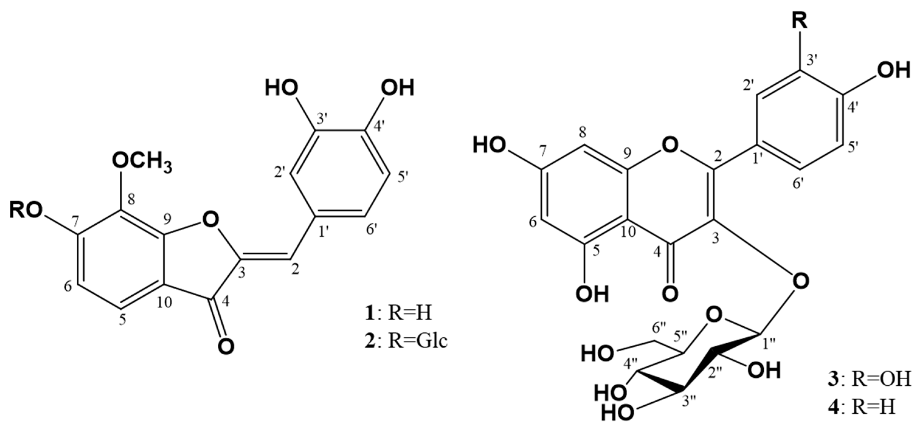
Figure 1. Molecular structures of compounds 14 from Coreopsis lanceolata flowers.
Molecules 26 06098 g001
Figure 2. HPLC chromatograms from Coreopsis lanceolata L. flowers.
Molecules 26 06098 g002
Figure 3. Recovery effects of flavonoids 14 (F1~F4) on intracellular oxidative stress in: (A) Caco-2 epithelial cells; (B) RAW264.7 macrophages; (C) PC-12 neurons; (D) HepG2 hepatocytes. Lowercase letters on the bars indicate significant differences according to the Tukey–Kramer HSD test (p < 0.05).
Molecules 26 06098 g003
Figure 4. Inhibition effect of extract and solvent fractions on nitric oxide (NO) production in RAW 264.7 cells: (A) 25 µg/mL; (B) 50 μg/mL; (C) 100 μg/mL. Lowercase letters on the bars indicate significant differences according to the Tukey–Kramer HSD test (p < 0.05). CLF: C. lanceolata flowers 80% methanol extract, CLFE: C. lanceolata flowers EtOAc fraction, CLFB: C. lanceolata flowers n-BuOH fraction, CLFW: C. lanceolata flowers H2O fraction.
Molecules 26 06098 g004
Figure 5. Inhibition effect of flavonoids 14 (F1~F4) on nitric oxide (NO) production in RAW 264.7 cells: (A) 25 μM; (B) 50 μM; (C) 100 μM. Lowercase letters on the bars indicate significant differences according to the Tukey–Kramer HSD test (p < 0.05).
Molecules 26 06098 g005
Figure 6. Effects of ethyl acetate fraction (CLFE), and flavonoids 14 on levels of tumor necrosis TNF-α (A), IL-1β (B), and IL-6 (C) inlipopolysaccharide (LPS)-stimulated RAW264.7 cells. Cells were pretreated for 3 h with indicated concentrations of samples and stimulated for 24 h with LPS (1 μg/mL). ELISA analysis was performed, as described in Materials and Methods. Lowercase letters on the bars indicate significant differences according to the Tukey–Kramer HSD test (p < 0.05).
Molecules 26 06098 g006
Figure 7. Inhibition effect of extract, solvent fractions, and flavonoids 14 (F1–F4) on the expression of iNOS and COX-2 in RAW264.7 cells: (A) Immunoblotting method for extract and solvent fractions; (B) Immunoblotting method for flavonoids 14 (F1–F4); (C) iNOS/β-actin ratio for extract and solvent fractions; (D) COX-2/β-actin ratio for extract and solvent fractions; (E) iNOS/β-actin ratio for flavonoids 14 (F1–F4); (F) COX-2/β-actin ratio for flavonoids 14 (F1~F4). Lowercase letters on the bars indicate significant differences according to the Tukey–Kramer HSD test (p < 0.05). CLF: C. lanceolata flowers 80% methanol extract, CLFE: C. lanceolata flowers EtOAc fraction, CLFB: C. lanceolata flowers n-BuOH fraction, CLFW: C. lanceolata flowers H2O fraction.
Molecules 26 06098 g007
Figure 8. Protective effect of extract, solvent fractions, and flavonoids 14 from Coreopsis lanceolata flowers on damaged PIs in zebrafish treated by alloxan and action of diazoxide (DZ) for PIs damaged by alloxan (AX) in zebrafish. (A) Size of the PIs for protective effects; (B) Size of the PIs for action of DZ; (C) PI image: NOR: normal group, AX: alloxan group, GLM: glimepiride + AX group, CLF: extract + AX, CLFE: EtOAc fraction + AX, CLFB: n-BuOH fraction + AX, CLFW: H2O fraction + AX, F1–4: flavonoids 14 + AX. (### p < 0.001, ## p < 0.01; compared to the normal group), (* p < 0.05, ** p < 0.01, *** p < 0.001 compared to the alloxan-treated group). Scale bar = 100 μm.
Molecules 26 06098 g008
Figure 9. Isolation procedures of compounds 14 from Coreopsis lanceolata flowers. fr.: fraction; CC: column chromatography; SiO2: silica gel; ODS: octadecyl silica gel.
Molecules 26 06098 g009
Table 1. Quantitative HPLC analysis for the contents of flavonoids 14 in MeOH extracts from Coreopsis lanceolata flowers.
FlavonoidsRT 1Regression Equationr2C 2
135.72y = 31643x + 257020.99952.8 ± 0.3
217.53y = 2369.8x + 680.750.999818.0 ± 0.9
316.80y = 7887x − 610480.99903.0 ± 0.2
419.48y = 1620.4x − 101010.999110.9 ± 0.9
1 Retention time (min); 2 Contentrations in extracts from C. lanceolata flowers (mg/g).
Table 2. DPPH and ABTS radical-scavenging activity of extract, solvent fractions, and flavonoids 14 from Coreopsis lanceolata flowers. CLF: C. lanceolata flowers 80% methanol extract, CLFE: C. lanceolata flowers EtOAc fraction, CLFB: C. lanceolata flowers n-BuOH fraction, CLFW: C. lanceolata flowers H2O fraction.
SamplesDPPH Radical
(mg VCE/100g DW) 1
ABTS Radical
(mg VCE/100g DW) 1
182.5 ± 5.3 a21084.4 ± 0.3 a
238.7 ± 3.2 b551.7 ± 0.8 b
322.7 ± 1.9 d439.2 ± 0.5 c
418.9 ± 2.0 e306.5 ± 0.2 d
CLF18.8 ± 0.8 e306.4 ± 0.3 d
CLFE32.8 ± 1.6 c549.9 ± 2.0 b
CLFB12.2 ± 2.1 f223.1 ± 0.7 e
CLFW6.5 ± 0.6 g189.4 ± 0.1 f
1 Milligrams of vitamin C equivalent (VCE)/100g of dry weight; 2 Data are presented as the mean ± standard deviation (n = 3); Means with different superscripts in the same column differ significantly different by Tukey-Kramer’s HSD test (p < 0.05).
Table 3. Physico-chemical and spectroscopic characteristics of flavonoids from Coreopsis lanceolata flowers.
Comp.1Crystals Characteristicsm.p. (°C)[α]D 2FAB/MS 3IR 4
1Red amorphous powder252−254-301 [M + H]+3366, 1661, 1604
2Red amorphous powder229−231−62.3°463 [M + H]+3360, 1659, 1617
3Yellow amorphous powder230−233−66.2°465 [M + H]+3366, 1660, 1607, 1501
4Yellow amorphous powder218−220−69.9°449 [M + H]+3364, 1656, 1607, 1506
1 Compound; 2 c = 0.10, CH3OH; 3 positive, m/z; 4 CaF2, v, cm−1.
Table 4. 1H NMR data for compounds 14. δH in ppm, coupling pattern, J in Hz.
No. of H1 a2 b3 c4 c
26.70, s6.70, s--
57.33, d, 9.07.41, d, 8.4--
66.72, d, 9.07.09, d, 8.46.24, br. s6.69, br. s
8--6.32, br. s6.71, br. s
2′7.46, d, 1.27.45, d, 1.27.70, br. s8.45, d, 8.8
3′---7.19, d, 8.8
5′6.83, d, 8.47.10, d, 8.06.88, d, 8.07.19, d, 8.8
6′7.26, dd, 8.4, 1.27.26, dd, 8.0, 1.27.57, br. d, 8.08.45, d, 8.8
8-OCH34.18, s4.12, s--
1″-5.10, d, 7.05.18, d, 7.06.21, d, 7.0
2″-3.57, dd, 7.0, 7.03.55, dd, 7.0, 7.04.20 #
3″-3.50 #3.49, dd, 7.0, 7.04.19 #
4″-3.45, dd, 7.0, 7.03.43 #4.01, dd, 7.0, 7.0
5″-3.49 #3.47 #4.18 #
6″-3.92, dd, 12.0, 1.2
3.75, dd, 12.0, 5.2
3.98, br. d, 12.0
3.84, dd, 12.0, 5.6
4.35, br. d, 12.0
4.21, dd, 12.0, 5.6
a CD3OD, 600 MHz; b CD3OD, 400 MHz; c pyridine-d5, 400 MHz; # overlapped.
Table 5. 13C NMR data for compounds 14. δC in ppm.
No. of C1 a2 b3 c4 c
2115.23115.81158.91161.25
3147.62147.09135.66135.64
4184.64184.73179.32181.33
5120.81120.11160.17157.21
6114.70113.7896.7594.16
7159.62158.11165.98166.01
8133.94136.1798.5299.46
9159.92159.42159.95165.52
10114.87115.55105.66103.71
8-OCH361.7261.88--
1′125.63125.82123.23121.62
2′119.26118.97115.99131.44
3′146.89146.92145.82115.63
4′149.71149.87150.00164.17
5′116.78116.61117.60115.59
6′126.42126.75123.21131.41
1″-102.54105.64106.12
2″-74.7975.1375.69
3″-78.4578.3578.56
4″-71.2371.1171.31
5″-77.9178.0078.01
6″-62.4262.3762.36
a CD3OD, 150 MHz; b CD3OD, 100 MHz; c pyridine-d5, 100 MHz.
Table 6. Solvent gradient for quantitative analysis of isolated flavonoids from Coreopsis lanceolata flowers using HPLC.
Time (min)Flow rate (mL/min)Solvent A (%)Solvent B (%)
01955
31955
618515
1218020
3518020
3711090
401955
451955
Publisher’s Note: MDPI stays neutral with regard to jurisdictional claims in published maps and institutional affiliations.