
Rhytidhylides A and B, Two New Phthalide Derivatives from the Endophytic Fungus Rhytidhysteron sp. BZM-9

Abstract
:
1. Introduction
2. Results and Discussion
3. Materials and Methods
3.1. General Experimental Procedures
3.2. Fungal Material
3.3. Fermentation, Extraction and Isolation of Compounds
3.4. X-ray Crystallographic Analysis
3.5. Quantum Chemistry Calculations
3.6. Cytotoxic Activity Assay
3.7. Antimicrobial Activity Assay
4. Conclusions
Supplementary Materials
Author Contributions
Funding
Institutional Review Board Statement
Informed Consent Statement
Data Availability Statement
Conflicts of Interest
Sample Availability
References
- Kuzniar, A.; Wlodarczyk, K.; Wolinska, A. Agricultural and other biotechnological applications resulting from trophic plant-endophyte interactions. Agronomy 2019, 9, 779. [Google Scholar] [CrossRef] [Green Version]
- Leon, A.; Del-Angel, M.; Avila, J.L.; Delgado, G. Phthalides: Distribution in nature, chemical reactivity, synthesis, and biological activity. Prog. Chem. Org. Nat. Prod. 2017, 104, 127–246. [Google Scholar] [CrossRef]
- Brady, S.F.; Wagenaar, M.M.; Singh, M.P.; Janso, J.E.; Clardy, J. The cytosporones, new octaketide antibiotics isolated from an endophytic fungus. Org. Lett. 2000, 2, 4043–4046. [Google Scholar] [CrossRef]
- Katoh, N.; Nakahata, T.; Kuwahara, S. Synthesis of novel antifungal phthalides produced by a wheat rhizosphere fungus. Tetrahedron 2008, 64, 9073–9077. [Google Scholar] [CrossRef]
- Miyazawa, M.; Tsukamoto, T.; Anzai, J.; Ishikawa, Y. Insecticidal effect of phthalides and furanocoumarins from Angelica acutiloba against Drosophila melanogaster. J. Agric. Food Chem. 2004, 52, 4401–4405. [Google Scholar] [CrossRef] [PubMed]
- Mullady, E.L.; Millett, W.P.; Yoo, H.D.; Weiskopf, A.S.; Chen, J.; DiTullio, D.; Knight-Connoni, V.; Hughes, D.E.; Pierceall, W.E. A phthalide with in vitro growth inhibitory activity from an oidiodendron strain. J. Nat. Prod. 2004, 67, 2086–2089. [Google Scholar] [CrossRef] [PubMed]
- Tianpanich, K.; Prachya, S.; Wiyakrutta, S.; Mahidol, C.; Ruchirawat, S.; Kittakoop, P. Radical scavenging and antioxidant activities of isocoumarins and a phthalide from the endophytic fungus Colletotrichum sp. J. Nat. Prod. 2011, 74, 79–81. [Google Scholar] [CrossRef] [PubMed]
- Mahajan, V.K.; Sharma, V.; Prabha, N.; Thakur, K.; Sharma, N.L.; Rudramurthy, S.M.; Chauhan, P.S.; Mehta, K.S.; Abhinav, C. A rare case of subcutaneous phaeohyphomycosis caused by a Rhytidhysteron species: A clinico-therapeutic experience. Int. J. Dermatol. 2014, 53, 1485–1489. [Google Scholar] [CrossRef] [PubMed]
- Pudhom, K.; Teerawatananond, T.; Chookpaiboon, S. Spirobisnaphthalenes from the mangrove-derived fungus Rhytidhysteron sp. AS21B. Mar. Drugs 2014, 12, 1271–1280. [Google Scholar] [CrossRef] [Green Version]
- Pudhom, K.; Teerawatananond, T. Rhytidenones A-F, Spirobisnaphthalenes from Rhytidhysteron sp. AS21B, an endophytic fungus. J. Nat. Prod. 2014, 77, 1962–1966. [Google Scholar] [CrossRef]
- Chokpaiboon, S.; Choodej, S.; Boonyuen, N.; Teerawatananond, T.; Pudhom, K. Highly oxygenated chromones from mangrove-derived endophytic fungus Rhytidhysteron rufulum. Phytochemistry 2016, 122, 172–177. [Google Scholar] [CrossRef] [PubMed]
- Siridechakorn, I.; Yue, Z.W.; Mittraphab, Y.; Lei, X.G.; Pudhom, K. Identification of spirobisnaphthalene derivatives with anti-tumor activities from the endophytic fungus Rhytidhysteron rufulum AS21B. Bioorgan. Med. Chem. 2017, 25, 2878–2882. [Google Scholar] [CrossRef] [PubMed]
- Zhang, S.; Kang, F.H.; Tan, J.B.; Chen, D.K.; Kuang, M.; Wang, W.X.; Xu, K.P.; Zou, Z.X. (±)-Rhytidhymarins A and B, two pairs of new isocoumarin derivatives from endophytic fungus Rhytidhysteron sp. BZM-9. New J. Chem. 2021, 45, 12700–12704. [Google Scholar] [CrossRef]
- Zhang, S.; Wang, W.X.; Tan, J.B.; Kang, F.H.; Chen, D.K.; Xu, K.P.; Zou, Z.X. Rhytidhyesters A-D, four new chlorinated cyclopentene derivatives from the endophytic fungus Rhytidhysteron sp. BZM-9. Planta Med. 2021, 87, 489–497. [Google Scholar]
- Klaiklay, S.; Rukachaisirikul, V.; Aungphao, W.; Phongpaichit, S.; Sakayaroj, J. Depsidone and phthalide derivatives from the soil-derived fungus Aspergillus unguis PSU-RSPG199. Tetrahedron Lett. 2016, 57, 4348–4351. [Google Scholar] [CrossRef]
- Sommart, U.; Rukachaisirikul, V.; Tadpetch, K.; Sukpondma, Y.; Phongpaichit, S.; Hutadilok-Towatana, N.; Sakayaroj, J. Modiolin and phthalide derivatives from the endophytic fungus Microsphaeropsis arundinis PSU-G18. Tetrahedron 2012, 68, 10005–10010. [Google Scholar] [CrossRef]
- Tang, Y.J.; Wang, Y.M.; Wang, Z.H.; Zhang, J.; Qin, J.C.; Wang, Q.K.; Yang, L.H.; Xu, L.Z.; Ding, Y.L.; Guo, Y.; et al. Novel antimicrobial metabolites produced by Sika deer-associated Actinomyces sp. JN411010. Nat. Prod. Res. 2013, 27, 2183–2189. [Google Scholar] [CrossRef]
- Martin, J.A.; Vogel, E. The synthesis of zinniol. Tetrahedron 1980, 36, 791–794. [Google Scholar] [CrossRef]
- Kimura, Y.; Mizuno, T.; Nakajima, H.; Hamasaki, T. Altechromones A and B, new plant growth regulators produced by the fungus, Alternaria sp. Biosci. Biotech. Biochem. 1992, 56, 1664–1665. [Google Scholar] [CrossRef] [Green Version]
- Xu, W.F.; Chen, G.; Li, Z.Q.; Lu, X.; Pei, Y.H. Isolation and identification of chemical constituents from Rheum palmatum L. Shenyang Yaoke Daxue Xuebao 2013, 30, 837–839. [Google Scholar]
- Khamthong, N.; Rukachaisirikul, V.; Tadpetch, K.; Kaewpet, M.; Phongpaichit, S.; Preedanon, S.; Sakayaroj, J. Tetrahydroanthraquinone and xanthone derivatives from the marine-derived fungus Trichoderma aureoviride PSU-F95. Arch. Pharm. Res. 2012, 35, 461–468. [Google Scholar] [CrossRef]
- Chang, C.W.; Chang, H.S.; Cheng, M.J.; Liu, T.W.; Hsieh, S.Y.; Yuan, G.F.; Chen, I.S. Inhibitory effects of constituents of an endophytic fungus Hypoxylon investiens on nitric oxide and interleukin-6 production in RAW264.7 macrophages. Chem. Biodivers. 2014, 11, 949–961. [Google Scholar] [CrossRef] [PubMed]
- Hassan, W.; Edrada, R.; Ebel, R.; Wray, V.; Proksch, P. New alkaloids from the mediterranean sponge Hamigera hamigera Mar. Drugs 2004, 2, 88–100. [Google Scholar]
- Li, F.; Li, K.; Li, X.M.; Wang, B.G. Chemical constituents of marine algal-derived endophytic fungus Exophiala oligosperma EN-21. Chin. J. Oceanol. Limnol. 2011, 29, 63–67. [Google Scholar] [CrossRef]
- DeLeo, F.R.; Otto, M.; Kreiswirth, B.N.; Chambers, H.F. Community-associated meticillin-resistant Staphylococcus aureus. Lancet 2010, 375, 1557–1568. [Google Scholar] [CrossRef] [Green Version]
- Lu, H.M.; Jiang, X.L. Structure and properties of bacterial cellulose produced using a trickling bed reactor. Appl. Biochem. Biotechnol. 2014, 172, 3844–3861. [Google Scholar] [CrossRef]
- Li, C.R.; Zhai, Q.Q.; Wang, X.K.; Hu, X.X.; Li, G.Q.; Zhang, W.X.; Pang, J.; Lu, X.; Yuan, H.; Gordeev, M.F.; et al. In vivo antibacterial activity of MRX-I, a new oxazolidinone. Antimicrob. Agents Chemother. 2014, 58, 2418–2421. [Google Scholar] [CrossRef] [Green Version]


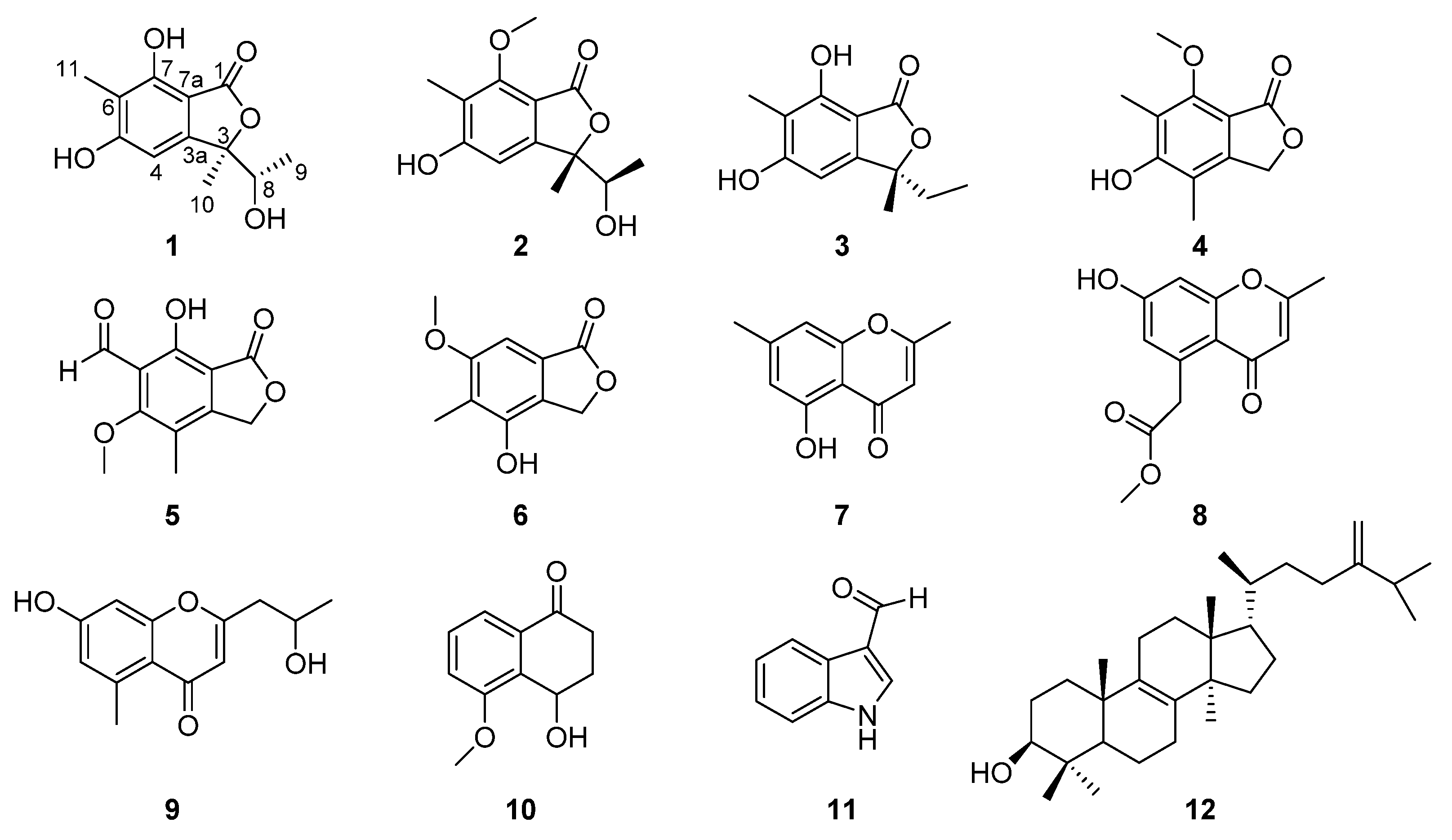
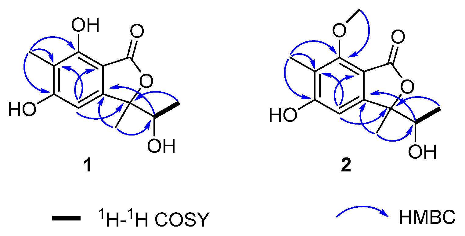
| Position | 1 (CD3OD) | 2 (DMSO-d6) | ||
|---|---|---|---|---|
| δH | δC | δH | δC | |
| 1 | 171.6 | 167.6 | ||
| 3 | 89.5 | 87.2 | ||
| 4 | 6.41 (1H, s) | 99.9 | 6.69 (1H, s) | 104.5 |
| 5 | 163.3 | 164.1 | ||
| 6 | 111.2 | 118.0 | ||
| 7 | 155.4 | 157.5 | ||
| 8 | 3.87 (1H, q, 6.5) | 71.1 | 3.84 (1H, q, 6.0) | 70.5 |
| 9 | 1.08 (3H, d, 6.5) | 16.1 | 0.90 (3H, d, 6.0) | 17.9 |
| 10 | 1.57 (3H, s) | 20.7 | 1.47 (3H, s) | 23.6 |
| 11 | 2.06 (3H, s) | 6.4 | 2.00 (3H, s) | 9.0 |
| 3a | 151.6 | 153.6 | ||
| 7a | 102.8 | 107.8 | ||
| -OCH3 | 3.86 (3H, s) | 61.7 | ||
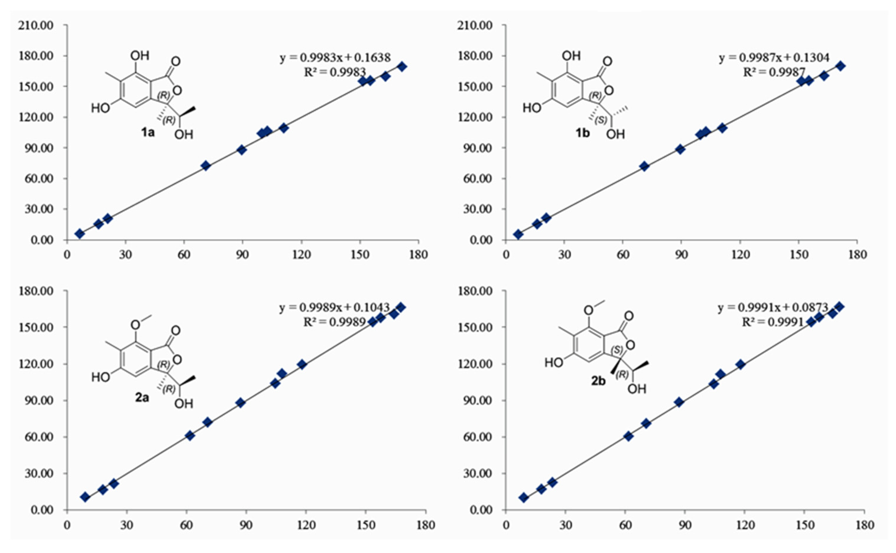
| Exptl. | 1 | Exptl. | 2 | ||||||
|---|---|---|---|---|---|---|---|---|---|
| 1a | dev | 1b | dev | 2a | dev | 2b | dev | ||
| 171.5 | 169.08 | 2.42 | 169.41 | 2.09 | 167.6 | 166.27 | 1.33 | 166.58 | 1.02 |
| 89.3 | 87.98 | 1.32 | 88.47 | 0.83 | 87.2 | 87.86 | 0.66 | 88.21 | 1.01 |
| 100.4 | 103.67 | 3.27 | 102.82 | 2.42 | 104.5 | 103.97 | 0.53 | 103.25 | 1.25 |
| 163.3 | 159.66 | 3.64 | 159.94 | 3.36 | 164.1 | 160.75 | 3.35 | 161.08 | 3.02 |
| 111.1 | 108.95 | 2.15 | 108.95 | 2.15 | 118 | 119.44 | 1.44 | 119.51 | 1.51 |
| 155.1 | 155.49 | 0.39 | 155.69 | 0.59 | 157.5 | 157.92 | 0.42 | 158.15 | 0.65 |
| 70.9 | 72.32 | 1.42 | 71.78 | 0.88 | 70.5 | 71.59 | 1.09 | 71.06 | 0.56 |
| 16.1 | 15.42 | 0.68 | 15.64 | 0.46 | 17.9 | 16.63 | 1.27 | 16.75 | 1.15 |
| 21.6 | 20.84 | 0.76 | 21.63 | 0.03 | 23.6 | 21.54 | 2.06 | 22.41 | 1.19 |
| 6.4 | 5.80 | 0.60 | 5.39 | 1.01 | 9 | 10.31 | 1.31 | 9.99 | 0.99 |
| 151.2 | 154.90 | 3.70 | 154.78 | 3.58 | 153.6 | 154.37 | 0.77 | 154.10 | 0.50 |
| 103.2 | 105.98 | 2.78 | 105.61 | 2.41 | 107.8 | 111.73 | 3.93 | 111.45 | 3.65 |
| 61.7 | 60.63 | 1.07 | 60.46 | 1.24 | |||||
| MAE a | 1.93 | MAE a | 1.65 | MAE a | 1.48 | MAE a | 1.37 | ||
| RMS b | 2.26 | RMS b | 2.00 | RMS b | 1.79 | RMS b | 1.63 | ||
| Pmean | 19.63% | Pmean | 27.76% | Pmean | 26.40% | Pmean | 32.40% | ||
| Prel | 1.54% | Prel | 98.46% | Prel | 6.51% | Prel | 93.49% | ||
| Compounds | MIC (ug/mL) | |
|---|---|---|
| MRSA | E. coli | |
| 1–2, 5, 7 | >500 | >500 |
| 3 | 250 | >500 |
| 4 | 250 | >500 |
| 6 | 125 | >500 |
| 8 | 125 | >500 |
| 9 | 125 | >500 |
| 10 | 125 | >500 |
| 11 | 500 | >500 |
| 12 | 62.5 | >500 |
| Vancomycin a | 1.25 | ≥40 |
Publisher’s Note: MDPI stays neutral with regard to jurisdictional claims in published maps and institutional affiliations. |
© 2021 by the authors. Licensee MDPI, Basel, Switzerland. This article is an open access article distributed under the terms and conditions of the Creative Commons Attribution (CC BY) license (https://creativecommons.org/licenses/by/4.0/).
Share and Cite
Zhang, S.; Chen, D.; Kuang, M.; Peng, W.; Chen, Y.; Tan, J.; Kang, F.; Xu, K.; Zou, Z. Rhytidhylides A and B, Two New Phthalide Derivatives from the Endophytic Fungus Rhytidhysteron sp. BZM-9. Molecules 2021, 26, 6092. https://doi.org/10.3390/molecules26206092
Zhang S, Chen D, Kuang M, Peng W, Chen Y, Tan J, Kang F, Xu K, Zou Z. Rhytidhylides A and B, Two New Phthalide Derivatives from the Endophytic Fungus Rhytidhysteron sp. BZM-9. Molecules. 2021; 26(20):6092. https://doi.org/10.3390/molecules26206092
Chicago/Turabian StyleZhang, Sha, Dekun Chen, Min Kuang, Weiwei Peng, Yan Chen, Jianbing Tan, Fenghua Kang, Kangping Xu, and Zhenxing Zou. 2021. "Rhytidhylides A and B, Two New Phthalide Derivatives from the Endophytic Fungus Rhytidhysteron sp. BZM-9" Molecules 26, no. 20: 6092. https://doi.org/10.3390/molecules26206092
APA StyleZhang, S., Chen, D., Kuang, M., Peng, W., Chen, Y., Tan, J., Kang, F., Xu, K., & Zou, Z. (2021). Rhytidhylides A and B, Two New Phthalide Derivatives from the Endophytic Fungus Rhytidhysteron sp. BZM-9. Molecules, 26(20), 6092. https://doi.org/10.3390/molecules26206092