Article

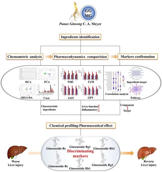
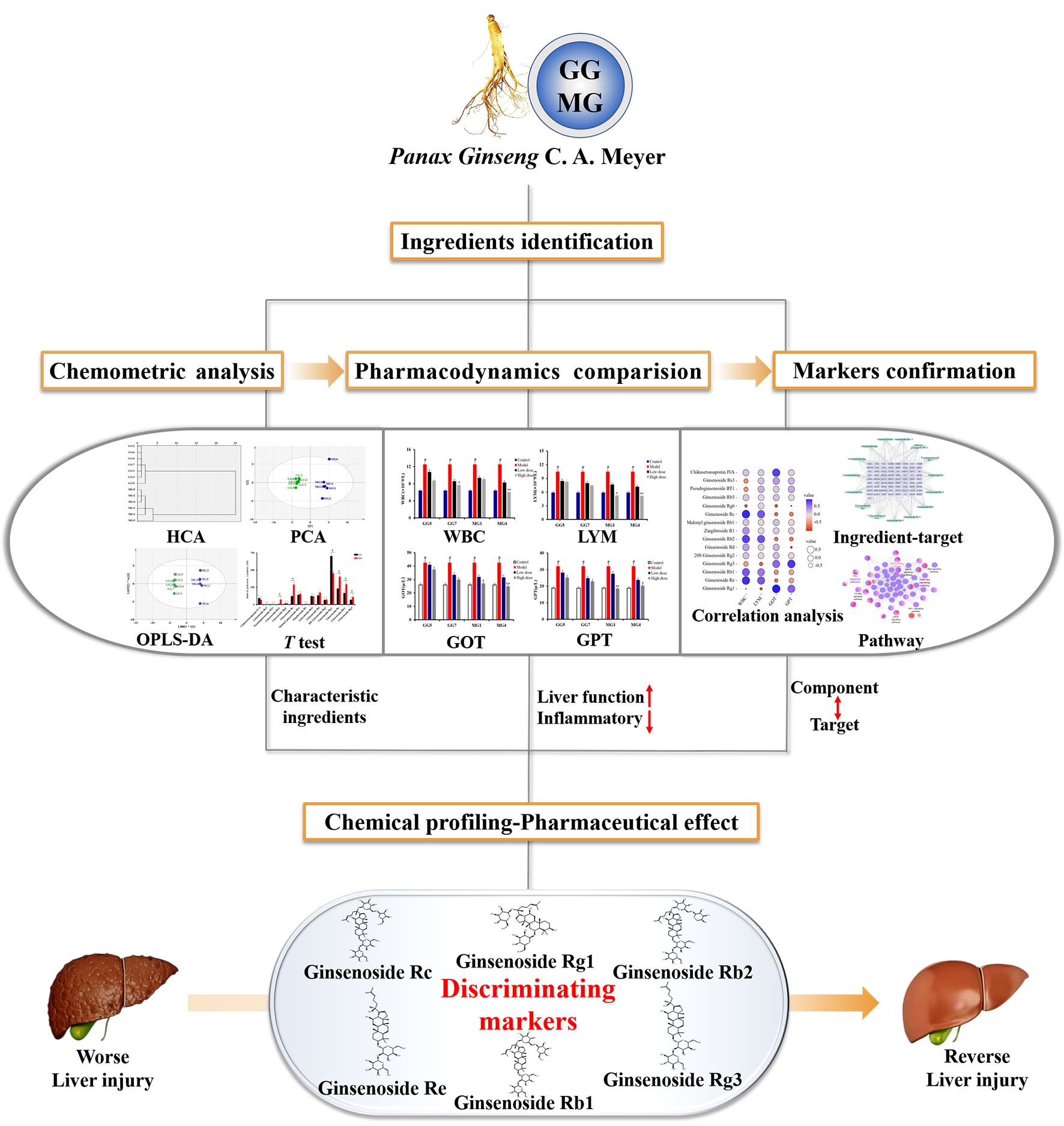
An Integrated Mutually Oriented “Chemical Profiling–Pharmaceutical Effect” Strategy for Screening Discriminating Markers of Underlying Hepatoprotective Effects to Distinguish Garden-Cultivated from Mountain-Cultivated Ginseng

School of Pharmacy, Shenyang Pharmaceutical University, Shenyang 110016, China
*
Author to whom correspondence should be addressed.
These authors contributed equally to this work.
Molecules 2021, 26(18), 5456; https://doi.org/10.3390/molecules26185456
Submission received: 30 July 2021 / Revised: 30 August 2021 / Accepted: 31 August 2021 / Published: 8 September 2021
(This article belongs to the Special Issue State-of-the-Art Analytical Technologies for Natural Products)

Abstract

:
Garden-cultivated Ginseng (GG) and mountain-cultivated Ginseng (MG) both belong to Panax Ginseng C. A. Meyer. However, the effective substances which can be used to distinguish GG from MG remain obscure. Therefore, the purpose of this study was to screen for discriminating markers that can assist in the correct identification of GG and MG. HPLC Q-TOF/MS and various chemometrics methods were used to analyze the chemical profiles of 13 batches of Ginseng and to explore the characteristic constituents of both GG and MG. The hepatocyte-protecting effects of GG and MG were investigated through a paclitaxel-induced liver injury model. Through a combination of correlation analysis and bioinformatic techniques, markers for differentiation between GG and MG were ascertained. A total of 40 and 41 compounds were identified in GG and MG, respectively, and 15 characteristic ingredients contributed significantly to the discrimination of GG from MG. Correlation analysis and network pharmacology were applied and ginsenosides Rg1, Re, Rb1, Rc, Rb2, and Rg3 were found to be discriminating markers of GG and MG. Six markers for the identification of GG and MG were screened out by a step-wise mutually oriented “chemical profiling–pharmaceutical effect” correlation strategy, which is of great significance for future quality assessment of Ginseng products.

Graphical Abstract

1. Introduction

Panax Ginseng C. A. Meyer (Ginseng), of genus Panax and family Araliaceae, has been used in traditional Chinese medicines (TCMs) for thousands of years throughout Asia to reinforce vital energy and tonify the spleen [1,2]. Based on extensive studies, Ginseng is rich in polysaccharides, ginsenosides, and volatile oils. More recently, numerous pharmacological studies have demonstrated that Ginseng possesses various biological activities, including antitumor, anti-inflammation, antioxidation, and immunoregulatory properties, among others [3,4,5]. Due to its diverse pharmacodynamics, Ginseng has been utilized in varied health products and dietary nutrients to relieve fatigue, deodorize, and ameliorate the toxic side effects of chemotherapy [6,7]. Recently, Ginseng has been clinically recognized as a hepatic protectant for its significant hepatoprotective effects and ability to improve liver function, as revealed by multitudinous studies [8,9,10,11].
Over recent years, inconsistency in the quality and therapeutic efficacy of available Ginseng products has become an intricate issue due to disparate growth environments and cultivation conditions. In the Chinese Pharmacopoeia (2020 edition), Ginseng was classified as either garden-cultivated Ginseng (GG, artificially cultivated) or mountain-cultivated Ginseng (MG, grown naturally under mountainous forest). In general, GG can be harvested after four to six years, while MG requires 10–20 years or longer. Both modern pharmacological research and traditional clinical experience indicate that the medicinal value and health function of MG is significantly better than that of GG [12,13]; thus, MG is consistently more expensive than GG. On account of the obvious differences in time investment and economic benefit, substitution of MG with GG has been increasingly rampant in the market, which is a primary cause of the current difficulty in maintaining the quality and effectiveness of Ginseng products. Thus, recent studies concerning the discrimination of GG from MG have attracted increasing attention. Previous research mainly focused on the contrastive qualitative-quantitative analysis of their chemical components in vitro [14,15,16]; however, it has been recognized that pharmaceutical efficacy is essentially the nucleus of TCM [17,18,19,20]. Unfortunately, few studies have sought to determine differentiating markers of GG and MG on the basis of their biological activity. Therefore, it is essential to integrate multiple analytical, pharmacological, and statistical approaches to establish a systematic strategy to explore the distinctive bioactive markers of GG and MG for their identification and differentiation.
In the present study, pharmacology-based markers for distinguishing GG from MG were first suggested by analyzing the chemical components of GG and MG using efficiency indexes. In brief, the ingredient database of Ginseng was obtained from seven batches of GG and six batches of MG analyzed in Liaoning by an established HPLC Q-TOF/MS method. Then, a variety of chemometric methods including hierarchical cluster analysis (HCA), principal component analysis (PCA), and orthogonal partial least squares discriminant analysis (OPLS-DA) were applied to explore the characteristic components of GG and MG. A contrastive study on the hepatoprotective function of GG and MG utilized a liver injury model induced by paclitaxel [21], which indicated that the liver-protective efficacy of MG was superior to that of GG. In order to further investigate the ingredients contributing to the different pharmacodynamics of GG and MG, we carried out a correlation analysis between the characteristic constituents and the efficiency indexes. Furthermore, in consideration of the “multi components-multi targets” characteristic of TCMs, network pharmacology was adopted to comprehensively screen for the effective discriminating markers of Ginseng. Above all, a step-wise mutually oriented “chemical profiling–pharmaceutical effect” strategy was established, and six chemical components—including ginsenosides Rg1, Re, Rb1, Rc, Rb2, and Rg3—were ascertained to be key markers, underlying the difference in hepatoprotective efficacy, for differentiating GG from MG. This study is of great significance in setting a more scientific benchmark for the analysis of Ginseng, and offers an alternative quality assessment system for TCMs.

2. Results and Discussion

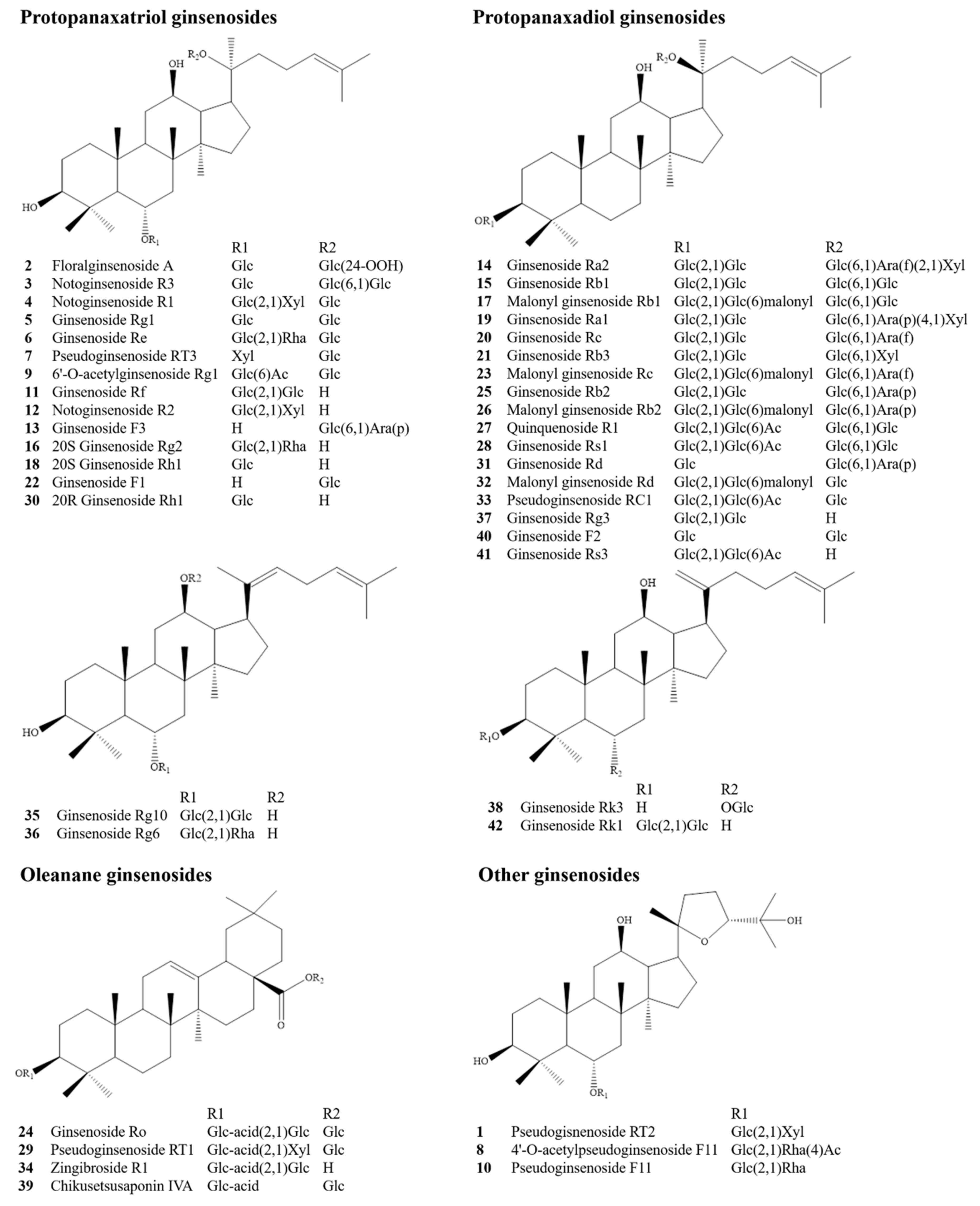
2.1. Qualitative and Semi-Quantitative Analysis of Ingredients in Ginseng with HPLC Q-TOF/MS

An established HPLC Q-TOF/MS method using digoxin as an internal standard (IS) was adopted to conduct the qualitative and semiquantitative analysis of the ingredients in Ginseng for seven batches of GG and six batches of MG, whose detailed information is presented in Table 1. The chemical components were identified by calculating possible chemical formulas based on comparisons between the determined molecular weight and chromatographic retention behavior and reference data [22,23,24]. As a result, a total of 42 chemical components were identified, including 19 protopanaxadiol ginsenosides (PPD-type), 16 protopanaxatriol ginsenosides (PPT-type), 4 oleanane ginsenosides, and 3 other ginsenosides; their structures are presented in Figure 1. A total of 40 and 41 compounds were identified in GG and MG, respectively, 40 of which were common to both, as shown in Table 2. Moreover, among them, 12 chemical ingredients were accurately identified by comparison with the reference substances (extraction ion chromatograms of reference substances and Ginseng sample solution are presented in Figure S1A–C).

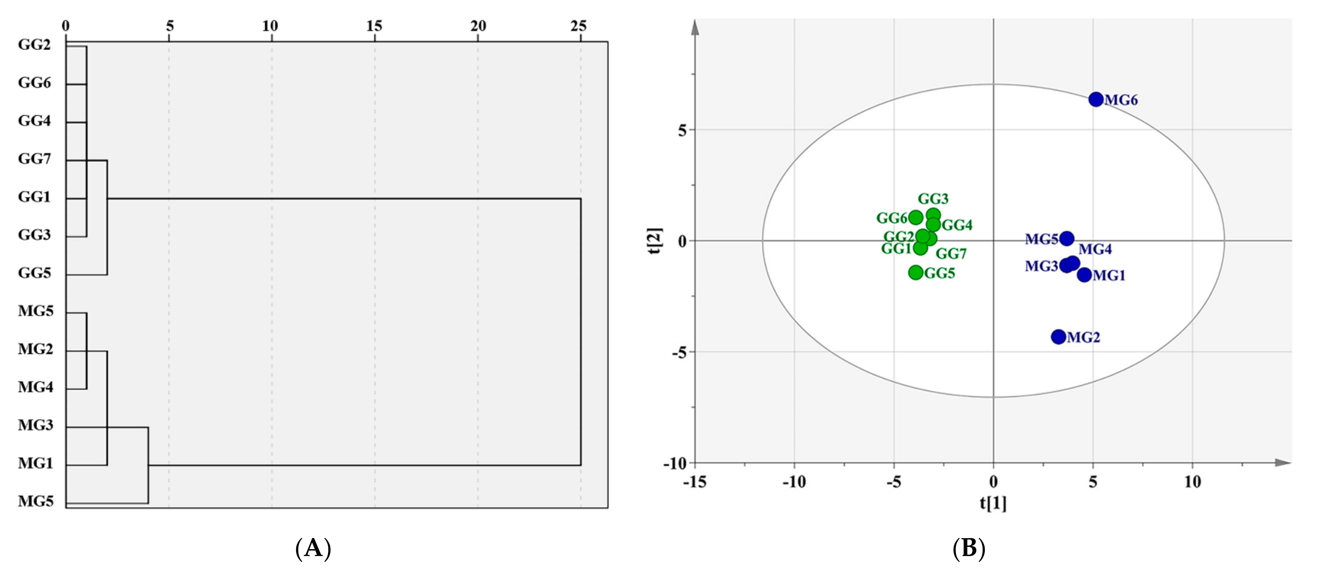
2.2. Exploration of Characteristic Components for Differentiating GG from MG by Chemometric Analysis

Characteristic components for the identification of GG and MG were explored using multifarious chemometric analysis methods. Internal standard normalization (via digoxin) was adopted in this study. The peak areas of all 42 compounds were calibrated to the IS and the ratio of the peak areas were used for chemometric analysis.
As seen in the dendrogram Figure 2A, 13 batches of Ginseng from Liaoning could be divided into two categories using HCA, of which GG was clustered into one group, and MG into another. Moreover, the PCA score plot illustrated that there was an efficacious separation between groups G1–G7 and M1–M6, as shown as Figure 2B, which was consistent with the results of HCA. In order to clarify the characteristic components that contributed greatly to this distinction, OPLS-DA was applied. The score plot, as shown in Figure 3A, indicated that the two groups were highly distinct with R2Y = 0.999 and Q2 = 0.973, and 15 components demonstrated VIP ≥ 1, as shown in Figure 3B. However, among them, only six constituents—ginsenosides Rg1, Re, Rb1, Rc, Rb2, and Rg3—revealed significant differences between GG and MG (* p < 0.05) according to the results of a Student’s t-test, as shown in Figure 4.
As above, all of the 15 chemometric components were common ingredients of GG and MG. Six ginsenosides were established as the qualitative and quantitative markers for in vitro identification of GG and MG. It was concluded that these six ingredients could be considered as preliminary marker candidates to distinguish GG from MG. Although these six components had significant differences in content, this seldom indicates the quality of TCMs. The selected markers, determined via efficacy mining, were considered to be more representative of quality.

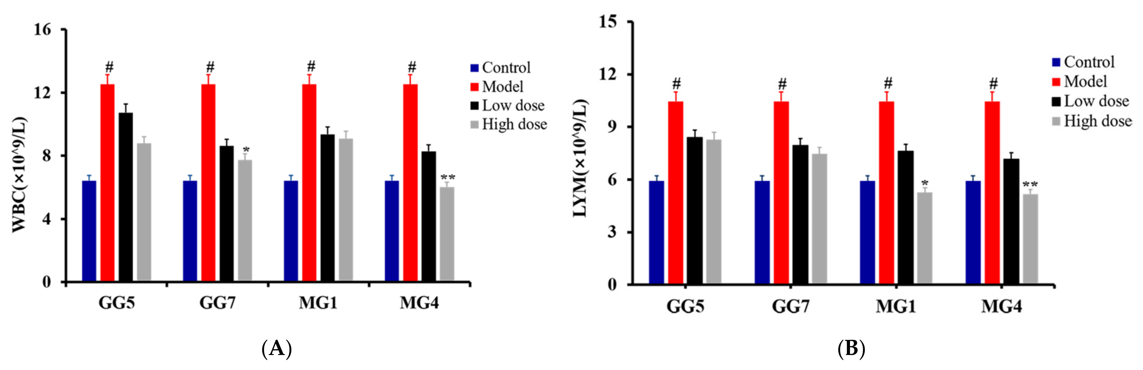
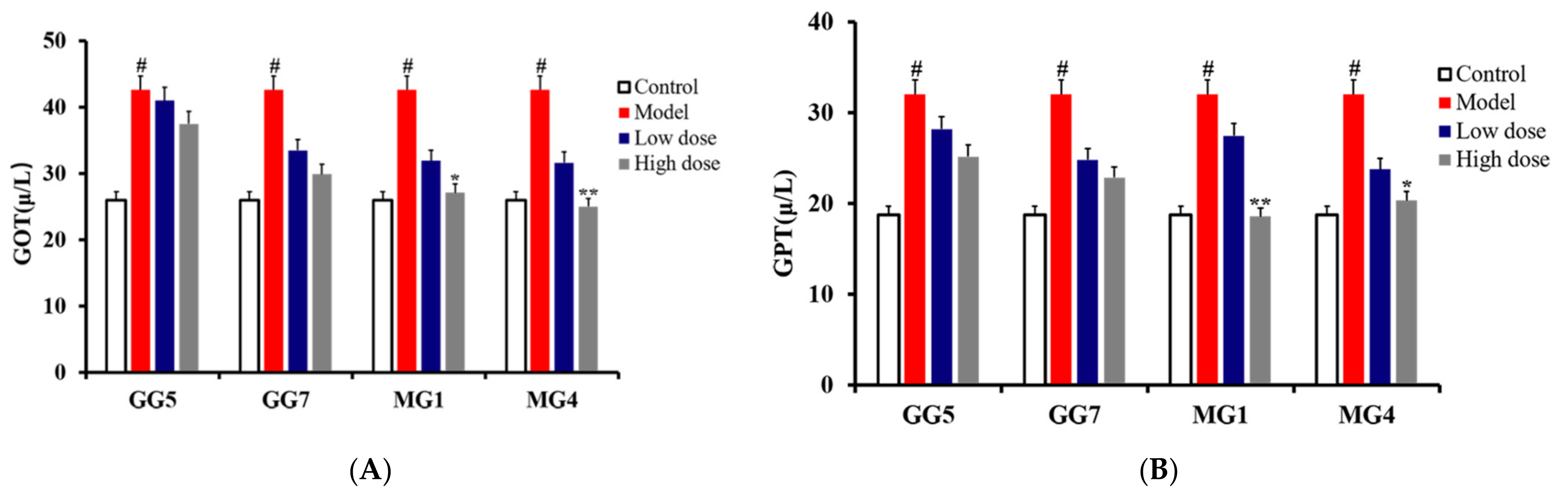
2.3. Comparative Study of the Pharmacodynamics of GG and MG in Paclitaxel-Induced Liver Injury

To further explore the discriminating markers of GG and MG underlying their respective pharmacodynamics, we conducted a comparative study of their liver-protective efficacies using a paclitaxel-induced liver injury rat model. Batches of GG5 and GG7, and MG1 and MG4, were included in the contrastive pharmacodynamic analysis as the contents of the 15 chemometric components in these batches were close to their average values in GG and MG, respectively.
As shown in Figure 5, an obvious elevation in white blood cell count (WBC) and lymphocyte count (LYM) could be observed in the model group compared with the control group (# p < 0.05), indicating abnormal regulation of autoimmunity and the development of inflammation. Furthermore, expression of the liver function indicators glutamic-oxaloacetic transaminase (GOT) and glutamic-pyruvic transaminase (GPT) were also increased markedly in the model group compared with the control group, as shown as Figure 6 (# p < 0.05), suggesting a decrease in liver function and the occurrence of parenchymal hepatic disease [25,26,27]. Through the above, it could be concluded that the liver injury model was successfully established. These escalating alterations were all consistently restored after treatment with GG and MG; however, MG exerted stronger therapeutic effects than GG based on the results of a t-test (* p < 0.05; ** p < 0.01). Therefore, our research demonstrates for the first time that the hepatoprotective effects of MG are superior to those of GG through in vivo pharmacology experiments.

2.4. Discovery of Pharmacodynamic-Based Markers to Distinguish GG from MG

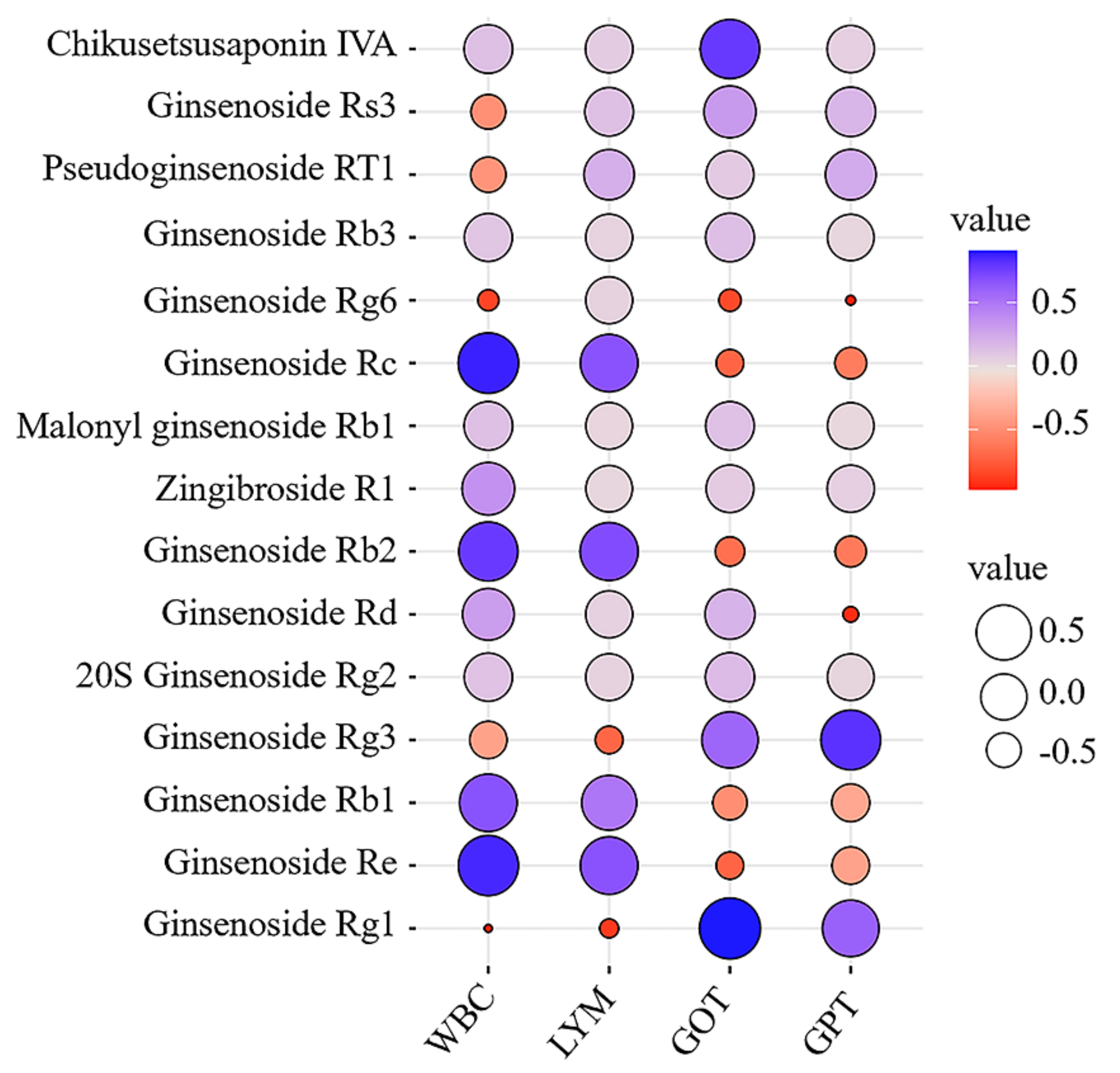
2.4.1. Correlation Analysis between Characteristic Components and Pharmacodynamic Indicators

Based on pharmacological comparison, a correlation analysis of the relative peak areas of the 15 characteristic components and the levels of the four pharmacodynamic indicators was performed with the corresponding GG5, GG7, MG1, and MG4 batch sample data. Evaluation of the correlation degree by the absolute value of the Pearson correlation coefficient: 0.6 ≤ |γ| ≤ 1 indicated correlation. Ginsenosides Re, Rb1, Rc, and Rb2 were positively correlated with WBC and LYM, while ginsenosides Rg1 and Rg3 were positively correlated with GOT and GPT, as shown in Figure 7 and Table S1, indicating that ginsenosides Rg1, Re, Rb1, Rc, Rb2, and Rg3 could participate in regulation of the milieu interne in morbid state and represent candidate markers for the distinction of GG from MG based on their underlying pharmacodynamics.

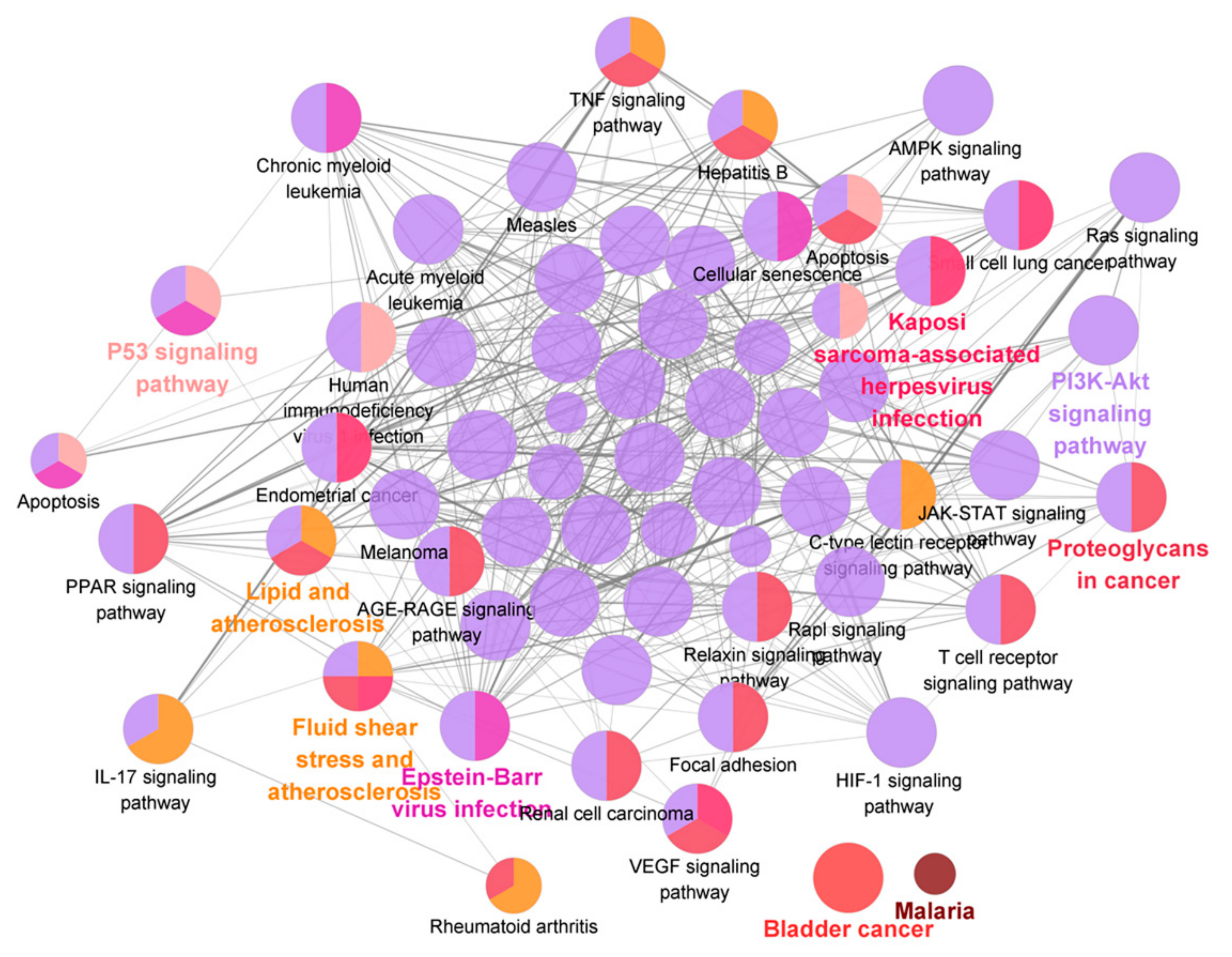
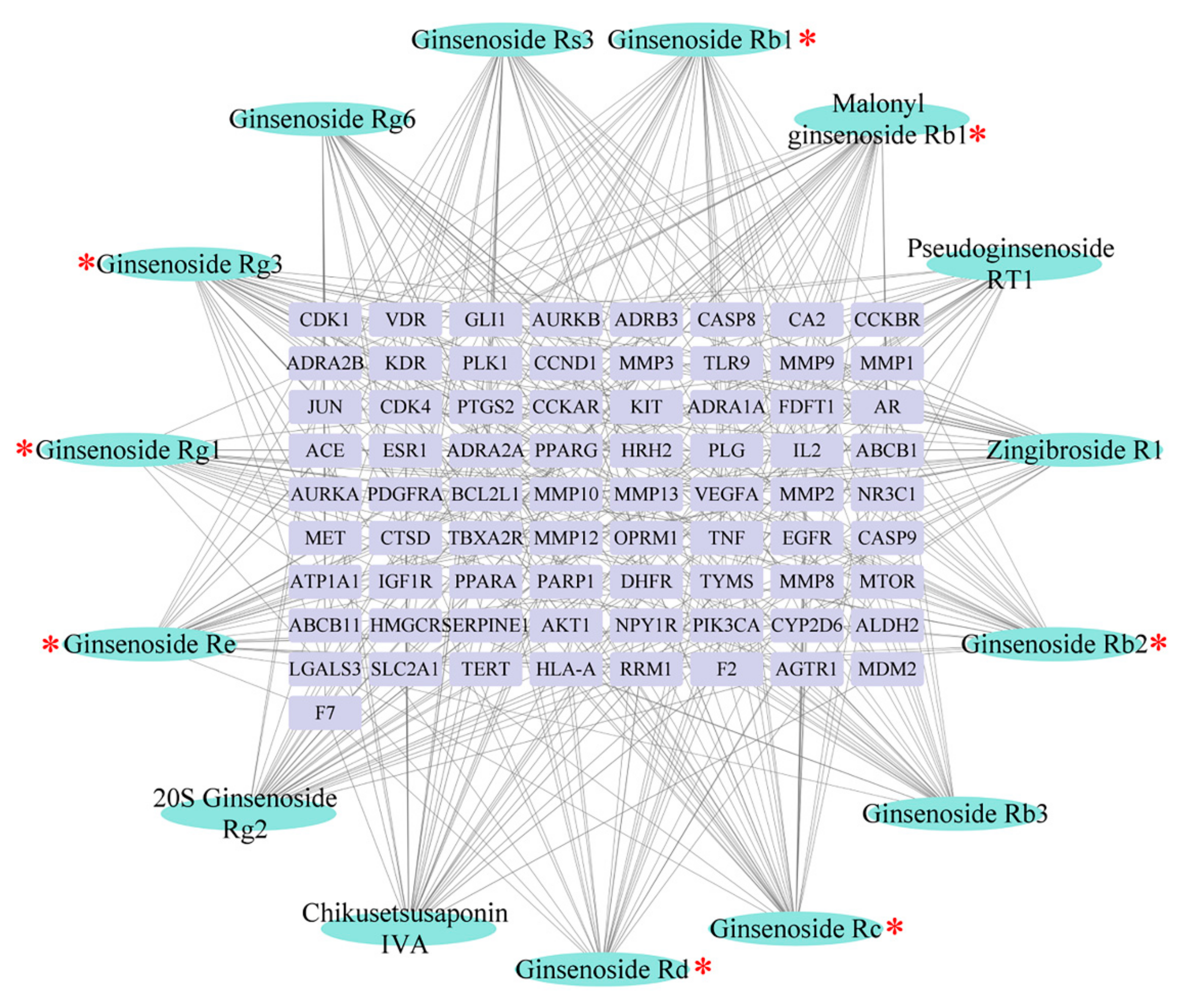
2.4.2. Identification of Effective Representative Substances of GG and MG Using Bioinformatic Analysis

Bioinformatic analysis was applied to visualize the large-scale data of “components–targets–pathways” and for virtual excavation of the bioactive markers capable of differentiating GG from MG, in consideration of the “multi components-multi targets” characteristic of TCMs.
Target genes of the 15 characteristic components were predicted by the SwissTargetPrediction database. Pathway enrichment analysis was performed with Cytoscape. A total of 73 targets and 62 pathways with significant differences were obtained. As shown in Figure 8 and Figure 9, Ginseng mainly participated in the regulation of inflammation and liver function through modulation of the pathways of PI3K-Akt, Ras, HIF-1, TNF, PPAR, AMPK, and p53, in addition to various pathways related to bile acid and lipid metabolism. These crucial targets and pathways were involved in the anti-liver injury effects of Ginseng [28,29,30,31,32,33]. Additionally, the degree value is a key parameter to assess the interconnectedness of components and their corresponding symptoms; the degree values of ginsenosides Rg1, Re, Rb1, Rc, Rb2, Rd, and Rg3, as well as malonyl ginsenoside Rb1, were higher than the average degree value (mean value = 27.4; as shown as Table S2).
Integrating all above results, the ginsenosides Rg1, Re, Rb1, Rc, Rb2, and Rg3 could be regarded as effective markers for distinguishing GG from MG. Moreover, in consideration of previous studies, it has been widely reported that ginsenoside are the most important components of Ginseng contributing to its beneficial properties. Ginsenosides Rg1, Re, and Rg3 act on the PI3K–Akt signaling pathway to reduce oxidative stress response [34,35,36]. In addition, ginsenosides Rb1, Rc, and Rb2 possess anti-inflammatory activities through the inhibition of both MAPK signaling pathways and the expression of TNF [37,38,39]. In agreement, our research identified these six ginsenoside compounds as pharmacodynamic-based markers to distinguish GG from MG.

3. Materials and Methods

3.1. Chemicals and Reagents

A total of 13 batches of Ginseng collected from Liaoning (including 7 batches of GG and 6 batches of MG) of different ages were obtained from Tong-Ren-Tang TCMs store (Shenyang, China). HPLC-grade acetonitrile, methanol, and formic acid were all purchased from Fisher Scientific (Fair Lawn, NJ, USA); all other reagents were analytical grade (Shandong Yuwang Industrial Co., Ltd., Yucheng, China). The GOT/GPT commercial enzyme-linked immunosorbent assay (ELISA) kits were purchased from the Nanjing Jiancheng Institute of Biological Engineering (Nanjing, China).

3.2. HPLC Q-TOF/MS Analysis

3.2.1. Sample Preparation

Ginseng powder (1 g, passed through a 5-mesh sieve) was extracted under reflux with 10 times the volume of water for 1 h, and the progress was repeated twice. A solution with concentration 50 mg/mL (medicinal material dosage/volume) was obtained by combining the extracts. Then, the solution was centrifuged at 12,000 rpm for 10 min and filtered through a 0.22 μm filter membrane for HPLC Q-TOF/MS analysis. Appropriate amounts of the reference substance ginsenosides Re, Rf, Rb1, Rc, F1, Ro, Rb2, Rd, Rg3, F2, 20R-ginsenoside Rh1, and chikusetsusaponin Iva (ShanghaiyuanyeBio-Technology Co., Ltd., Shanghai, China) were weighed and dissolved in methanol to prepare a mixed reference solution containing the 12 chemical components. Digoxin (25 ng/mL, Chengdu Chroma-Biotechnology Co., Ltd., Sichuan, China) was used as an IS for semiquantitative analysis. All samples were kept at 4 °C during analysis.

3.2.2. HPLC Q-TOF/MS Conditions

Analyses were performed on a 1260 Infinity HPLC system (Agilent Technology, Santa Clara, CA, USA). A reverse-phase column of Phenomenex Kinetex XB C18 (100 mm × 4.6 mm, 2.6 μm) was used for the chromatographic separations with a flow rate of 0.5 mL/min at 30 °C. The mobile phase consisted of 0.1% formic acid-water (A) and 0.1% formic acid-acetonitrile (B) with a gradient procedure as follows: 0–13 min, 5–30% B; 13–27 min, 30–40% B; 27–30 min, 40–70% B; 30–31 min, 70–90% B; 31.1–31.1 min, 90–5% B; 31.1–35 min, 5–5% B. The injection volume of each sample was 5 µL and the temperature of the sample plate was maintained at 4 °C.
A quadrupole time-of-flight mass spectrometry system (Triple TOF 5600, AB SCIEX Corporation, Foster City, CA, USA) was used to obtain the total ion chromatograms (TIC) in negative electrospray ionization (ESI) mode. The optimal instrument parameters were as follows: spray voltage of 4500 V, a source heater temperature of 550 °C, the range of m/z set to 100–2000 and nitrogen was used as the atomizing gas and other auxiliary gases. PeakView software (version 1.2.1, SCIEX) was used for data analysis and the precision error threshold was fixed at 5 ppm.

3.3. Animals

Healthy Sprague-Dawley rats (200–220 g; 8 weeks old; NO.SCXK(Liao)2020---0001) were provided by the Experimental Animal Center of Shenyang Pharmaceutical University and housed in a specialized pathogen free (SPF) standard environment (ambient temperature 22 ± 2 °C, with relative humidity of 50 ± 10 % and a natural light-dark cycle). All experiments complied with the Animal Experiment Code of Shenyang Pharmaceutical University and approval was obtained from the animal ethics committee of the institution (Ethical Approval number: SYPU-IACUC-G2P-2021-68). After a week of domestication of the rats, they were randomly divided into six groups: control, model, and both low (300 mg/kg) and high doses (800 mg/kg) of the 2 batches of GG (GG5/GG7) and 2 batches of MG (MG1/MG4) (n = 6). Food and water were available ad libitum.
Paclitaxel (10 mg/kg, Haikou Pharmaceutical Factory Co., Ltd, Hainan, China) was given intraperitoneally to induce liver injury once a day for two weeks. In the meantime, the rats in the medication administration groups were gavaged with the predetermined dose of Ginseng 1 h after the injection of paclitaxel, while rats in the control and model groups received the same volume of normal saline. One hour after the final administration, two blood samples (each 1.5 mL, one sample transferred into the tubes with anticoagulants directly, the other transferred into the heparinized tubes to obtain serum samples) were collected from the suborbital vein of each rat and stored at −80 °C until required.

3.4. Detection of Pharmacodynamic Indicators

In order to detect the levels of blood routine parameters WBC and LYM, a 1.5 mL blood sample of each rat was measured with an automatic animal blood cell analyzer (BC2800Vet Shenzhen).
Serum samples were used to measure the expression of liver function indicators, including GOT and GPT, according to the manufacturer’s instructions.

3.5. Bioinformatics Analysis Process

Network pharmacology analysis was performed with the characteristic ingredients in this study. The PubChem (https://pubchem.ncbi.nlm.nih.gov/ accessed on 30 July 2021) and SwissTargetPrediction (http://www.swisstargetprediction.ch/ accessed on 30 July 2021) databases were integrated to obtain the corresponding targets of each ingredient. The targets closely associated with “liver injury” were retrieved from the Therapeutic Target (http://db.idrblab.net/ttd/ accessed on 30 July 2021), GeneCards (https://www.genecards.org/ accessed on 30 July 2021), and Comparative Toxicogenomics Databases (http://ctdbase.org/ accessed on 30 July 2021). Finally, Cytoscape (3.8.2, USA) software was used to visualize the “component–target–pathway” network.

3.6. Statistical and Data Analysis

All values are presented as means ± SD. The data were analyzed using SIMCA14.0 (UmetricsAB, Umea, Sweden) and SPSS (SPSS Inc., Chicago, IL, USA). Data from two groups were analyzed using a two-tailed Student’s t-test and p values lower than 0.05 were considered significant.

4. Conclusions

In the present study, an integrated mutually oriented “chemical profiling–pharmaceutical effect” strategy was proposed and successfully applied to identify distinct bioactive markers for the identification of GG and MG for the first time. First, we established a chemical component database of Ginseng using HPLC Q-TOF/MS, which contained 40 compounds from GG and 41 compounds from MG. Multiple chemometric methods were further applied to discover the characteristic components. We then conducted a comparative study of the pharmacodynamics of GG and MG, which indicated that MG may exert superior therapeutic effects to GG. Furthermore, in order to explore the components responsible for this difference in efficacy, correlation analysis combined with network pharmacology technology was applied. Ginsenosides Rg1, Re, Rb1, Rc, Rb2, and Rg3 were identified as markers underlying the difference in hepatoprotective efficacy which can be used to distinguish GG from MG. These findings may contribute to the development of quality control and thus enhance the clinical efficacy of Ginseng.

Supplementary Materials

The following are available online, Figure S1: (A) Extracted ion chromatograms of 12 reference substances: 1. Ginsenoside Re; 2. Ginsenoside Rf; 3. Ginsenoside Rb1; 4. Ginsenoside Rc; 5. Ginsenoside F1; 6. Ginsenoside Ro; 7. Ginsenoside Rb2; 8. 20R Ginsenoside Rh1; 9. Ginsenoside Rd; 10. Ginsenoside Rg3; 11. Chikusetsusaponin IVa; 12. Ginsenoside F2; (B) The total ion chromatograms of GG5; (C) The total ion chromatograms of MG1, Table S1: Pearson correlation coefficient between the 15 characteristic ingredients and 4 pharmacodynamic indexes, Table S2: The degree value of 15 characteristic components.

Author Contributions

Conceptualization, methodology and software, S.L.; Writing—review and editing, supervision, Y.Z.; Writing—original draft and data curation, P.Y.; Writing—review and editing, M.T.; Resources and investigation, L.X.; Validation, Q.Z.; Supervision, K.B.; Funding acquisition, writing—review and editing, supervision, Q.L. All authors have read and agreed to the published version of the manuscript.

Funding

This research received no external funding.

Institutional Review Board Statement

The study was conducted according to the guidelines of the Declaration of Helsinki, and approved by the Ethics Committee of Shenyang Pharmaceutical University (protocol code: 68; date of approval: 11 March 2021).

Informed Consent Statement

Not applicable.

Data Availability Statement

Not applicable.

Acknowledgments

This work was supported by the National Natural Science Funds of China (Grant No. 81973464/H3203); China Postdoctoral Science Foundation (2020M680989); Liaoning Distinguished Professor Project for Qing Li (2017); Shenyang Science and Technology Innovation Project for Young; Middle-aged Talents (RC190505); Liaoning BaiQianWan Talents Program in 2019(A-37) and Liaoning key Research and Development Program (2018).

Conflicts of Interest

The authors declare no conflict of interest.

Sample Availability

Samples of the compounds are available from the authors.

References

  1. Guo, M.; Shao, S.; Wang, D.; Zhao, D.; Wang, M. Recent progress in polysaccharides from Panax ginseng C. A. Meyer. Food Funct. 2021, 12, 494–518. [Google Scholar] [CrossRef]
  2. Ki, S.H.; Yang, J.H.; Ku, S.K.; Kim, S.C.; Kim, Y.W.; Cho, I.J. Red ginseng extract protects against carbon tetrachloride-induced liver fibrosis. J. Ginseng Res. 2013, 37, 45–53. [Google Scholar] [CrossRef] [Green Version]
  3. Zhou, X.; Liu, H.; Zhang, M.; Li, C.; Li, G. Spectrum-effect relationship between UPLC fingerprints and anti-lung cancer effect of Panax ginseng. Phytochem. Anal. 2020, 32, 339–346. [Google Scholar] [CrossRef] [PubMed]
  4. Park, S.B.; Park, G.H.; Um, Y.; Kim, H.N.; Song, H.M.; Kim, N.; Kim, H.-S.; Jeong, J.B. Wood-cultivated ginseng exerts anti-inflammatory effect in LPS-stimulated RAW264.7 cells. Int. J. Biol. Macromol. 2018, 116, 327–334. [Google Scholar] [CrossRef] [PubMed]
  5. Kim, H.M.; Song, Y.; Hyun, G.H.; Long, N.P.; Park, J.H.; Hsieh, Y.S.Y.; Kwon, S.W. Characterization and Antioxidant Activity Determination of Neutral and Acidic Polysaccharides from Panax Ginseng C. A. Meyer. Molecules 2020, 25, 791. [Google Scholar] [CrossRef] [PubMed] [Green Version]
  6. Ji, Y.C.; Tae, S.W.; Seo, Y.Y.; Ike, C.d.P.; Yoon, J.C.; Hyung, S.A.; Yong, S.L.; Gu, Y.Y.; Jae, H.C. Red Ginseng Supplementation More Effectively Alleviates Psychological than Physical Fatigue. J. Ginseng Res. 2011, 35, 331–338. [Google Scholar]
  7. Zhu, H.; Long, M.-H.; Wu, J.; Wang, M.-M.; Li, X.-Y.; Shen, H.; Xu, J.-D.; Zhou, L.; Fang, Z.-J.; Luo, Y.; et al. Ginseng alleviates cyclophosphamide-induced hepatotoxicity via reversing disordered homeostasis of glutathione and bile acid. Sci. Rep. 2015, 5, 17536. [Google Scholar] [CrossRef] [PubMed]
  8. Lam, P.; Cheung, F.; Tan, H.Y.; Wang, N.; Yuen, M.F.; Feng, Y. Hepatoprotective Effects of Chinese Medicinal Herbs: A Focus on Anti-Inflammatory and Anti-Oxidative Activities. Int. J. Mol. Sci. 2016, 17, 465. [Google Scholar] [CrossRef] [PubMed] [Green Version]
  9. Ahmed, A.-H.; Shimaa, I.S.; Safaa, I.K.; Eman, S.E.-S.; Hosny, A.E.F.; Shafika, A.E. Panax ginseng is superior to vitamin E as a hepatoprotector against cyclophosphamide-induced liver damage. Complement. Ther. Med. 2019, 46, 95–102. [Google Scholar]
  10. Wang, W.; Wang, S.; Liu, J.; Cai, E.; Zhu, H.; He, Z.; Gao, Y.; Li, P.; Zhao, Y. Sesquiterpenoids from the root of Panax Ginseng protect CCl 4 –induced acute liver injury by anti-inflammatory and anti-oxidative capabilities in mice. Biomed. Pharmacother. 2018, 102, 412–419. [Google Scholar] [CrossRef]
  11. Wang, W.; Zhang, Y.; Li, H.; Zhao, Y.; Cai, E.; Zhu, H.; Li, P.; Liu, J. Protective Effects of Sesquiterpenoids from the Root of Panax ginseng on Fulminant Liver Injury Induced by Lipopolysaccharide/D-galactosamine. J. Agric. Food Chem. 2018, 66, 7758–7763. [Google Scholar] [CrossRef]
  12. Pan, H.-Y.; Qu, Y.; Zhang, J.-K.; Kang, T.-G.; Dou, D.-Q. Antioxidant activity of Ginseng cultivated under mountainous forest with different growing years. J. Ginseng Res. 2013, 37, 355–360. [Google Scholar] [CrossRef] [PubMed] [Green Version]
  13. Kim, J.; Yoo, J.-M.; Kim, J.S.; Kim, S.-G.; Park, J.E.; Seok, Y.M.; Son, J.-H.; Kim, H.J.; Faurot, K.R. Anticancer Effect of Mountain Ginseng on Human Breast Cancer: Comparison with Farm-Cultivated Ginseng. Alternat. Med. 2020, 2020, 2584783. [Google Scholar] [CrossRef] [PubMed]
  14. Xu, X.-F.; Cheng, X.-L.; Lin, Q.-H.; Li, S.-S.; Jia, Z.; Han, T.; Lin, R.-C.; Wang, D.; Wei, F.; Li, X.-R. Identification of mountain-cultivated ginseng and cultivated ginseng using UPLC/oa-TOF MSE with a multivariate statistical sample-profiling strategy. J. Ginseng Res. 2016, 40, 344–350. [Google Scholar] [CrossRef]
  15. Zhu, L.; Luan, X.; Yuan, Y.; Dou, D.; Huang, L. The characteristics of ginsenosides and oligosaccharides in mountain- and garden-cultivated Ginseng. J. Sci. Food Agric. 2021, 101, 1491–1498. [Google Scholar] [CrossRef] [PubMed]
  16. Lee, D.-Y.; Kim, J.; Shrestha, S.; Seo, K.-H.; Lee, Y.-H.; Noh, H.-J.; Kim, G.-S.; Kim, Y.-B.; Kim, S.-Y.; Baek, N.-I. Quality Evaluation of Panax ginseng Roots Using a Rapid Resolution LC-QTOF/MS-Based Metabolomics Approach. Molecules 2013, 18, 14849–14861. [Google Scholar] [CrossRef] [PubMed] [Green Version]
  17. Zhang, Y.; Li, S.; Liang, Y.; Liu, R.; Lv, X.; Zhang, Q.; Xu, H.; Bi, K.; Li, Z.; Li, Q. A systematic strategy for uncovering quality marker of Asari Radix et Rhizoma on alleviating inflammation based chemometrics analysis of components. J. Chromatogr. A 2021, 1642, 461960. [Google Scholar] [CrossRef]
  18. Liang, G.; Yang, J.; Liu, T.; Wang, S.; Wen, Y.; Han, C.; Huang, Y.; Wang, R.; Wang, Y.; Hu, L.; et al. A multi-strategy platform for quality control and Q-markers screen of Chaiqin chengqi decoction. Phytomedicine 2021, 85, 153525. [Google Scholar] [CrossRef]
  19. Wu, P.; Dong, X.-M.; Song, G.-Q.; Wei, M.-M.; Fang, C.; Zheng, F.-B.; Zhao, Y.-J.; Lu, H.-Q.; Cheng, L.-H.; Zhou, J.-L.; et al. Bioactivity-guided discovery of quality control markers in rhizomes of Curcuma wenyujin based on spectrum-effect relationship against human lung cancer cells. Phytomedicine 2021, 86, 153559. [Google Scholar] [CrossRef]
  20. Chen, R.; He, J.; Tong, X.; Tang, L.; Liu, M.; Lam, C.W.K. The Hedyotis diffusa Willd. (Rubiaceae): A Review on Phytochemistry, Pharmacology, Quality Control and Pharmacokinetics. Molecules 2016, 21, 710. [Google Scholar] [CrossRef] [PubMed]
  21. Dilek, K.; Banu, E.; Osman, N.K. The protective effect of beta-1,3-D-glucan on taxol-induced hepatotoxicity: A histopathological and stereological study. Drug Chem. Toxicol. 2010, 33, 8–16. [Google Scholar]
  22. Wang, X.; Liu, J.; Yang, X.; Zhang, Q.; Zhang, Y.; Li, Q.; Bi, K. Development of a systematic strategy for the global identification and classification of the chemical constituents and metabolites of Kai-Xin-San based on liquid chromatography with quadrupole time-of-flight mass spectrometry combined with multiple data-processing approaches. J. Sep. Sci. 2018, 41, 2672–2680. [Google Scholar] [PubMed]
  23. Zuo, T.; Zhang, C.; Li, W.; Wang, H.; Hu, Y.; Yang, W.; Jia, L.; Wang, X.; Gao, X.; Guo, D. Offline two-dimensional liquid chromatography coupled with ion mobility-quadrupole time-of-flight mass spectrometry enabling four-dimensional separation and characterization of the multicomponents from white ginseng and red ginseng. J. Pharm. Anal. 2020, 10, 597–609. [Google Scholar] [CrossRef] [PubMed]
  24. Lee, J.W.; Ji, S.-H.; Lee, Y.-S.; Jin, C.D.; Choi, B.-R.; Kim, G.-S.; Baek, N.-I.; Lee, D.Y. Mass Spectrometry Based Profiling and Imaging of Various Ginsenosides from Panax ginseng Roots at Different Ages. Int. J. Mol. Sci. 2017, 18, 1114. [Google Scholar] [CrossRef] [PubMed] [Green Version]
  25. Yen, F.-L.; Wu, T.-H.; Lin, L.-T.; Lin, C.-C. Hepatoprotective and antioxidant effects of Cuscuta chinensis against acetaminophen-induced hepatotoxicity in rats. J. Ethnopharmacol. 2007, 111, 123–128. [Google Scholar] [CrossRef]
  26. Chen, T.-M.; Subeq, Y.-M.; Lee, R.-P.; Chiou, T.-W.; Hsu, B.-G. Single dose intravenous thioacetamide administration as a model of acute liver damage in rats. Int. J. Exp. Pathol. 2008, 89, 223–231. [Google Scholar] [CrossRef]
  27. Liu, X.; Zhang, Y.; Liu, L.; Pan, Y.; Hu, Y.; Yang, P.; Liao, M. Protective and therapeutic effects of nanoliposomal quercetin on acute liver injury in rats. BMC Pharmacol. Toxicol. 2020, 21, 11. [Google Scholar] [CrossRef] [Green Version]
  28. Badr, G.; Sayed, E.A.; Waly, H.; Hassan, K.A.-H.; Mahmoud, M.H.; Selamoglu, Z. The Therapeutic Mechanisms of Propolis Against CCl4 -Mediated Liver Injury by Mediating Apoptosis of Activated Hepatic Stellate Cells and Improving the Hepatic Architecture through PI3K/AKT/mTOR, TGF-β/Smad2, Bcl2/BAX/P53 and iNOS Signaling Pathways. Cell. Physiol. Biochem. 2019, 53, 301–322. [Google Scholar]
  29. Nesma, M.E.A.E.-N.; Dalia, O.S.; Sawsan, S.M.; Salwa, M.N.; Rania, M.A.; Marwa, M.S.; Hanan, S.E.-A. Olmesartan attenuates type 2 diabetes-associated liver injury: Cross-talk of AGE/RAGE/JNK, STAT3/SCOS3 and RAS signaling pathways. Eur. J. Pharmacol. 2020, 874, 173010. [Google Scholar]
  30. Zhu, Q.; Wang, H.; Jiang, B.; Ni, X.; Jiang, L.; Li, C.; Wang, X.; Zhang, F.; Ke, B.; Lu, L. Loss of ATF3 exacerbates liver damage through the activation of mTOR/p70S6K/ HIF-1α signaling pathway in liver inflammatory injury. Cell Death Dis. 2018, 9, 910. [Google Scholar] [CrossRef] [Green Version]
  31. Jing, Z.-T.; Liu, W.; Xue, C.-R.; Wu, S.-X.; Chen, W.-N.; Lin, X.-J.; Lin, X. AKT activator SC79 protects hepatocytes from TNF-α-mediated apoptosis and alleviates d-Gal/LPS-induced liver injury. Am. J. Physiol.-Gastr. L 2019, 316, 387–396. [Google Scholar] [CrossRef]
  32. Orfila, C.; Lepert, J.-C.; Alric, L.; Carrera, G.; Béraud, M.; Pipy, B. Immunohistochemical distribution of activated nuclear factor kappaB and peroxisome proliferator-activated receptors in carbon tetrachloride-induced chronic liver injury in rats. Histochem. Cell Biol. 2005, 123, 585–593. [Google Scholar] [CrossRef]
  33. Yao, Y.-L.; Han, X.; Song, J.; Zhang, J.; Li, Y.-M.; Lian, L.-H.; Wu, Y.-L.; Nan, J.-X. Acanthoic acid protectsagainst ethanol-induced liver injury: Possible role of AMPK activation and IRAK4 inhibition. Toxicol. Lett. 2017, 281, 127–138. [Google Scholar] [CrossRef] [PubMed]
  34. Ning, C.; Gao, X.; Wang, C.; Huo, X.; Liu, Z.; Sun, H.; Yang, X.; Sun, P.; Ma, X.; Meng, Q.; et al. Protective effects of ginsenoside Rg1 against lipopolysaccharide/d-galactosamine-induced acute liver injury in mice through inhibiting toll-like receptor 4 signaling pathway. Int. Immunopharmacol. 2018, 61, 266–276. [Google Scholar] [CrossRef] [PubMed]
  35. Xie, W.; Zhou, P.; Qu, M.; Dai, Z.; Zhang, X.; Zhang, C.; Dong, X.; Sun, G.; Sun, X. Ginsenoside Re Attenuates High Glucose-Induced RF/6A Injury via Regulating PI3K/AKT Inhibited HIF-1a/VEGF Signaling Pathway. Front. Pharmacol. 2020, 11, 695. [Google Scholar] [CrossRef]
  36. Yang, J.; Li, S.; Wang, L.; Du, F.; Zhou, X.; Song, Q.; Zhao, J.; Fang, R. Ginsenoside Rg3 Attenuates Lipopolysaccharide-Induced Acute Lung Injury via MerTK-Dependent Activation of the PI3K/AKT/mTOR Pathway. Front. Pharmacol. 2018, 9, 850–863. [Google Scholar] [CrossRef] [PubMed] [Green Version]
  37. Ren, S.; Leng, J.; Xu, X.-Y.; Jiang, S.; Wang, Y.-P.; Yan, X.-T.; Liu, Z.; Chen, C.; Wang, Z.; Li, W. Ginsenoside Rb1, A Major Saponin from Panax ginseng, Exerts Protective Effects Against Acetaminophen-Induced Hepatotoxicity in Mice. Am. J. Chin. Med. 2019, 47, 1815–1831. [Google Scholar] [CrossRef]
  38. Dai, S.; Hong, Y.; Xu, J.; Lin, Y.; Si, Q.; Gu, X. Ginsenoside Rb2 promotes glucose metabolism and attenuates fat accumulation via AKT-dependent mechanisms. Biomed. Pharmacother 2018, 100, 93–100. [Google Scholar] [CrossRef]
  39. Tao, Y.; Man, H.R.; Lee, J.; Kim, S.H.; Yang, Y.; Kim, H.G.; Kim, Y.; Kim, C.; Kwak, Y.-S.; Kim, J.-H.; et al. Ginsenoside Rc from Korean Red Ginseng (Panax ginseng C.A. Meyer) Attenuates Inflammatory Symptoms of Gastritis, Hepatitis and Arthritis. Am. J. Chin. Med. 2016, 44, 595–615. [Google Scholar]
Figure 1. Chemical structures of the 42 components.
Molecules 26 05456 g001
Figure 2. (A) HCA dendrogram of the 13 batches of Ginseng; (B) PCA score plots of the 13 batches of Ginseng.
Molecules 26 05456 g002
Figure 3. (A) OPLS-DA score plots of the 13 batches of Ginseng; (B) The value of VIP of constituents (* p < 0.05).
Molecules 26 05456 g003
Figure 4. t-test analysis results of the 15 identified components (* p < 0.05).
Molecules 26 05456 g004
Figure 5. Effect of Ginseng on WBC (A) and LYM (B) in the blood of rats with liver injury induced by paclitaxel (compared with control group, # p < 0.05; compared with model group, * p < 0.05 and ** p < 0.01).
Molecules 26 05456 g005
Figure 6. Effect of Ginseng on the activity of GOT (A) and GPT (B) in the serum of rats with liver injury induced by paclitaxel (compared with control group, # p < 0.05; compared with model group, * p < 0.05 and ** p < 0.01).
Molecules 26 05456 g006
Figure 7. Bubble chart of Pearson correlation coefficients.
Molecules 26 05456 g007
Figure 8. “Ingredient–target” network diagram (blue, ingredient; purple, target), * indicates ingredients with a higher than the average degree value (mean value = 27.4).
Molecules 26 05456 g008
Figure 9. Pathway enrichment analysis of 15 characteristic components using Cytoscape.
Molecules 26 05456 g009
Table 1. A summary of the tested samples of Ginseng.
NO.OriginsYears
MG1Liaoning, Benxi8
MG2Liaoning, Benxi8–9
MG3Liaoning, Huanren14
MG4Liaoning, Huanren17
MG5Liaoning, Shenyang10
MG6Liaoning, Shenyang15
GG1Liaoning, Jinzhou4
GG2Liaoning, Fushun4
GG3Liaoning, Jinzhou5
GG4Liaoning, Fushun5
GG5Liaoning, Jinzhou6
GG6Liaoning, Jinzhou4
GG7Liaoning, Fushun6
Table 2. Detected compounds in Ginseng by HPLC Q-TOF/MS.
No.IdentificationFormulaAdduct Ionm/zError (ppm)Retention Time
(min)
Fragment Ions (m/z)
1Pseudogisnenoside RT2C41H70O14[COOH]+831.4726−1.314.45161.0451, 491.3708, 653.4280, 785.4686
2Floralginsenoside AC42H72O16[H]831.4726−2.714.51161.0451, 491.3708, 653.4280, 785.4686
3Notoginsenoside R3C48H82O19[COOH]+1007.542−0.515.03637.4349, 781.4764, 799.4853, 961.5411
4Notoginsenoside R1C47H80O18[COOH]+977.5311−0.515.34131.0352, 475.3777, 637.4310, 799.4866
5Ginsenoside Rg1C42H72O14[COOH]+845.48980.515.87161.0455, 475.3782, 637.4317, 799.4862
6Ginsenoside ReC48H82O18[COOH]+991.54780.516.07161.0462, 179.0564, 475.3810, 619.4249
7Pseudoginsenoside RT3C41H70O13[COOH]+815.47920.617.82391.2720, 475.3806, 553.3349, 637.4354
84’-O-acetylpseudoginsenoside F11C44H74O15[COOH]+887.50060.817.92161.0434, 391.2863, 475.3884, 619.4274
96’-O-acetylginsenoside Rg1C44H74O15[COOH]+887.50141.819.22475.3800, 619.4013, 637.4266, 781.4766
10Pseudoginsenoside F11 *C42H72O14[H]799.4850.220.21161.0455, 415.3218, 491.3768, 653.4331
11Ginsenoside RfC42H72O14[COOH]+845.48990.620.57161.0468,415.3225, 653.4201, 799.4903
12Notoginsenoside R2C41H70O13[COOH]+815.47930.721.49475.3778, 619.4207, 637.4388, 769.4760
13Ginsenoside F3C41H70O13[COOH]+815.47930.721.58161.0463, 457.3778, 475.3778, 619.4207
14Ginsenoside Ra2 **C58H98O26[H]1209.62921.521.83149.0419, 323.1010, 621.4420, 783.4917
15Ginsenoside Rb1C54H92O23[COOH]+1153.599−0.922.50161.0469, 221.0674, 459.3756, 621.4323
1620S Ginsenoside Rg2C42H72O13[COOH]+829.49510.822.67391.2899, 475.3792, 619.4224, 637.4360
17Malonyl ginsenoside Rb1C57H94O26[H]1193.595−1.122.93179.0562, 783.4874, 927.5361, 945.5423
1820S Ginsenoside Rh1C36H62O9[COOH]+683.43690.622.94391.2749, 457.3619, 475.3830, 637.4316
19Ginsenoside Ra1 **C58H98O26[H]1209.62820.723.08621.4382, 783.4733, 945.5652, 1077.5902
20Ginsenoside RcC53H90O22[COOH]+1123.59023.44149.0461, 621.4406, 765.4835, 783.4940
21Ginsenoside Rb3C53H90O22[COOH]+1123.589−0.923.85621.4402, 783.4904, 945.5373, 1077.5851
22Ginsenoside F1C36H62O9[COOH]+683.4365−0.123.91161.0456, 391.2859, 475.3801, 637.4345
23Malonyl ginsenoside RcC56H92O25[H]1163.584−0.923.94783.4904, 945.5373, 1059.5737, 1077.5851
24Ginsenoside RoC48H76O19[COOH]+1001.4960.924.4455.3498, 523.3764, 569.3854, 613.3544
25Ginsenoside Rb2C53H90O22[COOH]+1123.590.224.8621.4420, 783.4967, 945.5501, 1077.5889
26Malonyl ginsenoside Rb2C56H92O25[H]1163.584−0.924.83459.3823, 621.4387, 783.4917, 945.5454
27Quinquenoside R1C56H94O24[COOH]+1195.609−0.925.05179.0564, 323.0999, 621.4222, 783.4717
28Ginsenoside Rs1C55H92O23[H]1119.593−2.125.41077.5807, 945.5515, 783.4877, 621.4542
29Pseudoginsenoside RT1C47H74O18[H]925.4797−0.525.48523.4065, 569.3840, 613.3785, 701.4410
3020R Ginsenoside Rh1C36H62O9[COOH]+683.4370.725.89391.2854, 457.3704, 475.3805, 637.4319
31Ginsenoside RdC48H82O18[COOH]+991.54740.226.65161.0465, 621.4410, 783.4907,945.5418
32Malonyl ginsenoside RdC51H84O21[H]1031.543−0.626.97621.4394, 765.4816, 783.4937, 927.5336
33Pseudoginsenoside RC1C50H84O19[COOH]+1033.552−5.427.44161.0453, 459.3802, 621.4449, 765.4792
34Zingibroside R1C42H66O14[H]793.4373−0.827.45455.3508, 569.3859, 613.3767, 631.3851
35Ginsenoside Rg10C42H70O13[COOH]+827.4781−0.829.98161.0415, 221.0695, 619.4169, 781.4760
36Ginsenoside Rg6C42H70O12[COOH]+811.48420.531.86161.0447, 457.3617, 601.4126, 619.4218
37Ginsenoside Rg3C42H72O13[COOH]+829.49490.632.10161.0468, 459.3864, 621.4404,783.4958
38Ginsenoside Rk3C36H60O8[COOH]+665.42620.432.37161.0457, 457.3588, 619.4184, 665.4284
39Chikusetsusaponin IVAC42H66O14[H]793.438032.59455.3530, 569.3874, 613.3763, 793.4408
40Ginsenoside F2C42H72O13[COOH]+829.49490.632.73161.0460, 459.3853, 621.4396,783.4938
41Ginsenoside Rs3C44H74O14[COOH]+871.5011−4.432.92161.0461, 375.2889, 459.3834, 621.4326
42Ginsenoside Rk1C42H70O12[COOH]+811.48450.834.20161.0460, 537.3988, 603.4275, 765.4791
* indicates ingredients belong to GG; ** indicates ingredients belong to MG.
Publisher’s Note: MDPI stays neutral with regard to jurisdictional claims in published maps and institutional affiliations.

