Article

Pouteria lucuma Pulp and Skin: In Depth Chemical Profile and Evaluation of Antioxidant Activity

Dipartimento di Farmacia, Università degli Studi di Salerno, via Giovanni Paolo II n. 132, 84084 Fisciano, SA, Italy
*
Author to whom correspondence should be addressed.
Molecules 2021, 26(17), 5236; https://doi.org/10.3390/molecules26175236
Submission received: 1 August 2021 / Revised: 24 August 2021 / Accepted: 25 August 2021 / Published: 29 August 2021

Abstract

:
Pouteria lucuma Ruiz and Pav., known as the ‘Gold of the Incas’ or ‘lucuma’, is a subtropical fruit belonging to the Sapotaceae family, with a very sweet flavor, used to prepare cakes, ice creams as well as in the baking and dairy industries. To date, the content of primary metabolites is known, but little information is reported about the composition in specialized metabolites. Moreover, no study is reported on skin which represent an important agricultural waste due to the high demand for lucuma. In order to have a preliminary metabolite profile of Pouteria lucuma, the extracts of pulp and skin have been analyzed by LC-ESI/LTQOrbitrap/MS/MS in negative ion mode. The careful analysis of the accurate masses, of the molecular formulas and of the ESI/MS spectra allowed to identify specialized metabolites belonging to phenolic, flavonoid and polar lipid classes. The LC-MS/MS analysis guided the isolation of compounds occurring in the pulp extract whose structures have been characterized by spectroscopic methods including 1D- and 2D-NMR experiments and ESI-MS analysis. Furthermore, the phenolic content of the extracts along with the antioxidant activity of extracts and isolated compounds was evaluated.

1. Introduction

The genus Pouteria is a pan tropical group consisting of 325 species, many of which produce high-quality timber and edible fruit, represent great economic value. In addition to their commercial significance, several species have been used in folk medicine for several purposes, due to their biological activities including antioxidant, anti-inflammatory, antibacterial and antifungal properties [1,2].
Pouteria lucuma Ruiz and Pav. (Sapotaceae) is a subtropical fruit known as the ‘Gold of the Incas’ or ‘lucuma’ which is native to the Andean region and is found in Peru, Chile and Ecuador [3,4]. P. lucuma was a food of ancient cultivation [5], as suggested by ceramic representations of lucuma dating back to the Nazca and Moche pre-Inca civilizations. Lucuma has an ovoid or elliptical shape depending on the cultivar, a diameter variable between 7.5 and 10 cm, thin green or yellow-green skin and sweet yellow-orange flesh. Lucuma possesses a sweet taste and wonderful flavor and aroma described as ‘caramel like, maple-like with a bit of pumpkin-like taste’ [5]. For these characteristics, it is used to prepare cakes, ice creams and is mainly processed into frozen fruit or pulp and flour and then used in the baking and dairy industries both in Peru and in other countries such as the USA and members of the European Union. In Peru and Chile, ‘lucuma’ flavor ice cream is very popular. Recently, lucuma has attracted the attention of researchers because it constitutes a source of various compounds of interest for their antioxidant properties such as carotenoids and phenolic compounds [3,5,6]. Therefore, the taste along with its beneficial properties gained the attention of consumers and increased the trade of lucuma, which is widely distributed also in online markets. There are few studies focused on the content of this fruit [3,5,6,7], and no studies have been reported on its skin, which represents an important agricultural waste due to the high demand for lucuma.
Herein, the total phenolic content of pulp and skin extracts was determined by Folin-Ciocalteu assay and the antioxidant activity was evaluated by 2,2-diphenyl-1-picrylhydrazyl (DPPH) and trolox equivalent antioxidant capacity (TEAC) assays. The n-BuOH extract of the pulp and the methanol extract of the skin of P. lucuma fruits were preliminarily investigated by liquid chromatography coupled to high resolution mass spectrometry (ESI-Orbitrap-MS) in negative ion mode. The high resolution mass spectrometry is an advanced and informative analytical technique used for the metabolite profiling of plant extracts [8,9], and so far HR-LC-MS has not been employed for the chemical investigation of lucuma pulp. A recent investigation of lucuma seeds, aimed at valorizing agricultural industry waste, was performed by LC-MS/MS. Compounds tentatively identified as aminoacids, organic acids, nucleosides, phenolic acids, phenolic alcohols, flavonoids, and lipids along with some unknown compounds were reported [10]. Previous investigations on lucuma mainly reported the identification of primary metabolites [3,5,6] and the analysis of different biotypes, highlighting differences in the content of sugars, organic acids, total phenolics, total carotenoids while few phenolic compounds were identified by HPLC-DAD [3,5]. Moreover, volatile compounds responsible for the lucuma aroma were identified [6,11]. To the best of our knowledge, the chemical composition of the skin of lucuma has never been investigated before. Therefore, this work presents the first comprehensive metabolite profiling of P. lucuma pulp and skin polar extracts performed by an advanced technique like HR-LC-ESI-Orbitrap-MS along with NMR spectroscopy that provides an unambiguous structural elucidation of compounds. The LC-MS profile guided the isolation of compounds occurring in the pulp extract, of which the structures were elucidated by 1D- and 2D-NMR experiments. Finally, the antioxidant activity of isolated phenolic compounds was also investigated.

2. Results and Discussion

2.1. Evaluation of the Total Phenolic Content and Antioxidant Activity of P. lucuma Pulp and Skin

To evaluate the total phenolic content in lucuma extracts, a Folin-Ciocalteu assay was carried out [12]. For the n-BuOH extract of the pulp and the methanol extract of the skin, the total phenolic content was determined using the regression equation y = 0.0027x + 0.0982 (R2 = 0.993) where x is the concentration of gallic acid (μg/mL) and y is the absorbance measured at 760 nm; the results are reported in Table 1. n-BuOH extract of the pulp showed six times lower phenolic content in comparison to the methanol extract of the skin. The antioxidant activity of the extracts was evaluated by DPPH assay. For tested extracts, the inhibitory concentration (IC50) value was calculated and the results are reported in Table 1. The methanol extract of skin exerted a stronger antioxidant activity if compared to the n-BuOH extract of the pulp. Moreover, ABTS•+ radical scavenging activity was carried out [13]. Standard Trolox solutions were employed to obtain the calibration curve, resulted to be y = 20.345x + 1.2234 (R2 = 0.998). The extracts were then tested, and the obtained results were used to evaluate their antioxidant activity, expressed as TEAC, defined as the concentration (mM) of a standard Trolox solution exerting the same antioxidant activity of a 1 mg/mL solution of the tested extract.
Additionally, in this case the antioxidant activity evaluated by this method showed a higher activity for the skin extract than the pulp extract.
Comparing the obtained results with those reported in the literature, the total phenolic content of the pulp as well as the antioxidant activity are in agreement with those reported for some lucuma varieties [5]. Regarding the skin, this is the first report of the total phenolic content and the antioxidant activity. In the literature, a recent work reported their investigation of the seeds aimed at valorizing this waste [10]. Of note also is that the lucuma skin could represent an interesting by-product which is useful for the preparation of food supplements, considering that skin showed a total phenolic content as well as a DPPH and TEAC activity higher than seeds [10].

2.2. LC-MS Analysis of Specialized Metabolites Occurring in P. lucuma Pulp

Preliminary metabolite profiles of pulp n-BuOH extract of P. lucuma and skin MeOH extract were obtained by HR-LC-ESI-Orbitrap-MS analysis in negative ion mode. The LC-MS profile of n-BuOH extract of pulp (Figure 1) showed 36 main peaks corresponding to flavonoids, phenolics, as well as polar lipids derivatives. HR-LC-ESI-Orbitrap-MS/MS experiments using “data dependent scan” mode were performed by acquiring MS/MS spectra of the first and the second most intense ions produced during the HRMS. Some of the main peaks were tentatively attributed according to the accurate masses, characteristic fragmentation patterns, and retention times and by comparison with the literature data on P. lucuma. In detail, compounds 1, 6, 8, and 14 showed a fragmentation pattern typically ascribable to phenolic derivatives in which the main product ion (at m/z 169, 163, 193, and 227, respectively) was generated by the neutral loss of 162 Da ascribable to a hexose unit. Therefore, compounds 1, 6, 8, and 14 were tentatively identified as galloyl glycoside, p-coumaroyl glycoside, p-feruloyl glycoside, and resveratrol glycoside, respectively.
Furthermore, LC-MS analysis highlighted some flavonoids tentatively identified as belonging to the class of flavan-3-ols: gallocatechin (2), epigallocatechin (5), catechin (7), epicatechin (10), gallocatechin gallate (11); dihydroflavonols: ampelopsin (12) and taxifolin (15), as well as flavonols (13, 16 and 19) [14]. Glycosylated flavonols were identified according to the fragmentation patterns evidencing the loss of 146 Da, corresponding to a deoxyhexose unit; in this way, compounds 13 and 16 were determined as myricetin-O-deoxyglycoside and quercetin-O-deoxyglycoside, respectively.
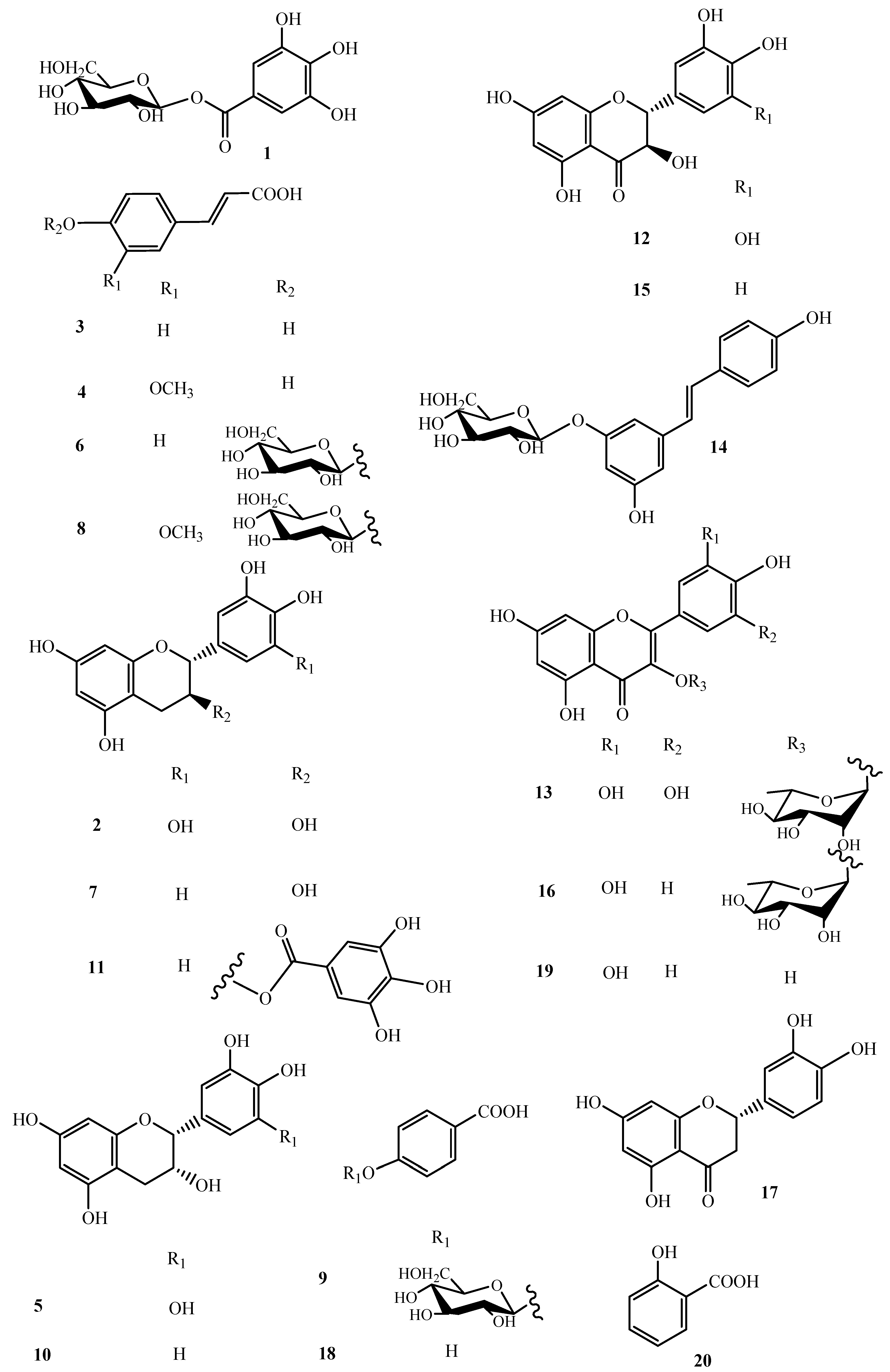
With the aim to obtain an in depth knowledge of the polar constituents of the pulp and to unambiguously attribute the peaks occurring in the HR-LC-ESI-Orbitrap-MS profile, a phytochemical investigation of the pulp n-BuOH extract was performed. n-BuOH extract was fractionated directly by HPLC-UV (Figure S1) to afford pure compounds, of which the structures were elucidated by 1D- and 2D-NMR experiments (Figure 2 and Figures S2–S21). In this way, galloyl 1-O-glucopyranoside (1), gallocatechin (2), p-coumaric acid (3), p-ferulic acid (4), epigallocatechin (5), p-coumaroyl 4-O-β-d-glucopyranoside (6), catechin (7), p-feruloyl-4-O-β-d-glucopyranoside (8), 4-hydroxybenzoic acid 4-O-β-d-glucopyranoside (9), epicatechin (10), gallocatechin-gallate (11), ampelopsin (12), myricetin 3-O-α-l-rhamnopyranoside (13), resveratrol-3-O-β-d-glucopyranoside (14), taxifolin (15), quercetin-3-O-α-l-rhamnopyranoside (16), eriodictyol (17), p-hydroxy benzoic acid (18), quercetin (19) and salycilic acid (20) were identified by comparison of their spectroscopic data with those reported in the literature [15,16,17,18,19,20].
In previous reports, some derivatives of gallocatechin, epigallocatechin, catechin and epicatechin, gallic acid, ellagic acid and derivative of hesperetin were only tentatively identified in lucuma on the basis of their UV spectra [3,5], thus this is the first report of their exact structures by isolation and structural elucidation. Eriodictyol (17) and quercetin (19) were reported in lucuma seeds [10]. Many compounds (1, 3, 4, 6, 8, 9, 1119), including salycilic acid (20), are reported here in lucuma pulp for the first time.

2.3. LC-MS Qualitative Analysis of Polar Lipids in P. lucuma Pulp n-BuOH Extract

2.3.1. Identification of Oxylipins (2125)

The analysis of the LC-ESI/LTQOrbitrap/MS spectra in negative ionization mode of the n-BuOH pulp extract showed pseudomolecular ions characterized by a molecular formula C18H36−2nO2m with n = 1, 2, 3 or 4 and m = 1, 2 or 3 and a fragmentation pattern attributable to the class of oxylipins (Table 2 and Table S1). These compounds are hydroxy fatty acids deriving from the oxidative metabolism of PUFAs such as linolenic acid (ALA, C18:3) and linoleic acid (LA, C18:2), differing in the unsaturation degree and number of hydroxyl groups [21,22]. Oxylipins yielded highly diagnostic MS2 fragmentation patterns (Table 2). According to the literature data [19,23], the position on the acyl chain of both hydroxyl groups and double bonds could be putatively assigned on the basis of characteristic product ions and diagnostic neutral losses generated by molecular rearrangements involving the head and the end of the acyl chain, respectively. Product ions generated by one or more consecutive neutral losses of 18 Da allowed to ascertain the number of hydroxyl groups occurring in the oxylipin structure. The detection of characteristic product ions, such as those at nominal m/z 171 (C9H15O3) and m/z 201 (C10H17O4), or those at nominal m/z 229 (C12H21O4), and 199 (C11H19O3), allowed to locate hydroxyl groups in the head (precisely at C9 and C10 positions) or in the tail of oxylipin (precisely at C12, C13, C15 and C16), respectively [19]. In particular, the MS/MS spectrum of compounds 21, 22, 24 and 25 showed diagnostic fragments at m/z 171, corresponding to the shortened acyl chain having the carboxyl group as COO and an aldehyde as terminal group originating from the rearrangement of the hydroxyl function on C9 following the cleavage of the C9–C10 bond [24]. Moreover, along with the molecular formula and the Ring Double Bond equivalent value (RDB), the fragmentation pattern allowed in some cases to establish the double bond position.
All this diagnostic information permitted us to establish the structure of compounds 21 ([M − H] at m/z 327.2171) and 22 ([M − H] at m/z 329.2326) differing for 2 Da but showing the same product ion at nominal m/z 229. This was formed by neutral loss of 98 Da (for compound 21) and 100 Da (for compound 22) from the corresponding molecular ion by rearrangement of the acyl chain end-part and breakdown of the C12–C13 bond, highlighting the presence of an additional double bond in the end-part of compound 21 in comparison with 22.
By these considerations, compounds 2125 could be putatively defined as reported in Table 2.

2.3.2. Identification of Galactolipids (2632, 34 and 36)

The analysis of the LC-ESI/LTQOrbitrap/MS profile highlighted the presence of pseudomolecular ions characterized by a molecular formula and a fragmentation pattern corresponding to glycolipids [25]. The LC-ESI HR/MS profile of P. lucuma pulp n-BuOH extract highlighted pairs of peaks—26 and 27, 28 and 29, 30 and 32—corresponding to the same molecular formula. The three pairs differed for the unsaturation degree.
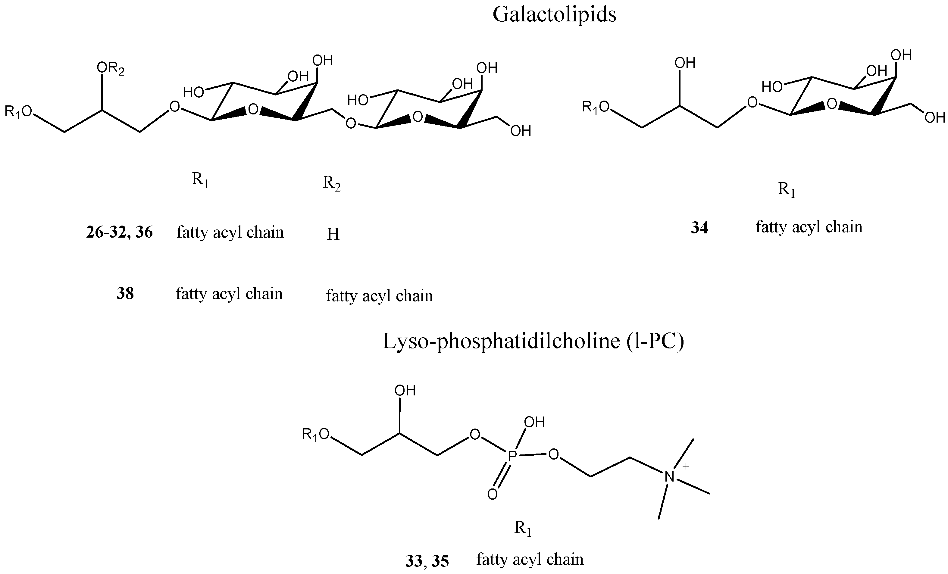
In the MS/MS spectrum of 2630 and 32, the same product ions at m/z 415, 397, and 235, leading to the assumption that 2630 and 32 had a common structural backbone could be observed. The product ions at m/z 397 and 415 derived from the [M − H] ion by neutral losses differing by a water molecule, respectively, corresponding to 278 and 260 Da for 26 and 27, to 280 and 262 Da for 28 and 29 and to 282 and 264 Da for 30 and 32. They could be referred to [(M-RCOOH) − H] and [(M-R=CO) − H] ions, as confirmed by the presence of the RCOO fatty acyl ion at m/z 277, 279 and 281, respectively. The common product ion at m/z 235 originated from the consecutive neutral losses of a fatty acid unit and one hexose moiety (162 Da), and could be assigned to the glycosylated form of a mono-dehydrated glycerol backbone. According to the literature data and their molecular formulae [23], compounds 2630, 31 and 32 could be tentatively identified as digalactosylmonoacylglycerol (DGMG) species differing in the unsaturation degree and/or regiospecificity of the fatty acyl group (Figure 3).
Moreover, the analysis of LC-MS/MS data allowed to assign compound 31 as a digalactosylmonoacylglycerols. Analogously, compound 34 could be identified as a monogalactosylmonoacylglycerols (MGMG), which was glycosylated with only one sugar unit (Figure 3).

2.3.3. Identification of Phosphatidylcholine Derivatives (33 and 35)

Analysis of the LC-ESI/LTQOrbitrap/MS profile in negative ionization mode of the pulp n-BuOH extract showed the presence of pseudomolecular ions corresponding to a molecular formula containing phosphorus heteroatom (Table 2 and Table S1). The fragmentation pattern of these ions showed diagnostic fragments of the polar heads attributable to the class of phosphatidylcholine derivatives [23]. For both compounds 33 and 35, in agreement with the literature data, the formation in negative ion mode of [(M + FA) − H] formic acid adducts for phosphatidylcholine derivatives was evident. Moreover, in tandem mass spectra of compounds 33 and 35, a product ion formed by the neutral loss of 60 Da from [(M + FA) − H] was observed, which allowed to identify this compound as l-PC (lyso-form of phosphatidylcholine) (Figure 3). This product ion could be attributed to the [(M-15) − H] ion, in which the l-PC derivative lost a methyl group from the choline head group to generate formic acid methyl ester [15].

2.4. LC-MS Analysis of P. lucuma Skin MeOH Extract

A preliminary metabolite fingerprint of P. lucuma skin MeOH extract was obtained by HR-LC-ESI-Orbitrap-MS analysis in negative ion mode. The LC-MS profile (Figure 1) showed eight main ion peaks which, on the basis of molecular formula, mass fragmentation behavior, and the literature data, could be defined as phenolics, distinguishable into galloyl 1-O-glucopyranoside (1), ellagic acid (37), flavonoids (15, 17 and 19), as well as polar lipids characterized by the presence of oxylipins (21 and 22) and a galactolipid (38) (Table 2 and Table S1). Among these metabolites, compounds 37 and 38 were not detected in the HR-LC-ESI-Orbitrap-MS profile of the pulp extract. Compound 37 showed a precursor ion [M − H] at m/z 300.0253, corresponding to the molecular formula C15H8O7 attributed to ellagic acid, previously reported in lucuma [3,5]. As evident in the HR-LC-ESI-Orbitrap-MS profile of P. lucuma skin MeOH extract, reported in Figure 1, the main compound is represented by taxifolin (15) and in smaller extent by eriodictyol (17).
Moreover, the analysis of LC-MS/MS data allowed to assign compound 38 as digalactosyldiacylglycerols (DGDG) [25], by considering the presence in its spectra of the characteristic product ions at m/z 415 and 397, corresponding to the digalactosylglycerol anion in the whole and in monodehydrated form, respectively (Table 2).

2.5. Antioxidant Activity of Isolated Metabolites by P. lucuma

The antioxidant capacity of isolated compounds was determined by TEAC assay (Table 3). Gallocatechin (2), epigallocatechin (5) and gallocatechin gallate (11) showed the strongest activity when compared with rutin, used as a reference compound. Taxifolin (15), the main compound occurring in the skin extract, showed a good activity with a value of 3.53 mM. This feature gives an interesting opportunity to valorize the skins for alternative uses, allowing companies to reduce their manufacturing costs.

3. Materials and Methods

3.1. Reagents

HPLC-grade and extraction solvents were purchased by VWR International PBI (Milano, Italy). LC-MS grade solvents were purchased by Merck (Darmstadt, Germany). 2,2-diphenyl-1-picrylhydrazyl (DPPH), 2,2′-azino-bis-(3-ethylbenzothiazoline-6-sulphonic acid (ABTS•+), potassium persulfate (K2S2O8), Folin-Ciocalteu reagent, Trolox, phosphate-buffered saline (PBS) solution, gallic acid and MeOH-d4 were purchased from Sigma-Aldrich (Darmstadt, Germany).

3.2. Plant Material and Extraction

Pouteria lucuma fruits have been purchased on Fruttaweb. The skins have been removed by pulp manually and pulp was separated from the seeds. A voucher specimen has been deposited in this Department (n. 155).
P. lucuma pulps and skins were stored in the freezer at temperature −5 °C, after some days they were submitted to lyophilization to obtain 250 g and 71.65 g, respectively.
Pulps were extracted by maceration employing solvent with increasing polarity. A pre-extraction procedure was used to remove undesirable components, such as fatty acids, employing n-hexane (900 mL, three times for three days) and chloroform (900 mL, three times for three days). Afterward, extraction with methanol (900 mL, three times for three days) was performed. After filtration and evaporation of the solvent to dryness in vacuo 35.41 g of MeOH crude extracts were obtained. The analysis carried out by TLC (Thin Layer Chromatography) showed an important presence of sugars; thus, two repartitions with n-BuOH and water (1:1) were carried out. 19.19 g of extract were used, solubilized in 200 mL of water and 200 mL of butanol, obtaining 18.40 mg of the water component and 0.73 mg of the BuOH component.
P. lucuma skins (71.65 g) were extracted in the same way by maceration employing n-hexane (155 mL, three times for three days), chloroform (155 mL, three times for three days), and methanol (155 mL, three times for three days). After filtration and evaporation of the solvent to dryness in vacuo, 240 mg of MeOH crude extract was obtained.

3.3. Total Phenolic Content

The total phenolic content of the extracts was determined by Folin-Ciocalteu assay following the procedure previously reported with slight modifications [12].
The extracts of pulp and skin have been dissolved in methanol to reach a concentration of 0.5 mg/mL. In the centrifuge tubes, Folin-Ciocalteu phenol reagent (0.5 mL), extract (0.5 mL), saturated sodium carbonate solution (1 mL) have been mixed and the volume has been adjusted with distilled water to 10 mL. The mixtures were allowed to react at room temperature for 45 min (until the characteristic blue color developed) and then centrifuged at 1046 RCF for 5 min. Absorbance of the clear supernatant has been measured at 517 nm on a UV-visible spectrophotometer (Thermo Scientific Multiskan Go, Waltham, MA, USA). A control without FC reagent and a blank with methanol instead of sample have been included in the assay.
A calibration curve of the standard gallic acid was used, following the same procedure used for the extracts. For gallic acid, the calibration equation was was y = 0.0027x + 0.0982 (R2 = 0.993). All the experiments were performed in triplicate, and results were expressed as a mean of gallic acid equivalents (GAE mg/g dried extract).

3.4. DPPH Radical Scavenging Activity

DPPH radical scavenging activity of the extracts was evaluated by employing the protocol previously described [26] with slight modifications. In brief, stock solutions (10 mg/mL) of the different typologies of the extracts were used to obtain six different concentrations: from 10 to 500 μg/mL of pulp and skin extracts. The samples were stirred vigorously for 10 s and kept in the dark for 30 min; subsequently, the samples were put in a multiwall plate, in detail in each well has been added 244 µL of DPPH and 6 µL of each sample. The absorbance was measured at 517 nm using a Thermo Scientific Multiskan Go Spectrophotometer. A control solution was prepared by replacing the tested extracts with MeOH. The percentage of DPPH radical scavenging activity of the extracts was calculated as follows:
DPPH free radical scavenging activity (I%) = [(A0 − A)/A0] × 100. Where A0 is the absorbance of the control solution, and A is the absorbance of the DPPHsolution containing the extract. The percentage of DPPH radical scavenging activity (%) was plotted against the extract concentration (μg/mL) to determine the IC50. All the experiments were performed in triplicate.

3.5. ABTS•+ Radical Scavenging Activity

ABTS•+ radical scavenging activity was evaluated by using the method previously reported [17] with slight modifications. The radical ABTS•+ has been generated chemically by the oxidation of ABTS 2 mM (50 mL) with K2S2O8 70 mM (0.2 L) after for 16 h of incubation in the dark at room temperature. Then, the mixture has been diluted with a phosphate-buffered saline (PBS) solution (pH = 7.4) to an absorbance of 0.700 ± 0.020 measured at 734 nm.
30 μL of four concentrations of the extracts (250, 500, 750, 1000 μg/mL) were added to 300 µL of ABTS•+ radical solution in each well and their absorbance was measured at 734 nm by employing Thermo Scientific Multiskan Go Spectrophotometer. A negative control was prepared by using MeOH instead of the extract. Trolox was used as a reference standard, and results were expressed as TEAC value [17]. Each determination was performed in triplicate.

3.6. HR-LC-ESI-Orbitrap-MS and HR-LC-ESI-Orbitrap-MS/MS Analysis

HR-LC-ESI-Orbitrap-MS/MS analyses were performed in negative ion mode, employing a Thermo Scientific Accela HPLC System (Thermo Scientific, Dreieich, Germany) equipped with a Phenomenex (Torrance, CA, USA) Kinetex EVO C-18 column (150 × 2.10 mm, 5 µm), coupled to a LTQ-Orbitrap XL mass spectrometer (Thermo Fisher Scientific, San Jose, CA, USA) [15].
The employed mobile phases were water-formic acid (A, 99.9:0.1, v/v) and acetonitrile-formic acid (B, 99.9:0.1, v/v). The gradient conditions used for LC separation were the following: 0 min 5% B, 25 min 80% B (held for 3 min), 35 min 5% B (held for 10 min). The flow rate was 0.2 mL/min, injection volume was 4 µL (0.5 mg/mL) and the column was kept at room temperature.
The Orbitrap mass analyzer was calibrated according to the manufacturer’s directions [25]. The scan was collected in the Orbitrap at a resolution of 30,000 in a m/z range of 150–2000 Da. The m/z of each identified compound was calculated to 4 decimal places and measured with a mass accuracy < 3.88 ppm. The (−)ESI parameter settings were: capillary temperature at 280 °C, sheath gas flow at 15 (arbitrary units), auxiliary gas flow at 5 (arbitrary units), source voltage at 3.5 kV, capillary voltage at −48 V, and tube lens offset at −176.47 V. In LC-(−)ESI/HRMS experiments, the Total Ion Current (TIC) profile was produced by monitoring the intensity of all the ions produced and acquired in every scan during the chromatographic run. A normalized collision energy at 30%, a minimum signal threshold at 250, and an isolation width at 2.0 were used. MS/MS data were acquired by using the Data-Dependent Scan experiment, allowing to select the precursor ion as the most intense peak during LC-MS analyses. Xcalibur software (version 2.1, Dreieich, Germany) was used for instrument control, data acquisition and data analysis.

3.7. Isolation of Secondary Metabolites from P. lucuma Pulp Extract

n-BuOH extract of P. lucuma pulp was chromatographed by an Agilent 1260 Infinity system (Agilent Technologies, Palo Alto, CA, USA), equipped with a binary pump (G-1312C) and a UV detector (G-1314B), Phenomenex C18 Synergi-Hydro-RP (250 × 10 mm; 10 μm) column and a Rheodyne injector were used.
The sample was solubilized in methanol to obtain a 15 mg /100 μL and 100 μL were injected for a total of 14 chromatographic runs.
The following solvents were used: solvent (A) water-formic acid (0.1%) and (Bacetonitrile-formic acid (0.1%), with a flow of 2 mL/min, at a wavelength of 310. For all the chromatographic runs the following gradient was used: the first step involved the use of a 5% of B maintained for 5 min, in 45 min 80 % of B was reached, in 10 min it arrived at 100% of B, the latter % was held for 10 min.
In this way the following compounds 1 (1.8 mg, tR = 5.2 min), 2 (2.1 mg, tR = 8.6 min), 3 (2.2 mg, tR = 23.0 min), 4 (2.0 mg, tR = 24.5 min), 5 (1.2 mg, tR = 8.8 min), 6 (1.7 mg, tR R = 18.5 min), 7 (2.1 mg, tR = 12.2 min), 8 (1.8 mg, tR = 19.2 min), 9 (1.9 mg, tR = 23.5 min), 10 (1.9 mg, tR = 12.6 min), 11 (1.6 mg, tR = 6.5 min), 12 (2.8 mg, tR = 16.5 min), 13 (1.6 mg, tR = 17.8 min), 14 (2.6 mg, tR = 19.6 min), 15 (3.1 mg, tR = 20.8 min), 16 (1.3 mg, tR = 21.2 min), 17 (2.0 mg, tR = 24.6 min), 18 (2.3 mg, tR = 24.0 min), 19 (1.2 mg, tR = 23.3 min), and 20 (2.3 mg, tR = 29.5 min) were isolated. The structures of these compounds have been unambiguously elucidated by NMR research.

3.8. NMR Analysis

NMR spectroscopic data were acquired in MeOH-d4 (99.95%, Sigma-Aldrich) on a Bruker DRX-600 spectrometer (Bruker BioSpin GmBH, Rheinstetten, Germany) equipped with a Bruker 5 mm TCI CryoProbe at 300 K. Data processing was carried out with Topspin 3.2 software (Bruker BioSpin, Rheinstetten, Germany) Standard pulse sequences and phase cycling were used for DQF-COSY, HSQC, HMBC, and ROESY spectra.

4. Conclusions

In the present study, a total of 36 compounds were detected in P. lucuma pulp n-BuOH extract using a LC-ESI/LTQOrbitrap/MS/MS analysis. The phenolic compounds were isolated and their structures were unambiguously identified by NMR experiments. So far, the fruits have been studied mainly for their content of sugars, organic acids, total phenolics and total carotenoids and this is the first accurate investigation highlighting the structures of the compounds occurring in the extract.
A large number of papers on food plant metabolomics often describing incomplete characterization of plant constituents or tentative structures have been reported. It is evident that the putative attribution of structures by the only MS analysis should be confirmed by isolation of compounds and structure elucidation by NMR experiments. In this work, a strategy based on the combination of LC-MS and NMR techniques has been used as a powerful tool to achieve a deeper knowledge of lucuma metabolite profile.
Some of the isolated compounds showed a radical scavenging activity higher than rutin, used as a reference compound. On the basis of the obtained results, lucuma might be considered as a rich source of bioactives with antioxidant properties.
Analysis of the LC-ESI/LTQOrbitrap profile, accurate mass measurements, fragmentation pattern analyses, and comparison with literature data allowed to putatively identify 16 lipid compounds belonging to oxylipin, glycolipid and phospholipid classes, all of them for the first time described in lucuma. It is noteworthy that the extract contained a wide range of polar lipids, along with phenolic compounds. Considering the biological activities reported for lipid classes and their effects on human health [25], these data reinforce the use of lucuma in human nutrition as a food rich in different classes of bioactive and healthy lipids with beneficial effects.
To encourage the recycling and exploitation of lucuma by-products, preliminarily the total phenolic content of the skin extract was evaluated (560.69 mg GAE/g extract). In order to correlate the total phenolic content of the extract to its chemical composition, a LC-ESI/LTQOrbitrap/MS/MS analysis was carried out on P. lucuma skin extract. Taxifolin (15) represents the predominant compound along with eriodictyol (17). Moreover, taxifolin (15) showed a strong radical scavenging activity with a TEAC value of 3.53. This work demonstrated that lucuma skins, which represent an industry waste from lucuma processing could be useful for the preparation of food supplements.

Supplementary Materials

The following materials are available online: Table S1. Molecular formula [M − H], [(M + HCOOH) − H], characteristic product ions (molecular formula and intensity) occurring in P. lucuma pulp n-BuOH extract and P. lucuma skin MeOH extract, identified by LCESI/LTQOrbitrap/MS/MS (negative ion mode); Figure S1. Chromatogram of P. lucuma pulp extract on the C18 Synergi-Hydro-RP column; Figures S2–S21. 1H NMR Spectrum (600 MHz, CD3OD) of compound 120.

Author Contributions

Conceptualization, C.P. and S.P.; methodology, A.C. and M.M.; formal analysis, A.C. and M.M.; investigation, A.C.; resources, S.P.; data curation, A.C. and M.M.; writing—original draft preparation, A.C. and M.M.; writing—review and editing, S.P.; supervision, C.P. and S.P.; project administration, S.P.; funding acquisition, S.P. All authors have read and agreed to the published version of the manuscript.

Funding

This research received no external funding.

Institutional Review Board Statement

Not applicable.

Informed Consent Statement

Not applicable.

Data Availability Statement

The data presented in this study are available in supplementary materials.

Conflicts of Interest

The authors declare no conflict of interest.

Sample Availability

Samples of the compounds 120 are available from the authors.

References

  1. Silva, C.A.M.; Simeoni, L.A.; Silveira, D. Genus Pouteria: Chemistry and biological activity. Rev. Bras. Farmacogn. 2009, 19, 501–509. [Google Scholar] [CrossRef] [Green Version]
  2. Ma, J.; Yang, H.; Basile, M.J.; Kennelly, E.J. Analysis of polyphenolic antioxidants from the fruits of three pouteria species by selected ion monitoring liquid chromatography-mass spectrometry. J. Agric. Food Chem. 2004, 52, 5873–5878. [Google Scholar] [CrossRef]
  3. Garcia-Rios, D.; Aguilar-Galvez, A.; Chirinos, R.; Pedreschi, R.; Campos, D. Relevant physicochemical properties and metabolites with functional properties of two commercial varieties of Peruvian Pouteria lucuma. J. Food Process. Preserv. 2020, 44, e14479. [Google Scholar] [CrossRef]
  4. Rojo, L.E.; Villano, C.M.; Joseph, G.; Schmidt, B.; Shulaev, V.; Shuman, J.L.; Lila, M.A.; Raskin, I. Wound-healing properties of nut oil from Pouteria lucuma. J. Cosmet. Dermatol. 2010, 9, 185–195. [Google Scholar] [CrossRef] [Green Version]
  5. Fuentealba, C.; Galvez, L.; Cobos, A.; Olaeta, J.A.; Defilippi, B.G.; Chirinos, R.; Campos, D.; Pedreschi, R. Characterization of main primary and secondary metabolites and in vitro antioxidant and antihyperglycemic properties in the mesocarp of three biotypes of Pouteria lucuma. Food Chem. 2016, 190, 403–411. [Google Scholar] [CrossRef] [PubMed]
  6. Aguilar-Galvez, A.; Garcia-Rios, D.; Janampa, C.; Mejia, C.; Chirinos, R.; Pedreschi, R.; Campos, D. Metabolites, volatile compounds and in vitro functional properties during growth and commercial harvest of Peruvian lucuma (Pouteria lucuma). Food Biosci. 2021, 40, 100882. [Google Scholar] [CrossRef]
  7. Maza-De la Quintana, R.; Paucar-Menacho, L.M. Lucuma (Pouteria lucuma): Composition, bioactive components, antioxidant activity, uses and beneficial properties for health. Sci. Agropecu. 2020, 11, 135–142. [Google Scholar] [CrossRef] [Green Version]
  8. Masullo, M.; Cerulli, A.; Montoro, P.; Pizza, C.; Piacente, S. In depth LC-ESIMSn-guided phytochemical analysis of Ziziphus jujuba Mill. leaves. Phytochemistry 2019, 159, 148–158. [Google Scholar] [CrossRef] [PubMed]
  9. Masullo, M.; Mari, A.; Cerulli, A.; Bottone, A.; Kontek, B.; Olas, B.; Pizza, C.; Piacente, S. Quali-quantitative analysis of the phenolic fraction of the flowers of Corylus avellana, source of the Italian PGI product “Nocciola di Giffoni”: Isolation of antioxidant diarylheptanoids. Phytochemistry 2016, 130, 273–281. [Google Scholar] [CrossRef]
  10. Guerrero-Castillo, P.; Reyes, S.; Robles, J.; Simirgiotis, M.J.; Sepulveda, B.; Fernandez-Burgos, R.; Areche, C. Biological activity and chemical characterization of Pouteria lucuma seeds: A possible use of an agricultural waste. Waste Manag. 2019, 88, 319–327. [Google Scholar] [CrossRef]
  11. Taiti, C.; Colzi, I.; Azzarello, E.; Mancuso, S. Discovering a volatile organic compound fingerprinting of Pouteria lucuma fruits. Fruits 2017, 72, 131–138. [Google Scholar] [CrossRef]
  12. Masullo, M.; Cerulli, A.; Mari, A.; de Souza Santos, C.C.; Pizza, C.; Piacente, S. LC-MS profiling highlights hazelnut (Nocciola di Giffoni PGI) shells as a byproduct rich in antioxidant phenolics. Food Res. Int. 2017, 101, 180–187. [Google Scholar] [CrossRef]
  13. Montoro, P.; Teyeb, H.; Masullo, M.; Mari, A.; Douki, W.; Piacente, S. LC-ESI-MS quali-quantitative determination of phenolic constituents in different parts of wild and cultivated Astragalus gombiformis. J. Pharm. Biomed. Anal. 2013, 72, 89–98. [Google Scholar] [CrossRef] [PubMed]
  14. Hamed, A.I.; Al-Ayed, A.S.; Moldoch, J.; Piacente, S.; Oleszek, W.; Stochmal, A. Profiles analysis of proanthocyanidins in the argun nut (Medemia argun-an ancient Egyptian palm) by LC-ESI-MS/MS. J. Mass Spectrom. 2014, 49, 306–315. [Google Scholar] [CrossRef] [PubMed]
  15. Cerulli, A.; Napolitano, A.; Masullo, M.; Hosek, J.; Pizza, C.; Piacente, S. Chestnut shells (Italian cultivar “Marrone di Roccadaspide” PGI): Antioxidant activity and chemical investigation with in depth LC-HRMS/MS(n) rationalization of tannins. Food Res. Int. 2020, 129, 108787. [Google Scholar] [CrossRef]
  16. Mahmood, N.; Piacente, S.; Burke, A.; Khan, A.; Pizza, C. Constituents of Cuscuta reflexa are anti-HIV agents. Antivir. Chem. Chemother. 1997, 8, 70–74. [Google Scholar] [CrossRef] [Green Version]
  17. Bottone, A.; Masullo, M.; Montoro, P.; Pizza, C.; Piacente, S. HR-LC-ESI-Orbitrap-MS based metabolite profiling of Prunus dulcis Mill. (Italian cultivars Toritto and Avola) husks and evaluation of antioxidant activity. Phytochem. Anal. 2019, 30, 415–423. [Google Scholar] [CrossRef] [PubMed]
  18. Funari, C.S.; Passalacqua, T.G.; Rinaldo, D.; Napolitano, A.; Festa, M.; Capasso, A.; Piacente, S.; Pizza, C.; Young, M.C.M.; Durigan, G.; et al. Interconverting flavanone glucosides and other phenolic compounds in Lippia salviaefolia Cham. ethanol extracts. Phytochemistry 2011, 72, 2052–2061. [Google Scholar] [CrossRef] [PubMed]
  19. D’Urso, G.; Napolitano, A.; Cannavacciuolo, C.; Masullo, M.; Piacente, S. Okra fruit: LC-ESI/LTQOrbitrap/MS/MS(n) based deep insight on polar lipids and specialized metabolites with evaluation of anti-oxidant and anti-hyperglycemic activity. Food Funct. 2020, 11, 7856–7865. [Google Scholar] [CrossRef]
  20. Benavides, A.; Montoro, P.; Bassarello, C.; Piacente, S.; Pizza, C. Catechin derivatives in Jatropha macrantha stems: Characterisation and LC/ESI/MS/MS quali-quantitative analysis. J. Pharm. Biomed. Anal. 2006, 40, 639–647. [Google Scholar] [CrossRef]
  21. Geng, P.; Harnly, J.M.; Chen, P. Differentiation of Whole Grain from Refined Wheat (T. aestivum) Flour Using Lipid Profile of Wheat Bran, Germ, and Endosperm with UHPLC-HRAM Mass Spectrometry. J. Agric. Food Chem. 2015, 63, 6189–6211. [Google Scholar] [CrossRef]
  22. Klockmann, S.; Reiner, E.; Bachmann, R.; Hackl, T.; Fischer, M. Food Fingerprinting: Metabolomic Approaches for Geographical Origin Discrimination of Hazelnuts (Corylus avellana) by UPLC-QTOF-MS. J. Agric. Food Chem. 2016, 64, 9253–9262. [Google Scholar] [CrossRef] [PubMed]
  23. Napolitano, A.; Cerulli, A.; Pizza, C.; Piacente, S. Multi-class polar lipid profiling in fresh and roasted hazelnut (Corylus avellana cultivar “Tonda di Giffoni”) by LC-ESI/LTQOrbitrap/MS/MS(n). Food Chem. 2018, 269, 125–135. [Google Scholar] [CrossRef]
  24. Levandi, T.; Püssa, T.; Vaher, M.; Toomik, P.; Kaljurand, M. Oxidation products of free polyunsaturated fatty acids in wheat varieties. Eur. J. Lipid Sci. Technol. 2009, 111, 715–722. [Google Scholar] [CrossRef]
  25. Cerulli, A.; Napolitano, A.; Hosek, J.; Masullo, M.; Pizza, C.; Piacente, S. Antioxidant and In Vitro Preliminary Anti-Inflammatory Activity of Castanea sativa (Italian Cultivar “Marrone di Roccadaspide” PGI) Burs, Leaves, and Chestnuts Extracts and Their Metabolite Profiles by LC-ESI/LTQOrbitrap/MS/MS. Antioxidants 2021, 10, 278. [Google Scholar] [CrossRef] [PubMed]
  26. Kilinc, H.; Masullo, M.; D’Urso, G.; Karayildirim, T.; Alankus, O.; Piacente, S. Phytochemical investigation of Scabiosa sicula guided by a preliminary HPLC-ESIMS(n) profiling. Phytochemistry 2020, 174, 112350. [Google Scholar] [CrossRef] [PubMed]
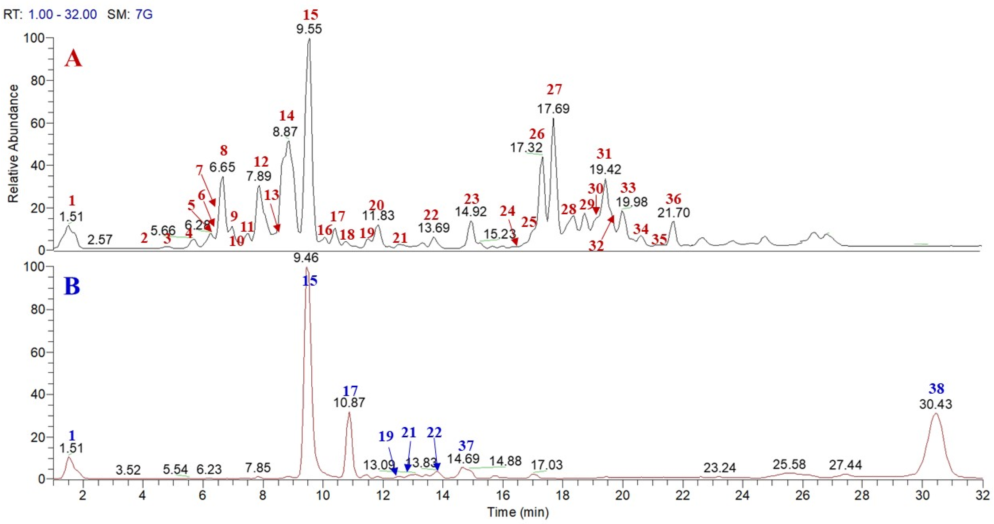
Figure 1. HR-LC-ESI-Orbitrap-MS profile of pulp n-BuOH extract (A) and skin methanol extract (B) of P. lucuma.
Molecules 26 05236 g001
Figure 2. Compounds isolated from P. lucuma pulp n-BuOH extract.
Molecules 26 05236 g002
Figure 3. Polar lipids (galactolipids and Lyso-phosphatidilcholine (l-PC) isolated from P. lucuma pulp n-BuOH extract.
Molecules 26 05236 g003
Table 1. Total phenolic content (TPC), DPPH and ABTS•+ radical scavenging activity of the extracts of P. lucuma pulp and skin evaluated by spectrophotometric assays.
P. lucumaTPC aDPPH•bABTS•+c
MeOH extract of skin *560.69 ± 4.7652.71 ± 1.47 *3.67 ± 0.27
n-BuOH extract of pulp **93.53 ± 4.83150.00 ± 2.55 **2.24 ± 0.12
Vitamin C 4.85 ± 0.05-
rutin (mM) 4.65 ± 0.15
* Range concentration 10–200 (μg/mL); ** Range concentration 50–500 (μg/mL). a Values are expressed as gallic acid equivalents (GAE) mg/g of dried extract. b Values are expressed as IC50 in μg/mL. c Values are expressed as concentration (mM) of a standard Trolox solution exerting the same antioxidant activity of a 1 mg/mL solution of the tested extract. Results are expressed as mean of three experiments.
Table 2. Compounds identified in P. lucuma pulp n-BuOH extract and P. lucuma skin MeOH extract by LCESI/LTQOrbitrap/MS/MS (negative ion mode).
MeOH Extract of P. lucuma Pulp
CompoundtR (min)Molecular Formula[(M + HCOOH) − H][M − H]Δ ppmProduct IonsClassification
1galloyl 1-O-glucopyranoside1.51C13H16O10 331.06641.35169phenolic
2gallocatechin4.84C15H14O7 305.065611.61287, 261, 221, 175, 125flavanol
3p-coumaric acid5.60C9H8O3209.0451 2.97145, 119phenolic
4p-ferulic acid5.66C10H10O4239.0554 1.78149, 133phenolic
5epigallocatechin6.28C15H14O7 305.06590.34287, 261, 221, 175, 125flavanol
6p-coumaroyl 4-O-β-d-glucopyranoside6.33C15H18O8 325.09280.98163, 145phenolic
7catechin6.53C15H14O6 289.07131.96179, 151, 137flavanol
8p-feruloyl-4-O-β-d-glucopyranoside6.65C16H20O9 355.10250.48193, 175phenolic
94-hydroxybenzoic acid 4-O-β-d-glucopyranoside6.96C13H16O8 299.07630.56137phenolic
10epicatechin7.33C15H14O6 289.07152.01179, 151, 137flavanol
11gallocatechin-gallate7.64C22H18O11 457.07690.75287, 169flavanol
12ampelopsin7.89C15H12O8 319.04562.24301, 193dihydroflavonol
13myricetin 3-O-α-l-rhamnopyranoside8.38C21H20O12 479.08240.72317flavonol
14resveratrol-3-O-β-d-glucopyranoside8.87C20H22O8 389.12320.14227phenolic
15taxifolin9.55C15H12O7 303.05031.16285, 177, 125dihydroflavonol
16quercetin 3-O-β-d-rhamnopyranoside10.41C21H20O11 447.0927−0.20301flavonol glycoside
17eriodictyol10.91C15H12O6 287.05500.02179, 163, 153dihydroflavonol
18p-hydroxy benzoic acid11.53C7H6O3 137.02183.1093phenolic
19quercetin11.70C15H10O7 301.03502.46179, 151flavonol
20salycilic acid11.83C7H6O3 137.02183.1093phenolic
21TriHoDe13.01C18H32O5 327.21710.51309, 291, 273, 229, 221, 171oxylipin
22TriHoMe13.69C18H34O5 329.23260.32311, 293, 229, 211, 199, 171oxylipin
23TriHoDe14.92C18H28O4 307.19090.48289, 271, 243, 235, 209oxylipin
24hydroxy-epoxy-octadecadienoic acid16.34C18H30O4 309.20650.50291, 273, 201, 171oxylipin
25hydroxy-epoxy-octadecadienoic acid isomer16.71C18H30O4 309.20650.5291, 273, 201, 171oxylipin
26DGMG (18:3)17.32C33H56O14721.3634 −2.54675, 415, 397, 305, 277, 235, 205galactolipid
27DGMG (18:3)17.69C33H56O146721.3631 −2.98675, 415, 397, 305, 277, 235, 205galactolipid
28DGMG (18:2)18.30C33H58O14723.3796 −3.47677, 415, 397, 279, 235galactolipid
29DGMG (18:2)18.74C33H58O14723.3795 −3.47677, 415, 397, 279, 235galactolipid
30DGMG (18:1)19.17C33H60O14725.3937 −3.87679, 415, 397, 281, 235galactolipid
31DGMG (16:0)19.42C31H58O14699.3788 −1.40415, 397, 235galactolipid
32DGMG (18:1)19.60C33H60O14725.3937 −3.87679, 415, 397, 281, 235galactolipid
33l-PC (16:0)19.98C24H50O7NP540.3296 0.028480, 255phosphatidylcholine
34MGMG (16:3)20.59C25H42O9 485.2744−0.27235galactolipid
35l-PC (18:1)20.59C26H52O7NP566.3451 −0.17506, 281phosphatidylcholine
36DGMG (18:0)21.70C33H62O14727.4096 −1.98681, 397galactolipid
MeOH extract of P. lucuma skin
1galloyl 1-O-glucopyranoside1.51C13H16O10 331.06681.39169phenolic
15taxifolin9.46C15H12O7 303.05031.16285, 177, 125dihydroflavonol
17eriodictyol10.87C15H12O6 287.05582.67179, 163, 153dihydroflavonol
19quercetin12.58C15H10O7 301.03461.00179, 151flavonol
21TriHoDe12.90C18H32O5 327.21690.88229, 211, 171oxylipin
22TriHOME13.83C18H34O5 329.23261.18293, 229, 211, 199, 171oxylipin
37ellagic acid14.69C14H6O8 300.9983−3.85257, 201phenolic
38DGDG (18.3, 16:0)30.50C49H86O15 959.5934−0.35913, 415, 397, 277, 255galactolipid
Table 3. ABTS•+ radical scavenging activity of isolated compounds 120.
CompoundTEAC Value (mM ± SD)
1galloyl 1-O-glucopyranoside3.24 ± 0.07
2gallocatechin5.12 ± 0.21
3p-coumaric acid0.70 ± 0.03
4p-ferulic acid1.93 ± 0.01
5epigallocatechin5.29 ± 0.30
6p-coumaroyl 4-O-β-d-glucopyranoside0.79 ± 0.01
7catechin2.56 ± 0.27
8p-feruloyl-4-O-β-d-glucopyranoside2.01 ± 0.05
94-hydroxybenzoic acid 4-O-β-d-glucopyranoside2.46 ± 0.07
10epicatechin2.14 ± 0.24
11gallocatechin-gallate5.92 ± 0.16
12ampelopsin2.29 ± 0.03
13myricetina 3-O-α-l-rhamnopyranoside4.41 ± 0.23
14resveratrol-3-O-β-d-glucopyranoside2.00 ± 0.40
15taxifolin3.53 ± 0.29
16quercetin 3-O-β-d-rhamnopyroside4.85 ± 0.10
17eriodictyol2.12 ± 0.06
18p-hydroxy benzoic acid2.25 ± 0.09
19quercetin4.71 ± 0.27
20salycilic acid2.12 ± 0.03
rutin4.65 ± 0.15
Results are expressed as mean of three experiments.
Publisher’s Note: MDPI stays neutral with regard to jurisdictional claims in published maps and institutional affiliations.