Article

Differential Expression of Yersinia pseudotuberculosis General Porin Genes during Short- and Long-Term Antibiotic Stresses

1
G.B. Elyakov Pacific Institute of Bioorganic Chemistry, Far Eastern Branch, Russian Academy of Sciences, 159, Pr. 100 Let Vladivostoku, 690022 Vladivostok, Russia
2
School of Biomedicine, Far Eastern Federal University, 8 Sukhanova St., 690090 Vladivostok, Russia
3
Friedrich-Loeffler-Institut, Federal Research Institute for Animal Health, Institute for Bacterial Infections and Zoonoses, Naumburger Str. 96a, D-07743 Jena, Germany
*
Author to whom correspondence should be addressed.
Molecules 2021, 26(13), 3956; https://doi.org/10.3390/molecules26133956
Submission received: 2 June 2021 / Revised: 21 June 2021 / Accepted: 23 June 2021 / Published: 28 June 2021

Abstract

:
Here, we investigated general porin regulation in Yersinia pseudotuberculosis 488, the causative agent of Far Eastern scarlet-like fever, in response to sublethal concentrations of antibiotics. We chose four antibiotics of different classes and measured gene expression using qRT-PCR and GFP reporter systems. Our data showed temporal regulation of the general porin genes ompF and ompC caused by antibiotic stress. The porin transcription initially decreased, providing early defensive response of the bacterium, while it returned to that of the untreated cells on prolonged antibiotic exposure. Unlike the major porin genes, the transcription of the alternative porin genes ompX and lamB was increased. Moreover, a short-term ompR- and marA-mediated porin regulation was observed. The main finding was a phenotypic heterogeneity of Y. pseudotuberculosis population manifested in variable porin gene expression under carbenicillin exposure. This may offer adaptive fitness advantages for a particular bacterial subpopulation.

1. Introduction

The global rise of antibiotic resistance in pathogens represents a serious threat to medicine, as it is the predominant cause of treatment failure and increased mortality [1]. Recent evidence suggests that sublethal concentrations of antimicrobials frequently found in the human body and nature play an important role in the development of antibiotic resistance [2]. Initially, susceptible bacterial populations survive and propagate under such conditions due to adaptive gene expression responses that have been observed in a variety of bacterial species [1,3,4,5]. In Gram-negative bacteria, altering the gene expression of outer membrane proteins or porins, resulting in decreased membrane permeability, is considered one of the first defense mechanisms of adaptation to antibiotic stress [1].
Yersinia pseudotuberculosis, a Gram-negative bacterium belonging to the Enterobacteriaceae family, is a human pathogen causing pseudotuberculosis infection. In Russia and Japan, it causes outbreaks of the disease known as Far Eastern scarlet-like fever (FESLF), with serious systemic inflammatory symptoms [6].
Y. pseudotuberculosis produces two major nonspecific porins, OmpF and OmpC, which we study intensively [7,8,9,10,11]. These porins consist of 16-stranded β-barrel trimers, each of which forms a central channel [12]. The channels control the permeability of the cell envelope for low molecular compounds, including β-lactam, tetracycline, chloramphenicol, and fluoroquinolone antibiotics [13,14,15,16]. Thus, the porins provide cell defense against certain antibiotics and subsequently mediate antimicrobial resistance by downregulating their gene expression or inducing beneficial mutations [17]. Moreover, the reversible phenotypical response offers advantages over conventional irreversible mutations [18]. Furthermore, transcriptional porin regulation is considered a rapid adaptive mechanism to environmental and antibiotic stresses occurring within the first 60 min [19]. The porin-mediated stress response has been widely studied in various enterobacteria, including Escherichia coli, Klebsiella pneumonia, Salmonella enterica, Serratia marcescens, and others [20,21,22]. Still, little is known about the effect of antibiotic stress on the general porin regulation in Y. pseudotuberculosis. Most research focuses on the effect of lethal antibiotic concentrations on bacterial physiology and resistance, while subinhibitory antimicrobial doses have been mostly disregarded [23]. Y. pseudotuberculosis genetic response to low concentrations of antibiotics is of particular interest because successful treatment of pseudotuberculosis infection largely depends on antibiotic therapy to maintain lethal drug concentrations in different human tissues.
Therefore, we aim to investigate the role of general porin regulation in Y. pseudotuberculosis adaptation to sublethal concentrations of four antibiotics utilizing porin channels for cell entrance.

2. Results and Discussion

2.1. Antibiotics and Y. pseudotuberculosis 488 Susceptibility

Y. pseudotuberculosis 488 (= strain 117), serotype O:1b, was isolated from a patient with FESLF in the Russian Far East region. To elucidate Y. pseudotuberculosis porin regulation under antibiotic stress, we chose carbenicillin, tetracycline, kanamycin, and chloramphenicol. These antibiotics have been shown to use porin channels to enter into a bacterial cell [13,14]. Several studies have demonstrated that porin regulation, resulting in decreased penetration of antimicrobials into the cell, contributes to bacterial adaptive resistance [24]. We determined the MIC ranges of the selected antibiotics for Y. pseudotuberculosis 488 at 27 °C (optimal for bacterial cultivation) and 37 °C (human body temperature) since it is known that porin expression is tightly regulated by environmental factors [25,26], predominantly by temperature [27,28]. Such regulation is shown for Y. pseudotuberculosis strains in the interactive RNA atlas www.pathogenex.org (ompF YPK_2649; ompC YPK_2839).
Y. pseudotuberculosis 488 showed high susceptibility to the tested antibiotics (Table S1). However, cells cultivated at 27 °C exhibited a 2-fold increased resistance to kanamycin and tetracycline compared with 37 °C, whereas the MICs of carbenicillin and chloramphenicol were not affected by incubation temperature. To induce antibiotic stress, we used subinhibitory concentrations (sub-MICs) of antibiotics corresponding to ½ MICs. Such sublethal antibiotic concentrations reflect the conditions that bacteria may encounter in the natural environments and the human body [29]. Moreover, exposure to sublethal concentration has been reported to play a protective role in the bacterial cell against a wide range of antimicrobials and alter the expression of various bacterial genes that can lead to nonspecific resistance [22].

2.2. An Early Transcriptional Response of General Porin Genes to Antibiotic Stress

Rapid modulation of porin gene expression is the first line of bacterial defense against harmful compounds, including antibiotics, which increases the chances of survival and adaptation of microorganisms to stressful conditions [19].
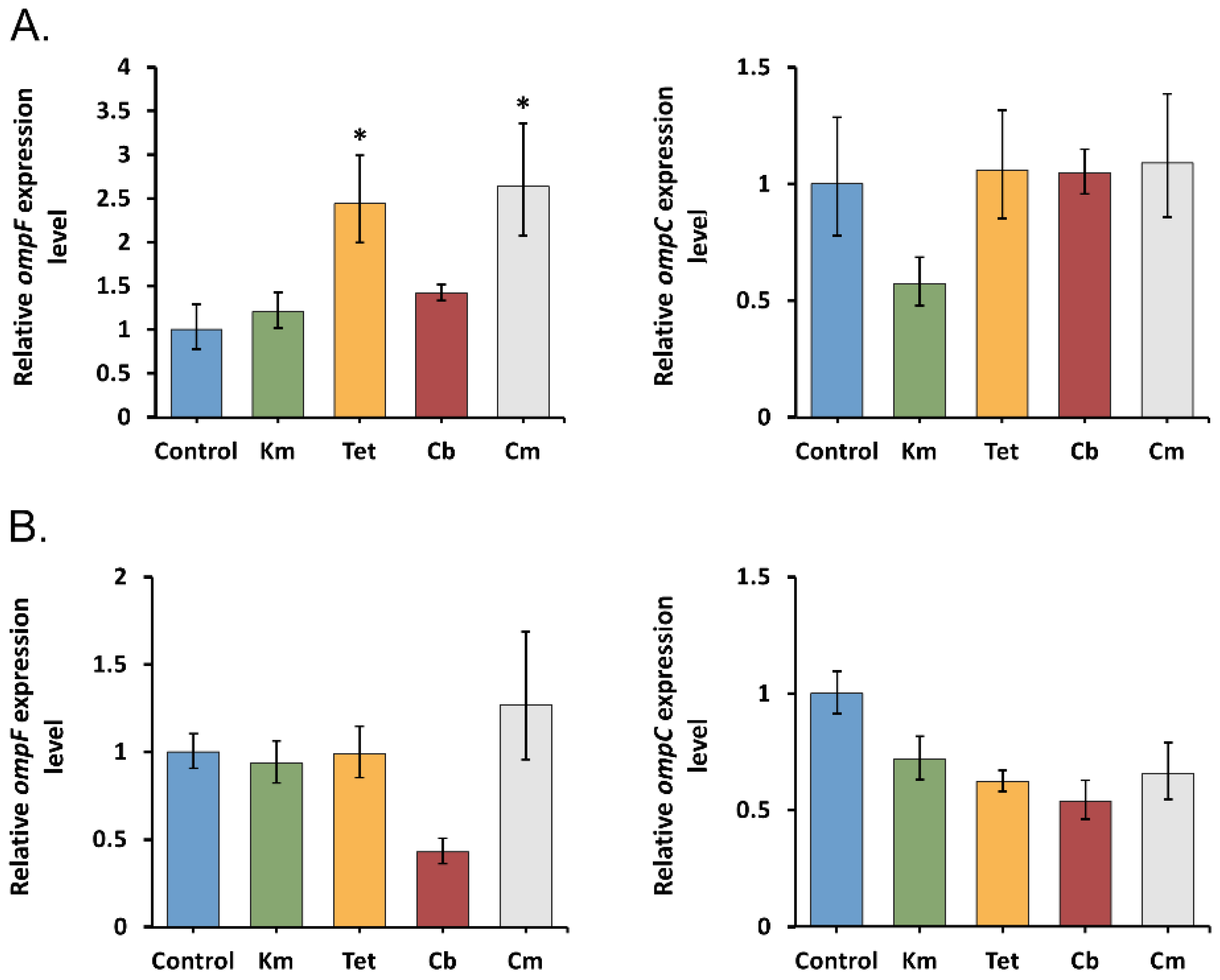
To study the early porin response in Y. pseudotuberculosis 488, we treated bacterial cells by sub-MICs of antibiotics for an hour, followed by measuring the level of ompF and ompC transcripts using qRT-PCR. The results obtained (Figure 1) indicate that kanamycin and tetracycline cause a transcription decrease in both general porins, regardless of temperature. ompF and ompC expression were downregulated 2.3- to 4-fold and 2.2- to 4.4-fold, respectively, in the presence of these antibiotics. It is not surprising because the reduced porin expression is considered to be a common strategy for protection against antibiotics. However, tetracycline and kanamycin downregulated only OmpF synthesis and upregulated OmpC production in Escherichia coli [30,31,32,33]. Nevertheless, Agafitei et al. have shown that reduced expression of the OmpC porin contributes to kanamycin resistance [34]. Viveiros et al. also found a coupled downregulation of the E. coli OmpF with the OmpC during exposure to increasing concentrations of tetracycline [24].
Interestingly, two other antibiotics caused different transcriptional responses of the porins in Y. pseudotuberculosis 488. After one-hour exposure of cells to carbenicillin, the expression level of ompF showed a 2.3- to 3-fold decrease, while the expression of ompC was not changed significantly (Figure 1). This result supports the findings that OmpF is the preferred route of entry for β-lactams [35]. We suppose that a decrease of ompF mRNA level observed at both temperatures could be a protective response of Y. pseudotuberculosis cells to the carbenicillin stress. This opinion agrees with the previous finding that lack or inhibition of OmpF contributes to increased resistance to β-lactams [36,37]. It was also unexpected that under short-term chloramphenicol stress, the ompF expression was not significantly altered, whereas the ompC expression was downregulated 3.9 to 4.6 times (Figure 1). Chloramphenicol has been reported, unlike β-lactams, to utilize both porins equally well [16]. However, there is no observation to indicate any effect of a reduced level of OmpC alone on the penetration of this antibiotic [38].
Considering the qRT-PCR results, we assumed that the general porins OmpF and OmpC of Y. pseudotuberculosis are involved in the early response to various antibiotic stresses caused by sublethal concentrations and provide the cells with the first defense mechanism by reducing the transcription level of one or both of their genes depending on the antibiotic type.

2.3. Transcriptional Response of General Porin Genes to Prolonged Antibiotic Stress

Several studies consider the modulation of porin gene expression as a rapid but temporary response of the cell to stress, which develops within the first 60 min [19,23]. However, in the case of antibiotic treatment, this mechanism can be replaced by more specific ones over time [1]. The data on the porin gene expression in time series are important for a comprehensive understanding of Y. pseudotuberculosis defense against different antimicrobials.
To evaluate the effect of prolonged antibiotic exposure on the porin gene expression, Y. pseudotuberculosis 488 was cultivated in the presence of sub-MICs of these antibiotics for 16 h. In contrast to the early porin response, here we did not see significant changes in the expression of ompF and ompC genes in most samples treated with antibiotics (Figure 2). The data obtained indicate that transcription of general porins in Y. pseudotuberculosis cells transiently decreased within the first hour and subsequently returned to levels comparable to those of the untreated cells.
Previously, Viveiros et al. observed that ompF and ompC transcript levels in E. coli remained comparable to the untreated controls after prolonged exposure to tetracycline [24]. However, post-translational marA-mediated porin regulation to antibiotic stress has been shown to inhibit their synthesis.
Surprisingly, we found a 2.4- and 2.6-fold increase in the expression of ompF under prolonged tetracycline and chloramphenicol stresses at 27 °C, which no one had reported earlier. At the same time, incubating cells with these two antibiotics at 37 °C did not affect porin transcription. One of the possible reasons for the ompF upregulation in the presence of tetracycline and chloramphenicol could be the decreased expression of alternative porins.
We hypothesized that observed changes in the porin regulation under antibiotic stress over time could be caused by several factors. For example, stationary-phase bacteria must respond and adapt to a variety of environmental stresses (nutrient exhaustion, pH changes, oxygen limitation, and others). In such a case, the regulation of porins provides the balance of outer membrane permeability between self-defense and competitiveness [39,40]. The significantly reduced expression of general porins, protecting cells from antibiotics within the first hour, might entail high metabolic costs at the stationary phase, since vital nutrients are simultaneously excluded.
It should be noted that in addition to the transcriptional regulation of porin genes, post-transcriptional regulation might play a major role in the physiological adaptation to prolonged antibiotic exposure, as was mentioned previously [24].
Moreover, the acquisition of mutations in porin genes, leading to loss of proteins, size, and conductance modification of their channels or decreasing their expression level, is known as another common mechanism for reducing the sensitivity of bacteria to antibiotics. It was observed that porin-related mutations substantially influence resistance to β-lactams, fluoroquinolones, tetracycline, and chloramphenicol [1]. However, we performed ompF and ompC sequence analysis of Y. pseudotuberculosis samples after prolonged antibiotic treatment and did not reveal any changes within the porin-encoding parts and their regulatory regions.

2.4. ompR and marA Effect on Porin Response under Antibiotic Stress

Next, we investigated the contribution of transcriptional factors in the regulation of porin genes.
The best-described input into controlling porin levels involves OmpR, which regulates ompC and ompF directly through OmpR–promoter DNA association [41]. The OmpR/EnvZ two-component system may play a major role in antibiotic resistance by modulating porin expression [42]. It was shown that OmpR deficiency in Y. pseudotuberculosis and Y. enterocolitica reduces bacterial sensitivity to the β-lactams, tetracycline, and polymyxin B, caused by the lack of OmpF and OmpC porins [43,44].
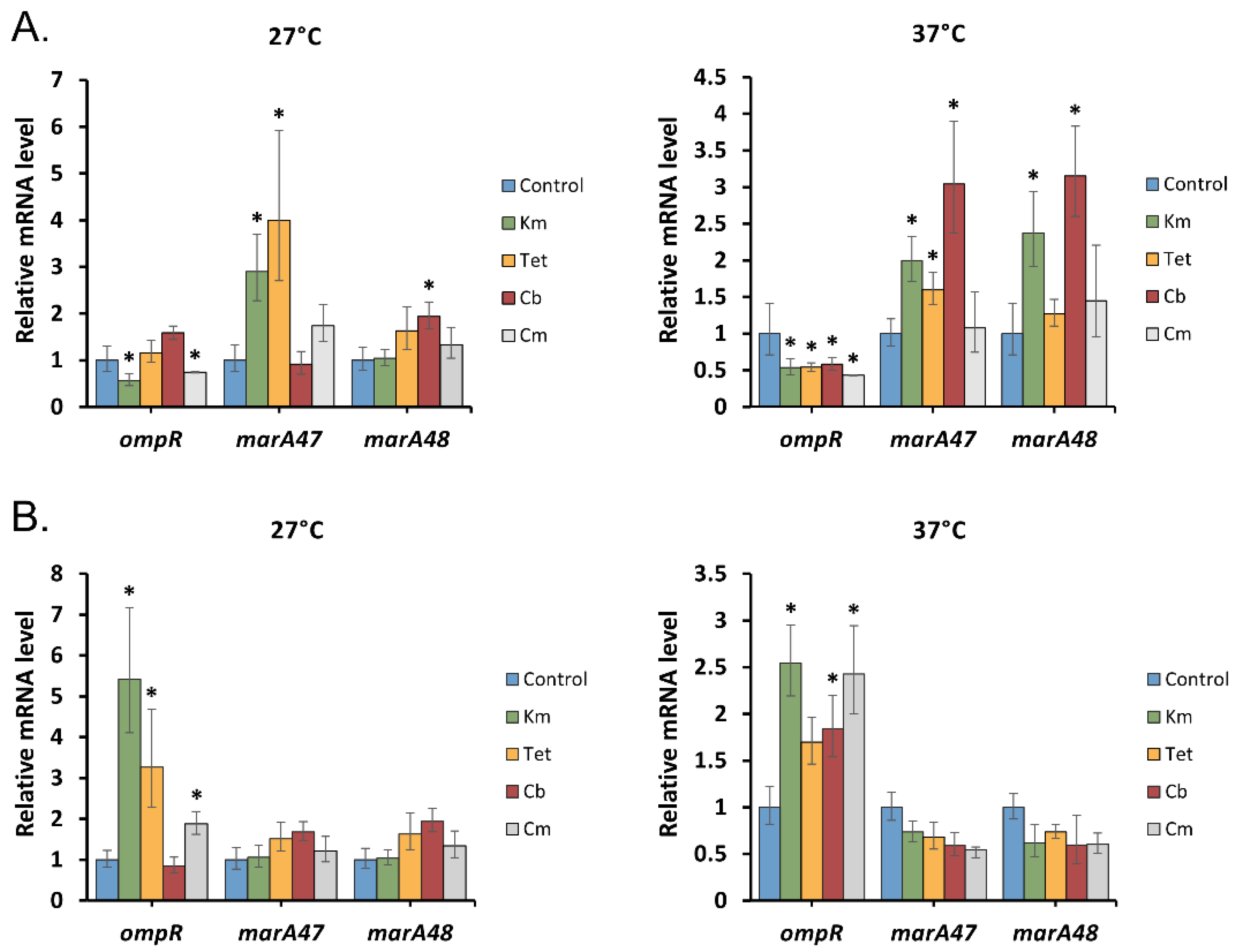
qRT-PCR analysis revealed differential expression of ompR in Y. pseudotuberculosis depending on the duration of antibiotic stress (Figure 3). Short exposure to antibiotics excluding carbenicillin and tetracycline at 27 °C slightly reduced (1.7- to 2.3-fold) its transcription associated with the downregulation of porin genes. On the contrary, prolonged antibiotic stress increased the level of ompR transcripts up to 5.3-fold; however, this did not affect the expression of ompF and ompC. A similar observation was described in the work of Viveiros et al., where the response of the ompC and ompF genes, despite the increased expression of ompR, remained comparable to their levels in E. coli cells not exposed to tetracycline [24]. Moreover, it was shown that OmpR positively controls the expression of the AcrAB-TolC efflux pump involved in the adaptive response of the Y. enterocolitica strain to different chemical stressors [45]. It is tempting to speculate that a high level of ompR transcription in Y. pseudotuberculosis may be required to induce this efflux pump system as another general defense mechanism against antibiotics.
Multiple studies have shown that general porin expression is post-transcriptionally regulated by global regulator MarA, involved in multidrug resistance response during antibiotic stress [46,47]. marA locus can directly or indirectly increase the level of micF, a non-coding RNA stress response gene, causing a post-transcriptional downregulation of ompF mRNA and reducing the OmpF synthesis [47]. Two MarA-like proteins have been reported in Yersinia pestis: MarA47 and MarA48 [48,49].
To test their involvement in the regulation of Y. pseudotuberculosis porin expression under short- and long-term exposure to antibiotics, we measured the levels of marA47 and marA48 mRNA using qRT-PCR (Figure 3). We found different activities of marA transcripts depending on the time of antibiotic exposure. A short treatment of cells with the antibiotics caused upregulation of at least one marA gene, except for chloramphenicol, which did not affect the marA expression. In the case of long-term exposure to the tested antibiotics, the level of marA expression remained comparable to those of the control samples.
The obtained results led us to conclude that regulation of Y. pseudotuberculosis general porins mediated by MarA was observed only for early antibiotic stress caused by sublethal concentrations of tetracycline, kanamycin, and carbenicillin. During long-term antibiotic presence, marA transcription remained unaltered and therefore MarA did not play a significant role in porin regulation at the post-transcriptional level.

2.5. Transcriptional Response of Y. pseudotuberculosis Alternative Porins to Prolonged Antibiotic Stress

In addition to major porins, Enterobacteriaceae can express alternative outer membrane proteins that may contribute to the adaptation of bacteria to antibiotic stress by modulating OmpF and OmpC levels or directly changing the cell permeability [35,50].
Therefore, to better understand the mechanism of Y. pseudotuberculosis adaptation, we investigated the transcriptional response of the genes coding for OmpX, OmpA, LamB, and OmpY porins to prolonged antibiotic stress.
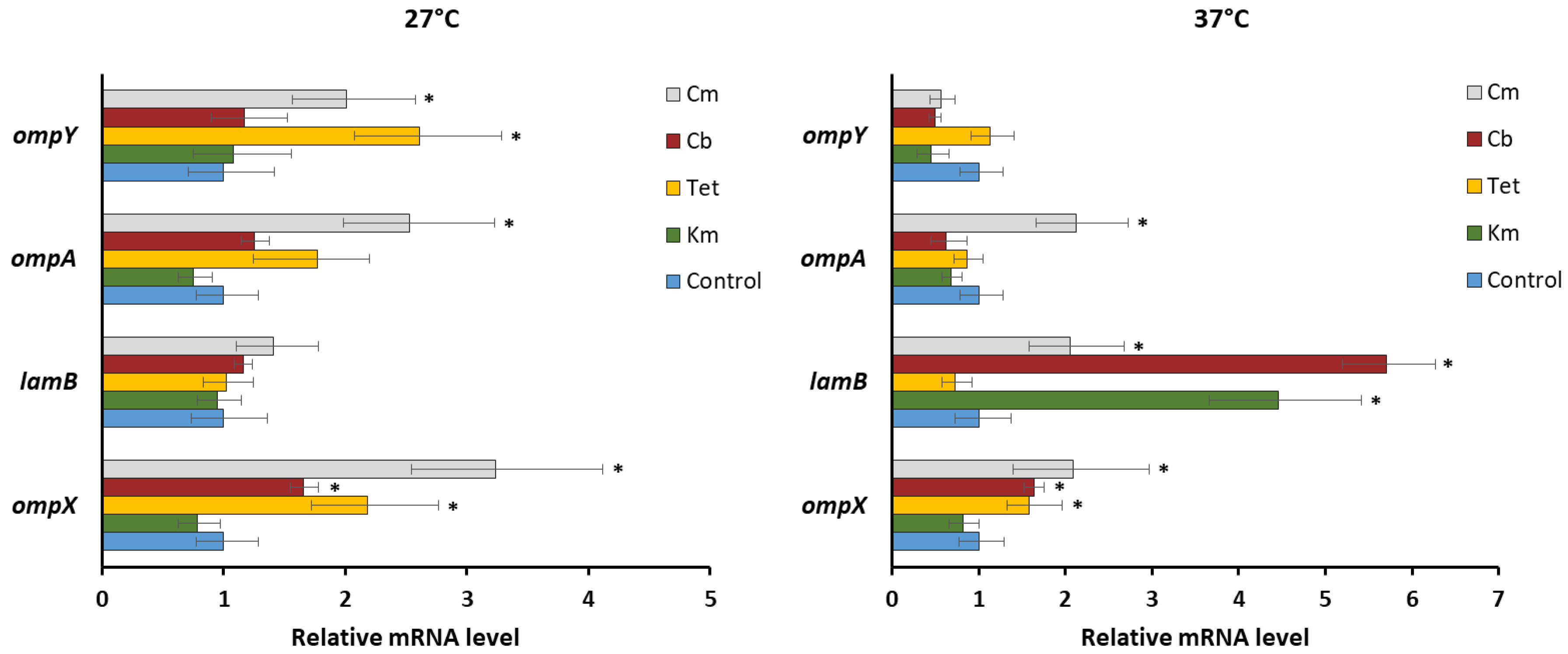
Using qRT-PCR, we observed the increased activity of ompX transcripts (1.7–3.2-fold) in the presence of most antibiotics (Figure 4), which is consistent with previously reported results [50]. It is believed that overexpression of OmpX may impair the normal assembly of major porins, which leads to their subsequent degradation by Deg proteases [51]. From this, we hypothesized that OmpF and OmpC expression of Y. pseudotuberculosis could be regulated by this mechanism at the post-translational level.
We also observed the induction of lamB transcription in the presence of kanamycin, carbenicillin, and chloramphenicol at 37 °C (Figure 4), when its initial mRNA level was significantly lower compared to 27 °C (data not shown). It is interesting to note that overexpression of LamB previously found for Enterobacter aerogenes-resistant isolates was associated with a low amount of its major porins [52].
The transcription of ompA, playing a more important role in the maintenance of membrane integrity, rather than antibiotic transport [35], remained unaltered in Y. pseudotuberculosis, except for chloramphenicol exposure, where the ompA expression increased up to 2.5-fold (Figure 4).
Previously, we described a new quiescent porin OmpY in Y. pseudotuberculosis initially predicted from the genomic data [53]. To date, our knowledge of such porins has been very limited. Both the functional role of OmpY and the conditions significantly affecting its expression in Yersinia are still unknown. qRT-PCR analysis revealed ompY upregulation under chloramphenicol and tetracycline exposure at 27 °C, while at 37 °C its level did not change (Figure 4). Several studies have shown that the expression of quiescent porins was induced by antibiotics; however, the impact of such regulation on antibiotic susceptibility remains questionable [20,54].

2.6. ompF and ompC Expression within Y. pseudotuberculosis Population

Current experimental evidence suggests that bacterial cultures are constituted of heterogeneous subpopulations [55] with differential responses to environmental stresses [56,57]. For instance, Sánchez-Romero and Casadesús have observed the population heterogeneity of S. enterica cells in the expression level of ompC under kanamycin exposure [57]. Moreover, a low level of ompC correlates with high kanamycin resistance of the cell.
Our qRT-PCR results based on average Ct values did not reveal any significant transcriptional response of porins to long-term antibiotic exposure for most samples. They said nothing about the variability of ompF and ompC expression in the Y. pseudotuberculosis population or the amount of their gene products in the cell. To answer these questions, we first developed fluorescent reporter strains of Y. pseudotuberculosis 488. The GFP reporter systems were constructed by fusion of the ompF/ompC regulatory regions, including binding sites for transcription and translation factors (5′UTRs and signal sequences for sRNAs, C-terminal YQF amino acids for proteases), to the GFP reporter gene in a low copy number plasmid pACYC184 (Figure S1). Thus, the fluorescence of Y. pseudotuberculosis transformants potentially not only provided a high-resolution method for assaying ompF/ompC promoter activity but also indirectly indicated OmpF and OmpC amounts. We excluded chloramphenicol from the experiments due to the plasmid containing the gene for its resistance. However, we additionally verified that the presence of this antibiotic in the medium did not affect the level of sample fluorescence (Figure S2).
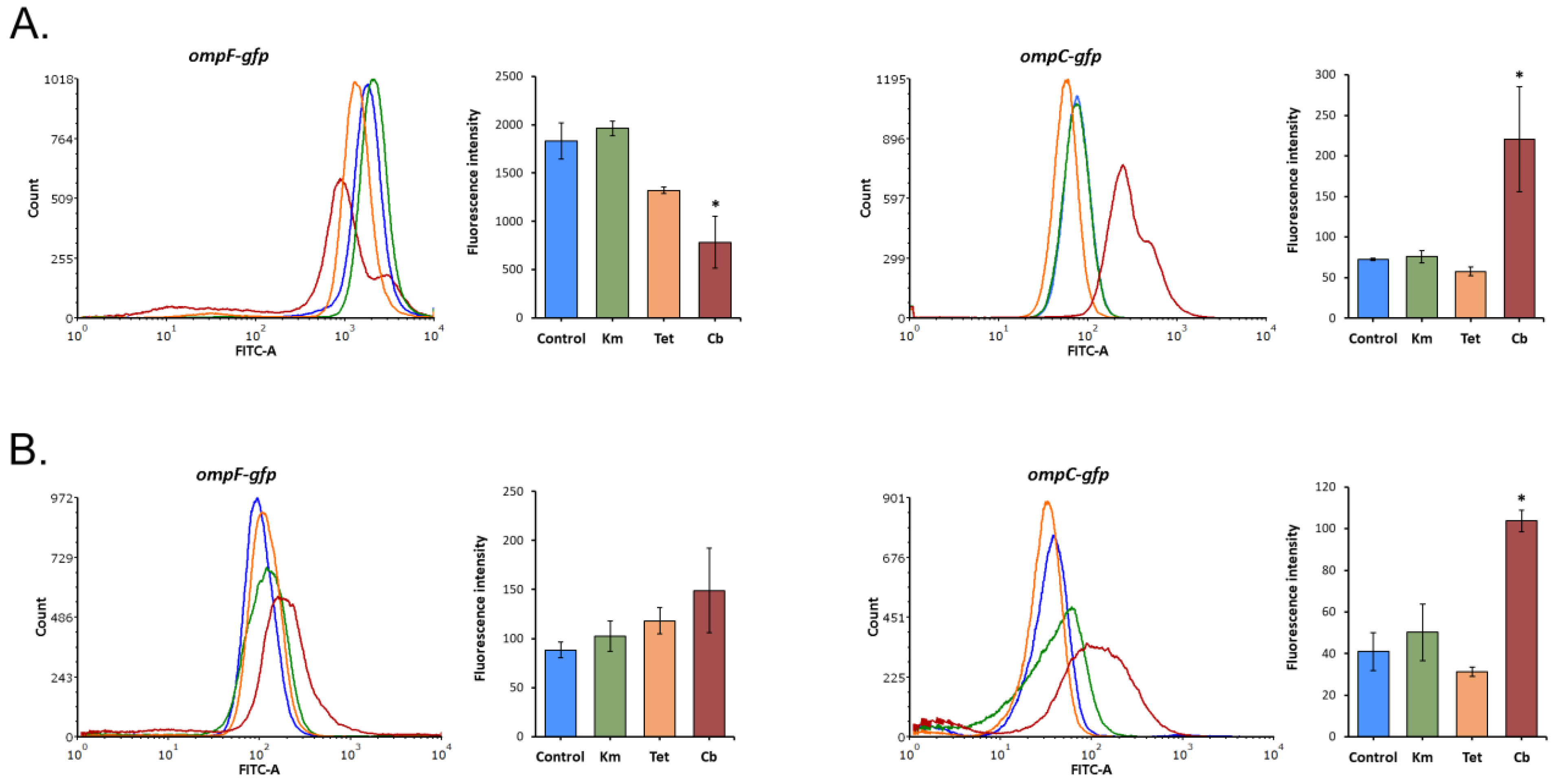
There was little evidence of any change in the mean fluorescence intensity, and hence OmpF and OmpC expression, after prolonged exposure to tetracycline and kanamycin (Figure 5), while qRT-PCR revealed the ompF upregulation in the presence of tetracycline at 27 °C (Figure 2). This difference indicates an additional post-transcriptional regulation of OmpF.
After treatment with sub-MIC of carbenicillin at 27 °C, there was a 2.3-fold decrease in the ompF level and, on the contrary, a 2.5–3-fold increase in the ompC level. This is consistent with the previous observation that adaptation to β-lactams can involve increased expression of OmpC relative to OmpF since an increase in OmpC proportion hinders their penetration [37]. It is worth noting that carbenicillin inhibited the expression of ompF only at 27 °C. We observed that this transcript was prevailing compared to ompC in the untreated samples at both incubation temperatures; however, at 37 °C, its level decreased by more than 18 times (Figure S3). This initially low amount of ompF transcripts and additional nutrient deficiencies due to the stationary growth phase could prevent a further decrease in its expression at 37 °C under carbenicillin stress.
The distribution of GFP fluorescence within the sample cultures under kanamycin and tetracycline exposure was comparable to the untreated controls. Only carbenicillin presence led to the appearance of two Y. pseudotuberculosis subpopulations with different fluorescence intensities, most notably at 27 °C (Figure 6). F1 subpopulation responded to carbenicillin stress by 1.9-fold inhibition of ompF expression, while the cells from F2 increased it by 1.8 times. ompC was upregulated under carbenicillin exposure in both subpopulations; however, in C2, its level was significantly higher. Moreover, most of the cells formed subpopulations with a lower level of both ompF and ompC expression (71% and 78% of the total cell amount, respectively; Figure 6C).
We thus propose that modification of the bacterial envelope by reduced ompF/increased ompC expression confers adaptive fitness advantages to carbenicillin in a fraction of the bacterial population.
Previously it was shown that heterogenous responses improve the survival of bacterial cultures under the effect of stress factors [58]. Moreover, gene expression variability, including porin genes, is considered to underly adaptive resistance in phenotypically heterogeneous microbial populations [57,59].

3. Materials and Methods

3.1. Bacterial Strain and Growth Conditions

Y. pseudotuberculosis 488 strain (= 117 strain, O:1b serotype) isolated from a patient with FESLF in the Russian Far East region was used in the present study.
To study short-term porin response to antibiotic stress, bacterial cells were grown in LB broth overnight at 27 °C with shaking. The next day, overnight cultures were diluted at a ratio of 1:50 into fresh LB medium and incubated with agitation at 27 °C (optimal growth temperature) and 37 °C (host temperature) to mid-exponential growth phase (OD600 = 0.4–0.6). Then, the antibiotics kanamycin (Sigma-Aldrich, St. Louis, MA, USA), tetracycline (Sigma-Aldrich, St. Louis, MA, USA), chloramphenicol (Sigma-Aldrich, St. Louis, MA, USA), and carbenicillin (Sigma-Aldrich, St. Louis, MA, USA) were added to the media in sublethal concentrations (0.5 MIC). After a 1 h antibiotic treatment (180 rpm, 27 °C and 37 °C), the cells were mixed with RNAprotect Bacteria Reagent (Qiagen, Hilden, Germany) and centrifuged.
To study porin response to long-term antibiotic exposure, overnight cultures were diluted and inoculated into LB broth media to obtain a final concentration of 5 × 105 CFU/mL. Bacteria were incubated with sublethal concentrations of antibiotics (kanamycin, tetracycline, chloramphenicol, and carbenicillin) for 16 h at 27 °C and 37 °C. RNAprotect Bacteria Reagent (Qiagen, Hilden, Germany) was added to the culture samples.
Three independent bacterial cultures for each test or control condition were prepared as biological replicates for RNA isolation.

3.2. MIC Determination

The MIC for antibiotics against Y. pseudotuberculosis strain was determined by the broth microdilution method in a 96-well microplate. Tetracycline (Sigma-Aldrich, St. Louis, MA, USA), carbenicillin (Sigma-Aldrich, St. Louis, MA, USA), kanamycin (Sigma-Aldrich, St. Louis, MA, USA), and chloramphenicol (Sigma-Aldrich, St. Louis, MA, USA) were tested in the concentration range of 0.12–256 μg/mL obtained after a series of 2-fold dilutions in the LB broth. Bacterial colonies were cultured in LB broth at 27 °C to reach McFarland standard 0.5. The suspensions were further diluted and inoculated into the wells with antibiotics to obtain a final concentration of 5 × 105 CFU/mL. The plates were incubated for 16 h at 27 °C and 37 °C. The MIC value was determined as the lowest concentration of an antibiotic at which visible growth was inhibited.

3.3. Gene Expression Measurement by qRT-PCR

The expression of ompF, ompC, ompA, ompX, ompY, ompR, marA47, and marA48 genes in Y. pseudotuberculosis 488 was studied using qRT-PCR. Total RNA was isolated from the prepared bacterial pellets using the Aurum total RNA mini kit (Bio-Rad, Hercules, CA, USA) according to the manufacturer’s protocol. The RNA concentration and purity were assessed by electrophoresis and a microvolume spectrophotometer (Implen GmbH, Munich, Germany). Of the purified total RNA, 2 μg was reverse-transcribed into cDNA with MMLV RT Kit (Evrogen, Moscow, Russia) and random hexamer primers (Evrogen, Moscow, Russia). The cDNAs were subsequently used to quantify the relative level of ompF, ompC, ompA, ompX, ompY, ompR, marA47, and marA48 by qRT-PCR in Light Cycler 96 (Roche, Basel, Switzerland). The nucleotide sequences of the studied genes are listed in Table S2. The gene-specific primers used in this experiment are listed in Table S3.
RT-PCR was carried out with HS GoTaq Polymerase (Promega, Madison, WI, USA) and the dye Eva Green (Biotium, Fremont, CA, USA). The following thermal cycling parameters were used for the reaction: initial denaturation at 95 °C for 8 min; and 40 cycles of amplification: 95 °C for 15 s, 55 °C for 10 s, and 72 °C for 20 s followed by fluorescence reading. A melting curve was drawn at the end to evaluate the specificity of the PCR. Quantification for each target gene expression was determined by the 2−ΔΔCT method [60] using the 16S rDNA gene as a reference.
qRT-PCR was carried out on three independent biological replicates, each containing two technical replicates. The results are presented in Table S4 as mean ± SD. One-way ANOVA was performed to assess the differences between the means of the test and control groups, with a p-value of ≤ 0.05 considered significant.

3.4. Porin Gene Sequencing

The coding and regulatory parts of ompF and ompC genes were amplified using OmpF/C-Bam_for and OmpF/C-Hind_rev primers and sequenced with internal primers (Table S3) to cover all regions. DNA sequencing was done with an ABI 3130 xl automated sequencer (Applied Biosystems, Waltham, MA, USA) and the ABI Prism dye terminator sequencing kit (Applied Biosystems, Waltham, MA, USA) according to the manufacturer’s directions.

3.5. GFP Reporter System Construction

The recombinant plasmids pACYC184-F-GFP and pACYC184-C-GFP containing the Y. pseudotuberculosis ompF and ompC regulatory regions fused to GFP were constructed as previously described [7]. To obtain the coding region of GFP, the plasmid DNA pTurboGFP (Evrogen, Moscow, Russia) was digested with BamHI and HindIII (Fermentas, Waltham, MA, USA), and the resulting gfp fragment was dephosphorylated with CAP alkaline phosphatase (Fermentas, Waltham, MA, USA). The ompF/ompC promoter, and terminator regions were amplified using specific primers, which contained BamHI and HindIII restriction sites, respectively (Table S3). The resulting PCR products were digested with the same endonucleases and ligated together (separately for ompF and ompC) with gfp in a three-way ligation. Next, ligation mixtures were used as templates for amplifying ompF-gfp and ompC-gfp reporter constructions with OmpF-Bam_for/OmpF-Hind_rev or OmpC-Bam_for/OmpC-Hind_rev primers (Table S3). The PCRs were carried out using the following reagents: 2 U of GoTaq DNA polymerase (Promega, Madison, WI, USA), 1× buffer for GoTaq DNA polymerase, 0.2 mM dNTPs, 0.5 μM primers, and 25–50 ng of the template. The following reaction conditions were used: 1 cycle of 5 min at 95 °C for denaturation, 30 cycles of 20 s at 94 °C, 30 s at 55 °C, 1 min 30 s at 72 °C, and 1 final cycle of 5 min at 72 °C. The PCR fragments of expected lengths (1300–1400 bp) were cut from the agarose gel and phosphorylated with T4 polynucleotide kinase (Fermentas, Waltham, MA, USA). The resulting reporter cassettes were inserted in the dephosphorylated vector pACYC184 via EcoRV (Figure S1). Recombinant plasmids were transformed into E. coli DH5alpha strain by electroporation. The colonies were selected on LB agar plates supplemented with chloramphenicol. All constructs were verified by PCR and DNA sequencing with pACYCSal_seq and pACYCEcV_seq primers (Table S2). The final recombinant plasmids were introduced into competent cells of the Y. pseudotuberculosis 488 using electroporation.

3.6. Flow Cytometry

To obtain comparable measurements with flow cytometry, the Y. pseudotuberculosis 488 containing recombinant plasmid pACYC184-F-GFP or pACYC184-C-GFP were grown in the same conditions as for qRT-PCR analysis. All cultures were incubated in 96-well plates without/with a sublethal concentration of antibiotics (Tet, Km, and Cb) and the addition of chloramphenicol (10 μg/mL) at 37 °C and 27 °C with shaking. After 16 h of cultivation, the cells were washed with PBS, pH 7, and diluted to an OD600 of 0.01. The flow cytometry measurements were made with a FACSCanto flow cytometer (BD Biosciences, San Jose, CA, USA). The excitation beam for the GFP was set at 488 nm, and the emission signal was captured with a 530/30 nm bandpass filter. The signals were amplified with the logarithmic mode for SSC, FSC, and fluorescence. For each sample, 100,000 events were recorded. The data were captured with the BD FACSdiva software (version 6.1.2) (BD Biosciences, San Jose, CA, USA), and for data analysis FCS Express 6 (De Novo Software Inc., Los Angeles, CA, USA) was used. The mean fluorescence value of the whole population was chosen as a global indicator of porin expression level in each bacterial culture. Y. pseudotuberculosis 488 without plasmids was used as a negative control to exclude background fluorescence.

4. Conclusions

In summary, we revealed temporal regulation of Y. pseudotuberculosis generalporin genes ompF and ompC caused by sublethal concentrations of different antibiotics. The porin transcription initially decreased, providing early defensive response of the bacterium, while it returned to that of the untreated cells on prolonged antibiotic exposure. Unlike the major porin genes, the transcription of the alternativeporin genes ompX and lamB was increased. Such modulation of porin transcription reflects the model of Y. pseudotuberculosis physiological adaptation to antibiotic exposure.
The main finding was a phenotypic heterogeneity of Y. pseudotuberculosis population manifested in variable porin gene expression under carbenicillin exposure. To verify whether such phenotypic heterogeneity offers a competitive advantage and contributes to low carbenicillin susceptibility for a particular Y. pseudotuberculosis subpopulation, future research is warranted.

Supplementary Materials

The following are available online, Figure S1: GFP reporter constructions; Figure S2: GFP fluorescence intensity in Y. pseudotuberculosis 488 transformed with ompC/ompF promoter-fused GFP reporter constructs under increasing Cm exposure; Figure S3: GFP fluorescence intensity in Y. pseudotuberculosis 488 transformed with ompF/ompC promoter-fused GFP reporter constructs at 27 °C and 37 °C incubation temperature; Table S1: MIC of antibiotics against Y. pseudotuberculosis 488; Table S2: Nucleotide sequences of Y. pseudotuberculosis 488 genes used in the study; Table S3: Oligonucleotide primers used in the study; Table S4: Average Ct values for ompF, ompC, and 16S rRNA of Y. pseudotuberculosis 488.

Author Contributions

Conceptualization, M.I. and A.R.; methodology, M.I., A.S., N.C., and E.B.; software, E.B.; investigation, E.B., N.C., K.G., and A.S.; resources, M.I. and A.R.; data curation, M.I.; writing—original draft preparation, E.B. and M.I.; writing—review and editing, M.I. and A.R.; supervision, M.I.; funding acquisition, M.I. and E.B. All authors have read and agreed to the published version of the manuscript.

Funding

This research received no external funding.

Institutional Review Board Statement

Not applicable.

Informed Consent Statement

Not applicable.

Data Availability Statement

Unpublished data are available from the authors.

Acknowledgments

We are grateful to the anonymous reviewers and editors that have improved our manuscript.

Conflicts of Interest

The authors declare no conflict of interest.

References

  1. Fernandez, L.; Hancock, R.E. Adaptive and mutational resistance: Role of porins and efflux pumps in drug resistance. Clin. Microbiol. Rev. 2012, 25, 661–681. [Google Scholar] [CrossRef] [Green Version]
  2. Andersson, D.I.; Hughes, D. Microbiological effects of sublethal levels of antibiotics. Nat. Rev. Microbiol. 2014, 12, 465–478. [Google Scholar] [CrossRef]
  3. Braoudaki, M.; Hilton, A.C. Adaptive resistance to biocides in Salmonella enterica and Escherichia coli o157 and cross-resistance to antimicrobial agents. J. Clin. Microbiol. 2004, 42, 73–78. [Google Scholar] [CrossRef] [PubMed] [Green Version]
  4. Daikos, G.L.; Lolans, V.T.; Jackson, G.G. First-exposure adaptive resistance to aminoglycoside antibiotics in vivo with meaning for optimal clinical use. Antimicrob. Agents Chemother. 1991, 35, 117–123. [Google Scholar] [CrossRef] [Green Version]
  5. Viveiros, M.; Jesus, A.; Brito, M.; Leandro, C.; Martins, M.; Ordway, D.; Molnar, A.M.; Molnar, J.; Amaral, L. Inducement and reversal of tetracycline resistance in Escherichia coli K-12 and the expression of proton gradient dependent multidrug efflux pump genes. Antimicrob. Agents Chemother. 2005, 49, 3578–3582. [Google Scholar] [CrossRef] [Green Version]
  6. Somova, L.M.; Antonenko, F.F.; Timchenko, N.F.; Lyapun, I.N. Far Eastern Scarlet-Like Fever is a Special Clinical and Epidemic Manifestation of Yersinia pseudotuberculosis Infection in Russia. Pathogens 2020, 9, 436. [Google Scholar] [CrossRef]
  7. Bystritskaya, E.P.; Stenkova, A.M.; Portnyagina, O.Y.; Rakin, A.V.; Rasskazov, V.A.; Isaeva, M.P. Regulation of Yersinia pseudotuberculosis major porin expression in response to antibiotic stress. Mol. Gen. Microbiol. Virol. 2014, 29, 63–68. [Google Scholar] [CrossRef]
  8. Khomenko, V.A.; Portnyagina, O.Y.; Novikova, O.D.; Isaeva, M.P.; Kim, N.Y.; Likhatskaya, G.N.; Vostrikova, O.P.; Solov’eva, T.F. Isolation and characterization of recombinant OmpF-like porin from the Yersinia pseudotuberculosis outer membrane. Russ. J. Bioorg. Chem. 2008, 34, 162–168. [Google Scholar] [CrossRef]
  9. Novikova, O.D.; Khomenko, V.A.; Emelyanenko, V.I.; Likhatskaya, G.N.; Zelepuga, E.A.; Kim, N.Y.; Isaeva, M.P.; Portnyagina, O.Y.; Vostrikova, O.P.; Sidorova, O.V.; et al. OmpC-like porin from Yersinia pseudotuberculosis: Molecular characteristics, physico-chemical and functional properties. Biochem. Suppl. Ser. A Membr. Cell Biol. 2011, 5, 263–277. [Google Scholar]
  10. Stenkova, A.M.; Isaeva, M.P.; Shubin, F.N.; Rasskazov, V.A.; Rakin, A.V. Trends of the major porin gene (OmpF) evolution: Insight from the genus Yersinia. PLoS ONE 2011, 6, e20546. [Google Scholar] [CrossRef] [Green Version]
  11. Stenkova, A.M.; Bystritskaya, E.P.; Guzev, K.V.; Rakin, A.V.; Isaeva, M.P. Molecular Evolution of the Yersinia Major Outer Membrane Protein C (OmpC). Evol. Bioinform. 2016, 12, 185–191. [Google Scholar] [CrossRef] [Green Version]
  12. Cowan, S.W.; Schirmer, T.; Rummel, G.; Steiert, M.; Ghosh, R.; Pauptit, R.A.; Jansonius, J.N.; Rosenbusch, J.P. Crystal structures explain functional properties of two E. coli porins. Nature 1992, 358, 727–733. [Google Scholar] [CrossRef]
  13. James, C.E.; Mahendran, K.R.; Molitor, A.; Bolla, J.-M.; Bessonov, A.N.; Winterhalter, M.; Pagès, J.-M. How beta-lactam antibiotics enter bacteria: A dialogue with the porins. PLoS ONE 2009, 4, e5453. [Google Scholar] [CrossRef]
  14. Bafna, J.A.; Sans-Serramitjana, E.; Acosta-Gutiérrez, S.; Bodrenko, I.V.; Hörömpöli, D.; Berscheid, A.; Brötz-Oesterhelt, H.; Winterhalter, M.; Ceccarelli, M. Kanamycin uptake into Escherichia coli is facilitated by OmpF and OmpC porin channels located in the outer membrane. ACS Infect. Dis. 2020, 6, 1855–1865. [Google Scholar] [CrossRef]
  15. Neves, P.; Berkane, E.; Gameiro, P.; Winterhalter, M.; De Castro, B. Interaction between quinolones antibiotics and bacterial outer membrane porin OmpF. Biophys. Chem. 2005, 113, 123–128. [Google Scholar] [CrossRef]
  16. Mortimer, P.G.; Piddock, L.J. The accumulation of five antibacterial agents in porin-deficient mutants of Escherichia coli. J. Antimicrob. Chemother. 1993, 32, 195–213. [Google Scholar] [CrossRef]
  17. Masi, M.; Réfrigiers, M.; Pos, K.M.; Pagès, J.M. Mechanisms of envelope permeability and antibiotic influx and efflux in Gram-negative bacteria. Nat. Microbiol. 2017, 2, 17001. [Google Scholar] [CrossRef] [PubMed]
  18. Sousa, A.M.; Machado, I.; Pereira, M.O. Phenotypic switching: An opportunity to bacteria thrive. In Science Against Microbial Pathogens: Communicating Current Research and Technological Advances; Formatex Research Center: Badajoz, Spain, 2012; pp. 252–262. [Google Scholar]
  19. Dupont, M.; James, C.E.; Chevalier, J.; Pagès, J.-M. An Early Response to Environmental Stress Involves Regulation of OmpX and OmpF, Two Enterobacterial Outer Membrane Pore-Forming Proteins. Antimicrob. Agents Chemother. 2007, 51, 3190–3198. [Google Scholar] [CrossRef] [Green Version]
  20. Doménech-Sánchez, A.; Hernández-Allés, S.; Martínez-Martínez, L.; Benedí, V.J.; Albertí, S. Identification and characterization of a new porin gene of Klebsiella pneumoniae: Its role in beta-lactam antibiotic resistance. J. Bacteriol. 1999, 181, 2726–2732. [Google Scholar] [CrossRef] [Green Version]
  21. Knopp, M.; Andersson, D.I. Amelioration of the fitness costs of antibiotic resistance due to reduced outer membrane permeability by upregulation of alternative porins. Mol. Biol. Evol. 2015, 32, 3252–3263. [Google Scholar] [CrossRef] [Green Version]
  22. Chetri, S.; Singha, M.; Bhowmik, D.; Nath, K.; Chanda, D.D.; Chakravarty, A.; Bhattacharjee, A. Transcriptional response of OmpC and OmpF in Escherichia coli against differential gradient of carbapenem stress. BMC Res. Notes 2019, 12, 138. [Google Scholar] [CrossRef]
  23. Dam, S.; Pagès, J.-M.; Masi, M. Stress responses, outer membrane permeability control and antimicrobial resistance in Enterobacteriaceae. Microbiology 2018, 164, 260–267. [Google Scholar] [CrossRef] [PubMed]
  24. Viveiros, M.; Dupont, M.; Rodrigues, L.; Couto, I.; Davin-Regli, A.; Martins, M.; Pagès, J.-M.; Amaral, L. Antibiotic stress, genetic response and altered permeability of E. coli. PLoS ONE 2007, 2, e365. [Google Scholar] [CrossRef]
  25. Deighan, P.; Free, A.; Dorman, C.J. A role for the Escherichia coli H-NS-like protein StpA in OmpF porin expression through modulation of micF RNA stability. Mol. Microbiol. 2000, 38, 126–139. [Google Scholar] [CrossRef] [Green Version]
  26. Andersen, J.; Forst, S.A.; Zhao, K.; Inouye, M.; Delihas, N. The function of micF RNA. micF RNA is a major factor in the thermal regulation of OmpF protein in Escherichia coli. J. Biol. Chem. 1989, 264, 17961–17970. [Google Scholar] [CrossRef]
  27. Begic, S.; Worobec, E.A. Regulation of Serratia marcescens ompF and ompC porin genes in response to osmotic stress, salicylate, temperature and pH. Microbiology 2006, 152, 485–491. [Google Scholar] [CrossRef] [Green Version]
  28. Bystritskaya, E.; Stenkova, A.; Chistuylin, D.; Chernysheva, N.; Khomenko, V.; Anastyuk, S.; Novikova, O.; Rakin, A.; Isaeva, M. Adaptive responses of outer membrane porin balance of Yersinia ruckeri under different incubation temperature, osmolarity, and oxygen availability. Microbiologyopen 2016, 5, 597–603. [Google Scholar] [CrossRef] [PubMed] [Green Version]
  29. Narimisa, N.; Amraei, F.; Kalani, B.S.; Mohammadzadeh, R.; Jazi, F.M. Effects of sub-inhibitory concentrations of antibiotics and oxidative stress on the expression of type II toxin-antitoxin system genes in Klebsiella pneumoniae. J. Glob. Antimicrob. Resist. 2020, 21, 51–56. [Google Scholar] [CrossRef] [PubMed]
  30. Zhang, D.-F.; Jiang, B.; Xiang, Z.-M.; Wang, S.-Y. Functional characterisation of altered outer membrane proteins for tetracycline resistance in Escherichia coli. Int. J. Antimicrob. Agents 2008, 32, 315–319. [Google Scholar] [CrossRef]
  31. Chang, C.C.; Yan, C.W.; Onal, R.; Zhang, Y. Genetic characterization and investigation of kanamycin susceptibility of ompC and ompF single gene deletion mutants of Escherichia coli K-12. J. Exp. Microbiol. Immunol. 2018, 22, 1–9. [Google Scholar]
  32. Hu, W.-C.; MacDonald, R.; Oosthuizen, J.L.; Soeren, M.V. Sub-inhibitory kanamycin changes outer membrane porin ratios in Escherichia coli B23 by increasing the level of OmpC. J. Exp. Microbiol. Immunol. 2011, 15, 96–102. [Google Scholar]
  33. Fei, Y.; Ma, V.; Maerkl, N.; You, P. The down regulation of E. coli OmpF in response to sub-inhibitory concentrations of kanamycin is not mediated by MarA. J. Exp. Microbiol. Immunol. 2012, 16, 101–107. [Google Scholar]
  34. Agafitei, O.; Kim, E.J.; Maguire, T.; Sheridan, J. The role of Escherichia coli porins OmpC and OmpF in antibiotic cross resistance induced by sub-inhibitory concentrations of kanamycin. J. Exp. Microbiol. Immunol. 2010, 14, 34–39. [Google Scholar]
  35. Choi, U.; Lee, C.-R. Distinct roles of outer membrane porins in antibiotic resistance and membrane integrity in Escherichia coli. Front. Microbiol. 2019, 10, 953. [Google Scholar] [CrossRef]
  36. Moya-Torres, A.; Mulvey, M.R.; Kumar, A.; Oresnik, I.J.; Brassinga, A.K. The lack of OmpF, but not OmpC, contributes to increased antibiotic resistance in Serratia marcescens. Microbiology 2014, 160 Pt 9, 1882–1892. [Google Scholar] [CrossRef]
  37. Igwe, J.C.; Olayinka, B.O.; Ehnimidu, J.O.; Onaolapo, J.A. Impact of outer membrane protein Ompc and Ompf on antibiotics resistance of E. coli isolated from UTI and diarrhoeic patients in Zaria, Nigeria. Clin. Microbiol. 2016, 5, 1000267. [Google Scholar]
  38. Toro, C.S.; Lobos, S.R.; Calderón, I.; Rodríguez, M.; Mora, G.C. Clinical isolate of a porinless Salmonella typhi resistant to high levels of chloramphenicol. Antimicrob. Agents. Chemother. 1990, 34, 1715–1719. [Google Scholar] [CrossRef] [PubMed] [Green Version]
  39. Fajardo-Lubián, A.; Zakour, N.L.B.; Agyekum, A.; Qi, Q.; Iredell, J.R. Host adaptation and convergent evolution increases antibiotic resistance without loss of virulence in a major human pathogen. PLoS Pathog. 2019, 15, e1007218. [Google Scholar] [CrossRef] [Green Version]
  40. Ferenci, T.; Phan, K. How porin heterogeneity and trade-offs affect the antibiotic susceptibility of Gram-Negative bacteria. Genes 2015, 6, 1113–1124. [Google Scholar] [CrossRef] [Green Version]
  41. Liu, X.; Ferenci, T. Regulation of porin-mediated outer membrane permeability by nutrient limitation in Escherichia coli. J. Bacteriol. 1998, 180, 3917–3922. [Google Scholar] [CrossRef] [Green Version]
  42. Tierney, A.R.; Rather, P.N. Roles of two-component regulatory systems in antibiotic resistance. Future Microbiol. 2019, 14, 533–552. [Google Scholar] [CrossRef] [PubMed]
  43. Flamez, C.; Ricard, I.; Arafah, S.; Simonet, M.; Marceau, M. Phenotypic analysis of Yersinia pseudotuberculosis 32777 response regulator mutants: New insights into two-component system regulon plasticity in bacteria. Int. J. Med. Microbiol. 2008, 298, 193–207. [Google Scholar] [CrossRef]
  44. Nieckarz, M.; Raczkowska, A.; Jaworska, K.; Stefańska, E.; Skorek, K.; Stosio, D.; Brzostek, K. The role of OmpR in the expression of genes of the KdgR regulon involved in the uptake and depolymerization of oligogalacturonides in Yersinia enterocolitica. Front. Cell Infect. Microbiol. 2017, 7, 366. [Google Scholar] [CrossRef]
  45. Raczkowska, A.; Trzos, J.; Lewandowska, O.; Nieckarz, M.; Brzostek, K. Expression of the AcrAB components of the AcrAB-TolC multidrug efflux pump of Yersinia enterocolitica is subject to dual regulation by OmpR. PLoS ONE 2015, 10, e0124248. [Google Scholar] [CrossRef]
  46. Masi, M.; Pagès, J.-M. Structure, function and regulation of outer membrane proteins involved in drug transport in Enterobactericeae: The OmpF/C—TolC case. Open Microbiol. J. 2013, 7, 22–33. [Google Scholar] [CrossRef] [Green Version]
  47. Cohen, S.P.; McMurry, L.M.; Levy, S.B. MarA locus causes decreased expression of OmpF porin in multiple-antibiotic-resistant (Mar) mutants of Escherichia coli. J. Bacteriol. 1988, 170, 5416–5422. [Google Scholar] [CrossRef] [Green Version]
  48. Lister, I.M.; Mecsas, J.; Levy, S.B. Effect of MarA-like proteins on antibiotic resistance and virulence in Yersinia pestis. Infect. Immun. 2010, 78, 364–371. [Google Scholar] [CrossRef] [Green Version]
  49. Udani, R.A.; Levy, S.B. MarA-like regulator of multidrug resistance in Yersinia pestis. Antimicrob. Agents Chemother. 2006, 50, 2971–2975. [Google Scholar] [CrossRef] [PubMed] [Green Version]
  50. Dupont, M.; Dé, E.; Chollet, R.; Chevalier, J.; Pagès, J.-M. Enterobacter aerogenes OmpX, a cation-selective channel mar- and osmo-regulated. FEBS Lett. 2004, 569, 27–30. [Google Scholar] [CrossRef] [PubMed] [Green Version]
  51. Castillo-Keller, M.; Vuong, P.; Misra, R. Novel mechanism of Escherichia coli porin regulation. J. Bacteriol. 2006, 188, 576–586. [Google Scholar] [CrossRef] [Green Version]
  52. Gayet, S.; Chollet, R.; Molle, G.; Pagès, J.-M.; Chevalier, J. Modification of outer membrane protein profile and evidence suggesting an active drug pump in Enterobacter aerogenes clinical strains. Antimicrob. Agents Chemother. 2003, 47, 1555–1559. [Google Scholar] [CrossRef] [Green Version]
  53. Solov’eva, T.F.; Likhatskaya, G.N.; Khomenko, V.A.; Stenkova, A.M.; Kim, N.Y.; Portnyagina, O.Y.; Novikova, O.D.; Trifonov, E.V.; Nurminski, E.A.; Isaeva, M.P. A novel OmpY porin from Yersinia pseudotuberculosis: Structure, channel-forming activity and trimer thermal stability. J. Biomol. Struct. Dyn. 2011, 28, 517–533. [Google Scholar] [CrossRef]
  54. Dam, S.; Pagès, J.-M.; Masi, M. Dual Regulation of the small RNA MicC and the quiescent porin OmpN in response to antibiotic stress in Escherichia coli. Antibiotics 2017, 6, 33. [Google Scholar] [CrossRef] [PubMed] [Green Version]
  55. Ambriz-Aviña, V.; Contreras-Garduño, J.A.; Pedraza-Reyes, M. Applications of flow cytometry to characterize bacterial physiological responses. BioMed Res. Int. 2014, 2014, 461941. [Google Scholar] [CrossRef]
  56. de Jong, I.G.; Veening, J.-W.; Kuipers, O.P. Single cell analysis of gene expression patterns during carbon starvation in Bacillus subtilis reveals large phenotypic variation. Environ. Microbiol. 2012, 14, 3110–3121. [Google Scholar] [CrossRef]
  57. Sánchez-Romero, M.A.; Casadesús, J. Contribution of phenotypic heterogeneity to adaptive antibiotic resistance. Proc. Natl. Acad. Sci. USA 2014, 111, 355–360. [Google Scholar] [CrossRef] [Green Version]
  58. Magdanova, L.A.; Golyasnaya, N.V. Heterogeneity as an adaptive trait of microbial populations. Microbiology 2013, 82, 1–10. [Google Scholar] [CrossRef]
  59. Erickson, K.E.; Otoupal, P.B.; Chatterjee, A. Gene expression variability underlies adaptive resistance in phenotypically heterogeneous bacterial populations. ACS Infect. Dis. 2015, 1, 555–673. [Google Scholar] [CrossRef]
  60. Livak, K.J.; Schmittgen, T.D. Analysis of relative gene expression data using real-time quantitative PCR and the 2−ΔΔCT method. Methods 2001, 25, 402–408. [Google Scholar] [CrossRef] [PubMed]
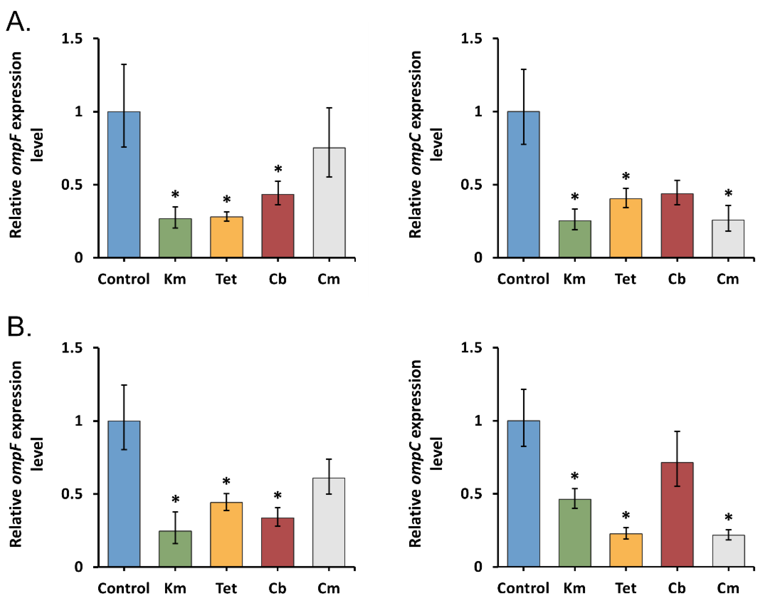
Figure 1. Relative expression levels of Y. pseudotuberculosis ompF and ompC after short-term antibiotic exposure detected by qRT-PCR. (A) 27 °C incubation temperature; (B) 37 °C incubation temperature. All results are expressed as mean ± standard deviation from three independent experiments. An asterisk indicates p-value < 0.05 vs. respective untreated control. Significance was calculated using one-way ANOVA. Km—kanamycin, Tet—tetracycline, Cb—carbenicillin, and Cm—chloramphenicol.
Molecules 26 03956 g001
Figure 2. Relative expression levels of Y. pseudotuberculosis ompF and ompC after prolonged antibiotic exposure detected by qRT-PCR. (A) 27 °C incubation temperature; (B) 37 °C incubation temperature. All results are expressed as mean ± standard deviation from three independent experiments. An asterisk indicates p-value < 0.05 vs. respective untreated control. Significance was calculated using one-way ANOVA. Km—kanamycin, Tet—tetracycline, Cb—carbenicillin, and Cm—chloramphenicol.
Molecules 26 03956 g002
Figure 3. Relative expression levels of Y. pseudotuberculosis ompR, marA47, and marA48 under antibiotic stress detected by qRT-PCR. (A) 1 h antibiotic exposure, (B) 16 h antibiotic exposure. All results are expressed as mean ± standard deviation from three independent experiments. An asterisk indicates p-value < 0.05 vs. respective untreated control. Significance was calculated using one-way ANOVA. Km—kanamycin, Tet—tetracycline, Cb—carbenicillin, and Cm—chloramphenicol.
Molecules 26 03956 g003
Figure 4. Relative expression levels of Y. pseudotuberculosis ompX, lamB, ompA, and ompY under prolonged antibiotic stress detected by qRT-PCR. All results are expressed as mean ± standard deviation from three independent experiments. An asterisk indicates p-value < 0.05 vs. respective untreated control. Significance was calculated using one-way ANOVA. Km—kanamycin, Tet—tetracycline, Cb—carbenicillin, and Cm—chloramphenicol.
Molecules 26 03956 g004
Figure 5. GFP fluorescence intensity in Y. pseudotuberculosis 488 transformed with ompF/ompC promoter-fused GFP reporter constructs following 16 h antibiotic treatment monitored by flow cytometry. (A) 27 °C incubation temperature; (B) 37 °C incubation temperature. All results are expressed as mean ± standard deviation between three experimental trials. An asterisk indicates p-value < 0.05 vs. respective control. Significance was calculated using one-way ANOVA. The histograms from a single representative experiment are shown. Control—without antibiotic treatment, Km—kanamycin, Tet—tetracycline, and Cb—carbenicillin.
Molecules 26 03956 g005
Figure 6. Fluorescence heterogeneity of Y. pseudotuberculosis 488 transformed with ompF/ompC promoter-fused GFP reporter constructs following treatment with a sublethal dose of carbenicillin monitored by flow cytometry. (A) Fluorescence histograms; (B) fluorescent intensity of subpopulations; (C) the percentage of cells in each subpopulation. All results are expressed as mean ± standard deviation between three experimental trials. An asterisk indicates p-value < 0.05 between groups. Significance was calculated using one-way ANOVA. The histograms from a single representative experiment are shown. F1, F2, and C1, C2—subpopulations of Y. pseudotuberculosis 488 with low/high GFP fluorescence.
Molecules 26 03956 g006
Publisher’s Note: MDPI stays neutral with regard to jurisdictional claims in published maps and institutional affiliations.