Perspective

Toxic Metal Species and ‘Endogenous’ Metalloproteins at the Blood–Organ Interface: Analytical and Bioinorganic Aspects

by
Tristen G. Bridle
1,
Premkumari Kumarathasan
2 and
Jürgen Gailer
1,*
1
Department of Chemistry, 2500 University Drive NW, University of Calgary, Calgary, AB T2N 1N4, Canada
2
Environmental Health Science and Research Bureau, Health Canada, Ottawa, ON K1A 0K9, Canada
*
Author to whom correspondence should be addressed.
Molecules 2021, 26(11), 3408; https://doi.org/10.3390/molecules26113408
Submission received: 7 April 2021 / Revised: 19 May 2021 / Accepted: 1 June 2021 / Published: 4 June 2021

Abstract

:
Globally, human exposure to environmental pollutants causes an estimated 9 million deaths per year and it could also be implicated in the etiology of diseases that do not appear to have a genetic origin. Accordingly, there is a need to gain information about the biomolecular mechanisms that causally link exposure to inorganic environmental pollutants with distinct adverse health effects. Although the analysis of blood plasma and red blood cell (RBC) cytosol can provide important biochemical information about these mechanisms, the inherent complexity of these biological matrices can make this a difficult task. In this perspective, we will examine the use of metalloentities that are present in plasma and RBC cytosol as potential exposure biomarkers to assess human exposure to inorganic pollutants. Our primary objective is to explore the principal bioinorganic processes that contribute to increased or decreased metalloprotein concentrations in plasma and/or RBC cytosol. Furthermore, we will also identify metabolites which can form in the bloodstream and contain essential as well as toxic metals for use as exposure biomarkers. While the latter metal species represent useful biomarkers for short-term exposure, endogenous plasma metalloproteins represent indicators to assess the long-term exposure of an individual to inorganic pollutants. Based on these considerations, the quantification of metalloentities in blood plasma and/or RBC cytosol is identified as a feasible research avenue to better understand the adverse health effects that are associated with chronic exposure of various human populations to inorganic pollutants. Exposure to these pollutants will likely increase as a consequence of technological advances, including the fast-growing applications of metal-based engineering nanomaterials.

Graphical Abstract

1. Introduction

Ever since Earth came into being 4.5 billion years ago, ‘Panta rhei’ (attributed to Heraclitus), which means ‘everything flows’, has described the dynamic mingling of chemical elements and their species between the geosphere, the hydrosphere, the atmosphere and the biosphere (i.e., all living organisms). Owing to three revolutions—the agricultural, the cognitive and the industrial—Homo sapiens has since multiplied to an extent that all anthropogenic activities cumulatively perturb the global biogeochemical cycles of at least 11 potentially toxic chemical elements [1]. This development has necessitated biogeochemists to add the ‘anthrosphere’ as another environmental compartment to accurately describe the dynamic cycling of chemical elements on the surface of our planet and has—quite appropriately—prompted some scientists to claim that we live in ‘the Anthropocene’, defined as a geological age in which human activity has been the dominant influence on climate and the environment. Certain anthropogenic activities, such as the metallurgy industry, are known to dramatically affect ecosystems [2]. In northern Siberia, for example, 24,000 km2 of Taiga (roughly 1/3 of the size of Austria) represent the most polluted area in the world where there is essentially no tree growth due to the emission of 1.8 million t of pollutants (98% SO2 as well as Cu and Ni) in 2018 [3]. Anthropogenic inorganic pollutants that are released into the global environment due to technological advances and fossil fuel consumption can also enter the biological system by different exposure routes to adversely affect human health. One of the main routes by which inorganic pollutants are absorbed is the gastrointestinal (GI) tract. Stretched out, the GI tract of a human adult is 8 m long and its gut mucosa covers a surface area of 260–300 m2, which corresponds roughly to the area of a tennis court. The GI tract mediates the efficient absorption of all nutrients, including proteins, vitamins, carbohydrates, fatty acids and essential trace elements (e.g., Cu, Fe, Zn) [4], from our diet into the systemic blood circulation for subsequent distribution to organs to maintain health and well-being [5]. Ultimately, however, the GI tract is a double-edged sword as it will also absorb bioavailable pollutants from our diet [6,7] and transfer them into the bloodstream to various degrees [8]. While organic pollutants, such as polycyclic aromatic hydrocarbons (PAHs), certainly need to be considered as well, the primary focus of this paper will be on inorganic pollutants, including environmental metals and organometallic species, such as AsIII, AsV, Cd2+, CH3Hg+, Hg2+ and Pb2+ to which essentially all humans are chronically exposed [8], including children, pregnant women and the elderly, who are particularly vulnerable populations [9]. Toxic metals and metalloid compounds, once introduced into the environment, are fundamentally different from organic pollutants in that they cannot be degraded and they can therefore linger for hundreds of years [10]. Internalized toxic metals can access target organs by molecular mimicry, participate in redox cycles (e.g., transition metals), contribute to oxidative stress and form DNA and/or protein adducts, which can have considerable adverse implications for the organism [11,12].
In the biosphere, an important biological compartment where molecules literally move continuously is the bloodstream, which from an inorganic perspective includes nutrient metals (e.g., Cu, Fe, Zn) and environmental metal pollutants (AsIII, AsV, Cd2+, CH3Hg+ Hg2+ and Pb2+). Crucially, the bloodstream needs to maintain a constant flux of energy and essential trace element ‘building blocks’ for the continuous assembly of biochemically vital metalloproteins within organs (e.g., the liver [13]) and it also receives biomolecular products from tissue/organs [14], which can provide useful information about the health status of an organism [15,16,17]. Despite this, the dynamic exchange of essential and toxic metal species at the blood–organ interface is often neglected even though the human bloodstream itself—owing to the presence of up to 10,000 plasma proteins [18], >400 small molecular weight metabolites [19], and oxidative (plasma) and reducing components (red blood cells)—provides a rich ‘playground’ for bioinorganic chemistry processes to unfold at 37 °C. Accordingly, the bloodstream should not only be regarded as a ‘pipe’ which merely mediates the translocation of essential trace elements (beneficial) and inorganic pollutants (detrimental) to organs [20], and vice versa since it also represents a biological compartment where bioinorganic chemical reactions of toxicological relevance unfold. The bloodstream must therefore be considered in the development of physiologically based pharmacokinetic models [4,21,22] (Figure 1). Likewise, the intravenous administration of highly cytotoxic anticancer active metallodrugs is of toxicological relevance and the interested reader is referred to a recent review [23]. Given the inherent complexity of the bloodstream, the number of inorganic pollutants of interest and the large variety of biomolecules which may be adversely affected [24], two toxicology-related research goals can be identified at the blood–organ interface which both rely on the application of advanced analytical methods [25] to determine metalloentities to tackle the biological complexity [21]: one which has a clear analytical flavor and another one with a strong bioinorganic one.
The analytical problem refers to the quantification of ‘endogenous’ metalloproteins, which are proteins that contain one or more bound essential metal/metalloid and are present in blood plasma (e.g., transport proteins) as well as RBC cytosol. Just like several other plasma proteins that are routinely quantified for diagnostic purposes [26], the quantification of metalloproteins has inherent diagnostic value [25,27,28] and can be useful in diagnosing genetic as well as non-genetic disorders [18]. At the same time, metalloprotein concentrations in plasma and RBC cytosol may be potentially useful as indirect indicators of the cumulative environmental exposure to individual or multiple inorganic pollutants.
The bioinorganic problem refers to the in vivo formation of metalloentities in plasma and/or RBC cytosol which contain an essential metal and a toxic metal/metalloid [29]. While the formation of these species is of inherent toxicological relevance, not all of these species necessarily have a half-life in the bloodstream that qualifies them as potential exposure biomarkers [30]. Nevertheless, knowledge about their transient formation in the bloodstream is crucial as these bioinorganic processes can affect the secretion of metalloproteins from organs to the bloodstream, which therefore directly relates to the analytical problem of quantifying endogenous metalloproteins in plasma. In addition, information on the stability of bioinorganic complexes in different biological compartments is of importance in testing for associations between pollutant exposure and adverse health effects in epidemiological studies in order to choose the proper biological compartment for the appropriate exposure period (e.g., acute or chronic exposure).
Since considerable progress has been made in the quantification of metalloentities in the bloodstream, it appears timely to assess their utility as potentially useful ‘read-outs’ to gain insight into the inherently complex interaction between mammalian organisms and their environment (Figure 1). After a brief overview of the endogenous metalloproteins which are present in human blood plasma and RBC cytosol (i.e., lysate), we will identify the biochemical processes by which chronic exposure to inorganic pollutants may result in increased/decreased metalloprotein concentrations in plasma and/or RBC lysate. We will not elaborate on the available instrumental analytical approaches that can be employed for their determination as this information is available in several comprehensive reviews [25,27,28,31,32,33,34]. Thereafter, we will identify bi- and trimetallic complexes which contain essential and toxic metals and are formed in blood plasma and/or RBC cytosol. We will also briefly elaborate on their bioinorganic mechanisms of formation. Last but not least, we will discuss the important role that dynamic interactions between plasma proteins and small molecular weight (SMW) metabolites play in the disposition of toxic metal species to target organs. Systems toxicology will be identified as a useful approach to obtain deeper insight into how the chronic exposure of human populations to toxic metals is potentially linked to disease processes [35].

2. Metalloproteins Containing a Single Metal as Biomarker

2.1. Blood Plasma/Serum

Although plasma and serum are readily available biological matrices for biomarker discovery, the dynamic range of the plasma protein concentrations makes the detection of low-abundance proteins, which have great diagnostic potential, often quite difficult. Conversely, approximately 10 metalloproteins that contain transition metals are present in plasma/serum (Table 1), which can be readily determined [36].
The Cu-containing ceruloplasmin (Cp, 132 kDa) and the highly abundant Fe-containing transferrin (Tf, 79.7 kDa) have long been known to be integral constituents of these biological fluids for >60 years [38,39]. Both metalloproteins are secreted from the liver and represent key players in the tissue homeostasis of the corresponding transition metals [40,41,42]. It is therefore not surprising that Cp and Tf are routinely quantified in plasma/serum in clinical biochemistry laboratories using a variety of immunoassays, immunoturbidometric or other inexpensive methods. The concentration range of Cp in plasma in healthy adults, for example, is 0.20–0.60 g/L, and while higher values may be attributed to nine possible causes, including rheumatoid arthritis and leukemia, lower values are associated with nine other possible causes [26], including Wilson’s disease [43] and Menke’s disease [44]. This example illustrates that the plasma concentrations of individual metalloproteins are useful in revealing ‘systemic biochemical perturbations’ in an organism, but it has also been pointed out that the quantification of only a single metalloprotein (i.e., Cp or Tf) is often inconclusive in terms of gaining insight into the essential trace element status of an organism [45].
In contrast, the simultaneous quantification of several endogenous plasma/serum metalloproteins which contain Cu, Fe and Zn of an organism would effectively correspond to a ‘snapshot’ of the dynamic equilibrium of each of these transition metals in the environment (i.e., the diet)–bloodstream–organ system [46] and thus effectively provide insight into the homeostatic regulation of each of these transition metals [47]. The ‘snapshot’ approach would therefore provide a more comprehensive measure of the trace element status of an organism as the analysis of blood plasma would allow one to observe increased and/or decreased plasma concentrations of individual metalloproteins pertaining to one transition metal (i.e., Cu, Fe and Zn), provided that reference concentration ranges for each detectable metalloprotein in healthy adults are available. Importantly, the capability to determine multiple metalloproteins of one particular transition metal would allow one to clearly distinguish between a diet-related nutritional deficiency (e.g., all metalloproteins are below their healthy ranges) and toxicologically relevant events, such as a particular toxic metal species-mediated specific inhibition of the assembly of a specific metalloprotein at the blood–organ interface (e.g., the concentration of one metalloprotein is below, but all others are within the healthy range). Although there is experimental evidence that individual plasma metalloproteins have potential as disease biomarkers in humans [48,49,50,51,52,53], their full potential for the diagnosis of neurodegenerative diseases has not been realized [54]. This fact is somewhat surprising given that the concentration of Cu, Fe and Zn in brain tissue is in the mM range [55], which makes the possibility to detect potential neurodegenerative disease-related metalloprotein signatures in blood plasma imminently feasible [56,57].
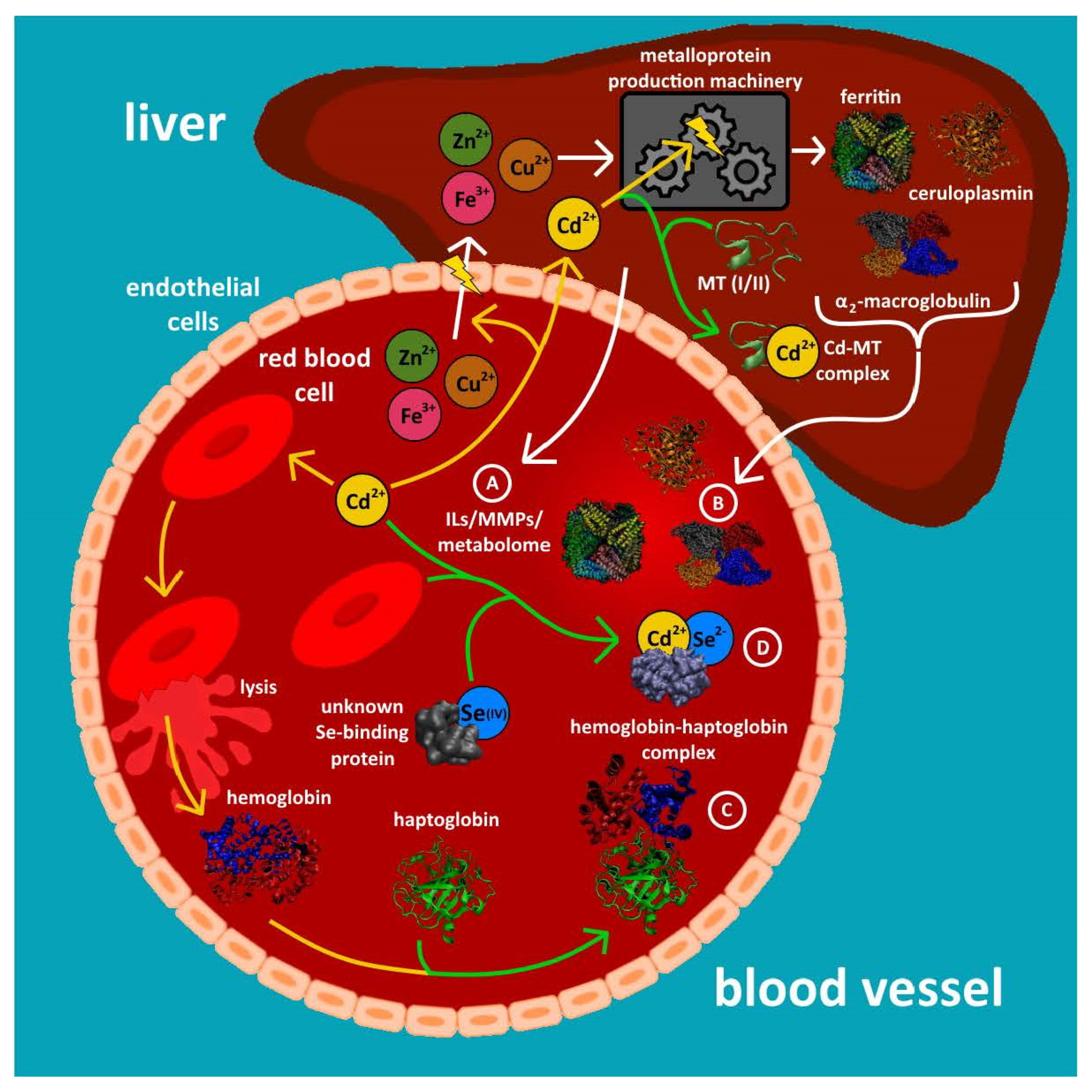
With regard to relevant metalloproteins in blood plasma ~10 major Cu-, Fe- and Zn-containing metalloentities including Cp, Tf, the Zn-protein α2 macroglobulin and the extracellular Cu, Zn superoxide dismutase (ex-Cu/Zn SOD) have been observed (Table 1) [18,36]. Moreover, matrix metalloproteinases (MMPs), which contain Zn in the catalytic site, have been reported in plasma [58,59]. MMPs are a class of multidomain Zn-dependent endopeptidases with different isoforms and are extracellular matrix modifiers. The activity of the various MMP isoforms has been used to distinguish health vs. disease status (e.g., cardiovascular, lung, infection, inflammatory, cancer) and also in pregnancy, and this set of markers has clinical value. Mn SOD, which is found mainly in mitochondria, has also been quantified in human plasma [60]. In addition, four selenoproteins have been detected in mouse plasma [61]. While some researchers have reported that the analysis of excised organs (e.g., the liver) from sentinel organisms that inhabit a particular area is useful in detecting toxic metal-containing exposure biomarkers [62], comparatively less is known about metal-containing biomarkers in the bloodstream, which is in constant contact with all organs. Based on these facts, it seems prudent to critically assess the inherent potential that metalloentities that are contained in plasma and/or RBC cytosol can offer in the context of assessing the chronic exposure of mammalian organisms, including humans, to inorganic environmental pollutants. To this end, hemoglobin adduct formation for the toxic metalloid species AsH3 has been previously reported [63]. Metabolomics approaches represent an alternative means to gain insight into the organ damage that is associated with the chronic exposure of mammals to toxic metalloid species [16], and proteomics approaches have similarly been used to identify maternal blood biomarkers to better understand the relationship between the chronic exposure of expecting mothers to toxic metal species and adverse pregnancy outcomes [64,65] (Figure 2A). The determination of endogenous metalloproteins (Figure 2B,C) and metabolites that contain essential and toxic metals in the bloodstream (Figure 2D), however, offers another useful approach to observe inorganic pollution-related perturbations at the systems level and to possibly identify metalloentities that could be used as indirect inorganic pollutant biomarkers because the metalloproteome is inherently less complex than the plasma proteome [66].

2.1.1. Decreased Plasma Concentrations of Individual Metalloproteins

The absence of individual metalloproteins from blood plasma can be indicative of genetic diseases. Wilson’s disease (WD), for example, can be diagnosed by observing negligible Cp concentrations in plasma [40] owing to the decreased assembly of this metalloprotein in the liver and its concomitantly reduced secretion into the systemic blood circulation. In an analogous manner, the chronic exposure of mammals to inorganic pollutants could similarly result in a gradual decrease of the plasma concentration of a particular metalloprotein over time since essentially all plasma metalloproteins are assembled in and secreted from the liver. A decreased plasma concentration of a particular metalloprotein could therefore be caused by a reduced translocation/flux of the corresponding essential element building block into the liver either by it forming a complex with a toxic metal in the bloodstream [67] or by it adversely affecting a molecular transporter that mediates the influx of a particular transition metal ion to the liver [68] (Figure 2B). In addition, the chronic exposure of mammals to one or more inorganic pollutants could selectively inhibit the biosynthesis of a particular metalloprotein within the liver similar to what has been observed in microbes [69,70]. Although cytosolic metallothioneins play an important role in sequestering certain toxic metals (e.g., Hg2+) within organs [71,72], some toxic metal species may target biomolecular chaperones that are critical for metalloprotein assembly [73] or otherwise interfere with a metalloprotein’s biosynthesis [74]. All of these aforementioned scenarios would decrease the expression of the corresponding metalloprotein over time with a concomitant decrease of its plasma concentration [18] until it is below the healthy control range [26]. Although this principal approach represents an indirect means to assess exposure, it offers two important advantages. Firstly, the quantification of plasma metalloproteins is inherently more practical than the more invasive approach to detect inorganic pollution-related metalloprotein biomarkers, such as Cd-metallothionein in liver biopsy samples [62] as blood plasma is readily accessible. Secondly, if multiple toxic metal species target the assembly of one particular metalloprotein, the decrease of its metalloprotein concentration in plasma would effectively represent an integral/cumulative measure of the exposure to all toxic metal species. Plasma selenoproteins appear to be interesting candidates to test this hypothesis since Hg2+ and AsIII have been shown to specifically target selenoprotein synthesis [29] and plasma selenoproteins can be routinely quantified [75]. Despite this, only a few studies have reported which demonstrate that the chronic exposure of mammals to a specific toxic metal species is associated with a temporal decrease of the concentration of a specific selenoprotein in plasma [76].

2.1.2. Increased Plasma Concentrations of Individual Metalloproteins

Two Fe-containing metalloproteins are released from tissues into the systemic blood circulation, namely ferritin (Ft), which is constantly released from endothelial and organ cells [77], and myoglobin, the plasma concentration of which is elevated after a heart attack as it is released from heart cells [78]. In an analogous manner, one may envision that the chronic exposure of a mammal to inorganic pollutants could result in organ damage which is associated with the release of metal-based tissue leakage products into the bloodstream. An important prerequisite for the use of Fe metalloproteins as indicators of exposure to inorganic pollutants is that only those metal-containing tissue leakage product(s) are of practical use that have an appropriate half-life and can be detected in the presence of the classical plasma metalloproteins (i.e., Cp, Tf).
The suicidal death of RBCs is referred to as eryptosis, which can be triggered by toxic metal/metalloid species including AsIII, Cd2+ and Hg2+ [79]. Thus, the rupture of RBCs also represents a potential source of metal-based ‘tissue’ leakage products in the blood plasma. To this end, a major plasma Fe-metalloprotein in human plasma was identified as a haptoglobin (Hp)–hemoglobin (Hb) complex [37]. This Hp–Hb complex is formed in plasma after RBCs rupture and the released cytosolic Hb readily reacts with the plasma protein Hp [37,80]. A disease that directly relates to this observation is a rare human disorder called paroxysmal nocturnal hemoglobinuria (NPH), which is associated with the increased rupture of erythrocytes during the night and afflicts about 0.5–1.5 per million people [81]. Since many inorganic pollutants are able to enter RBCs [82] over their lifetime of approximately 120 days, it is conceivable that chronic human exposure to these pollutants could destabilize RBC membranes and eventually result in their rupture [83]. This, in turn, would result in an increased plasma concentration of Hp–Hb complexes (Figure 2C) provided that their subsequent sequestration from the bloodstream by the spleen and by macrophages is sluggish. Based on these considerations, it is evident that more basic research needs to be conducted before the full potential of endogenous plasma metalloproteins as pollution biomarkers in humans as well as sentinel mammalian organisms can be realized [18]. In support of this notion, an association between maternal lead levels and increased MMPs in plasma has been observed [59].

2.2. Red Blood Cells

RBC cytosol contains the Fe-containing hemoglobin (Hb, 64 kDa) and catalase [84], the Zn-containing carbonic anhydrase (CA, 30 kDa), a Cu- and Zn-containing superoxide dismutase (SOD) [85] and an unidentified Mn-containing protein, possibly a Mn-containing SOD [85]. RBCs are assembled in the bone marrow and then released into the bloodstream where they can circulate for about 120 days. Therefore, the determination of Cu, Fe and Zn metalloproteins in RBC cytosol [86] could provide an excellent means to assess the chronic exposure of humans to inorganic pollutants, which accumulate therein [87]. For example, it is known that chronic lead (Pb2+) poisoning in mammals targets the heme biosynthetic pathway by inhibiting ferrochelatase, which inserts Fe into heme [88]. Accordingly, chronic exposure to Pb2+ eventually results in the formation of a Zn-protoporphyrin within RBCs [89]. The diagnostic relevance of determining the Cu, Fe and Zn metalloproteome ‘snapshot’ is, therefore, immediately apparent as a decreased Hb concentration and an increased cytosolic concentration of Zn-protoporphyrin (it has a different MW than CA) would allow one to diagnose chronic human exposure to Pb2+. Since many toxic metal/metalloid species are absorbed by RBCs through a variety of uptake mechanisms [68], the quantification of the Cu-, Fe- and Zn-containing metalloproteins in RBC cytosol has inherent potential to be of use in harboring information about the chronic exposure of a mammal to inorganic pollutants. Another lead-binding protein in the erythrocyte is delta-aminolevulinic acid dehydratase (ALAD), which is known to be inhibited by Pb exposure [90].

3. Bi-/Trimetallic Complexes Which Contain Essential and Toxic Metals as Biomarkers

3.1. Blood Plasma/Serum

Blood represents the first biochemically complex biological fluid that the inorganic nutrients (i.e., essential trace elements, e.g., Se) and pollutants (e.g., As, Cd and Hg) that are absorbed from the GI tract encounter. Direct experimental evidence for the formation of complexes in plasma which contain an essential and a toxic metal dates back to the mid-1970s when a Hg- and Se-containing complex with a molar ratio of 1:1 was observed [91]. The detection of a related Cd and Se complex (Figure 2D) with a similar stoichiometry suggested that the formation of both complexes is mechanistically related [61,92]. Subsequent studies employed X-ray absorption spectroscopy to structurally characterize the Hg-Se species which was formed in blood plasma 25 min after New Zealand white rabbits were intravenously injected with Hg2+ and sodium selenite (SeIV) as a (Hg-Se)100 species [93]. Interestingly, up to 30 of these Hg-Se nanoparticles bound to the plasma protein selenoprotein P [94]. The formed (Hg-Se)100–selenoprotein P adduct is stable in solution at pH 7.4 [95] and is likely the species that binds to the surface of RBCs [96] and is eventually deposited in the kidneys and the liver [97]. Thus, the (Hg-Se)100–selenoprotein P adduct fulfills all basic requirements for a useful biomarker [75], but its usefulness as an exposure biomarker for Hg2+ exposure requires studies to determine its half-life in the bloodstream. Seemingly unrelated to this it has been reported that forests are sinks for Hg and forest fires therefore release significant quantities of Hg that result in an enhanced Hg accumulation in fish [98]. Since fire fighters are exposed to Hg species when fighting forest fires and considering that they are known to suffer from distinct occupational diseases [99], it would be useful to establish a biomarker for their cumulative Hg exposure which can serve as a basis to estimate their exposure to other, more concerning pollutants. While the detection of (Hg-Se)100-selenoprotein P adducts in plasma could therefore be a useful biomarker to assess the exposure of firefighters to Hg0 and Hg2+, its formation in the bloodstream also implies a decreased influx of the essential trace element selenium into organs, which is likely to gradually decrease the assembly of selenoproteins therein. Thus, human exposure to Hg2+ and/or Hg0 will eventually result in a decreased secretion of selenoproteins into the systemic blood circulation.

3.2. Red Blood Cells

The trace element antagonism between AsIII and SeIV that was discovered >80 years ago in mammals was eventually demonstrated to be based on the formation of the seleno-bis (S-glutathionyl) arsinium ion [(GS)2AsSe] within RBCs after arsenite (AsIII) and selenite (SeIV) were added to RBC lysate [100]. Interestingly, the addition of CH3Hg+ to RBC cytosol which had been spiked with (GS)2AsSe resulted in the formation of the trimetallic species (GS)2AsSeHg-CH3 [101]. The analysis of this RBC cytosol by liquid chromatography, however, revealed (GS)2AsSeHg-CH3 to be rather unstable which makes its formation not a feasible biomarker. More recently, the addition of Hg2+, methylmercury (CH3Hg+) or thimerosal (it contains CH3CH2Hg+) to RBC cytosol revealed the formation of hemoglobin (Hb) species with distinct Hb–Hg bonds [86]. While the formation of these species in RBC cytosol has direct toxicological relevance as it was recently demonstrated that the binding of CH3CH2Hg+ to Hb will adversely affect the binding of O2 [102], their detection represents a convenient means to detect human exposure to the aforementioned Hg species.

4. Dynamic Interactions between Toxic Metal Species, Plasma Proteins and SMW Thiols

When toxic metal species enter the human bloodstream, they encounter not only thousands of proteins [14] but also over 400 small molecular weight (SMW) molecules and metabolites, including amino acids, peptides, fatty acids and nucleotides that are present at µM concentrations [19]. The variation in concentration of these biomolecules in the bloodstream has been demonstrated to be, in part, genetically determined [19] and is therefore possibly involved in the distribution of toxic metal species to target organ tissues [103]. Thus, the biological fate of toxic metal species at the blood–organ interface appears to be critically determined by their interactions with plasma proteins and SMW molecules/metabolites (Figure 3).
One SMW metabolite that could be an important player in the toxicology of metals is homocysteine (hCys), which is an intermediate metabolite that is formed by the de-methylation of methionine [105], and is present in blood plasma of healthy adults at concentrations of 8–12 µM [106]. Hyperhomocysteinemia is a disease that is characterized by elevated levels of hCys in blood plasma (e.g., >15 µM) and has been linked to the development of cardiovascular disease, stroke, and Alzheimer’s disease (AD) [105], potentially due to the accidental incorporation of hCys into protein structures instead of methionine during translation that subsequently causes protein damage/aggregation, a hallmark of many diseases. Evidence in support of hCys playing an important role in the toxicology of metals comes from in vivo studies [104] and in vitro studies, which have demonstrated that 500 µM of hCys in the phosphate buffered saline (PBS) mobile phase was able to abstract Cd2+ from HSA when a HSA–Cd complex was injected onto a SEC column [107]. Although this hCys concentration was higher than what is observed in hyperhomocysteinemia patients, the fact that the results were obtained under near physiological conditions provide a rationale to more systematically investigate the role that this metabolite may play in the translocation of this and other toxic metal species to target organs. Accordingly, an important research avenue is to connect the bioinorganic chemistry of toxic metal species in the bloodstream with uptake mechanisms on the surface of target organ cells and the biomolecular mechanisms of organ damage [35,108]. This strategy will require not only to determine the structure of the actual metal species that is uptaken into an organ [109], but also to obtain knowledge about their uptake into various organ cell types. Taken together, the integration of the bioinorganic chemistry that unfolds at the blood–organ interface has the potential to establish the mechanisms which may causally link the chronic exposure of humans to toxic metal species with adverse health effects and possibly also to the etiology of diseases.
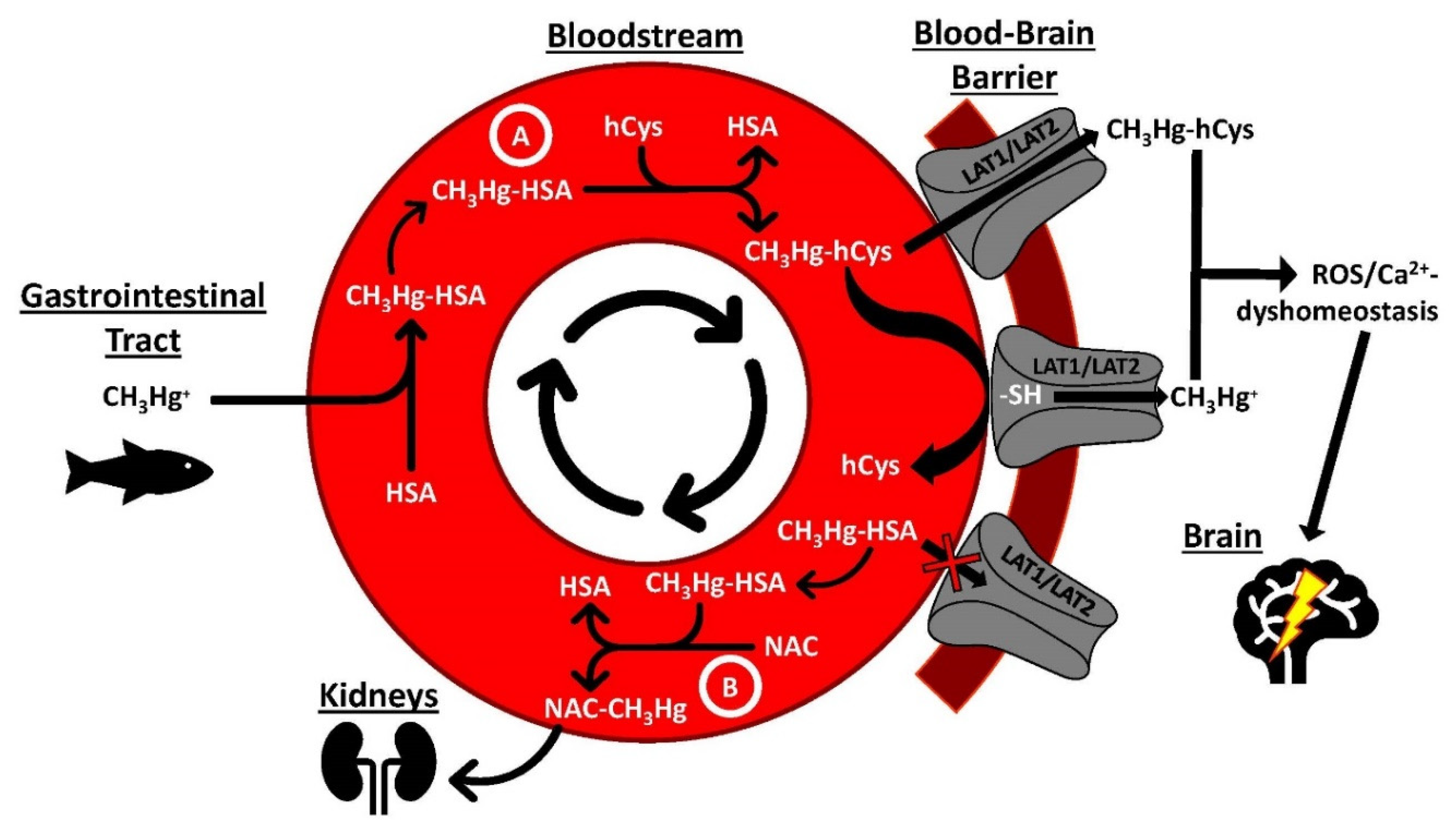
One toxic metal species that is of considerable public health relevance is the neurotoxin methylmercury (CH3Hg+), which has a strong affinity for thiol groups (-SH) on proteins and SMW thiols, including homocysteine (hCys) [110]. The competitive interaction of this toxic metal species with thiol groups located on proteins and SMW metabolites, however, is highly dynamic [111] and less well understood [112], but could play an important role in its translocation from plasma proteins across the blood–brain barrier (Figure 3A). To this end, it has been demonstrated that nearly 90% of the CH3Hg+ located in organ tissues of mice can be mobilized to urine by the oral administration of N-acetyl-L-cysteine (NAC) [113,114]. Studies that are designed to probe the interplay between CH3Hg+, plasma proteins, and SMW thiols under near-physiological conditions could shed important new light not only on the biomolecular mechanisms that are involved in the translocation of this metal species across the blood–brain and the placental barrier (Figure 3A and Figure 4), but also with regard to how the urinary excretion of CH3Hg+ may be deliberately enhanced in susceptible populations [115], such as the Inuit which consume comparatively large daily doses of this toxic metal species (Figure 3B) [116]. Probing the interactions between other toxic metal species, plasma proteins and SMW metabolites (e.g., hCys) in the bloodstream is also crucial to gain insight into the biomolecular mechanisms which deliver neurotoxic metal species to the brain [117] (Figure 4), where they may undergo toxicologically important biotransformations [118] and may initiate the apoptosis of astrocytes [119]. Establishing all sequential bioinorganic processes that unfold in the blood–organ system to facilitate the uptake of a toxic metal species into the brain with the biomolecular mechanisms which cause cell damage therein [120] thus offers the prospect of causally linking human environmental exposure to toxic metal species with the etiology of neurodegenerative diseases [121], including Alzheimer’s disease [122].

5. Conclusions

It is estimated that the emission of pollutants globally caused 9 million deaths in 2015 [127] and poses a significant burden on the global economy [128]. Even though a variety of efforts are underway to reduce human exposure to toxic metal species [129,130,131], their inadvertent introduction into the food chain is being increasingly recognized [132]. In addition, there is direct experimental evidence that human exposure to toxic metals adversely affects organs [133], pregnancy outcomes [64] and neurodevelopment in children [9]. Given the dynamic flow of inorganic and toxic elements through mammalian organisms and owing to the paucity of information on the mechanistic basis of environmental metal exposure and pregnancy outcomes [59] gaining insight into the environment–bloodstream–organ system [112] is critical. To this end, events that unfold in the bloodstream are of focal interest as they play an important role in determining the onset of organ damage [68,134] and may also be implicated in human diseases of unknown etiology [35,135]. Besides, blood plasma is also a valuable matrix to analyze tissue-specific markers and probably can be useful in screening for both exposure and effect markers in the same compartment simultaneously with the use of appropriate instrumental analysis methods. The analysis of blood plasma and RBC cytosol for endogenous metalloproteins and species which contain essential metal and toxic metals represents a feasible approach to gain insight into the dyshomeostasis of several essential metals and to observe toxic metal-containing biomarkers to better assess the health status of an organism. The integration of the bioinorganic chemistry that occurs in the bloodstream with biomolecular processes that unfold in organs emerges as an important research goal to detail our understanding of the environment–blood–organ system to possibly disentangle the environment vs. bad genes dichotomy to causally link human exposure to adverse health effects and possibly diseases [136]. Knowledge regarding the dynamics of the metalloproteome and their potential to serve as plasma/serum biomarkers can be useful in guiding future biomonitoring and epidemiological studies in choosing suitable bioinorganic species in the optimal biological compartment. Understanding the concerted biomolecular events which link exposure to disease is a critical prerequisite before environmental regulations can be further tightened to reduce the emission of toxic metals and metalloids into the environment to ascertain that future generations have the same opportunity to live a healthy life as we do.

Author Contributions

J.G. wrote the manuscript to which T.G.B. contributed Section 4. P.K. provided critical feedback on the entire manuscript. All authors have read and agreed to the published version of the manuscript.

Funding

Part of the research that is reviewed in this manuscript was supported by a Discovery Grant from the Natural Sciences and Engineering Research Council (NSERC) of Canada. T.G.B. was funded from this grant.

Institutional Review Board Statement

The research that was conducted at the University of Calgary was approved by the Life & Environmental Sciences Animal Care Committee (LESACC).

Informed Consent Statement

Informed consent was obtained from all subjects involved in studies conducted at the University of Calgary.

Data Availability Statement

The data presented in this study are available on request from the corresponding author.

Acknowledgments

Alexander Hill is greatly acknowledged for assembling Figure 2 and Marie Fraser (Department of Biological Sciences, University of Calgary) for producing Figure 4.

Conflicts of Interest

The authors declare no conflict of interest.

References

  1. Pacyna, J.M. Monitoring and assessment of metal contaminants in the air. In Toxicology of Metals; Chang, L.W., Ed.; CRC Press: Boca Raton, FL, USA, 1996; pp. 9–28. [Google Scholar]
  2. Izatt, R.M.; Izatt, S.R.; Bruening, R.L.; Izatt, N.E.; Moyer, B.A. Challenges to achievement of metal sustainability in our high-tech society. Chem. Soc. Rev. 2014, 43, 2451–2475. [Google Scholar] [CrossRef] [PubMed]
  3. Kirdyanov, A.V.; Krusic, P.J.; Shishov, V.V.; Vaganov, E.A.; Fertikov, A.I.; Myglan, V.S.; Barinov, V.V.; Brose, J.; Esper, J.; Ilyin, V.A.; et al. Ecological and conceptual consequences of Arctic pollution. Ecol. Lett. 2020, 23, 1827–1837. [Google Scholar] [CrossRef]
  4. Underwood, E.J. Trace metals in human and animal health. J. Hum. Nutr. 1981, 35, 37–48. [Google Scholar] [CrossRef]
  5. Bomer, N.; Beverborg, N.G.; Hoes, M.F.; Streng, K.W.; Vermeer, M.; Dokter, M.M.; Ijmker, J.; Anker, S.D.; Cleland, J.G.F.; Hillege, H.L.; et al. Selenium and outcome in heart failure. Eur. J. Heart Fail. 2019, 22, 1415–1423. [Google Scholar] [CrossRef] [PubMed] [Green Version]
  6. Sadiq, N.W.; Beauchemin, D. Simultaneous speciation analysis of arsenic, chromium and selenium in the bioaccessible fraction for realistic assessment of food safety. Anal. Chem. 2018, 257, 230–236. [Google Scholar] [CrossRef]
  7. McLaughlin, M.J.; Parker, D.R.; Clarke, J.M. Metals and micronutrients and food safety issues. Field Crop. Res. 1999, 60, 143–163. [Google Scholar] [CrossRef]
  8. Centers of Disease Control and Prevention. Fourth National Report on Human Exposure to Environmental Chemicals; Department of Health and Human Services, Centers for Disease Control and Prevention: Atlanta, GA, USA, 2009. [Google Scholar]
  9. Grandjean, P.; Landrigan, P.J. Developmental neurotoxicity of industrial chemicals. Lancet 2006, 368, 2167–2178. [Google Scholar] [CrossRef]
  10. Coulthard, T.J.; Macklin, M.G. Modeling long-term contamination in river systems from historical metal mining. Geology 2003, 31, 451–454. [Google Scholar] [CrossRef]
  11. Rodzik, A.; Pomastowski, P.; Sagandykova, G.M.; Buszewski, B. Interactions of whey proteins with metal ions. Int. J. Mol. Sci. 2020, 21, 2156. [Google Scholar] [CrossRef] [Green Version]
  12. Buszewski, B.; Žuvela, P.; Król-Górniak, A.; Railean-Plugaru, V.; Rogowska, A.; Wong, M.W.; Yi, M.; Rodzik, A.; Sprynskyy, M.; Pomastowski, P. Interactions of zinc aqua complexes with ovalbumin at the forfront of the Zn2+/ZnO-OVO hybrid complex formation mechanism. Appl. Surf. Sci. 2021, 542, 148641. [Google Scholar] [CrossRef]
  13. Zoroddu, M.A.; Aaseth, J.; Crisponi, G.; Medici, S.; Peana, M.; Nurchi, V.M. The essential metal for humans: A brief overview. J. Inorg. Biochem. 2019, 195, 120–129. [Google Scholar] [CrossRef] [PubMed]
  14. Anderson, N.L.; Anderson, N.G. The human plasma proteome: History, character, and diagnostic prospects. Mol. Cell. Proteom. 2002, 1, 845–867. [Google Scholar] [CrossRef] [PubMed] [Green Version]
  15. Zhou, Y.; Quin, S.; Sun, M.J.; Tang, L.; Yan, X.W.; Kim, T.K.; Caballero, J.; Glusman, G.; Brunkov, M.E.; Soloski, M.J.; et al. Measurement of organ-specific and acute-phase blood protein levels in early lyme disease. J. Proteome Res. 2020, 19, 346–359. [Google Scholar] [CrossRef] [PubMed]
  16. Wang, X.; Mu, X.; Zhang, J.; Huang, Q.; Alamdar, A.; Tian, M.; Liu, L.; Shen, H. Serum metabolites reveals that arsenic exposure disrupted lipid and amino acid metabolkism in rats: A step forward in understanding chronic arsenic toxicity. Metallomics 2015, 7, 544–552. [Google Scholar] [CrossRef]
  17. Dey, K.K.; Wang, H.; Niu, M.; Bai, B.; Wang, X.; Li, Y.; Cho, J.-H.; Tan, H.; Mishra, A.; High, A.A.; et al. Deep undepleted human serum proteome profiling toward biomarker discovery for Alzheimer’s disease. Clin. Proteom. 2019, 16, 1–13. [Google Scholar] [CrossRef] [PubMed] [Green Version]
  18. Manley, S.A.; Gailer, J. Analysis of the plasma metalloproteome by SEC-ICP-AES: Bridging proteomics and metabolomics. Expert Rev. Proteom. 2009, 6, 251–265. [Google Scholar] [CrossRef]
  19. Shin, S.-Y.; Fauman, E.B.; Petersen, A.-K.; Krumsiek, J.; Santos, R.; Huang, J.; Arnold, M.; Erte, I.; Forgetta, V.; Yang, T.-P.; et al. An atlas of genetic influences on human blood metabolites. Nat. Genet. 2014, 46, 543–550. [Google Scholar] [CrossRef] [Green Version]
  20. Lyon, T.D.B.; Aughey, E.; Scott, R.; Fell, G.S. Cadmium concentration in human kidney in the UK: 1978–1993. J. Environ. Monitor. 1999, 1, 227–231. [Google Scholar] [CrossRef]
  21. Hood, L. A systems approach to medicine will transform healthcare. In Physical Biology, from Atoms to Medicine; Zewail, A.H., Ed.; Imperial College Press: London, UK, 2008; pp. 337–366. [Google Scholar]
  22. Dede, E.; Tindall, M.J.; Cherrie, J.W.; Hankin, S.; Collins, C. Physiologically-based pharmacokinetic and toxicokinetic models for estimating human expaosure to five toxic elements through oral ingestion. Environ. Toxicol. Pharmacol. 2018, 57, 104–114. [Google Scholar] [CrossRef]
  23. Sarpong-Kumankomah, S.; Gailer, J. Application of a novel metallomics tool to probe the fate of metal-based anticancer drugs in blood plasma: Potential, challenges and prospects. Curr. Top. Med. Chem. 2021, 21, 48–58. [Google Scholar] [CrossRef]
  24. Sturla, S.J.; Boobis, A.R.; Fitzgerald, R.E.; Hoeng, J.; Kavlock, R.J.; Schirmer, K.; Whelan, M.; Wilks, M.F.; Peitsch, M.C. Systems toxicology: From basic research to risk assessment. Chem. Res. Toxicol. 2014, 27, 314–329. [Google Scholar] [CrossRef]
  25. Gomez-Ariza, J.L.; Jahromi, E.Z.; Gonzalez-Fernandez, M.; Garcia-Barrera, T.; Gailer, J. Liquid chromatography-inductively coupled plasma-based metallomic approaches to probe health-relevant interactions between xenobiotics and mammalian organisms. Metallomics 2011, 3, 566–577. [Google Scholar] [CrossRef]
  26. Craig, W.Y.; Ledue, T.B.; Ritchie, R.F. Plasma Proteins: Clinical Utility and Interpretation; Dade Behring Inc.: Newark, NJ, USA, 2000. [Google Scholar]
  27. Da Silva, M.A.O.; Sussulini, A.; Arruda, M.A.Z. Metalloproteomics as an interdisciplinary area involving proteins and metals. Expert Rev. Proteom. 2010, 7, 387–400. [Google Scholar] [CrossRef]
  28. Barnett, J.P.; Scanlan, D.J.; Blindauer, C.A. Protein fractionation and detection for metalloproteomics: Challenges and approaches. Anal. Bioanal. Chem. 2012, 402, 3311–3322. [Google Scholar] [CrossRef]
  29. Gailer, J. Arsenic-selenium and mercury-selenium bonds in biology. Coord. Chem. Rev. 2007, 251, 234–254. [Google Scholar] [CrossRef]
  30. Riuz-Laguna, J.; Garcia-Alfonso, C.; Peinado, J.; Moreno, S.; Leradi, L.A.; Cristaldi, M.; Lopez-Barea, J. Biochemical biomarkers of pollution in Algerian mouse (Mus spretus) to assess the effects of the Aznalcollar disaster on Donana Park (Spain). Biomarkers 2001, 6, 146–160. [Google Scholar]
  31. Sarpong-Kumankomah, S.; Miller, K.; Gailer, J. Biological chemistry of toxic metals and metalloids, such as arsenic, cadmium and mercury. In Encyclopedia of Analytical Chemistry; Meyers, R.A., Ed.; John Wiley and Sons Ltd.: Hoboken, NJ, USA, 2020. [Google Scholar]
  32. Sussulini, A.; Becker, J.S.; Becker, J.S. Laser ablation ICP-MS: Application in biomedical research. Mass Spectrom. Rev. 2017, 36, 47–57. [Google Scholar] [CrossRef]
  33. Becker, J.S.; Zoriy, M.; Matusch, A.; Wu, B.; Salber, D.; Palm, C.; Becker, J.S. Bioimaging of metals by laser ablation inductively coupled plasma mass spectrometry (LA-ICP-MS). Mass Spectrom. Rev. 2010, 29, 156–175. [Google Scholar] [CrossRef]
  34. Coufalikova, K.; Benesova, I.; Vaculovic, T.; Kanicky, V.; Preisler, J. LC coupled to ESI, MALDI and ICP MS—A multiple hypehnation for metallproteomic studies. Anal. Chim. Acta 2017, 968, 58–65. [Google Scholar] [CrossRef]
  35. Hill, A.; Gailer, J. Linking molecular targets of Cd in the bloodstream to organ-based adverse health effects. J. Inorg. Biochem. 2021, 217, 111279. [Google Scholar] [CrossRef]
  36. Manley, S.A.; Byrns, S.; Lyon, A.W.; Brown, P.; Gailer, J. Simultaneous Cu-, Fe-, and Zn-specific detection of metalloproteins contained in rabbit plasma by size-exclusion chromatography-inductively coupled plasma atomic emission spectroscopy. J. Biol. Inorg. Chem. 2009, 14, 61–74. [Google Scholar] [CrossRef] [PubMed]
  37. Sarpong-Kumankomah, S.; Gailer, J. Identification of a haptoglobin-hemoglobin complex in human blood plasma. J. Inorg. Biochem. 2019, 201, 110802. [Google Scholar] [CrossRef]
  38. Broman, L. Separation and characterization of two ceruloplasmins from human serum. Nature 1958, 182, 1655. [Google Scholar] [CrossRef]
  39. Morgan, E.H. Exchange of iron and transferrin across endothelial surfaces in rat and rabbit. J. Physiol. 1963, 169, 339–345. [Google Scholar] [CrossRef] [PubMed] [Green Version]
  40. Scheinberg, I.H.; Gitlin, D. Deficiency of ceruloplasmin in patients with hepatolenticular degeneration (Wilson’s Disease). Science 1952, 116, 484–489. [Google Scholar] [CrossRef]
  41. Rouault, T.A. The role of regulatory proteins in mammalian iron homeostasis and disease. Nat. Chem. Biol. 2006, 2, 406–414. [Google Scholar] [CrossRef]
  42. Campbell, C.H.; Brown, R.; Linder, M.C. Circulating ceruloplasmin is an important source of copper for normal and malignant animal cells. Biochim. Biophys. Acta 1981, 678, 27–38. [Google Scholar] [CrossRef]
  43. George, G.N.; Pickering, I.J.; Harris, H.H.; Gailer, J.; Klein, D.; Lichtmannegger, J.; Summer, K.-H. Tetrathiomolybdate causes formation of hepatic copper-molybdenum clusters in an animal model of Wilson’s Disease. J. Am. Chem. Soc. 2003, 125, 1704–1705. [Google Scholar] [CrossRef]
  44. Bull, P.C.; Thomas, G.R.; Rommens, J.M.; Forbes, J.R.; Cox, D.W. The Wilson disease gene is a putative copper transporting P-type ATPase similar to the Menkes gene. Nat. Genet. 1993, 5, 327–337. [Google Scholar] [CrossRef]
  45. Bonhorst, J.A.; Kipp, A.P.; Haase, H.; Meyer, S.; Schwerdtle, T. The crux of inept biomarkers for risks and benefits of trace elements. Trends Anal. Chem. 2018, 104, 183–190. [Google Scholar] [CrossRef]
  46. Cannon, W.B. Organization for physiological homeostasis. Physiol. Rev. 1929, 9, 399–431. [Google Scholar] [CrossRef]
  47. Coverdale, J.P.C.; Barnett, J.P.; Adamu, A.H.; Griffiths, E.J.; Stewart, A.J.; Blindauer, C.A. A metalloproteomic analysis of interactions between plasma proteins and zinc: Elevated fatty acid levels affect zinc distribution. Metallomics 2019, 11, 1805–1819. [Google Scholar] [CrossRef] [Green Version]
  48. Kimura, F.; Hasegawa, G.; Obayasji, H.; Adachi, T.; Hara, H.; Ohta, T.; Fukui, M.; Kitagawa, Y.; Park, H.; Nakamura, N.; et al. Serum extracellular superoxide dismutase in patients with type 2 diabetes. Diabetes Care 2003, 26, 1246–1250. [Google Scholar] [CrossRef] [PubMed] [Green Version]
  49. Adachi, T.; Inoue, M.; Maehata, E.; Suzuki, S. Relationship of plasma extracellular-superoxide dismutase level with insulin resistance in type 2 diabetic patients. J. Endocrinol. 2004, 181, 413–417. [Google Scholar] [CrossRef] [Green Version]
  50. Adachi, T.; Wang, J.; Wang, X.L. Age-related change of plasma extracellular-superoxide dismutase. Clin. Chim. Acta 2000, 290, 169–178. [Google Scholar] [CrossRef]
  51. Yamamoto, M.; Hara, H.; Adachi, T. Effects of homocysteine on the binding of extracellular-superoxide dismutase to the endothelial cell surface. FEBS Lett. 2000, 486, 159–162. [Google Scholar] [CrossRef] [Green Version]
  52. Gonzalez-Dominguez, R.; Garcia-Barrera, T.; Gomez-Ariza, J.L. Characterization of metal profiles in serum during the progression of Alzheimer’s disease. Metallomics 2014, 6, 292–300. [Google Scholar] [CrossRef]
  53. Zhang, R.; Wang, I.; Zhao, Q.H.; Jiang, R.; Gong, S.G.; Jiang, X.; Xu, X.Q.; He, Y.Y.; Li, Y.; Jing, Z.C. Alteration of extracellular superoxide dismutase in idiopathic pulmonary arterial hypertension. Front. Med. 2020, 7, 509. [Google Scholar] [CrossRef]
  54. Scassellati, C.; Bonvicini, C.; Benussi, L.; Ghidoni, R.; Squitti, R. Nejurodevelopmental disorders: Metallomics studies for the identification of potential biomarkersassociated to diagnostics and treatment. J. Trace Elem. Med. Biol. 2020, 60, 125499. [Google Scholar] [CrossRef] [PubMed]
  55. Adlard, P.A.; Bush, A.I. Metals and Alzheimer’s disease. J. Alzheimer’s Dis. 2006, 10, 145–163. [Google Scholar] [CrossRef]
  56. Hood, L.; Heath, J.R.; Phelps, M.E.; Lin, B. Systems biology and new technologies enable predictive and preventative medicine. Science 2004, 306, 640–643. [Google Scholar] [CrossRef] [Green Version]
  57. Price, N.D.; Magis, A.T.; Earls, J.C.; Glusman, G.; Levy, R.; Lausted, C.; McDonald, D.T.; Kuseback, U.; Moss, C.L.; Zhou, Y.; et al. A wellness study of 108 individuals using personal, dense, dynamic data clouds. Nat. Biotechnol. 2017, 35, 747–756. [Google Scholar] [CrossRef] [PubMed]
  58. Nagase, H.; Visse, R.; Murphy, G. Structure and function of matrix metalloproteinases and TIMPs. Cardiovasc. Res. 2006, 69, 562–573. [Google Scholar] [CrossRef] [Green Version]
  59. Au, F.; Bielecki, A.; Blais, E.; Fisher, M.; Cakmak, S.; Basak, A.; Gomes, J.; Arbuckle, T.E.; Fraser, W.D.; Vincent, R.; et al. Blood metal levels and third trimester maternal plasma matrix metalloproteinases (MMPs). Chemosphere 2016, 159, 506–515. [Google Scholar] [CrossRef] [PubMed]
  60. Wang, D.M.; Chen, D.C.; Wang, L.; Zhang, X.Y. Sex differences in the association between symptoms and superoxide dismutasein patients with never-treated first-episode schizophrenia. World J. Biol. Psychiatry 2020. [Google Scholar] [CrossRef]
  61. Garcia-Sevilliano, M.A.; Garcia-Barrera, T.; Navarro, F.; Gomez-Ariza, J.L. Cadmium toxicity in Mus musculus mice based on a metallomic study. Antagonistic interaction between Se and Cd in the bloodstream. Metallomics 2014, 6, 672–681. [Google Scholar] [CrossRef] [PubMed]
  62. Lopez-Barea, J.; Gomez-Ariza, J.L. Environmental proteomics and metallomics. Proteomics 2006, 6, S51–S62. [Google Scholar] [CrossRef] [PubMed]
  63. Yamauchi, T.; Yamano, Y.; Yamanaka, K.; Hata, A.; Nakadate, T.; Kuroda, Y.; Endo, Y.; Endo, G. Possible production of arsenic hemoglobin adducts via exposure to arsine. J. Occup. Health 2015, 57, 161–168. [Google Scholar] [CrossRef] [PubMed] [Green Version]
  64. Rahman, A.; Kumarathasan, K.; Gomes, J. Infant and mother related outcomes from exposure to metals with endocrine disrupting properties during pregnancy. Sci. Total Environ. 2016, 569, 1022–1031. [Google Scholar] [CrossRef]
  65. Gomes, J.; Au, F.; Basak, A.; Cakmak, S.; Vincent, R.; Kumarathasan, P. Maternal blood biomarkers and adverse pregnancy outcomes: A systematic review and meta-analysis. Crit. Rev. Toxicol. 2019, 49, 461–478. [Google Scholar] [CrossRef]
  66. Harding, A. Panning the proteome for biomarker gold. Science 2012, 337, 1120–1122. [Google Scholar] [CrossRef] [Green Version]
  67. Martelli, A.; Rousselet, E.; Dycke, C.; Bouron, A.; Moulis, J.M. Cadmium toxicity in animal cells by interference with essential elements. Biochimie 2006, 88, 1807–1814. [Google Scholar] [CrossRef] [PubMed]
  68. Sarpong-Kumankomah, S.; Gibson, M.A.; Gailer, J. Organ damage by toxic metals is critically determined by the bloodstream. Coord. Chem. Rev. 2018, 374, 376–386. [Google Scholar] [CrossRef]
  69. Xu, F.F.; Imlay, J.A. Silver(I), mercury(II), cadmium(II) and Zn(II) target exposed enzymic iron-sulfur clusters when they toxify Escherichia Coli. Appl. Environ. Microbiol. 2012, 78, 3614–3621. [Google Scholar] [CrossRef] [PubMed] [Green Version]
  70. Begg, S.L.; Eijkelkamp, B.A.; Luo, Z.; Counago, R.M.; Morey, J.R.; Maher, M.J.; Ong, C.-I.Y.; McEwan, A.G.; Kobe, B.; O’Mara, M.L.; et al. Dysregulation of transition metal ion homeostasis is the molecular basis for cadmium toxicity in Streptococcus pneumoniae. Nat. Commun. 2015, 6, 6418. [Google Scholar] [CrossRef] [Green Version]
  71. Zalups, R.K. Molecular interactions with mercury in the kidney. Pharmacol. Rev. 2000, 52, 113–143. [Google Scholar]
  72. Coyle, P.; Philcox, J.C.; Carey, L.C.; Rofe, A.M. Metallothionein. The multipurpose protein. Cell. Mol. Life Sci. 2002, 59, 627–647. [Google Scholar] [CrossRef]
  73. O’Halloran, T.V.; Culotta, V.C. Metallochaperones, an intracellular shuttle service for metal ions. J. Biol. Chem. 2000, 275, 25057–25060. [Google Scholar] [CrossRef] [Green Version]
  74. Garcia-Sevilliano, M.A.; Garcia-Barrea, T.; Navarro, F.; Gomez-Ariza, J.L. Absolute quantification of superoxide dismutase in cytosol and mitochondria of mice hepatic cells expsoed to mercury by a novel metallomic approach. Anal. Chim. Acta 2014, 842, 42–50. [Google Scholar] [CrossRef] [PubMed]
  75. Garcia-Sevilliano, M.A.; Rodriguesz-Moro, G.; Garcia-Barrea, T.; Navarro, F.; Gomez-Ariza, J.L. Biological interactions between mercury and seleniumin distribution and detoxifiction processes in mice under controlled exposure. Effects on selenoprotein. Chem. Biol. Interact. 2015, 229, 82–90. [Google Scholar] [CrossRef]
  76. Usuki, F.; Fujimura, M. Decreased plasma thiol antioxidant barrier amd selenoproteins as potential biomarkers for ongoing methylmercury intoxication and an individual protective capacity. Arch. Toxicol. 2016, 90, 917–926. [Google Scholar] [CrossRef] [PubMed]
  77. Kell, D.B.; Pretorius, E. Serum ferritin is an important inflammatory disease marker, as it is mainly a leakage product from damaged cells. Metallomics 2014, 6, 748–773. [Google Scholar] [CrossRef] [Green Version]
  78. Pur, M.R.K.; Hosseini, M.; Faridbod, F.; Ganjali, M.R. Highly sensitive label-free electrochemoluminescence aptasensor for early detection of myoglobin, a biomarker for myocardial infarction. Microchim. Acta 2017, 184, 3529–3537. [Google Scholar] [CrossRef]
  79. Lang, F.; Gulbins, E.; Lerche, H.; Huber, S.M.; Kempe, D.S.; Foeller, M. Eryptosis, a window to sytemic disease. Cell Physiol. Biochem. 2008, 22, 373–380. [Google Scholar] [CrossRef]
  80. Miller, K.; Sarpong-Kumankomah, S.; Egorov, A.; Gailer, J. Sample preparation of blood plasma enables baseline separation of iron metalloproteins by SEC-GFAAS. J. Chromatogr. B 2020, 1147, 122147. [Google Scholar] [CrossRef] [PubMed]
  81. Hill, A.; DeZern, A.E.; Kinoshita, T.; Brodsky, R.A. Paroxysmal nocturnal haemoglobinuria. Nat. Rev. Dis. Primers 2017, 3, 1–14. [Google Scholar] [CrossRef] [Green Version]
  82. Gailer, J. Probing the bioinorganic chemistry of toxic metals in the mammalian bloodstream to advance human health. J. Inorg. Biochem. 2012, 108, 128–132. [Google Scholar] [CrossRef] [PubMed]
  83. Moitra, S.; Brashier, B.B.; Sahu, S. Occupational cadmium exposure-associated oxidative stress and erythrocyte fragility among jewelry workers in India. Am. J. Ind. Med. 2014, 57, 1064–1072. [Google Scholar] [CrossRef]
  84. Ordonez, Y.N.; Montes-Bayon, M.; Blanco-Gonzalez, E.; Sanz-Medel, A. Quantitative analysis and simultaneous activity measurements of Cu, Zn-superoxide dismutase in red blood cells by HPLC-ICPMS. Anal. Chem. 2010, 82, 2387–2394. [Google Scholar] [CrossRef]
  85. Pomazal, K.; Prohaska, C.; Steffan, I.; Reich, G.; Huber, J.F.K. Determination of Cu, Fe, Mn, and Zn in blood fractions by SEC-HPLC-ICP-AES coupling. Analyst 1999, 124, 657–663. [Google Scholar] [CrossRef]
  86. Gibson, M.A.; Sarpong-Kumankomah, S.; Nehzati, S.; George, G.N.; Gailer, J. Remarkable differences in the biochemical fate of Cd2+, Hg2+, CH3Hg+ and thimerosal in red blood cell lysate. Metallomics 2017, 9, 1060–1072. [Google Scholar] [CrossRef] [PubMed]
  87. Jahromi, E.Z.; Gailer, J. Probing bioinorganic chemistry processes in the bloodstream to gain new insights into the origin of human diseases. Dalton Trans. 2010, 39, 329–336. [Google Scholar] [CrossRef] [PubMed] [Green Version]
  88. Warren, M.J.; Cooper, J.B.; Wood, S.P.; Shoolingin-Jordan, P.M. Lead poisoning, haem synthesis and 5-aminolaevulinic acid dehydratase. TIBS 1998, 23, 217–221. [Google Scholar] [CrossRef]
  89. Lamola, A.A.; Yamane, T. Zn protoporphyrin in the erythrocytes of patients with lead intoxication and iron deficiency anemia. Science 1974, 186, 936–938. [Google Scholar] [CrossRef]
  90. Gonick, H.C. Lead-binding proteins: A review. J. Toxicol. 2011, 2011, 686050. [Google Scholar] [CrossRef] [Green Version]
  91. Burk, R.F.; Foster, K.A.; Greenfield, P.M.; Kiker, K.W. Binding of simultaneously administered inorganic selenium and mercury to a rat plasma protein. Proc. Soc. Exp. Biol. Med. 1974, 145, 782–785. [Google Scholar] [CrossRef]
  92. Sasakura, C.; Suzuki, K.T. Biological interaction between transition metals (Ag, Cd and Hg), selenide/sulfide and selenoprotein P. J. Inorg. Biochem. 1998, 71, 159–162. [Google Scholar] [CrossRef]
  93. Gailer, J.; George, G.N.; Pickering, I.J.; Madden, S.; Prince, R.C.; Yu, E.Y.; Denton, M.B.; Younis, H.S.; Aposhian, H.V. Structural basis of the antagonism between inorganic mercury and selenium in mammals. Chem. Res. Toxicol. 2000, 13, 1135–1142. [Google Scholar] [CrossRef]
  94. Suzuki, K.T.; Sasakura, C.; Yoneda, S. Binding sites for the (Hg-Se) complex on selenoprotein P. Biochim. Biophys. Acta 1998, 1429, 102–112. [Google Scholar] [CrossRef]
  95. Khan, M.A.K.; Wang, F. Mercury-selenium compounds and their toxicological significance: Toward a molecular understanding of the mercury-selenium antagonism. Environ. Toxicol. Chem. 2009, 28, 1567–1577. [Google Scholar] [CrossRef]
  96. Cherdwongcharoensuk, D.; Oliveira, M.J.; Aguas, A.P. In vivo formation and binding of SeHg complexes to the erythrocyte surface. Biol. Trace Elem. Res. 2010, 136, 197–203. [Google Scholar] [CrossRef] [PubMed]
  97. Khan, M.A.K.; Wang, F. Reversible dossolution of glutathione-mediated HgSexS1−x nanoparticles and possible significance in Hg-Se antagonism. Chem. Res. Toxicol. 2009, 22, 1827–1832. [Google Scholar] [CrossRef] [PubMed]
  98. Kelly, E.N.; Schindler, D.W.; St. Louis, V.L.; Donald, D.B.; Vladicka, K.E. Forest fire increases mercury accumulation by fishes via food web restructuring and increased mercury inputs. Proc. Natl. Acad. Sci. USA 2006, 103, 19380–19385. [Google Scholar] [CrossRef] [PubMed] [Green Version]
  99. LeMasters, G.K.; Genaidy, A.M.; Succop, P.; Deddens, J.; Sobeih, T.; Barriera-Viruet, H.; Dunning, K.; Lockey, J. Cancer risk among firefighters: A review and metal-analysis of 32 studies. J. Occup. Environ. Med. 2006, 48, 1189–1202. [Google Scholar] [CrossRef] [PubMed]
  100. Manley, S.A.; George, G.N.; Pickering, I.J.; Glass, R.S.; Prenner, E.J.; Yamdagni, R.; Wu, Q.; Gailer, J. The seleno bis (S-glutathionyl) arsinium ion is assembled in erythrocyte lysate. Chem. Res. Toxicol. 2006, 19, 601–607. [Google Scholar] [CrossRef]
  101. Korbas, M.; Percy, A.J.; Gailer, J.; George, G.N. A possible molecular link between the toxicological effects of arsenic, selenium and methylmercury: Methylmercury(II) seleno bis (S-glutathionyl) arsenic(III). J. Biol. Inorg. Chem. 2008, 13, 461–470. [Google Scholar] [CrossRef]
  102. De Magalhanes Silva, M.; de Araujo Dantas, M.D.; de Sila Filho, R.C.; dos Santos Sales, M.V.; de Almeida Xavier, J.; Leite, A.C.R.; Goulart, M.O.F.; Grillo, L.A.M.; de Barros, W.A.; de Fatima, A.; et al. Toxicity of thimerosal in biological systems: Conformational changes in human hemoglobin, decrease of oxygen binding, increase of protein glycation and amyloid formation. Intern. J. Biol. Macromol. 2020, 154, 661–671. [Google Scholar] [CrossRef]
  103. Thomas, D.J.; Smith, J.C. Effects of coadministered low-molecular weight thiol compounds on short-term distribution of methylmercury in the rat. Toxicol. Appl. Pharmacol. 1982, 62, 104–110. [Google Scholar] [CrossRef]
  104. Simmons-Wills, T.A.; Koh, A.S.; Clarkson, T.W. Transport of a neurotoxicant by molecualr mimicry: The methylmercury-L-cysteine complex is a substrate for human L-type large neutral amino acid transporter (LAT) 1 and LAT 2. Biochem. J. 2002, 367, 239–246. [Google Scholar] [CrossRef] [PubMed]
  105. Jakubowski, H. Homocysteine modification in protein structure/function and human disease. Physiol. Rev. 2019, 99, 555–604. [Google Scholar] [CrossRef]
  106. Huang, J.; Williams, M. Total plasma homocysteine determination using ion-exchange chromatography. J. Liq. Chromatogr. Relat. Technol. 2000, 23, 3143–3153. [Google Scholar] [CrossRef]
  107. Sagmeister, P.; Gibson, M.A.; McDade, K.H.; Gailer, J. Physiologically relevant plasma D, L-homocysteine concentrations mobilize Cd from human serum albumin. J. Chromatogr. B 2016, 1027, 181–186. [Google Scholar] [CrossRef]
  108. Araujo, J.E.; Jorge, S.; Santos, H.M.; Chiechi, A.; Galstyan, A.; Lodeiro, C.; Diniz, M.; Kleinman, M.T.; Ljubimova, J.Y.; Capelo, J.L. Proteomic changes driven by urban pollution suggest particulate matter as a deregulator of energy metabolism, mitochondrial activity, and oxidative pathways in the rat brain. Sci. Total Environ. 2019, 687, 839–848. [Google Scholar] [CrossRef] [PubMed]
  109. Hoffmeyer, R.E.; Singh, S.P.; Doonan, C.J.; Ross, A.R.S.; Hughes, R.J.; Pickering, I.J.; George, G.N. Molecular mimicry in mercury toxicology. Chem. Res. Toxicol. 2006, 19, 753–759. [Google Scholar] [CrossRef]
  110. Ajsuvakova, O.P.; Tinkov, A.A.; Aschner, M.; Rocha, J.B.T.; Michalke, B.; Skalnaya, M.G.; Skaklny, A.V.; Butnariu, M.; Dadar, M.; Sarac, I.; et al. Sylfhydryl groups as targets of mercury toxicity. Coord. Chem. Rev. 2020, 417, 213343. [Google Scholar] [CrossRef]
  111. Rabenstein, D.L.; Evans, C.A. The mobility of methylmercury in biological systems. Bioinorg. Chem. 1978, 8, 107–114. [Google Scholar] [CrossRef]
  112. Nogara, P.A.; Oliveira, C.S.; Schmitz, G.L.; Piquini, P.C.; Farina, M.; Aschner, M.; Rocha, J.B.T. Methylmercury’s chemistry: From the environment to the mammalian brain. Biochim. Biophys. Acta 2019, 1863, 129284. [Google Scholar] [CrossRef]
  113. Ballatori, N.; Lieberman, M.W.; Wang, W. N-acetylcysteine as an antidote in methylmercury poisoning. Environ. Health Perspect. 1998, 106, 267–271. [Google Scholar] [CrossRef]
  114. Madejczyk, M.S.; Aremu, D.A.; Simmons-Willis, T.A.; Clarkson, T.W.; Ballatori, N. Accelerated urinary excretion of methylmercury following administration of its antidote N-acetylcysteine requires Mrp2/Abcc2, the apical multidrug resistance-associated protein. J. Pharmacol. Exp. Ther. 2007, 322, 378–384. [Google Scholar] [CrossRef] [PubMed] [Green Version]
  115. Koh, A.S.; Simmons-Willis, T.A.; Pritchard, J.B.; Grassl, S.M.; Ballatori, N. Identification of a mechanism by which the methylmercury antidotes N-acetylcysteine and dimercaptopropanesulfonate enhance urinary metal excretion: Transport by the renal organic anion transporter-1. Mol. Pharmacol. 2002, 62, 921–926. [Google Scholar] [CrossRef] [Green Version]
  116. Campbell, P.G.C.; Gailer, J. Effects of Non-essential Metal Releases on the Environment and Human Health. In Metal Sustainability: Global Challenges, Consequences and Prospects; Izatt, R.M., Ed.; John Wiley & Sons Ltd.: Chichester, UK, 2016; pp. 221–252. [Google Scholar]
  117. Charlet, L.; Chapron, Y.; Faller, P.; Kirsch, R.; Stone, A.T.; Baveye, P.C. Neurodegenerative diseases and exposure to the environmental metals Mn, Pb, and Hg. Coord. Chem. Rev. 2012, 256, 2147–2163. [Google Scholar] [CrossRef]
  118. Korbas, M.; O’Donoghue, J.L.; Watson, G.E.; Pickering, I.J.; Singh, S.P.; Myers, G.J.; Clarkson, T.W.; George, G.N. The chemical form of mercury in human brain following poisoning or environmental exposure. ACS Chem. Neurosci. 2010, 1, 810–818. [Google Scholar] [CrossRef] [Green Version]
  119. Im, J.-Y.; Paik, S.-G.; Han, P.-L. Cadmium-induced astroglial death proceeds via glutathione depletion. J. Neurosci. Res. 2006, 83, 301–308. [Google Scholar] [CrossRef]
  120. Farina, M.; Rocha, J.B.T.; Aschner, M. Mechanisms of methylmercury-induced neurotoxicity: Evidence from experimental studies. Life Sci. 2011, 89, 555–563. [Google Scholar] [CrossRef] [Green Version]
  121. Costa, L.G.; Aschner, M.; Vitalone, A.; Syversen, T.; Soldin, O.P. Developmental neuropathology of environmental agents. Annu. Rev. Pharmacol. Toxicol. 2004, 44, 87–110. [Google Scholar] [CrossRef] [Green Version]
  122. Bjorklund, G.; Tinkov, A.A.; Dadar, M.; Rahman, M.M.; Chirumbolo, S.; Skalny, A.V.; Skalnaya, M.G.; Haley, B.E.; Ajsuvakova, O.P.; Aaseth, J. Insights into the potential role of mercury in Alzheimer’s disease. J. Mol. Neurosci. 2019, 67, 511–533. [Google Scholar] [CrossRef]
  123. Jude, K.M.; Banerjee, A.L.; Haldar, M.K.; Manokaran, S.; Roy, B.; Mallik, S.; Srivastava, D.K.; Christianson, D.W. Ultrahigh resolution crystal structures of human carbonic anhydrase I and II complexed with two-prong inhibitors reveal the molecular basis of high affinity. J. Am. Chem. Soc. 2006, 128, 3011–3018. [Google Scholar] [CrossRef] [PubMed] [Green Version]
  124. Berman, H.M.; Westbrook, J.; Feng, Z.; Gilliland, G.; Bhat, T.N.; Weissig, H.; Shindyalov, I.N.; Bourne, P.E. The protein data bank. Nucleic Acids Res. 2000, 28, 235–242. [Google Scholar] [CrossRef] [PubMed] [Green Version]
  125. He, Y.; Ning, T.; Xie, T.; Qui, Q.; Zhang, L.; Sun, Y.; Jiang, D.; Fu, K.; Yin, E.; Zhang, W.; et al. Large-scale production of functional human serum albumin from transgenic rice seeds. Proc. Natl. Acad. Sci. USA 2011, 108, 19078–19083. [Google Scholar] [CrossRef] [Green Version]
  126. Guex, N.; Peitsch, M.C. SWISS-Model and the Swiss-PdbViewer: An environment for comparative protein modelling. Electrophoresis 1997, 18, 2714–2723. [Google Scholar] [CrossRef]
  127. Landrigan, P.J.; Fuller, R.; Acosta, N.J.R.; Adeyi, O.; Arnold, R.; Basu, N.; Balde, A.B.; Bertollini, R.; Bose-O’Reilly, S.; Boufford, J.I.; et al. The Lancet commission on pollution and health. Lancet 2018, 391, 462–512. [Google Scholar] [CrossRef] [Green Version]
  128. Trasande, L.; Liu, Y. Reducing the staggering costs of environmental disease in children, estimated at $76.6 billion in 2008. Health Aff. 2011, 30, 863–870. [Google Scholar] [CrossRef] [PubMed] [Green Version]
  129. Sebastian, A.; Prasad, M.N.V. Cadmium minimization in rice: A review. Agron. Sustain. Dev. 2014, 34, 155–173. [Google Scholar] [CrossRef]
  130. Puschenreiter, M.; Horak, O.; Friesl, W.; Hartl, W. Low-cost agricultural measures to reduce heavy metal transfer to the food chain—A review. Plant Soil Environ. 2005, 51, 1–11. [Google Scholar] [CrossRef] [Green Version]
  131. Lew, T.T.S.; Park, M.; Cui, J.; Strano, M.S. Plant nanobionic sensors for arsenic detection. Adv. Mater. 2020, 33, 2005683. [Google Scholar] [CrossRef] [PubMed]
  132. Nachman, K.E.; Punshon, T.; Rardin, L.; Signes-Pastor, A.J.; Murray, C.J.L.; Jackson, B.P.; Guerinot, M.L.; Burke, T.A.; Chen, C.Y.; Ahsan, H.; et al. Opportunities and challenges for dietary arsenic intervention. Environ. Health Perspect. 2018, 126, 084503. [Google Scholar] [CrossRef] [Green Version]
  133. Orr, S.E.; Bridges, C.C. Chronic kidney disease and exposure to nephrotoxic metals. Int. J. Mol. Sci. 2017, 18, 1039. [Google Scholar]
  134. Weiss, B.; Clarkson, T.W.; Simon, W. Silent latency period in methylmercury poisoning and in neurodegenerative disease. Environ. Health Perspect. 2002, 110, 851–854. [Google Scholar] [CrossRef] [Green Version]
  135. Marth, J.D. A unified vision of the building blocks of life. Nat. Cell Biol. 2008, 10, 1015–1016. [Google Scholar] [CrossRef]
  136. Rossignol, D.A.; Genius, S.J.; Frye, R.E. Environmental toxicants and autism spectrum disorders: A systematic review. Transl. Psychiatry 2014, 4, e360. [Google Scholar] [CrossRef]
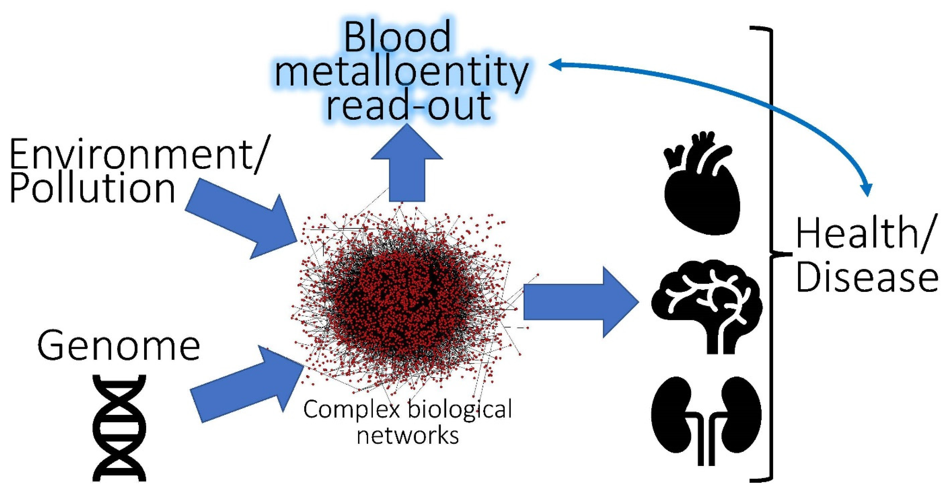
Figure 1. Conceptual depiction of the interaction between the environment and the genome of an organism that involves complex biological networks. The measurements of distinct metalloentities in the bloodstream may serve as biomarkers to assess the chronic exposure of an organism to individual or multiple toxic metal/metalloid species.
Molecules 26 03408 g001
Figure 2. Illustration of the principal mechanisms by which human exposure to toxic metal/metalloid species may result in increased or decreased concentrations of biomarkers in the bloodstream. Mechanism (A) refers to increased concentrations of biomarkers that are related to the stress of organs, which includes interleukins (IL) and/or matrix metalloproteinases (MMPs). Mechanisms (BD) refer to changes in the concentrations of metalloentities, which includes ‘endogenous’ metalloproteins (B), species which contain essential and toxic metals/metalloids (C) and a haptoglobin–hemoglobin complex that is formed in plasma following the rupture of RBCs (D).
Molecules 26 03408 g002
Figure 3. Conceptual illustration of bioinorganic chemistry processes which unfold between CH3Hg+, plasma proteins and SMW thiols/metabolites in the bloodstream and play a fundamental role in its translocation to the brain and/or its urinary excretion. The mobilization of CH3Hg+ from human serum albumin (HSA) by hCys may result in the formation of CH3Hg-hCys-adducts which may then be translocated across the blood–brain barrier (A) by L-type large neutral amino acid transporter (LAT) 1 and LAT 2 [104]. Alternatively, CH3Hg-hCys -adducts may donate CH3Hg+ to a thiol group on transmembrane proteins which ultimately mediate its uptake into the cytosol (B).
Molecules 26 03408 g003
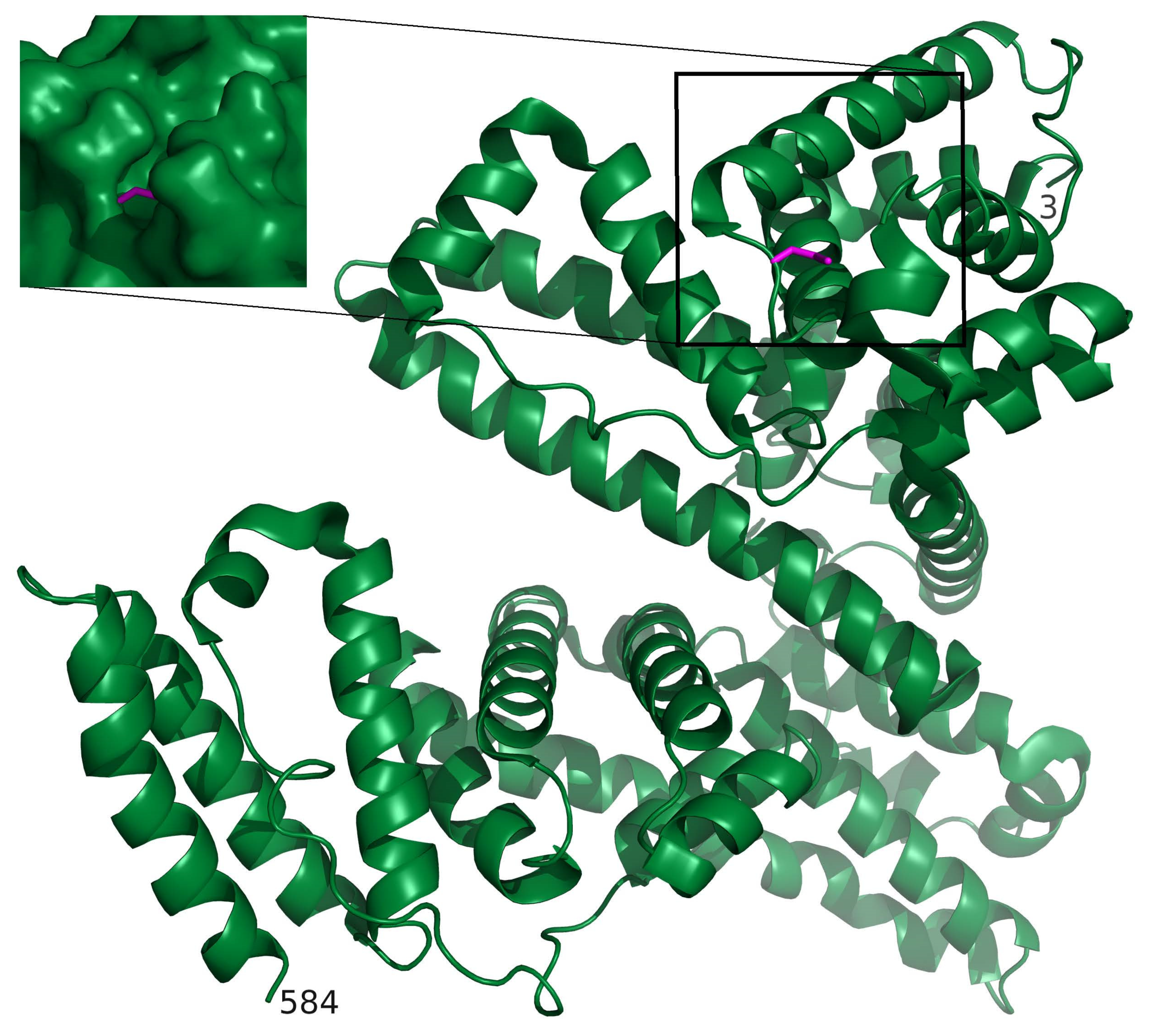
Figure 4. A model of CH3Hg+ bound to HSA was established by using CH3HgCys from the structure of human carbonic anhydrase (PDB identifier 2FOU [123,124]) which was superimposed on Cys-34 of HSA (PDB identifier 3SQJ [125]) using SWISS-MODEL/Swiss-pdbViewer [126]. The ribbon diagram of HSA shows where the binding site of CH3Hg+ is located and the amino- and carboxy-termini are indicated by residue numbers 3 and 584, respectively. The inset (left) shows a closeup of CH3Hg bound to Cys-34 (magenta) with a surface representation of the protein. PyMOL Molecular Graphics System (Version 1.4.1. Schroedinger L.L.C.) was used to establish the structural model. The location of CH3Hg+ within a cleft suggests that its direct translocation from HSA to LAT1 and/or LAT2 is unlikely and that SMW thiols may critically mediate this transfer.
Molecules 26 03408 g004
Table 1. Molecular properties of major metalloproteins and metallopeptides in human plasma/serum.
MetalMetalloprotein or Entity Which Contains Bound MetalMolecular Mass (kDa)Number of Metal Atoms Bound per ProteinReference
FeFerritin450<4500[36]
Transferrin79.92[36]
Haptoglobin–hemoglobin complex86–9002[37]
CuBlood coagulation factor V3301[36]
Transcuprein2700.5[36]
Ceruloplasmin1326[36]
Albumin661[36]
Extracellular superoxide dismutase1654[36]
Peptides and amino acids<5-[36]
Znα2 Macroglobulin7255[36]
Albumin661[36]
Extracellular superoxide dismutase1654[36]
Publisher’s Note: MDPI stays neutral with regard to jurisdictional claims in published maps and institutional affiliations.