Article

In Vitro and in Vivo Activity of mTOR Kinase and PI3K Inhibitors Against Leishmania donovani and Trypanosoma brucei

1
Leishmania Research Laboratory, Institut Pasteur Korea, 696 Sampyeong-dong, Bundang-gu, Seongnam-si, Gyeonggi-do 463–400, Korea
2
Screening Development Platform, Institut Pasteur Korea, 696 Sampyeong-dong, Bundang-gu, Seongnam-si, Gyeonggi-do 463–400, Korea
*
Author to whom correspondence should be addressed.
Molecules 2020, 25(8), 1980; https://doi.org/10.3390/molecules25081980
Submission received: 4 March 2020 / Revised: 2 April 2020 / Accepted: 21 April 2020 / Published: 23 April 2020

Abstract

:
Kinetoplastid parasites, including Leishmania and Trypanosoma spp., are life threatening pathogens with a worldwide distribution. Next-generation therapeutics for treatment are needed as current treatments have limitations, such as toxicity and drug resistance. In this study, we examined the activities of established mammalian target of rapamycin (mTOR)/phosphoinositide 3-kinase (PI3K) inhibitors against these tropical diseases. High-throughput screening of a library of 1742 bioactive compounds against intracellular L. donovani was performed, and seven mTOR/PI3K inhibitors were identified. Dose-dilution assays revealed that these inhibitors had half maximal effective concentration (EC50) values ranging from 0.14 to 13.44 μM for L. donovani amastigotes and from 0.00005 to 8.16 μM for T. brucei. The results of a visceral leishmaniasis mouse model indicated that treatment with Torin2, dactolisib, or NVP-BGT226 resulted in reductions of 35%, 53%, and 54%, respectively, in the numbers of liver parasites. In an acute T. brucei mouse model using NVP-BGT226 parasite numbers were reduced to under the limits of detection by five consecutive days of treatment. Multiple sequence and structural alignment results indicated high similarities between mTOR and kinetoplastid TORs; the inhibitors are predicted to bind in a similar manner. Taken together, these results indicated that the TOR pathways of parasites have potential for the discovery of novel targets and new potent inhibitors.

1. Introduction

Neglected tropical diseases including leishmaniasis, human African trypanosomiasis (HAT, or sleeping sickness), and Chagas disease are caused by the pathogenic protozoans Leishmania spp., Trypanosoma brucei subspecies, and Trypanosoma cruzi, respectively. Globally, there are nearly one hundred thousand deaths and over 22 million reported cases associated with these diseases annually [1,2]. There are three types of leishmaniasis: cutaneous leishmaniasis (CL), mucocutaneous leishmaniasis, and visceral leishmaniasis (VL) [3,4]. The disease is distributed throughout 97 countries, mainly in Africa, Asia, and Latin America, but the fatal form of VL mostly occurs in Ethiopia, South Sudan, India, Bangladesh, and Brazil [5,6]. Current treatment options include sodium stibogluconate, which is only available for administration via injection; amphotericin B, which is repurposed from antifungal treatment; and miltefosine, being the only orally administered drug [7]. HAT is caused by protozoa of the subspecies of T. brucei, including T. brucei gambiense and T. brucei rhodesiense in Africa. The disease is usually transmitted by the bite of an infected tsetse fly. It most commonly occurs in rural areas and can be fatal if not treated. The treatment used depends on the type and the stage of infection. Pentamidine or suramin are widely used for the treatment of acute-phase infections. Melarsoprol or eflornithine are usually used for treatment of the second, or neurological, phase of disease [8,9]. In 2009, Drugs for Neglected Diseases initiative developed the oral nifurtimox/intravenous eflornithine combination therapy (NECT) for the second stage HAT, and more recently, in 2019, an effective oral monotherapy of fexinidazole was launch for both disease stages [10,11]. Chagas disease (American trypanosomiasis) is caused by T. cruzi and is spread by members of the Triatominae subfamily (kissing bugs); the disease distribution includes Paraguay, Colombia, Venezuela, Argentina, Bolivia, Chile, and Brazil. Symptoms can be developed 10–30 years after infection, and nifurtimox and benznidazole are used to cure infected patients [12,13]. The drugs used to treat kinetoplastid diseases are often highly toxic, expensive, and limited by administration route. These characteristics are significant barriers to their use for treatment of patients in developing countries [14,15,16,17]. Despite an urgent need, the development of next-generation therapeutics has been halted by no or low financial profits, as these diseases occur mostly in the poorest parts of the world [18,19,20]. To reduce the costs and time-consuming safety and efficacy trials, repurposing established drugs used for other diseases is an attractive path to facilitate the development of anti-kinetoplastid therapeutics [21,22,23,24].
Target of rapamycin (TOR) is a serine/threonine protein kinase that is a key master regulator of multiple signaling pathways in eukaryotes. The mammalian TOR (mTOR) pathway is involved in various biological functions, such as cellular growth and proliferation, metabolism, survival, motility, and autophagy [25,26,27]. The mTOR protein has a central role in the pathway. It belongs to the family of phosphoinositide 3-kinase (PI3K)-related kinases, which is a protein family with roles in cellular responses to various types of stresses [28,29,30]. Extensive investigations have led to the identification and functional characterization of PI3Kα, β, γ, and δ isoforms; these isoforms are currently considered as attractive targets for new anti-cancer therapies [31,32,33,34]. There are three types of PI3K inhibitors, including dual mTOR/PI3K, pan-PI3K, and isoform-specific inhibitors [35]. Based on the target, more than 35 compounds are being tested in clinical trials for treatment of different types of cancers. Many of these compounds have not advanced to late-phase trials due to toxicity and limitations in activity. However, everolimus, temsirolimus, idelalisib, and copanlisib have been approved by the U.S. Food and Drug Administration for clinical use as cancer treatments [36]. Moreover, ridaforolimus for metastatic soft-tissue sarcoma (clinicaltrials.gov identifier: NCT00538239) and buparlisib for advanced-stage breast cancer (NCT01610284) completed phase III clinical trials. Furthermore, taselisib for breast cancer (NCT02340221), duvelisib for chronic lymphocytic leukemia (NCT02004522), and ipatasertib for advanced-stage breast cancer (NCT03800836) are undergoing phase III clinical trials advocating the potential use of mTOR and PI3K inhibitor for therapeutic interventions [37,38,39].
In T. brucei, two orthologues of mTOR, TbTOR1 and TbTOR2, were identified. TbTOR1 is involved in cell cycle regulation and protein synthesis, whereas TbTOR2 coordinates cell polarization and cytokinesis [40,41]. More recently, an unusual TOR exclusively present in kinetoplastids, TbTOR-like 1, was discovered and found to have a role in the control of polyphosphate levels and acidocalcisome maintenance [42]. With the presence of TOR, rapamycin exhibits potent activity against parasite growth via preventing formation of TbTOR complex 2; this mechanism is contrary to the mechanisms discovered in other eukaryotes [40]. Three TORs have been identified for Leishmania, and mutant studies revealed that TOR1 and TOR2 are essential for the survival of Leishmania major promastigotes, and similar to T. brucei, TOR3 is required for acidocalcisome biogenesis [43].
Based on the essentiality of TORs in kinetoplastid parasites, several groups have evaluated the use of mTOR kinase and PI3K inhibitors against the parasites [44,45]. NVP-BEZ235, an anti-cancer agent at the clinical trial stage, is a potent inhibitor of Leishmania spp. and Trypanosoma spp. in vitro and showed efficacy against T. brucei in an animal model [37]. In a CL animal model, it was found that treatment with rapamycin and GSK-2126458 resulted in significant decreases in footpad swelling and parasitemia in the draining lymph nodes in infected animals [38]. These compounds are shown to kill parasites directly. On the other hand, Khadem et al. have demonstrated that selective pharmacological inhibition of the host PI3Kδ with CAL-101 results in significant reductions of parasite burdens in VL and CL animal models [40]. These findings suggest that mTOR/PI3K inhibitors have potential applications for treatment of kinetoplastid diseases.
In this study, we report the results of an in vitro screening of bioactive compounds against intracellular L. donovani. We identified new mTOR/PI3K inhibitors for further assessment for use on different forms of kinetoplastid parasites. Potent compounds in vitro were subjected to in vivo models of VL and HAT. Structural modeling of identified inhibitors binding to the TORs of kinetoplastid parasites were predicted to provide insights on the potential mode of inhibition at molecular level.

2. Results and Discussion

2.1. Intracellular Leishmania Pilot Screening

We used a high-content screening system to assess a library of 1742 compounds with potential bioactivity against intracellular Leishmania. Human acute monocytic leukemia (THP-1) cells infected with L. donovani were treated with compounds; images were analyzed to quantify the numbers of parasites and host cells and infection ratios. The reproducibility and robustness of the assay was evaluated by duplicate runs. The correlation between two independent screenings indicated a high degree of linear relationship, with an R2 value of 0.922. There were 74 compounds that had activity in set 1 or set 2 (threshold > 60%), and 32 with activity in both sets (Figure 1A). To evaluate assay quality, the Z′ factor was calculated using the average values from duplicate testing. The Z′ factor was given by Z′ = 1 − 3(σc+ + σc)/(μc+ − μc), where σc+c were the standard deviation values of the positive/negative controls and μc+c were the corresponding mean values. The Z′ value of the screening using the infection ratio was 0.798; this result indicated excellent assay quality (Figure 1B).
The screening results indicated that 50 compounds were active, based on a >60% threshold of the inhibition of intracellular parasite survival (Figure 1B). Since this number included the compounds showing activity due to killing of the host cells (low selectivity), we plotted the parasite survival inhibition versus the host cell viability to further filter parasite-selective compounds. With >60% parasite survival inhibition and >60% host cell viability, a final number of 20 compounds was selected out of the 1742 with the overall hit rate of 1.15% (Figure 1C).

2.2. Hit Characteristics and Selection of mTOR/PI3K Inhibitor

Among the selected 20 hits, five compounds were in the category of mTOR/PI3K inhibitors. In the screening library, 58 mTOR/PI3K inhibitors were present, and by proportion, 8.62% (= 5/58) was found active within this category of inhibitor. This rate is relatively higher compared to the overall hit rate of 1.15% (Figure 1D). The mTOR complex inhibitors, such as rapamycin and its analogs deforolimus, temsirolimus, and everolimus, were included in the library, but did not show potent inhibition at the screening concentration (10 µM). Previously reported antileishmanial compounds, such as dactolisib (NVP-BEZ235) [37] and GSK2126458 [39], were found active, but the latter one showed some toxicity against the host cell. The compounds known to exhibit activity in vivo via modulation of the host immune response (i.e., CAL-101) had no direct effects on the parasite [38].
With two additional mTOR/PI3K inhibitors at the border of threshold, a total of 22 compounds were subjected to dose-dilution assays for activity confirmation. In the result, compounds such as dopamine uptake inhibitors (GBR12909 and 3-CPMT), protein kinase A inhibitor (H89), VEGFR inhibitor (cediranib and ENMD-2076), and EGFR inhibitor (afatinib) had either low activity (half maximal effective concentration, EC50) or high toxicity (CC50). Other compounds, including Y29794, metaphit, Ro 106–9920, SKF 96365, and NNC05–2090 were confirmed to be active, but did not group together into a category of inhibitor. In the case of the mTOR/PI3K inhibitors, all the compounds were well-confirmed with potent activities and sufficient selectivity index (SI) values (Table 1). Based on these results, we sought to focus the subsequent investigations on this class of inhibitors (Figure 2).

2.3. In Vitro Activity Against Kinetoplastid Parasite

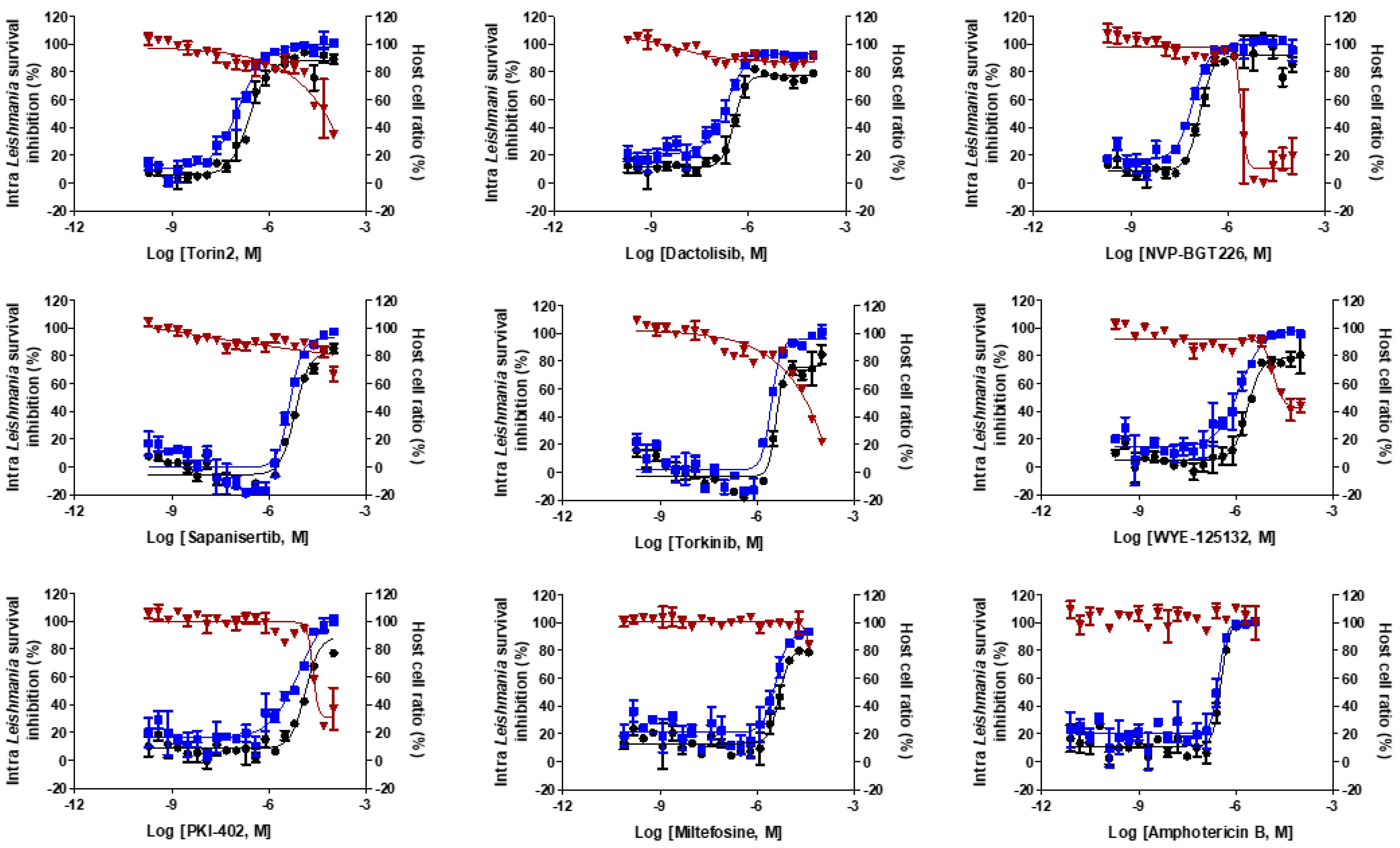
The confirmatory intracellular Leishmania assays found that the tested mTOR/PI3K inhibitors had EC50 values, ranging from 0.14 to 13.44 μM (Table 1 and Figure 3). The most potent compound was NVP-BGT226 with EC50 value of 0.14 μM, which is approximately 2.5 and 30 times more potent than amphotericin B and miltefosine, respectively. The structurally similar compounds, Torin2 (EC50 = 0.25 μM) and dactolisib (EC50 = 0.36 μM), exhibited activities in the submicromolar range. Torkinib and sapanisertib share the same scaffold, and PKI-402 and WYE-125132 are structurally similar. These inhibitors were moderately potent with EC50 values similar to that of miltefosine (Table 1). We then sought to check any relationship between the intracellular Leishmania survival inhibition values to mTOR/PI3K inhibition values from published data (Table S2) [46,47,48,49,50]. Interestingly, the inhibitors that are active against mammalian PI3K at low nanomolar range, such as Torin2, dactolisib, and NVP-BGT226, were mostly found active against the parasite except PKI-402.
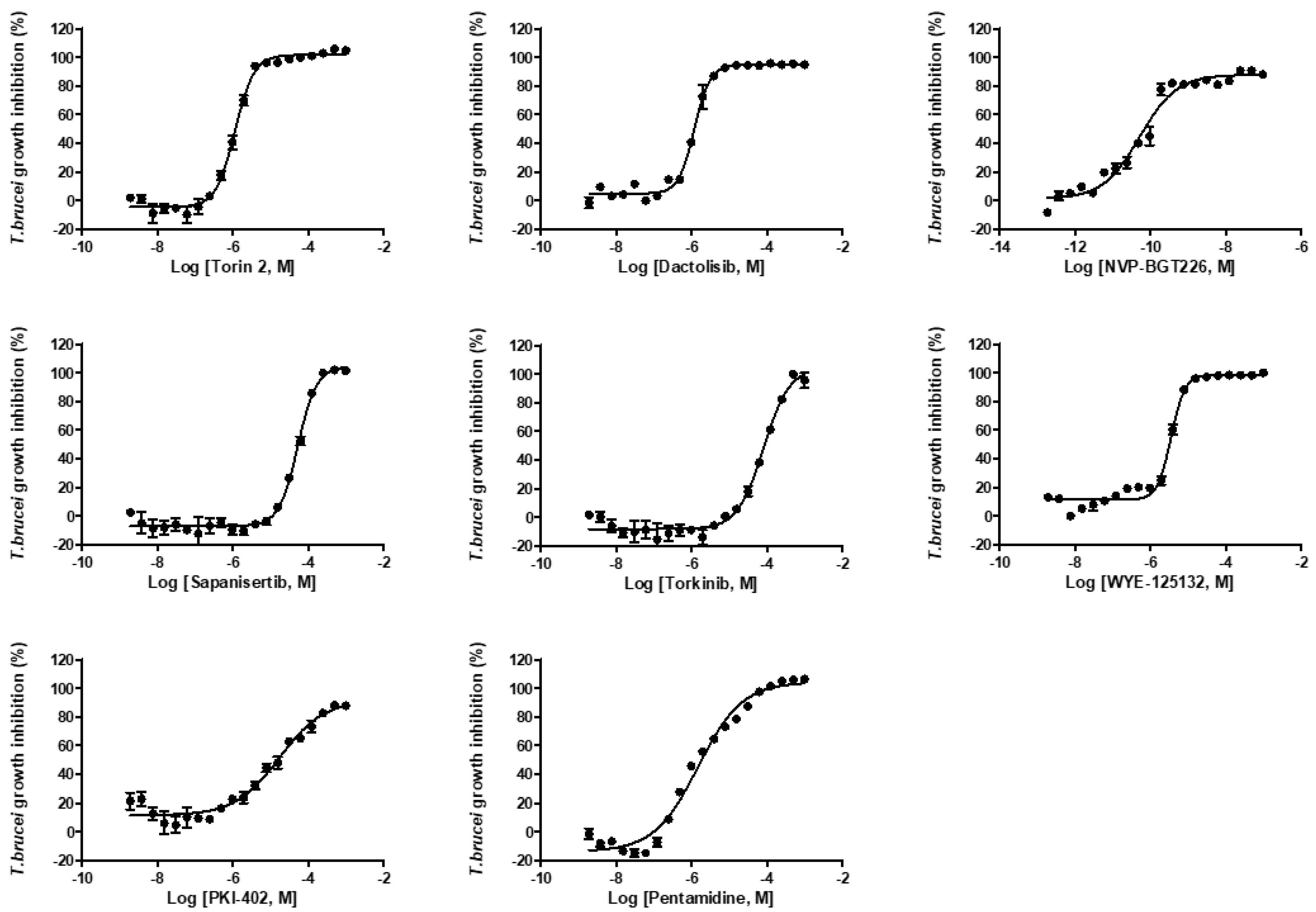
We further tested the seven compounds against L. donovani promastigotes (Figure S1), T. brucei bloodstream form (Figure 4) and intracellular T. cruzi. In the L. donovani promastigote growth inhibition assay, the EC50 values ranged from 0.009 to 7.46 μM; NVP-BGT226 was again the most potent compound (Table 1). The correlation of activity between the promastigote and amastigote assay was R2 = 0.64. Two structurally similar compounds, WYE-125132 and PKI-402, were 6.3- and 27.4-fold more potent against the promastigotes compared with the intracellular amastigotes (Figure 5A). In T. brucei assay, the compounds were generally more active and especially NVP-BGT226 had an EC50 value in the picomolar range (0.000054 μM). Except for sapanisertib and torkinib, all the compounds had submicromolar activities with values approximately similar to pentamidine. The extracellular forms of L. donovani and T. brucei displayed a high correlation of activity with R2 = 0.80 (Figure 5C), but the correlation between T. brucei and the amastigotes of L. donovani with R2 = 0.51 (Figure 5B). These differences may be due to genus-related differences between the parasites or to the effect of the extra host cell membrane on the penetration of compounds, or both. Furthermore, since dactolisib (NVP-BEZ235) showed varying range of in vitro activity depending on the subspecies of T. brucei in the previous report, the tested compounds in this study may exhibit different inhibitory activity to other subspecies of T. brucei or in different species of Leishmania [51,52]. One interesting point is the differences of antileishmanial and antitrypanosomal potency among Torin2, dactolisib and NVP-BGT225, in which the compounds share a similar structure. NVP-BGT226 and dactolisib share the same scaffold of 3-methyl-1-phenyl-8-(pyridin-3-yl)-1,3-dihydro-2H-imidazo[4,5-c]quinolin-2-one, and the corresponding structure in Torin2 is 1-phenyl-9-(pyridin-3-yl)benzo[h][1,6]naphthyridin-2(1H)-one. Since all three compounds differ by the substitutions on the phenyl and pyridine-3-yl group, the difference of potency by the sequence of NVP-BGT226 > Torin2 > dactolisib potentially have resulted from such substitutions [53,54,55,56]. The group led by Pollastri has shown that the corresponding substitutions can improve the activity of dactolisib against the in vitro growth of T. brucei as well as SI [37]. The same set of compounds was tested using a T. cruzi-infected human osteosarcoma cell line (U2OS) model. However, most of the compounds had high toxicity against the host, with a low selectivity index (data not shown). Based on the activity correlations, the compounds are expected to directly act on the parasite and share a similar mechanism of action or a target between different parasites.

2.4. In Vivo Efficacy of mTOR/IP3K Inhibitors in VL Mouse Model

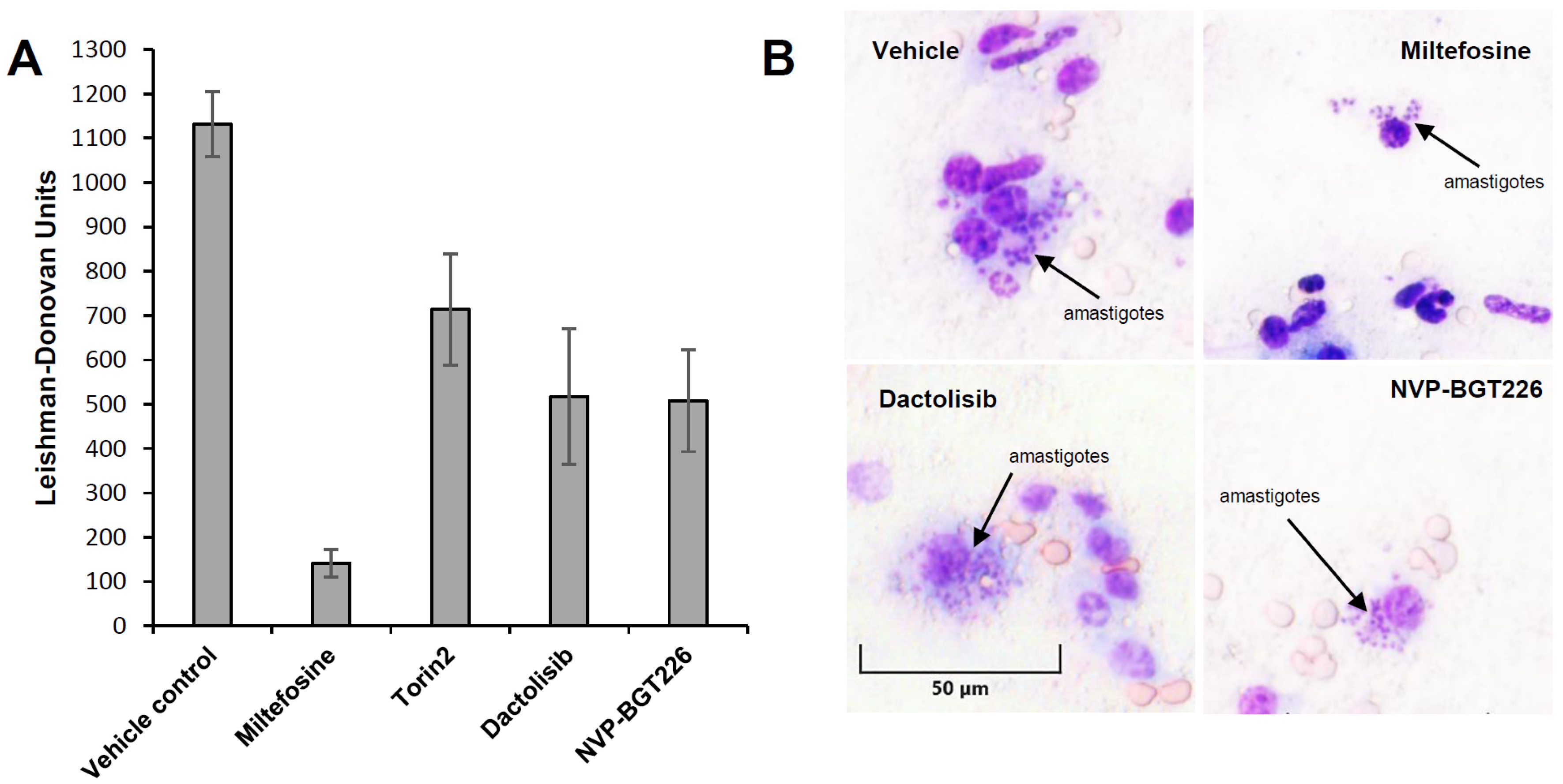
Commonly used animal models for evaluating compound efficacy in vivo are BALB/c (for acute) and hamster (for chronic) models [57,58,59,60,61,62]. For this proof of concept in vivo study, L. donovani infected BALB/c model was used to evaluate the ability of compound to reduce parasites in liver. Based on their EC50 and SI values from the intracellular Leishmania assay, we selected Torin2, dactolisib, and NVP-BGT226 (Table 1). Prior to the efficacy evaluation, the doses were selected based on the results of a literature search [45,63,64]. And in pilot toxicity study in mice all the compounds were well tolerated without displaying any overt signs of toxicity during the five consecutive days of treatment. Using the same dosing scheme, the efficacies of the compounds were evaluated in VL mouse model. Miltefosine-treated mice, showed 87 ± 2.13% inhibition of parasitemia in liver compared to the vehicle-treated control mice. In the test groups, significant decreases in parasitemia by 54 ± 3.18%, 53 ± 9.50%, and 35 ± 5.81% in the NVP-BGT226-, dactolisib-, and Torin2-treated mice, respectively (Table 2 and Figure 6), were observed and the weight of spleens were similar to that of miltefosine-treated control mice (Figure S2). In the Giemsa-stained liver smear, residual parasites not cleared by the tested compounds were observed inside host cells (Figure 6B). Even though some activity was present, the tested compounds did not show comparably improved inhibition over miltefosine at a given dose. Another point to mention is the dose used for this study. In the in vitro assay, NVP-BGT226 showed CC50 value of 2.66 μM, but no sign of toxicity was observed in vivo at 5 mg/kg dose. Since the dose was selected based on the previously reported literature that does not show adverse effect, the toxic effect by the compound was not observed in vivo [64]. For more thorough investigations, the maximum tolerated dose of compounds in infected animals can be determined to test efficacy of compounds with varying range of doses.

2.5. In Vivo Efficacy of NVP-BGT226 in T. brucei Mouse Model

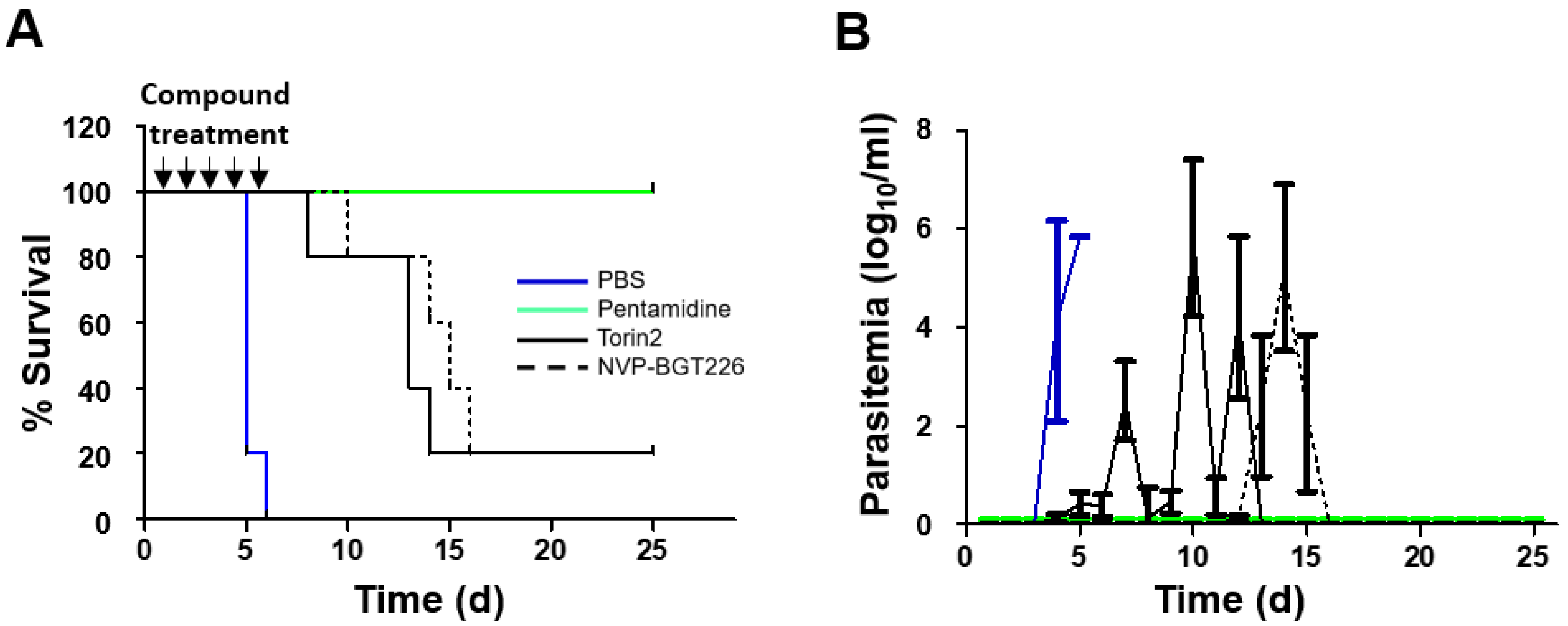
The Torin2 and NVP-BGT226 compounds had more potent activities compared with pentamidine and were thus chosen for testing in T. brucei acute infection model. Since the efficacy of NVP-BEZ235 was evaluated in the T. b. rhodesiense infected mouse model in the previous work by Pollastri et al., the compound was not selected for testing in this study [37,51,65]. T. brucei-infected mice with absence of any treatment all died within 6 days post-infection (Figure 7A). For the drug testing, mice were treated with pentamidine (control), Torin2, or NVP-BGT226 at 30, 15, or 5 mg/kg for 5 days via the per os route, respectively, and the survival and parasitemia results are presented in Figure 7. In the group treated with NVP-BGT226, 80% survived during 14 days post-infection followed by death afterwards, and 20% of the mice survived more than 25 days without parasitemia observed. In the Torin2-treated group, 80% of the mice survived for at least 13 days after infection, but parasitemia was observed still by day 5. NVP-BGT226 exhibited slightly improved efficacy over the Torin2-treated group with extend 1–2 days delay in survival. In addition, compared with the previous reports of NVP-BEZ235 [38], NVP-BGT226 exhibited somewhat improved efficacy in the T. brucei acute mouse infection model.

2.6. Prediction of Inhibitor Binding to Kinetoplastid TORs

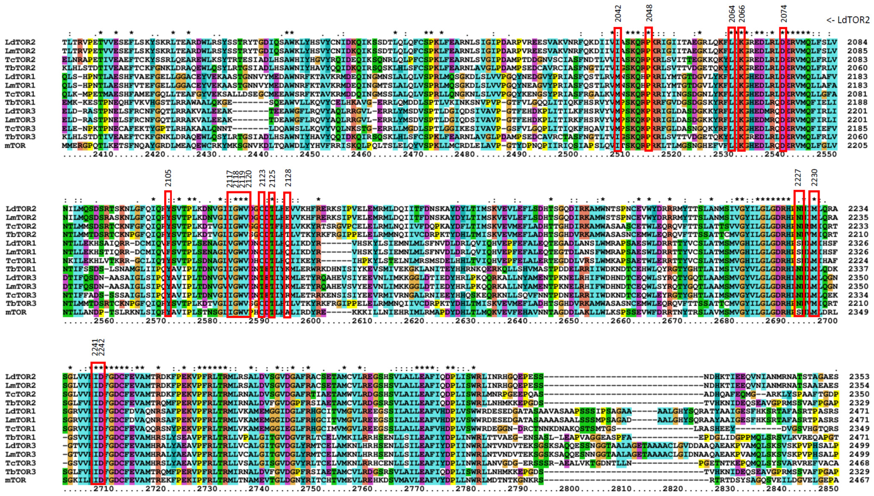
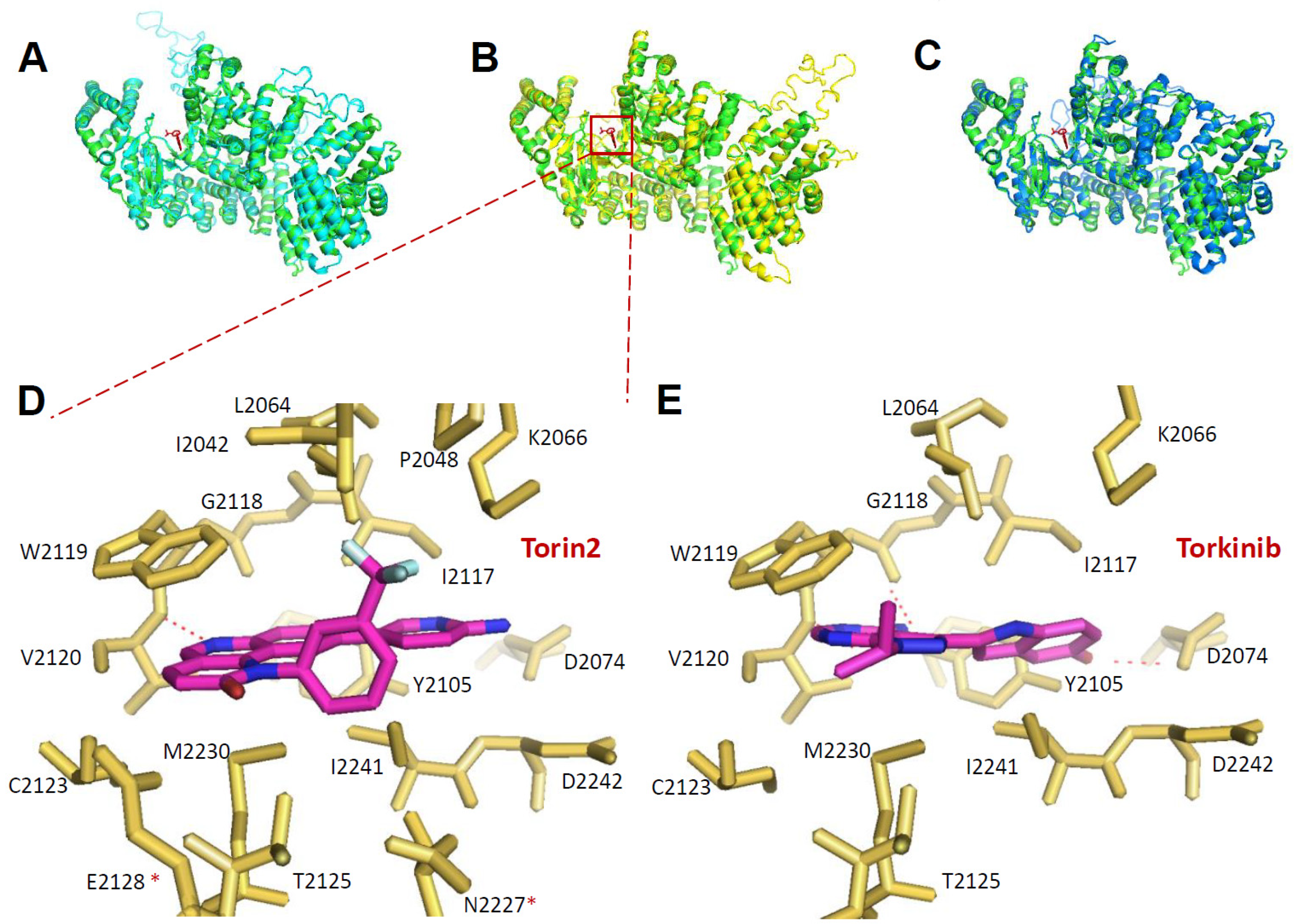
We then sought to further investigate the structural aspects of inhibitor binding. First, multiple sequence alignment analysis was performed between mTOR and kinetoplastid parasite TORs (Figure 8). In the case of L. donovani, LdTOR1 showed sequence identity of 39.77% to mTOR, followed by LdTOR2 at 36.77% and LdTOR3 at 32.08%. L. major, T. brucei, and T. cruzi TOR1, 2, and 3 had similar sequence identity to mTOR within the range of 30%–40% (Table 3). Then we build homology models of kinetoplastid TORs and compared the structural similarity with human mTOR using root mean square deviation (RMSD) values. LdTOR1, 2, and 3 had RMSD values of 0.374, 0.428, and 0.291, respectively, with the mTOR structure implying high degrees of structural similarity to the human mTOR structure (Table 3 and Figure 9A–C). Since LmTOR3 was found not essential for the survival of the parasites in the promastigote stage, further analysis of Torin2 and torkinib binding to LdTOR1 and 2 was performed by overlaying Leishmania TOR models onto inhibitor-mTOR structures [43]. Seventeen amino acid residues were located within 4Å of Torin2, and among them, 15 residues were found identical to that of human mTOR, except for the difference of Ala2248 to Glu2128 and Ser2342 to Asn2227 in LdTOR2. In terms of molecular interaction, a hydrogen bond of N in the tricyclic benzonaphthyridinone ring of Torin2 to the O of W2119 backbone (2.9 Å) was identified (Figure 9D). For torkinib, the -OH and –NH2 moieties were predicted to interact with D2074 and the backbone of G2118, respectively (Figure 9E). These interactions were also found in the mTOR structures, which suggests a high possibility of LdTOR1/2 inhibition by the inhibitors with similar binding modes.
In summary, using high-throughput screening, we identified seven mTOR/PI3K inhibitors with potent activity against the trypanosomatid parasites, L. donovani and T. brucei. Among these compounds, NVP-BGT226 was the most potent in vitro and was efficacious in VL and T. brucei animal models. The compound binding predictions based on the structural analysis suggests possible LdTOR1/2 inhibition by mTOR/PI3K inhibitors. This group of inhibitors was extensively developed for anti-cancer treatments, but currently hurdled by toxicity in human clinical trials. Due to this reason, a direct repurposing to kinetoplastids infections may not be readily feasible, but further development of inhibitors that are more selective to the kinetoplastid TORs would be possible. Another approach is utilizing the host immune response modulating properties of this class of compounds, as demonstrated by Khadem and colleagues, and further search for mTOR/PI3K inhibitors that well balance the direct and indirect killing effects. With limited number of highly potent inhibitors against Leishmania, these inhibitors are excellent chemical tools to decipher TOR pathways in kinetoplastid parasites which may lead to the discovery of new drug targets.

3. Materials and Methods

3.1. Ethics Statement

All animal studies were performed in strict accordance with the guidelines and principles established by the Korean Animal Protection Law (http://animalrightskorea.org). The use of animals was approved by the Institutional Animal Care and Use Committee (IACUC) of the Institut Pasteur Korea (IACUC approval number IPK-16003–3 for VL in vivo model and IPK-19002 for acute T. brucei in vivo model).

3.2. Inhibitors

The inhibitors used in this study were purchased from MedChem Express (Monmouth Junction, NJ, USA).

3.3. Parasite and Cell Cultures

L. donovani MHOM/SD/62/1S-CL2D parasites were cultured as promastigotes at 28 °C in M199 medium (Sigma-Aldrich, St. Louis, MO, USA) with 40 mM HEPES, 0.1 mM adenine, 0.0001% biotin, and 4.62 mM NaHCO3 supplemented with 10% fetal bovine serum (FBS, Gibco, Carlsbad, CA, USA), 100 µ/mL penicillin (Gibco), and 100 µg/mL streptomycin (Gibco). THP-1 cells (ATCC TIB-202) were cultured in RPMI-1640 medium containing 4.5 g/L glucose, 10 mM HEPES, 1 mM sodium pyruvate, and 10% FBS. The cells were maintained in tissue culture flasks (Nunc A/S, Roskilde, Denmark) in a 5% CO2 incubator at 37 °C. The T. b. brucei Lister strain 427 (bloodstream form: BSF) was cultivated in HMI-9 medium supplemented with 10% FBS, 100 µg/mL penicillin, and 100 µg/mL streptomycin at 37 °C and a 5% CO2 atmosphere. The parasites were sub-cultured every 3 or 4 days and were maintained for 10 passages.

3.4. Screening of Bioactive Compounds Against Intracellular Leishmania

PMA-treated THP-1 human monocytic cells were seeded at 0.8 × 10⁴ cells per well in a 384-well culture plate (Greiner Bio-One, Kremsmünster, Austria) in RPMI-1640 complete medium supplemented with 10% FBS. After 48 h of incubation at 37 °C in the presence of 5% CO2, the promastigotes of L. donovani that were incubated with lectin for 30 min at 28 °C were added to the cells at a parasite to cell ratio of 20:1. Infected THP-1 cells were treated with amphotericin B (at 4 μM, positive control), miltefosine (at 10 μM, positive control), and screening compounds (at 10 μM) [66,67,68]. The negative control consisted of THP-1 infected with the parasite with only 0.5% DMSO. After 72 h, the cells that were infected and treated with the drug were washed with serum-free RPMI-1640 medium. The cells and parasites were stained using 5 μM DRAQ5 and 4% PFA. The images were acquired based on reading using an Operetta® automated microscope (PerkinElmer, Inc., Waltham, MA 02451 USA). They were further analyzed using ColumbusTM (PerkinElmer, Inc. Waltham, MA, USA) software to quantify parasite numbers, host cell numbers, and infection ratios. In brief, large-sized nucleus of host cells was first detected using Draq-5 (Thermo Fisher, Rockford, IL, USA) signal and the host cell boundary masking was performed using the low-intensity signals from cytosols (additional feature of Draq-5). Then the small-sized nucleus signal by Draq-5 was used to identify parasites within the area of masked host cell. Infection ratio (IR) was determined with the value of the number of infected cells divided by total number of cells and the average number of parasites per macrophage (P/φ) was defined by the value of the number of parasites divided by the number of infected cells in the acquired image. The average IR value of the negative control wells was calculated as 0.53. Compounds selected based on the screening results were further assessed in a dose-dilution manner (two-fold serial dilution for 20 points starting from 100 μM) using the same method.

3.5. Parasite Growth Inhibition

L. donovani promastigote and T. b. brucei Lister 427 BSF growth inhibition were assayed by measuring the conversion of resazurin to resorufin. The assays were performed in 384-well plates that were seeded with L. donovani promastigotes or T. brucei 427 BSF (5 × 10⁴ cells per well). After seeding, the parasites were exposed to the compounds for 3 days. Resazurin sodium salt (200 μM; R7017; Sigma-Aldrich, St. Louis, MO, USA) was then added, and the samples were incubated for 5 h. After incubation, the parasites were fixed using 4% paraformaldehyde, and the plates were analyzed using a Victor3TM plate reader (PerkinElmer, Inc., Waltham, MA, USA) at 590 nm (emission) and 530 nm (excitation) [69]. Amphotericin B and miltefosine were used as the reference drugs for the L. donovani promastigote growth inhibition [67]. Pentamidine was used for the T. b. brucei Lister 427 BSF growth inhibition assay [68].

3.6. In Vivo Experiments

3.6.1. VL Mouse Model

Groups of five-week-old female BALB/c mice (five per group) were injected with 2 × 10⁷ hamster spleen-derived L. donovani amastigotes via the retro-orbital venous sinus route. From day 7 post-infection, groups of mice were treated using the drug vehicle only, miltefosine (30 mg/kg), Torin2 (15 mg/kg), dactolisib (50 mg/kg), or NVP-BGT226 (5 mg/kg). The highest dose for each compound without showing adverse effects was selected based on literature search [44,45,49]. The vehicle for the compounds was 5% N-methylpyrrolidone, 15% polyvinylpyrrolidone, and 80% deionized water. Drug dosing solutions were freshly prepared each day. All drugs were administered once daily for 5 days via the per os route. On day 16 post-infection, all animals were humanely euthanized and assessed microscopically using Giemsa-stained liver imprints. Parasite burdens were measured by counting (blinded to treatment) the number of amastigotes per 1000 cell nuclei and multiplying this number by the liver weight (mg) (Leishman-Donovan Unit: LDU) [70]. The LDU values for the drug-treated samples were compared to those of the untreated samples, and the percent inhibition values were calculated.

3.6.2. HAT Mouse Model

BALB/c mice were infected with T. b. brucei Lister 427 (4 × 10⁴ cells) by intraperitoneal (i.p.) injection. The mice were divided into groups (n = 5), and drug treatment was performed for five consecutive days by starting from day 1 post-infection administering 30 mg/kg, 15 mg/kg, and 5 mg/kg of pentamidine, Torin2, and NVP-BGT226, respectively. Parasitemia was evaluated daily for 2 weeks by blood collection from the mouse tail vein, and survival was monitored for 1 month. Mice showing impaired health status and/or with a parasite load > 108 cells per mL of blood were euthanized.

3.7. Prediction of Compound Binding Modes

The protein sequence for L. donovani TOR1 (LdBPK_366580.1), LdTOR2 (LdBPK_344160.1), LdTOR3 (LdBPK_343750.1), L. major TOR1 (LmjF.36.6320), LmTOR2 (LmjF.34.4530), LmTOR3 (LmjF34.3940), T. brucei TOR1 (Tb427.04.800), TbTOR2 (Tb427.04.420), TbTOR3 (Tb927.4.800), T. cruzi TOR1 (BCY_11356), TcTOR2 (BCY_20486), TcTOR3 (BCY_20528), and mTOR (NP_004949) were downloaded from the Kinetoplastid Genomic Resource database (https://tritrypdb.org/tritrypdb/) and the Proteins database (https://www.ncbi.nlm.nih.gov/). The sequence alignment was performed using ClustalX2.1 (Cambridgeshire, UK). The homology models of LdTOR1/2/3 were obtained using SWISS_MODEL (http://swissmodel.expasy.org) with the Torin2 bound mTOR structures (PDB ID: 4JSX, and 4JT5, http://www.rcsb.org) as templates [71,72]. The PyMOL 1.3 program (Palo Alto, CA, USA) was used to obtain the structural alignment results.

3.8. Statistical Analyses

All of the half maximal effective concentrations (EC50) and the half maximal cytotoxic concentrations (CC50) values were calculated using two independent experiments. The dose-response curves were fitted using GraphPad Prism 6 software (GraphPad Software, San Diego, CA, USA) by using a sigmoidal dose-response equation with a variable hill slope option.

Supplementary Materials

The following are available online, Figure S1: Dose-response curves of miltefosine, amphotericin B, and mTOR/PI3K compounds against L. donovani promastigote growth, Figure S2: Changes in spleen weight in L. donovani-infected BALB/c mice and effects of treatment with compound, Table S1: The list of 1742 compounds, Table S2: The relationships between mTOR/PI3K inhibition activity and intracellular L. donovani inhibition activity.

Author Contributions

Conceptualization, J.H.N.; Methodology, T.-N.P., K.-H.B., D.S.; Software, T.-N.P., J.H.N.; Formal Analysis, T.-N.P.; Resources, J.H.N.; Data Curation, T.-N.P., K.-H.B., N.L., S.Y.B.; Writing—Original Draft Preparation, T.-N.P., J.H.N.; Writing—Review & Editing, J.H.N.; Visualization, T.-N.P., J.H.N.; Supervision, D.S., J.H.N.; Project Administration, J.H.N.; Funding Acquisition, J.H.N. All authors have read and agreed to the published version of the manuscript.

Funding

This research was funded by the National Research Foundation of Korea, grant number NRF-2017M3A9G6068246 and 2017R1D1A1B03033204, through a grant funded by the government of the Republic of Korea (MSIP) and Gyeonggi-do.

Conflicts of Interest

The authors declare no conflicts of interest.

References

  1. Stuart, K.; Brun, R.; Croft, S.; Fairlamb, A.; Gürtler, R.E.; McKerrow, J.; Reed, S.; Tarleton, R.L. Kinetoplastids: Related protozoan pathogens, different diseases. J. Clin. Investig. 2008, 118, 1301–1310. [Google Scholar] [CrossRef] [Green Version]
  2. World Health Organization. Leishmaniasis. Fact sheet No. 375, 2017. World Health Organization. Available online: http://www.who.int/mediacentre/factsheets/fs375/en/ (accessed on 23 April 2020).
  3. Boelaert, M.; Criel, B.; Leeuwenburg, J.; Van Damme, W.; Le Ray, D.; Van Der Stuyft, P. Visceral leishmaniasis control: A public health perspective. Trans. R. Soc. Trop. Med. Hyg. 2000, 94, 465–471. [Google Scholar] [CrossRef]
  4. World Health Organization. Control of the Leishmaniases; WHO Technical Report Series 949; WHO: Geneva, Switzerland, 2010. [Google Scholar]
  5. Alvar, J.; Velez, I.D.; Bern, C.; Herrero, M.; Desjeux, P.; Cano, J.; Jannin, J.; Boer, M.D. Leishmaniasis Worldwide and Global Estimates of Its Incidence. PLOS ONE 2012, 7, e35671. [Google Scholar] [CrossRef]
  6. Pigott, D.M.; Bhatt, S.; Golding, N.; A Duda, K.; Battle, K.E.; Brady, O.J.; Messina, J.P.; Balard, Y.; Bastien, P.; Pratlong, F.; et al. Global distribution maps of the leishmaniases. eLife 2014, 3, 3. [Google Scholar] [CrossRef]
  7. No, J.H. Visceral leishmaniasis: Revisiting current treatments and approaches for future discoveries. Acta Trop. 2016, 155, 113–123. [Google Scholar] [CrossRef]
  8. Doua, F.; Miezan, T.W.; Singaro, J.R.S.; Yapo, F.B.; Baltz, T. The Efficacy of Pentamidine in the Treatment of Early-Late Stage Trypanosoma brucei gambiense Trypanosomiasis *. Am. J. Trop. Med. Hyg. 1996, 55, 586–588. [Google Scholar] [CrossRef]
  9. Kennedy, P.G. Clinical features, diagnosis, and treatment of human African trypanosomiasis (sleeping sickness). Lancet Neurol. 2013, 12, 186–194. [Google Scholar] [CrossRef]
  10. Alirol, E.; Schrumpf, D.; Heradi, J.A.; Riedel, A.; De Patoul, C.; Quere, M.; Chappuis, F. Nifurtimox-Eflornithine Combination Therapy for Second-Stage Gambiense Human African Trypanosomiasis: Médecins Sans Frontières Experience in the Democratic Republic of the Congo. Clin. Infect. Dis. 2012, 56, 195–203. [Google Scholar] [CrossRef]
  11. Lindner, A.K.; Lejon, V.; Chappuis, F.; Seixas, J.; Kazumba, L.; Barrett, M.P.; Mwamba, E.; Erphas, O.; A Akl, E.; Villanueva, G.; et al. New WHO guidelines for treatment of gambiense human African trypanosomiasis including fexinidazole: Substantial changes for clinical practice. Lancet Infect. Dis. 2020, 20, e38–e46. [Google Scholar] [CrossRef]
  12. Antinori, S.; Galimberti, L.; Bianco, R.; Grande, R.; Galli, M.; Corbellino, M. Chagas disease in Europe: A review for the internist in the globalized world. Eur. J. Intern. Med. 2017, 43, 6–15. [Google Scholar] [CrossRef] [PubMed]
  13. Zulantay, I.; Apt, W.; Ramos, D.; Godoy, L.; Valencia, C.; Molina, M.; Sepulveda, E.; Thieme, P.; Martinez, G.; Corral, G. The Epidemiological Relevance of Family Study in Chagas Disease. PLOS Neglected Trop. Dis. 2013, 7, e1959. [Google Scholar] [CrossRef] [PubMed] [Green Version]
  14. De Koning, H.P. Ever-increasing complexities of diamidine and arsenical crossresistance in African trypanosomes. Trends Parasitol. 2008, 24, 345–349. [Google Scholar] [CrossRef] [PubMed]
  15. Hamill, R.J. Amphotericin B Formulations: A Comparative Review of Efficacy and Toxicity. Drugs 2013, 73, 919–934. [Google Scholar] [CrossRef]
  16. Ponte-Sucre, A.; Gamarro, F.; Dujardin, J.-C.; Barrett, M.P.; Lopez-Vélez, R.; García-Hernández, R.; Pountain, A.W.; Mwenechanya, R.; Papadopoulou, B. Drug resistance and treatment failure in leishmaniasis: A 21st century challenge. PLOS Neglected Trop. Dis. 2017, 11, e0006052. [Google Scholar] [CrossRef]
  17. Wilkinson, S.R.; Taylor, M.C.; Horn, D.; Kelly, J.M.; Cheeseman, I.H. A mechanism for cross-resistance to nifurtimox and benznidazole in trypanosomes. Proc. Natl. Acad. Sci. USA 2008, 105, 5022–5027. [Google Scholar]
  18. Castro, J.; De Mecca, M.M.; Bartel, L. Toxic side effects of drugs used to treat Chagas’ disease (American trypanosomiasis). Hum. Exp. Toxicol. 2006, 25, 471–479. [Google Scholar] [CrossRef]
  19. Chappuis, F.; Sundar, S.; Hailu, A.; Ghalib, H.; Rijal, S.; Peeling, R.W.; Alvar, J.; Boelaert, M. Visceral leishmaniasis: What are the needs for diagnosis, treatment and control? Nat. Rev. Microbiol. 2007, 5, 873–882. [Google Scholar] [CrossRef]
  20. World Health Organization. Human African Trypanosomiasis: A Guide for Drug Supply, 2002. World Health Organization. Available online: https://apps.who.int/iris/handle/10665/67252 (accessed on 23 April 2020).
  21. Andrews, K.; Walduck, A.; Kelso, M.J.; Fairlie, D.P.; Saul, A.; Parsons, P.G.; Kelso, M.J. Anti-malarial effect of histone deacetylation inhibitors and mammalian tumour cytodifferentiating agents. Int. J. Parasitol. 2000, 30, 761–768. [Google Scholar] [CrossRef]
  22. Eastman, R.T.; White, J.; Hucke, O.; Bauer, K.; Yokoyama, K.; Nallan, L.; Chakrabarti, D.; Verlinde, C.; Gelb, M.H.; Rathod, P.K.; et al. Resistance to a Protein Farnesyltransferase Inhibitor inPlasmodium falciparum. J. Boil. Chem. 2005, 280, 13554–13559. [Google Scholar] [CrossRef] [Green Version]
  23. Frearson, J.A.; Brand, S.; McElroy, S.P.; Cleghorn, L.A.T.; Smid, O.; Stojanovski, L.; Price, H.P.; Güther, M.L.S.; Torrie, L.S.; Robinson, D.; et al. N-myristoyltransferase inhibitors as new leads to treat sleeping sickness. Nat. 2010, 464, 728–732. [Google Scholar] [CrossRef] [Green Version]
  24. Pollastri, M.P.; Campbell, R.K. Target repurposing for neglected diseases. Futur. Med. Chem. 2011, 3, 1307–1315. [Google Scholar] [CrossRef] [PubMed] [Green Version]
  25. Laplante, M.; Sabatini, D.M. mTOR signaling at a glance. J. Cell Sci. 2009, 122, 3589–3594. [Google Scholar] [CrossRef] [Green Version]
  26. Sarbassov, D.D.; Ali, S.M.; Sabatini, D.M. Growing roles for the mTOR pathway. Curr. Opin. Cell Boil. 2005, 17, 596–603. [Google Scholar] [CrossRef]
  27. Wullschleger, S.; Loewith, R.; Hall, M.N. TOR Signaling in Growth and Metabolism. Cell 2006, 124, 471–484. [Google Scholar] [CrossRef] [Green Version]
  28. Abraham, R.T. PI 3-kinase related kinases: ‘Big’ players in stress-induced signaling pathways. DNA Repair 2004, 3, 883–887. [Google Scholar] [CrossRef]
  29. Bakkenist, C.J.; Kastan, M.B. Initiating Cellular Stress Responses. Cell 2004, 118, 9–17. [Google Scholar] [CrossRef] [Green Version]
  30. Keith, C.T.; Schreiber, S.L. PIK-Related Kinases: DNA Repair, Recombination, and Cell Cycle Checkpoints. Sci. 1995, 270, 50. [Google Scholar] [CrossRef]
  31. Alzahrani, A.S. PI3K/Akt/mTOR inhibitors in cancer: At the bench and bedside. Semin. Cancer Boil. 2019, 59, 125–132. [Google Scholar] [CrossRef]
  32. Fruman, D.A.; Chiu, H.; Hopkins, B.D.; Bagrodia, S.; Cantley, L.C.; Abraham, R.T. The PI3K Pathway in Human Disease. Cell 2017, 170, 605–635. [Google Scholar] [CrossRef] [Green Version]
  33. Hillmann, P.; Fabbro, D. PI3K/mTOR Pathway Inhibition: Opportunities in Oncology and Rare Genetic Diseases. Int. J. Mol. Sci. 2019, 20, 5792. [Google Scholar] [CrossRef] [Green Version]
  34. Mayer, I.A.; Arteaga, C.L. The PI3K/AKT Pathway as a Target for Cancer Treatment. Annu. Rev. Med. 2016, 67, 11–28. [Google Scholar] [CrossRef]
  35. Yang, J.; Nie, J.; Ma, X.; Wei, Y.; Peng, Y.; Wei, X. Targeting PI3K in cancer: Mechanisms and advances in clinical trials. Mol. Cancer 2019, 18, 26. [Google Scholar] [CrossRef] [Green Version]
  36. Janku, F.; Yap, T.A.; Meric-Bernstam, F. Targeting the PI3K pathway in cancer: Are we making headway? Nat. Rev. Clin. Oncol. 2018, 15, 273–291. [Google Scholar] [CrossRef]
  37. Diaz-Gonzalez, R.; Kuhlmann, F.M.; Galan-Rodriguez, C.; da Silva, L.M.; Saldivia, M.; Karver, C.E.; Rodriguez, A.; Beverley, S.M.; Navarro, M.; Pollastri, M.P. The susceptibility of trypanosomatid pathogens to PI3/mTOR kinase inhibitors affords a new opportunity for drug repurposing. PLoS Negl Trop Dis. 2011, 5, e1297. [Google Scholar] [CrossRef] [Green Version]
  38. Khadem, F.; Jia, P.; Mou, Z.; Feiz Barazandeh, A.; Liu, D.; Keynan, Y.; Uzonna, J.E. Pharmacological inhibition of p110delta subunit of PI3K confers protection against experimental leishmaniasis. J. Antimicrob Chemother. 2017, 72, 467–477. [Google Scholar] [CrossRef] [Green Version]
  39. Khadir, F.; Shaler, C.R.; Oryan, A.; Rudak, P.T.; Mazzuca, D.M.; Taheri, T.; Dikeakos, J.D.; Haeryfar, S.M.M.; Rafati, S. Therapeutic control of leishmaniasis by inhibitors of the mammalian target of rapamycin. PLOS Neglected Trop. Dis. 2018, 12, e0006701. [Google Scholar] [CrossRef]
  40. Barquilla, A.; Crespo, J.L.; Navarro, M. Rapamycin inhibits trypanosome cell growth by preventing TOR complex 2 formation. Proc. Natl. Acad. Sci. USA 2008, 105, 14579–14584. [Google Scholar]
  41. Barquilla, A.; Navarro, M. Trypanosome TOR complex 2 functions in cytokinesis. Cell Cycle 2009, 8, 697–699. [Google Scholar] [CrossRef]
  42. De Jesus, T.C.L.; Tonelli, R.R.; Nardelli, S.C.; Augusto, L.D.S.; Motta, M.C.M.; Girard-Dias, W.; Miranda, K.; Ulrich, P.; Jimenez, V.; Barquilla, A.; et al. Target of Rapamycin (TOR)-like 1 Kinase Is Involved in the Control of Polyphosphate Levels and Acidocalcisome Maintenance in Trypanosoma brucei*. J. Boil. Chem. 2010, 285, 24131–24140. [Google Scholar] [CrossRef] [Green Version]
  43. Da Silva, L.M.; Beverley, S.M. Expansion of the target of rapamycin (TOR) kinase family and function in Leishmania shows that TOR3 is required for acidocalcisome biogenesis and animal infectivity. Proc. Natl. Acad. Sci. USA 2010, 107, 11965–11970. [Google Scholar]
  44. Liu, Q.; Wang, J.; Kang, S.A.; Thoreen, C.C.; Hur, W.; Ahmed, T.; Sabatini, D.M.; Gray, N.S. Discovery of 9-(6-Aminopyridin-3-yl)-1-(3-(trifluoromethyl)phenyl)benzo[h][1,6]naphthyridin-2(1H)-one (Torin2) as a Potent, Selective, and Orally Available Mammalian Target of Rapamycin (mTOR) Inhibitor for Treatment of Cancer. J. Med. Chem. 2011, 54, 1473–1480. [Google Scholar] [CrossRef] [Green Version]
  45. Maira, S.-M.; Stauffer, F.; Brueggen, J.; Furet, P.; Schnell, C.; Fritsch, C.; Brachmann, S.; Chène, P.; De Pover, A.; Schoemaker, K.; et al. Identification and characterization of NVP-BEZ235, a new orally available dual phosphatidylinositol 3-kinase/mammalian target of rapamycin inhibitor with potent in vivo antitumor activity. Mol. Cancer Ther. 2008, 7, 1851–1863. [Google Scholar] [CrossRef] [Green Version]
  46. Apsel, B.; Blair, J.A.; Gonzalez, B.Z.; Nazif, T.; Feldman, M.E.; Aizenstein, B.; Hoffman, R.; Williams, R.L.; Shokat, K.M.; Knight, Z.A. Targeted polypharmacology: Discovery of dual inhibitors of tyrosine and phosphoinositide kinases. Nat. Methods 2008, 4, 691–699. [Google Scholar] [CrossRef] [PubMed] [Green Version]
  47. Hsieh, A.C.; Liu, Y.; Edlind, M.P.; Ingolia, N.T.; Janes, M.R.; Sher, A.; Shi, E.Y.; Stumpf, C.R.; Christensen, C.; Bonham, M.J.; et al. The translational landscape of mTOR signalling steers cancer initiation and metastasis. Nature 2012, 485, 55–61. [Google Scholar] [CrossRef] [PubMed] [Green Version]
  48. Mallon, R.; Hollander, I.; Feldberg, L.; Lucas, J.; Soloveva, V.; Venkatesan, A.; Dehnhardt, C.; Santos, E.D.; Chen, Z.; Dos Santos, O.; et al. Antitumor Efficacy Profile of PKI-402, a Dual Phosphatidylinositol 3-Kinase/Mammalian Target of Rapamycin Inhibitor. Mol. Cancer Ther. 2010, 9, 976–984. [Google Scholar] [CrossRef] [Green Version]
  49. Markman, B.; Tabernero, J.; Krop, I.; Shapiro, G.I.; Siu, L.; Chen, L.C.; Mita, M.; Cuero, M.M.; Stutvoet, S.; Birle, D.; et al. Phase I safety, pharmacokinetic, and pharmacodynamic study of the oral phosphatidylinositol-3-kinase and mTOR inhibitor BGT226 in patients with advanced solid tumors. Ann. Oncol. 2012, 23, 2399–2408. [Google Scholar] [CrossRef] [PubMed]
  50. Yu, K.; Shi, C.; Toral-Barza, L.; Lucas, J.; Shor, B.; Kim, J.-E.; Zhang, W.-G.; Mahoney, R.; Gaydos, C.; Tardio, L.; et al. Beyond Rapalog Therapy: Preclinical Pharmacology and Antitumor Activity of WYE-125132, an ATP-Competitive and Specific Inhibitor of mTORC1 and mTORC2. Cancer Res. 2010, 70, 621–631. [Google Scholar] [CrossRef] [PubMed] [Green Version]
  51. Keiser, J.; Stich, A.; Burri, C. New drugs for the treatment of human African trypanosomiasis: Research and development. Trends Parasitol. 2001, 17, 42–49. [Google Scholar] [CrossRef]
  52. Seixas, J.D.; Luengo-Arratta, S.A.; Díaz, R.; Saldivia, M.; Rojas-Barros, D.I.; Manzano, P.; Gonzalez, R.D.; Berlanga, M.; Smith, T.K.; Navarro, M.; et al. Establishment of a Structure–Activity Relationship of 1H-Imidazo[4,5-c]quinoline-Based Kinase Inhibitor NVP-BEZ235 as a Lead for African Sleeping Sickness. J. Med. Chem. 2014, 57, 4834–4848. [Google Scholar] [CrossRef] [Green Version]
  53. Fang, B.; Kannan, A.; Guo, T.; Gao, L. Simultaneously targeting DNA damage repair pathway and mTORC1/2 results in small cell lung cancer growth arrest via ER stress-induced apoptosis. Int J. Biol Sci. 2018, 14, 1221–1231. [Google Scholar] [CrossRef]
  54. Kang, M.H.; Reynolds, C.P.; Maris, J.M.; Gorlick, R.; Kolb, E.A.; Lock, R.; Carol, H.; Keir, S.T.; Wu, J.; Lyalin, D.; et al. Initial testing (stage 1) of the investigational mTOR kinase inhibitor MLN0128 by the pediatric preclinical testing program. Pediatr. Blood Cancer 2014, 61, 1486–1489. [Google Scholar] [CrossRef] [Green Version]
  55. Okada, T.; Lee, A.; Qin, L.-X.; Agaram, N.; Mimae, T.; Shen, Y.; O’Connor, R.; López-Lago, M.A.; Craig, A.; Miller, M.L.; et al. Integrin-α10 Dependency Identifies RAC and RICTOR as Therapeutic Targets in High-Grade Myxofibrosarcoma. Cancer Discov. 2016, 6, 1148–1165. [Google Scholar] [CrossRef] [Green Version]
  56. Yamamoto, T.; Kanaya, N.; Somlo, G.; Chen, S. Synergistic anti-cancer activity of CDK4/6 inhibitor palbociclib and dual mTOR kinase inhibitor MLN0128 in pRb-expressing ER-negative breast cancer. Breast Cancer Res. Treat. 2019, 174, 615–625. [Google Scholar] [CrossRef] [PubMed]
  57. Gupta, S.; Yardley, V.; Vishwakarma, P.; Shivahare, R.; Sharma, B.; Launay, D.; Martin, D.; Puri, S.K. Nitroimidazo-oxazole compound DNDI-VL-2098: An orally effective preclinical drug candidate for the treatment of visceral leishmaniasis. J. Antimicrob. Chemother. 2014, 70, 518–527. [Google Scholar] [CrossRef] [PubMed] [Green Version]
  58. Khare, S.; Nagle, A.S.; Biggart, A.; Lai, Y.H.; Liang, F.; Davis, L.C.; Barnes, S.W.; Mathison, C.J.N.; Myburgh, E.; Gao, M.-Y.; et al. Proteasome inhibition for treatment of leishmaniasis, Chagas disease and sleeping sickness. Nat. 2016, 537, 229–233. [Google Scholar] [CrossRef]
  59. Manhas, R.; Tandon, S.; Sen, S.S.; Tiwari, N.; Munde, M.; Madhubala, R. Leishmania donovani Parasites Are Inhibited by the Benzoxaborole AN2690 Targeting Leucyl-tRNA Synthetase. Antimicrob. Agents Chemother. 2018, 62, AAC.00079–18. [Google Scholar] [CrossRef] [Green Version]
  60. Mowbray, C.; Braillard, S.; Speed, W.; Glossop, P.A.; Whitlock, G.A.; Gibson, K.R.; Mills, J.E.J.; Brown, A.D.; Gardner, J.M.F.; Cao, Y.; et al. Novel Amino-pyrazole Ureas with Potent In Vitro and In Vivo Antileishmanial Activity. J. Med. Chem. 2015, 58, 9615–9624. [Google Scholar] [CrossRef]
  61. Patterson, S.; Wyllie, S.; Norval, S.; Stojanovski, L.; Simeons, F.R.; Auer, J.L.; Osuna-Cabello, M.; Read, K.D.; Fairlamb, A. The anti-tubercular drug delamanid as a potential oral treatment for visceral leishmaniasis. eLife 2016, 5, 334. [Google Scholar] [CrossRef] [Green Version]
  62. Saha, S.; Acharya, C.; Pal, U.; Chowdhury, S.R.; Sarkar, K.; Maiti, N.C.; Jaisankar, P.; Majumder, H.K. A Novel Spirooxindole Derivative Inhibits the Growth of Leishmania donovani Parasites both In Vitro and In Vivo by Targeting Type IB Topoisomerase. Antimicrob. Agents Chemother. 2016, 60, 6281–6293. [Google Scholar] [CrossRef] [Green Version]
  63. Berry, T.; Luther, W.; Bhatnagar, N.; Jamin, Y.; Poon, E.; Sanda, T.; Pei, D.; Sharma, B.; Vetharoy, W.R.; Hallsworth, A.; et al. The ALK(F1174L) mutation potentiates the oncogenic activity of MYCN in neuroblastoma. Cancer Cell 2012, 22, 117–130. [Google Scholar] [CrossRef] [Green Version]
  64. Chang, K.-Y.; Tsai, S.-Y.; Wu, C.-M.; Yen, C.-J.; Chuang, B.-F.; Chang, J.-Y. Novel Phosphoinositide 3-Kinase/mTOR Dual Inhibitor, NVP-BGT226, Displays Potent Growth-Inhibitory Activity against Human Head and Neck Cancer Cells In Vitro and In Vivo. Clin. Cancer Res. 2011, 17, 7116–7126. [Google Scholar] [CrossRef] [Green Version]
  65. Myburgh, E.; Coles, J.A.; Ritchie, R.; Kennedy, P.G.E.; McLatchie, A.P.; Rodgers, J.; Taylor, M.C.; Barrett, M.P.; Brewer, J.M.; Mottram, J.C. In Vivo Imaging of Trypanosome-Brain Interactions and Development of a Rapid Screening Test for Drugs against CNS Stage Trypanosomiasis. PLOS Neglected Trop. Dis. 2013, 7, e2384. [Google Scholar] [CrossRef]
  66. Siqueira-Neto, J.; Moon, S.; Jang, J.; Yang, G.; Lee, C.; Moon, H.K.; Chatelain, E.; Genovesio, A.; Cechetto, J.; Freitas-Junior, L.H. An Image-Based High-Content Screening Assay for Compounds Targeting Intracellular Leishmania donovani Amastigotes in Human Macrophages. PLOS Neglected Trop. Dis. 2012, 6, e1671. [Google Scholar] [CrossRef] [PubMed] [Green Version]
  67. Vermeersch, M.; Da Luz, R.I.; Toté, K.; Timmermans, J.; Cos, P.; Maes, L. In Vitro Susceptibilities of Leishmania donovani Promastigote and Amastigote Stages to Antileishmanial Reference Drugs: Practical Relevance of Stage-Specific Differences. Antimicrob. Agents Chemother. 2009, 53, 3855–3859. [Google Scholar] [CrossRef] [PubMed] [Green Version]
  68. Yang, G.; Zhu, W.; Wang, Y.; Huang, G.; Byun, S.Y.; Choi, G.; Li, K.; Huang, Z.; Docampo, R.; Oldfield, E.; et al. In Vitro and in Vivo Activity of Multitarget Inhibitors against Trypanosoma brucei. ACS Infect. Dis. 2015, 1, 388–398. [Google Scholar] [CrossRef] [PubMed] [Green Version]
  69. Bowling, T.; Mercer, L.; Don, R.; Jacobs, R.; Nare, B. Application of a resazurin-based high-throughput screening assay for the identification and progression of new treatments for human African trypanosomiasis. Int. J. Parasitol. Drugs Drug Resist. 2012, 2, 262–270. [Google Scholar] [CrossRef] [PubMed] [Green Version]
  70. Stauber, L.A. Leishmaniasis in the hamster. In Some Physiological Aspects and Consequences of Parasitism; Rutgers University Press: New Brunswisck, NJ, USA, 1955. [Google Scholar]
  71. Choi, J.; Chen, J.; Schreiber, S.L.; Clardy, J. Structure of the FKBP12-Rapamycin Complex Interacting with Binding Domain of Human FRAP. Science 1996, 273, 239–242. [Google Scholar] [CrossRef] [PubMed]
  72. Veverka, V.; Crabbe, T.; Bird, I.; Lennie, G.; Muskett, F.W.; Taylor, R.J.; Carr, M. Structural characterization of the interaction of mTOR with phosphatidic acid and a novel class of inhibitor: Compelling evidence for a central role of the FRB domain in small molecule-mediated regulation of mTOR. Oncogene 2007, 27, 585–595. [Google Scholar] [CrossRef] [PubMed] [Green Version]
Sample Availability: Not available
Figure 1. Pilot screening results for intracellular L. donovani infection in THP-1 cell line. Red, negative control (DMSO); black, positive control (4 μM amphotericin B); gray, compound. A total of 1742 compounds were screened, with R2 = 0.922 (A), Z′ = 0.798 (B), and host cell survival and intracellular parasite survival inhibition > 60%, black squares (C), and cell ratio and intracellular parasite survival inhibition of 58 mammalian target of rapamycin (mTOR)/phosphoinositide 3-kinase (PI3K) compounds (D).
Molecules 25 01980 g001
Figure 2. Structures of compounds investigated in this work.
Molecules 25 01980 g002
Figure 3. Dose-response curves of miltefosine, amphotericin B, and mTOR/PI3K compounds against Table 1 cells infected with intracellular L. donovani. Inhibition infection ratio (%) (● black-filled circles); inhibition parasite number (%) ( blue-filled squares); and host cell ratio (%) ( red-filled triangles). The results are expressed as mean ± standard deviation values for duplicate experiments.
Molecules 25 01980 g003
Figure 4. Dose-response curves of pentamidine and mTOR/PI3K compounds against T. brucei bloodstream form growth. The results are expressed as mean ± standard deviation values for duplicate experiments.
Molecules 25 01980 g004
Figure 5. Correlation of pEC50 values for seven mTOR/PI3K inhibitors against L. donovani and T. brucei. (A) Correlation of pEC50 values against intracellular L. donovani and promastigotes. (B) Correlation of pEC50 values against intracellular L. donovani and T. brucei BSF. (C) Correlation of pEC50 values against L. donovani promastigotes and T. brucei BSF.
Molecules 25 01980 g005
Figure 6. Activity of miltefosine and mTOR/PI3K compounds in L. donovani-infected BALB/c mice. Animals were infected and treated as described, and numbers of LDUs on slides of liver smears were counted. All treatments began 7 days after infection. The results are expressed as mean ± standard deviation values. (A) Groups of animals (five mice per group) were treated with single 30 mg/kg doses of miltefosine, a vehicle control with PBS, or with Torin2, dactolisib, NVP-BGT226 formulations at 15, 50, and 5 mg/kg, respectively, for 5 days via the oral route. (B) Postmortem Giemsa-stained liver smears were obtained from mice after no treatment or exposure to the vehicle, miltefosine, or mTOR/PI3K compounds at the doses indicated.
Molecules 25 01980 g006
Figure 7. In vivo results for T. brucei mouse model of infection. (A) Mouse survival after treatment with 30 mg/kg pentamidine (solid green line), 15 mg/kg Torin2 (solid black line), 5 mg/kg NVP-BGT226 (dotted black line), or the PBS control (solid blue line) for 5 days. (B) Parasitemia as a function of time after treatment.
Molecules 25 01980 g007
Figure 8. Multiple sequence alignments of the FKBP-rapamycin-binding (FRB) domains of the TOR1/2/3 kinases of L. donovani, L. major, T. cruzi, and T. brucei, and human mTOR. Seventeen amino acid residues around binding sites are indicated using red squares.
Molecules 25 01980 g008
Figure 9. Modeling of L. donovani TOR domains. (AC) Structural alignment of LdTOR1/2/3 and mTORDeltaN-mLST8-Torin2 using PyMOL. The blue cartoon represents the structure of the LdTOR1, the yellow cartoon represents the structure of the LdTOR2, and the cyan cartoon represents the structure of the LdTOR3. The green cartoon represents the structure of the mTORDeltaN-mLST8 and the red stick is the Torin2 structure. (D) Structures of Torin2 inhibitor bound to the LdTOR2 catalytic cleft of L. donovani. Stick representation of Torin2 (C, red; N, blue; F, green) and of LdmTOR2 residues within 4 Å (except for Asp2074 and Asp2242). Red-dotted lines indicate atoms within hydrogen-bonding distance. (E) Torkinib-LdmTOR2 structure, represented as in (D), with stick representation of torkinib.
Molecules 25 01980 g009
Table 1. Efficacy of tested compounds in THP-1 cells infected with intracellular L. donovani, L. donovani promastigote growth; and T. brucei bloodstream form.
CompoundsIntracellular
L. donovani
EC50 ± SD a
(μM)
THP-1 Cell
CC50 ± SD a
(μM)
Selectivity Index
(SI)
L. donovani Promastigote
EC50 ± SD a
(μM)
T. brucei Lister 427 BSF
EC50 ± SD a
(μM)
Miltefosine4.81 ± 0.81>40>8.31.03 ± 0.63NA
Amphotericin B0.34 ± 0.04>4>11.80.015 ± 0.003NA
PentamidineNANANANA0.15 ± 0.01
Torin20.25 ± 0.0330.50 ± 1.31220.03 ± 0.0040.11 ± 0.01
Dactolisib (NVP-BEZ235)0.36 ± 0.07>100≥277.80.38 ± 0.060.12 ± 0.01
NVP-BGT2260.14 ± 0.022.66 ± 0.46190.009 ± 0.0010.000054 ± 0.000003
Sapanisertib6.39 ± 1.12>100≥397.46 ± 1.045.34 ± 0.98
Torkinib3.83 ± 0.9833.43 ± 1.48.73.86 ± 1.118.16 ± 1.52
WYE-1251322.26 ± 0.7143.27 ± 2.7190.36 ± 0.040.12 ± 0.01
PKI-40213.44 ± 2.3528.48 ± 1.22.10.49 ± 0.220.26 ± 0.02
a Shown are mean half maximal effective concentrations (EC50) ± SD (standard deviations) of data from duplicate measurements; NA, not applicable; Selectivity index (SI = THP-1 CC50/Intracellular L. donovani EC50).
Table 2. Efficacy of tested compounds in L. donovani—infected BALB/c mice.
CompoundsDose
(mg/kg)
LDU in the Liver
(Mean ± SD)
Inhibition of L. donovani In Vivo (%)
(Mean ± SD)
Vehicle1132 ± 730
Miltefosine30142 ± 2487 ± 2.13
Torin215735 ± 6635 ± 5.81
Dactolisib (NVP-BEZ235)50527 ± 10853 ± 9.50
NVP-BGT2265516 ± 3654 ± 3.18
Parasite burden (LDU, Leishman-Donovan Unit) was evaluated 7 days after compound administration. Data are presented as mean ± SD; NA, not applicable.
Table 3. The multiple sequence alignment between mTOR and kinetoplastid parasite TORs.
mTORL. donovaniL. majorT. cruziT. brucei
123123123123
Sequence identity (%)10039.7736.7732.0839.8336.9632.1838.1936.2333.6333.4735.1534.78
RMSD00.3740.4280.2910.1570.2960.2800.3600.3930.3920.2840.1730.422
Sequence identity (%) and root mean square deviation (RMSD) were calculated by using SWISS MODEL and PyMOL, respectively.