Article

Efficacy of Topical Treatment with (−)-Epigallocatechin Gallate, A Green Tea Catechin, in Mice with Cutaneous Leishmaniasis

by
Andrea M. Sosa
1,
Agustín Moya Álvarez
1,
Estefanía Bracamonte
1,
Masataka Korenaga
2,3,
Jorge D. Marco
1 and
Paola A. Barroso
1,*
1
Instituto de Patología Experimental, Facultad de Ciencias de la Salud, Universidad Nacional de Salta-CONICET, Salta 4400, Argentina
2
Department of Parasitology, Kochi Medical School, Kochi University, Okocho Kohasu, Nankoku, Kochi Prefecture 783-8505, Japan
3
Faculty of Health Sciences, Kochi Gakuen University, Asahi-Tenjincho, Kochi, Kochi Prefecture 780-0955, Japan
*
Author to whom correspondence should be addressed.
Molecules 2020, 25(7), 1741; https://doi.org/10.3390/molecules25071741
Submission received: 20 March 2020 / Revised: 8 April 2020 / Accepted: 9 April 2020 / Published: 10 April 2020
(This article belongs to the Special Issue Drug Discovery for Neglected Diseases)

Abstract

:
The treatment of leishmaniasis includes pentavalent antimony drugs but, because of the side effects, toxicity and cases of treatment failure or resistance, the search of new antileishmanial compounds are necessary. The aims of this study were to evaluate and compare the in vitro antileishmanial activity of four green tea catechins, and to assess the efficacy of topical (−)-epigallocatechin gallate in a cutaneous leishmaniasis model. The antileishmanial activity of green tea catechins was evaluated against intracellular amastigotes, and cytotoxicity was performed with human monocytic cell line. BALB/c mice were infected in the ear dermis with Leishmania (Leishmania) amazonensis and treated with topical 15% (−)-epigallocatechin gallate, intraperitoneal Glucantime, and control group. The efficacy of treatments was evaluated by quantifying the parasite burden and by measuring the lesions size. (−)-Epigallocatechin gallate and (−)-epigallocatechin were the most active compounds with IC50 values <59.6 µg/mL and with a selectivity index >1. Topical treatment with (−)-epigallocatechin gallate decreased significantly both lesion size and parasite burden (80.4% inhibition) compared to control group (p < 0.05), and moreover (−)-epigallocatechin gallate showed a similar efficacy to Glucantime (85.1% inhibition), the reference drug for leishmaniasis treatment.

1. Introduction

Cutaneous leishmaniasis (CL) is a widespread tropical infection caused by a protozoa parasite of Leishmania genus which is transmitted to humans by the bite of an infected sand fly [1]. In the New World, CL is produced by Leishmania (Leishmania) mexicana, L. (L.) amazonensis, L. (Viannia) braziliensis, L. (V.) panamensis, L. (V.) peruviana, and L. (V.) guyanensis. In general, CL is characterized by an ulcerative lesion (single or multiple), and depending of Leishmania species, the disease can progress to more severe manifestations such us mucocutaneous, diffuse leishmaniasis, disseminated leishmaniasis, and leishmaniasis recidivans [1]. The antimonials, sodium stibogluconate (Pentostam) or meglumine antimoniate (Glucantime), are the first choice for the treatment of all forms of leishmaniasis, but because of their toxicity, high cost and prolonged treatment regimens, are not satisfactory as an ideal drug. In addition, treatment failure was reported in a clinical trial where meglumine antimoniate showed a low efficacy against Leishmania Viannia species [2]. Therefore, there is a need for new antileishmanial drugs.
The green tea from the leaf of Camellia sinensis (Theaceae) is one of the most popular beverages worldwide, and for many years the health benefits of its polyphenol E (PE) fraction have been studied. The PE is constituted by (+)-catechin (C), (−)-epicatechin (EC), (−)-gallocatechin (GC), (−)-epigallocatechin (EGC), (−)-gallocatechin gallate (GCG), (−)-catechin gallate (CG) and (−)-epigallocatechin gallate (EGCG), which is the major one among of these components. The PE fraction has antioxidant activity, anti-inflammatory, antitumoral and antimicrobial effects [3,4,5]. In addition, EGCG administered by intraperitoneal injection showed anti-Trypanosoma cruzi activity decreasing significantly the parasite burden and increasing the mice survival [6]. These were consistent with the inhibition of T. cruzi amastigote replication in infected Vero cells by EGCG and GCG [7]. Then, our group studied the antileishmanial effect of seven catechins and the whole PE fraction from C. sinensis against two species, L. (L.) amazonensis and L. (V.) braziliensis [8]. EGCG, GCG, EGC, GC and GCG inhibited the growth of promastigotes of both Leishmania species, and C and EC were the less active compounds [8]. Moreover, the PE fraction showed activity against intracellular amastigotes of L. (L.) amazonensis [8]. Recently, it was reported that EGCG oral treatment showed high efficacy inhibiting the parasite burden of L. (V.) braziliensis (92.1%) and L. (L.) amazonensis in BALB/c mice [9,10]. Moreover, the antileishmanial activity of EGCG is related to the generation of oxygen reactive species (ROS) that alter the mitochondrial function and kill the parasite [11]. Another mechanism of action of EGCG is related to its capacity to inhibit leishmania arginase, increasing the nitric oxide toxic agent for the parasite [12].
There is a strong evidence that topical treatments for CL have a high cure rates, safety and effectiveness, with low side-effects, relapse and recurrence rates [13]. A previous study has shown that EGCG formulated with USP hydrophilic ointment has a good penetration into mouse and human skin [14]. Therefore, taking into account all the mentioned above, the aims of this study were to assess in vitro the antileishmanial activity of green tea catechins (GTC) (Figure 1) against intracellular amastigotes, and to evaluate the efficacy of a topical treatment with EGCG, the most abundant flavanol in green tea, in a L. (L.) amazonensis-infected BALB/c mice model of CL.

2. Results

2.1. Antileishmanial Activity Against L. (L.) Amazonensis

We investigated the activity of four GTC against intracellular amastigotes. These compounds showed antileishmanial activity in macrophages infected with L. (L.) amazonensis after 48 h of incubation, EGCG and EGC being the most active with IC50 values <59.6 µg/mL (Table 1). On the other hand, the IC50 of Glucantime (Gl), the reference drug for the treatment of leishmaniasis, was 6.5 µg/mL (Table 1).

2.2. Citotoxicity

The cytotoxicity of GTC was also evaluated in vitro on THP1 cells (human leukemia monocytic cells line). Selectivity indexes showed that EGCG, GCG, EGC and GC were more selective against Leishmania amastigotes than the macrophage cells (SI > 1) (Table 1). Gl showed lower toxicity on THP1 cells than catechins, with a 50% cytotoxic concentration (CC50) >400 µg Sbv/mL.
The selectivity index (SI) (CC50 for THP1/IC50 for Leishmania) was calculated. A SI higher than one, was considered more selective for activity against Leishmania, whereas a value lower than one was considered more selective for the activity against the host cells [15].

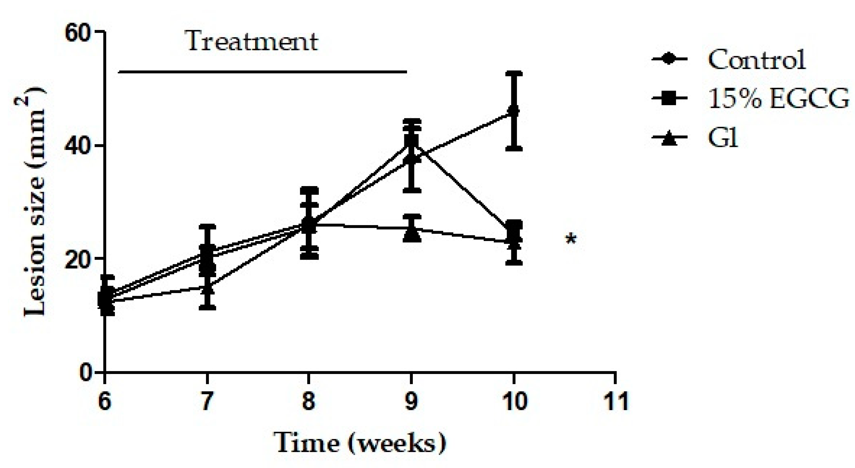
2.3. In Vivo Antileishmanial Activity of Catechin

The activity of EGCG, the main GTC, was evaluated in an L. (L.) amazonensis-BALB/c model. No significant differences were observed in the lesions’ sizes when the treatments were initiated (Figure 2). After one week of topical treatment with 15% EGCG, the edge of the lesions began to flatten. At the end of the experiments (week 10), EGCG promoted a significant reduction of the lesion size (p < 0.05), showing smaller lesions than control group. In the same manner, treatment with Gl, the reference drug for leishmaniasis, reduced the lesion size as was expected (p < 0.05) (Figure 2). The changes observed in the evolution of lesions in mice treated with topical EGCG were correlated with a significant decrease of the parasite burden (80.4% of inhibition) compared with control group (p < 0.05) (Figure 3 and Figure 4). In addition, EGCG showed a similar efficacy to intraperitoneal treatment with Gl (85.1% of inhibition) (Figure 4).

3. Discussion

The green tea from the leaf of Camellia sinensis is one of the most popular beverages worldwide, and for many years the health benefits of its PE fraction have been studied. The PE is constituted by catechins, and its biological activities, such us antimicrobial, anticancer and antiprotozoal, have been reported [3,4,5,6,7].
In a previous work, we showed the leishmanicidal activity of EGCG, EGC, GCG, GC and PE against promastigotes of L. (L.) amazonensis and L. (V.) braziliensis, being the former one, the more active catechin [6]. On the other hand, (+)-catechin, (−)-epicatechin and (−)-catechin gallate did not inhibit the growth of parasites in both species [8]. This prompted us to evaluate and compare the activity of EGCG, EGC, GCG and GC in macrophages infected with L. (L.) amazonensis. Among the catechins studied, ECGC and EGC were the most active ones against intracellular amastigotes with IC50 values <59.6 µg/mL. Other studies in vitro, reported the EGCG activity against intracellular amastigotes of L. (L.) amazonensis and L. (V.) braziliensis with IC50 values of 0.74 µg/mL and 1.57 µg/mL, respectively [10,11]. The low values of IC50 of EGCG, may be due to the experimental conditions used in the assays, such as the incubation temperature of infected macrophage cultures (37 °C). It is well known that amastigotes of L. (L.) amazonensis and L. (V.) braziliensis are sensitive to temperature [16]. Therefore, incubation at 37 °C can significantly inhibit parasite growth compared to that at 34 °C in the absence of antileishmanial drugs, giving a significantly lower IC50 in the presence of drugs [16]. On the other hand, EGCG and GCG showed antileishmanial activity against another species, L. (L.) donovani, the causal agent of visceral leishmaniasis, with IC50 values of 19.1 µg/mL and 8.9 µg/mL, respectively [17].
Besides the in vitro experiments, herein we performed an in vivo evaluation of the topical EGCG in hydrophilic ointment on L. (L.) amazonensis-BALB/c mice, since a topical treatment for CL is desirable due to the limited side effects and easy administration [13]. According to our knowledge, no previous study has reported the topical use of EGCG against CL. In addition, it was demonstrated that EGCG in hydrophilic ointment was efficiently absorbed into intact mouse skin with an uptake of up to 1%–20% of the applied dose [14]. In the present study, after 18 days of daily topical application with EGCG, both the lesions’ sizes and parasite burden were reduced significantly (80.4% inhibition) compared to the control group. Moreover, topical EGCG showed a similar efficacy to Gl, the reference drug for leishmaniasis treatment. It is important to mention that, in this study, the treatment with EGCG was initiated when the infection has already been established (week six post-infection). On the other hand, Inacio et al. reported the efficacy of oral treatment with EGCG in L. (L.) amazonensis in vivo model at the onset of the infection (week one post-infection); the treatment reduced significantly the parasite burden and lesion size compared with control group [10]. In addition, EGCG was also effective in mice infected with L. (V.) braziliensis but with a dose 3.3-fold higher (100 mg/kg/day) than the dose required for L. (L.) amazonensis (30 mg/kg/day) [10,11].
The antileishmanial activity of EGCG observed against different species of Leishmania in both the in vitro and in vivo model of infection can be explained by the inhibitory effect of EGCG on the enzyme arginase, involved in the polyamine synthesis pathway as demonstrated by dos Reis et al. [12]. Besides, this enzyme was considered a target to control Leishmania infection [12].
Thus, in the present work we demonstrated in vitro the antileishmanial activity of EGCG and EGC. Furthermore, topical treatment with EGCG showed a similar efficacy to Gl, the standard drug for treating leishmaniasis, in an animal model of cutaneous leishmaniasis caused by L. (L.) amazonensis.

4. Materials and Methods
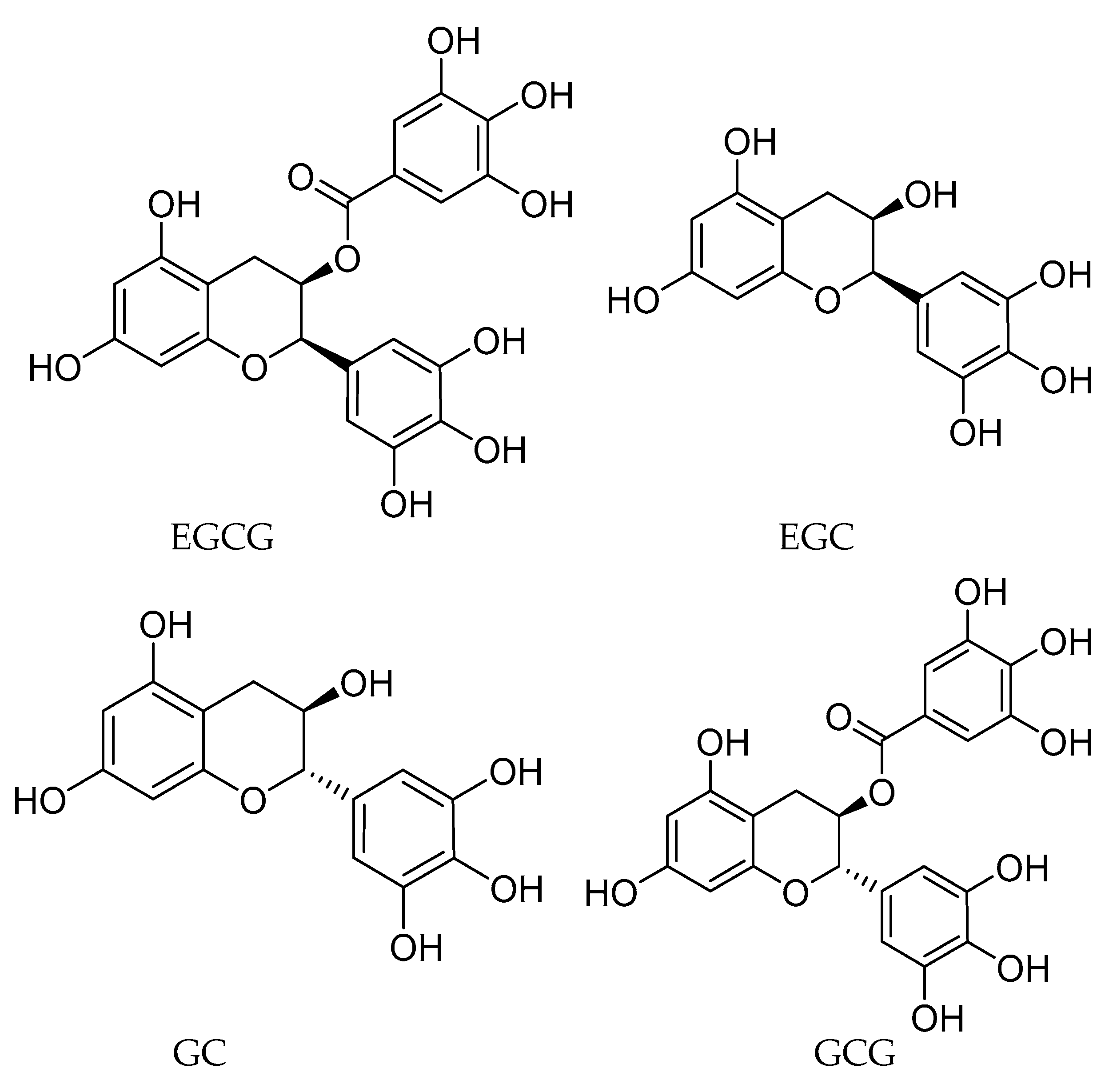
4.1. Compounds

(−)-Epigallocatechin gallate (EGCG), (−)-epigallocatechin (EGC), (−)-gallocatechin (GC) and (−)-gallocatechin gallate (GCG) were kindly supplied by Mitsui Norin (Shizuoka, Japan) and the purity of each compound was 98%. Meglumine antimoniate (Glucantime®, Aventis-Sanofi, São Paulo, Brazil) was kindly supplied by Ministerio de Salud de la Nación (Buenos Aires, Argentina).

4.2. Parasites

After several passages in mice, L. (L.) amazonensis (MHOM/BR/73/M2269) was isolated with sterile proline balanced salts solution (PBSS) containing 100 U/mL penicillin and 50 µg/mL streptomycin (P-E) and cultured in a Difco blood agar (USMARU) medium containing 20% defibrinated rabbit blood [18]. Then, the promastigotes were cultured in RPMI 1640 medium supplemented with 10% (v/v) heat-inactivated fetal bovine serum (FBS) and P-E. The parasites were maintained by successive passages at 23 °C.

4.3. Macrophage Cytotoxicity Screening

The cytotoxicity of EGCG, EGC, GCG and GC was evaluated in a human monocytic cell line derived from an acute monocytic leukemia patient (THP1). The cells (3 x 10⁴/150 µL) were first differentiated to macrophages with 10 ng/mL of phorbol myristate acetate for 24 h at 37 °C in a 5% CO2 95% air mixture. Then, the cells were treated with a different concentration (1.5 to 100 µg/mL) of EGCG, EGC, GCG and GC and Gl (12.5 to 400 µg Sbv/mL) during 48 h. The controls were incubated with RPMI 1640 medium (Gibco, Grand Island, NY, USA). The viability of the macrophages was determined by the MTT assay (Nacalai tesque, Kyoto, Japan). Briefly, 50 μL of MTT (2 mg/mL) was added to each well and, after 4 h of incubation at 37 °C, the reaction was stopped dissolving the formazan crystals with 100 μL/well of dimethyl sulfoxide (DMSO). The relative amount of formazan produced by viable macrophages was determined using a spectrophotometer at 570 nm. The percentage of viable cells was calculated according to the formula: OD treated culture–OD blank x 100/OD control–OD blank as was described previously [19]. The CC50 was calculated for each compound as the concentration of compound that reduces cell growth by 50%.

4.4. In Vitro Antiamastigote Activity

THP1 cells (2 × 10⁵/mL) were differentiated to macrophages with 10 ng/mL of phorbol myristate acetate and placed in Lab-Tek eight chamber slides for 24 h at 37 °C in a 5% CO2 95% air mixture. Adherent macrophages were infected with L. (L.) amazonensis at a ratio of four parasites per macrophage and incubated at 34 °C in a 5% CO2 95% air mixture during 24 h [16]. After infection, the cells were washed with pre-warmed PBS to remove free parasites. A new medium with different concentrations of EGCG, EGC, GCG and GC (2.3 to 130 µg/mL) and Gl (2.5 to 80 µg Sbv/mL) was added to each well. The positive controls were infected cells cultured under the presence of complete RPMI-1640 medium. The chambers were returned to the CO2 incubator for an additional 48 h at 34 °C, 5% CO2 [17]. The cells were stained with Diff Quik (Biopur S.R.L., Rosario, Argentina), and the results were expressed as the concentration of compound to inhibit the 50% of the growth (IC50) of the intracellular amastigotes.

4.5. Animals

BALB/c mice were bred at the mouse facility of Instituto de Patología Experimental, Faculty of Health Sciences, National University of Salta (Salta, Argentina). All procedures adopted in this study were according to ethical standards and the protocol was approved by the Animal Ethics Committee of the Faculty of Health Sciences, National University of Salta (Salta, Argentina) (Resolution 309–18).

4.6. In Vivo Studies

Female BALB/c mice of six weeks old (weight, 20 to 25 g) were randomly separated into three groups of five animals and were inoculated by intradermal via in their right ear with 5 × 10³ (20 µL) infective promastigotes of L. (L.) amazonensis (late log phase of growth). The animals were housed in groups of five in metal cages under a controlled environmental condition (12:12 h light/dark cycle and 23 °C room temperature) with food and water ad libitum.
At week six postinfection, mice received the treatments as follow: group 1, treated with EGCG in USP hydrophilic ointment (15%), group 2, treated with 120 mg Sbv/kg/d of Gl administered by intraperitoneal route, and group 3, control mice (without treatment). EGCG was uniformly mixed in USP hydrophilic cream (Yoshida Pharmaceutical Co., Saitama, Japan) (15% w/w) and was topically applied (20 mg/lesion). EGCG ointment was prepared daily. The treatments were given during 18 days, once a day, six times a week, until the end of the treatments (9th week). The efficacy of treatments was determined by measuring the lesions’ sizes (mm2) with a digital caliper weekly (150 mm; Digimess) and by quantifying the parasite burden with the limit dilution assay as was described previously [20].

4.7. Statistical Analysis

The GraphPad Prism 5.0 software (GraphPad Software Inc., San Diego, CA, USA) was employed to carry out calculations of the IC50 and CC50 values by a nonlinear regression. Multiple comparisons of lesions’ sizes between groups were made with a one-way analysis of variance (ANOVA) followed by Tukey test. The statistical analysis of the parasite burden data obtained by the limiting dilution assay was done using ELIDA software, which analyses data through the Poisson distribution and by the X2 test [21]. p < 0.05 was considered significant. The data are representative of two separate experiments in vivo.

5. Conclusions

The present work demonstrated the antileishmanial activity of EGCG and EGC were the most active catechins against intracellular amastigotes in an in vitro model. Moreover, topical EGCG showed a similar efficacy to the Gl treatment, promoting a significant size reduction in lesions and the parasite burden in an animal model of cutaneous leishmaniasis caused by L. (L.) amazonensis. In order to improve even more the efficacy of a topical treatment with EGCG, the synergism between EGCG and ECG should be evaluated in further studies.

Author Contributions

Conceptualization, A.M.S., J.D.M. and P.A.B.; methodology, A.M.S., A.M.Á., E.B., J.D.M. and P.A.B.; data curation, A.S., A.M.Á., and E.B.; formal analysis, A.S., J.D.M. and P.A.B.; writing—original draft preparation, A.M.S. and P.A.B.; writing—review and editing, A.M.S., M.K., J.D.M. and P.A.B.; supervision, P.A.B.; project administration, P.A.B.; funding acquisition, P.A.B. All authors have read and agreed to the published version of the manuscript.

Funding

This research was funded by The National Scientific and Technical Research Council (PIP 11220110101153).

Acknowledgments

The authors thank to Med. Vet. Maria Celia Mora and to the technicians Alejandro Uncos, Federico Ramos and Renato Uncos for the technical supports in the in vivo assays. We also thank to Dr. Carl Taswell who kindly provided us the ELIDA software.

Conflicts of Interest

The authors declare no conflict of interest.

References

  1. Burza, S.; Croft, S.L.; Boelaert, M. Leishmaniasis. Lancet 2018, 15, 951–970. [Google Scholar] [CrossRef]
  2. Palacios, R.; Osorio, L.E.; Grajalew, L.F.; Ochoa, M.T. Treatment failure in children in a randomized clinical trial with 10 and 20 days of meglumine antimonate for cutaneous leishmaniasis due to Leishmania viannia species. Am. J. Trop. Med. Hyg. 2001, 64, 187–193. [Google Scholar] [CrossRef] [PubMed] [Green Version]
  3. Fraga, C.G.; Martino, V.S.; Ferraro, G.E.; Coussio, J.D.; Boveris, A. Flavonoids as antioxidants evaluated by in vitro and in situ liver chemiluminescence. Biochem. Pharmacol. 1987, 36, 717–720. [Google Scholar] [CrossRef]
  4. Vijaya, K.; Ananthan, S.; Nalini, R. Antibacterial effect of theaflavin, polyphenon 60 (Camellia sinensis) and Euphorbia hirta on Shigella spp.—A cell culture study. J. Ethnopharmacol. 1995, 49, 115–118. [Google Scholar] [CrossRef]
  5. Manjeshwar, S.B.; Sreelatha, M.; Santosh, K.K. Growth inhibitory and antimetastatic effect of green tea polyphenols on metastasis-specific mouse mammary carcinoma 4T1 cells in vitro and in vivo systems. Clin. Cancer Res. 2005, 11, 1918–1927. [Google Scholar]
  6. Güida, M.C.; Esteva, M.I.; Camino, A.; Flawiá, M.M.; Torres, H.N.; Paveto, C. Trypanosoma cruzi: In vitro and in vivo antiproliferative effects of epigallocatechin gallate (EGCg). Exp. Parasitol. 2007, 117, 188–194. [Google Scholar] [CrossRef] [PubMed]
  7. Paveto, C.; Güida, M.C.; Esteva, M.I.; Martino, V.; Coussio, J.; Flawiá, M.; Torres, H.N. Anti-Trypanosoma cruzi activity of green tea (Camellia sinensis) catechins. Antimicrob. Agents. Chemother. 2004, 48, 69–74. [Google Scholar] [CrossRef] [PubMed] [Green Version]
  8. Barroso, P.A.; Marco, J.D.; Korenaga, M.; Hashiguchi, Y. Antileishmanial activity of green tea (Camellia sinensis) catechins against Leishmania (Leishmania) amazonensis and Leishmania (Viannia) braziliensis. Studies on New and Old World Leishmaniasis and their transmission, with particular reference to Ecuador, Peru, Argentina and Pakistan. Res. Rep. 2007, 8, 104–110. [Google Scholar]
  9. Inacio, J.D.; Canto-Cavalheiro, M.M.; Almeida-Amaral, E.E. In vitro and in vivo effects of (−)-epigallocatechin 3-O-gallate on Leishmania amazonensis. J. Nat. Prod. 2013, 25, 1993–1996. [Google Scholar] [CrossRef] [PubMed]
  10. Inacio, J.D.; Canto-Cavalheiro, M.M.; Menna-Barreto, R.F.; Almeida-Amaral, E.E. Mitochondrial damage contribute to epigallocatechin-3-gallate induced death in Leishmania amazonensis. Exp. Parasitol. 2012, 132, 151–155. [Google Scholar] [CrossRef] [PubMed] [Green Version]
  11. Inacio, J.D.; Gervazoni, L.; Canto-Cavalheiro, M.M.; Almeida-Amaral, E.E. The effect of (−)-epigallocatechin 3-O-gallate in vitro and in vivo in Leishmania braziliensis: Involvement of reactive oxygen species as a mechanism of action. PLoS Negl. Trop. Dis. 2014, 21, e3093. [Google Scholar] [CrossRef] [PubMed] [Green Version]
  12. Dos Reis, M.B.; Manjolin, L.C.; Maquiaveli, C.C.; Santos-Filho, O.A.; da Silva, E.R. Inhibition of Leishmania (Leishmania) amazonensis and rat arginases by green tea EGCG, (+)-catechin and (−)-epicatechin: A comparative structural analysis of enzyme-inhibitor interactions. PLoS ONE 2013, 8, e78387. [Google Scholar] [CrossRef] [PubMed]
  13. Wolf Nassif, P.; De Mello, T.F.P.; Navasconi, T.R.; Mota, C.A.; Demarchi, I.G.; Aristides, S.M.A.; Lonardoni, M.V.C.; Teixeira, J.J.V.; Silveira, T.G.V. Safety and efficacy of current alternatives in the topical treatment of cutaneous leishmaniasis: A systematic review. Parasitology 2017, 144, 995–1004. [Google Scholar] [CrossRef] [PubMed]
  14. Dvorakova, K.; Dorr, R.T.; Valcic, S.; Timmermann, B.; Alberts, D.S. Pharmacokinetics of the green tea derivative, EGCG, by the topical route of administration in mouse and human skin. Cancer Chemother. Pharmacol. 1999, 43, 331–335. [Google Scholar] [CrossRef] [PubMed]
  15. Tiuman, T.S.; Ueda-Nakamura, T.; Garcia Cortez, D.A.; Dias Filho, B.P.; Morgado-Díaz, J.A.; de Souza, W.; Nakamura, C.V. Antileishmanial activity of parthenolide, a sesquiterpene lactone isolated from Tanacetum parthenium. Antimicrob Agents Chemother. 2005, 49, 176–182. [Google Scholar] [CrossRef] [PubMed] [Green Version]
  16. Fernández, O.; Diaz-Toro, Y.; Valderrama, L.; Ovalle, C.; Valderrama, M.; Castillo, H.; Perez, M.; Saravia, N.G. Novel approach to in vitro drug susceptibility assessment of clinical strains of Leishmania spp. J. Clin. Microbiol. 2012, 50, 2207–2211. [Google Scholar] [CrossRef] [PubMed] [Green Version]
  17. Tasdemir, D.; Kaiser, M.; Brun, R.; Yardley, V.; Schmidt, T.J. Antitrypanosomal and antileishmanial activities of flavonoids and their analogues: In vitro, in vivo, structure-activity relationship, and quantitative structure-activity relationship studies. Antimicrob Agents Chemother. 2006, 50, 1352–1364. [Google Scholar] [CrossRef] [PubMed] [Green Version]
  18. Marco, J.D.; Barroso, P.A.; Calvopiña, M.; Kumazawa, H.; Furuya, M.; Korenaga, M.; Cajal, S.P.; Mora, M.C.; Rea, M.M.J.; Borda, C.E.; et al. Species assignation of Leishmania from human and canine American tegumentary leishmaniasis cases by multilocus enzyme electrophoresis in North Argentina. Am. J. Trop. Med. Hyg. 2005, 72, 606–611. [Google Scholar] [CrossRef] [PubMed]
  19. Adinehbeigi, K.; Razi Jalali, M.H.; Shahriari, A.; Bahrami, S. In vitro antileishmanial activity of fisetin flavonoid via inhibition of glutathione biosynthesis and arginase activity in Leishmania infantum. Pathog. Glob. Health. 2017, 111, 176–185. [Google Scholar] [CrossRef] [PubMed]
  20. Lima, H.C.; Bleyenberg, J.A.; Titus, R.G. A simple method for quantifying Leishmania in tissues of infected animals. Parasitol. Today. 1997, 13, 80–82. [Google Scholar] [CrossRef]
  21. Taswell, C. Limiting dilution assays for the separation, characterization and quantification of biologically active particles and their clonal progeny. In Cell Separation: Methods and Selected Applications; Pretlow, T.G., Pretlow, T.P., Eds.; Academic Press: New York, NY, USA, 1986; pp. 109–145. [Google Scholar]
Sample Availability: Samples of the compounds are not available from the authors.
Figure 1. Structures of the green tea catechins assayed.
Molecules 25 01741 g001
Figure 2. Evaluation of efficacy of topical treatment with 15% EGCG in BALB/c mice infected with L. (L.) amazonensis. Animals were treated with 15% EGCG during 18 days, once a day, six times a week and Gl was administered by intraperitoneal route, once a day, six times a week (120 mg Sbv/kg/d). Lesions’ sizes of mice treated with topical 15% EGCG decreased significantly at week 10 compared to control group (untreated mice) (p < 0.05). In addition, no difference in lesion size was observed between mice treated with 15% EGCG and Gl, the first-line drug for the treatment of leishmaniasis. Lesions’ sizes in the control group increased as was expected.
Molecules 25 01741 g002
Figure 3. Promastigotes of L. (L.) amazonensis (5 × 10³/20 µL) were inoculated in the ear dermis of BALB/c mice. After six weeks postinfection, mice were treated with topical 15% EGCG and Gl. The photomicrographs are showing the efficacy of 15% EGCG and Gl at the end of the experiment (week 10). (A) Lesion with elevated border and central crater of an untreated mouse. (B) Small lesion with thin border of a mouse treated with 15% EGCG. (C) Small papular lesion after treatment with Gl.
Molecules 25 01741 g003
Figure 4. Efficacy of topical treatment with 15% EGCG in BALB/c mice infected with L. (L.) amazonensis. Parasite burden decreased significantly after 18 days of treatment with topical EGCG with respect to control group (p < 0.05). In addition, EGCG showed a similar efficacy to the intraperitoneal Gl treatment. Results represent the mean ± standard deviation of two independent experiments.
Molecules 25 01741 g004
Table 1. Antileishmanial activity against intracellular amastigotes of L. (L.) amazonensis, cytotoxicity and selectivity index of green tea catechins after 48 h of treatment.
CompoundsIntracellular Amastigotes IC50 (µg/mL)THP1 CC50 (µg/mL)SI
EGCG59.6 ± 9.388.9 ± 21.91.5
EGC≥44.3120.7 ± 20≥2.7
GCG67.5 ± 9.594.9 ± 11.81.4
GC93.5 ± 5.0115.2 ± 37.11.2
Gl6.5 ± 1.7 >400>61.5
IC50: inhibitory concentration for 50% of parasites; CC50: cytotoxic concentration for 50% of cells; SI: selectivity index obtained from ratio CC50 THP1/IC50 Leishmania. Results represent the mean ± standard deviation of two independent experiments by duplicate.

