1. Introduction
Parkinson’s disease (PD) is characterized by the degeneration of dopaminergic neurons in the brain, especially in the substantia nigra, resulting in a decreased dopamine level in the striatum. Consequently, stimulation of the dopamine D
2 receptor (D
2R) is reduced and, further, the morphology of striatal neurons and synaptic connections is changing. These effects lead to motor dysfunctions like bradykinesia, tremor, rigor, and postural instability [
1,
2]. The standard therapy of PD is based on the pharmacological increase of dopamine levels either by administration of L-DOPA as metabolic precursor of dopamine or by dopamine agonists. Further strategies to raise or maintain the dopamine levels are based on inhibitors for dopamine reuptake and dopamine-degrading enzymes (e.g., monoamine oxidase B, catechol-
O-methyl transferase) [
2]. These dopamine- increasing drugs have only a positive effect on motor functions at short-term treatment but cause undesirable side effects at long-term treatment like dyskinesia, nausea, and hallucinations. Due to these limitations, the medicinal research is currently focusing on non-dopaminergic therapies of PD [
2,
3].
The adenosine A
2A receptor (A
2AR) is a G-protein coupled receptor that is highly expressed in the basal ganglia of the brain, mainly in striatopallidal GABAergic neurons. Furthermore, in this brain area the A
2AR and the D
2R are co-localized and form heteromeric receptor complexes while showing contradictory effects on motor functions [
2]. For example, an L-DOPA-induced increase of dopamine stimulates the D
2R leading to enhanced motor activity. Activation of the A
2AR by adenosine or appropriate agonists results in the inhibition of the D
2R and, thus, in reduced dopamine binding [
2,
4,
5]. Pharmacological inhibition of the A
2AR amplifies the D
2R-dependent signaling cascade and improves motor symptoms [
2,
3,
5,
6]. Since the A
2AR modulates the dopamine binding affinity of the D
2R, selective A
2AR antagonists have great potential as appropriate non-dopaminergic PD therapeutics [
2,
3,
6]. In clinical phase II and III trials, the A
2AR antagonists
istradefylline,
preladenant, and
tozadenant improved motor symptoms in PD patients [
7,
8,
9]. Out of these,
istradefylline (Nourianz
®) has already been approved in Japan [
10] and currently by the U.S. Food and Drug Administration (FDA) as an adjunctive treatment to L-DOPA.
Furthermore, a comparative neuropathological postmortem study on brain slices from healthy subjects and PD patients showed a 2.5-fold increased striatal A
2AR protein level already at early PD stages pointing out that A
2AR upregulation is an initial event in the pathogenesis of the disease [
11]. Though, it remains unclear whether there is a correlation between altered A
2AR expression and motor symptoms in PD.
Molecular imaging of the A
2AR with positron emission tomography (PET) enables the non-invasive investigation of pathological changes of the receptor level in the human brain [
12].
A clinical PET study using [
11C]
SCH442416 (
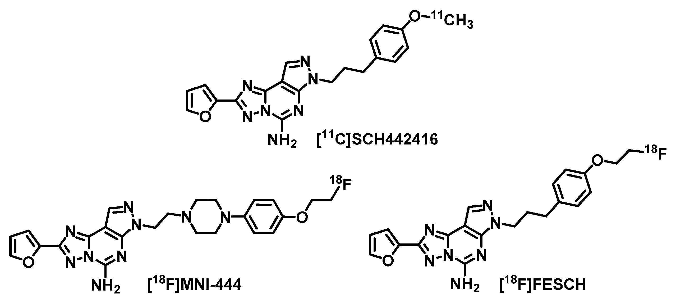
Figure 1) revealed a 70–80% increased A
2AR availability in the striatum of PD patients with L-DOPA-induced dyskinesia [
13]. This strongly indicates a key role of A
2AR not only in the development of PD [
11] but also in the progression of the disease and in association with the dopaminergic standard therapy [
2,
13]. Hence, A
2AR-PET is considered as an appropriate tool for early diagnosis and staging of PD as well as evaluation of potential A
2AR antagonists for PD treatment by means of receptor occupancy studies. The main disadvantage of
11C-labeled radiotracers for clinical routine applications is the short half-life of the nuclide (
t1/2 = 20.3 min). To date, there is only one
18F-labeled (
t1/2 = 109.7 min) A
2AR radiotracer, [
18F]
MNI-444 [
14,
15] (
Figure 1), that has already been evaluated in a clinical PET study. In healthy volunteers [
15], a high uptake of activity in the striatum has been observed. However, no follow-up studies have been published since 2015 and further data of [
18F]
MNI-444 regarding specificity of A
2AR binding in humans or selectivity over the adenosine A
1 receptor (A
1R), which is also highly expressed in the brain [
16,
17], are not yet available. For that reason, we selected the well-studied and highly A
2AR-selective radiotracer [
18F]
FESCH [
18,
19,
20,
21] (
Ki(
hA
2AR) = 12.4 nM,
Ki(
hA
1R) ~ 10 µM, former [
18F]
MRS5425 [
18]) for our purposes, which is the [
18F]fluoroethoxy analog of [
11C]
SCH442416 (
Figure 1). [
18F]
FESCH has been evaluated in healthy rats and in rats with unilateral PD symptoms by PET showing an A
2AR-specific binding in the striatum and a significantly increased A
2AR-mediated uptake in the 6-hydroxydopamine-lesioned hemisphere of 9–12% [
19,
21]. Therefore, [
18F]
FESCH has been stated as the most suitable radiotracer for quantification of the A
2AR in the brain with PET [
21].
The rotenone-based mouse model has been well established for pre-clinical investigations of PD [
22,
23,
24]. Chronic administration of the neurotoxin rotenone to mice leads to relapse of dopaminergic neurons and changes in motor functions similar to the symptoms in PD patients [
24,
25,
26]. In this small animal PET/MR imaging study with [
18F]
FESCH, we aimed to validate if rotenone-treated mice reflect the aspect of striatal A
2AR upregulation in PD. In addition, we investigated [
18F]
FESCH by in vitro autoradiography on mouse brain slices and in vivo metabolism as well as baseline and blocking PET studies in healthy CD-1 mice.
2. Results and Discussion
The A
2AR antagonist
FESCH and the required known phenol precursor desmethyl SCH442416 for
18F-labeling were synthesized in three or two steps, respectively, from commercially available 2-(furan-2-yl)-7
H-pyrazolo[4,3-
e][1,2,4]triazolo[1,5-
c]pyrimidine-5-amin and 1-(3-bromopropyl)-4- methoxybenzene according to procedures described in the literature [
18,
27,
28]. Evaluation of
FESCH in an established binding assay revealed high A
2AR affinity (
Ki(
hA
2AR) = 0.6 ± 0.1 nM, n = 4) and good A
1R selectivity (
Ki(
hA
1R) = 203 ± 40 nM, n = 4).
The initially published radiosynthesis of [
18F]
FESCH comprises a two-step two-pot procedure [
19,
20]. Briefly, the respective [
18F]fluoroethyl intermediate from the
18F-labeling step was isolated either by semi-preparative HPLC [
19] or solid-phase extraction on a cartridge [
20] and, only then, reacted with the phenol precursor desmethyl SCH442416. To simplify the radiosynthesis and avoid loss of activity through purification of the [
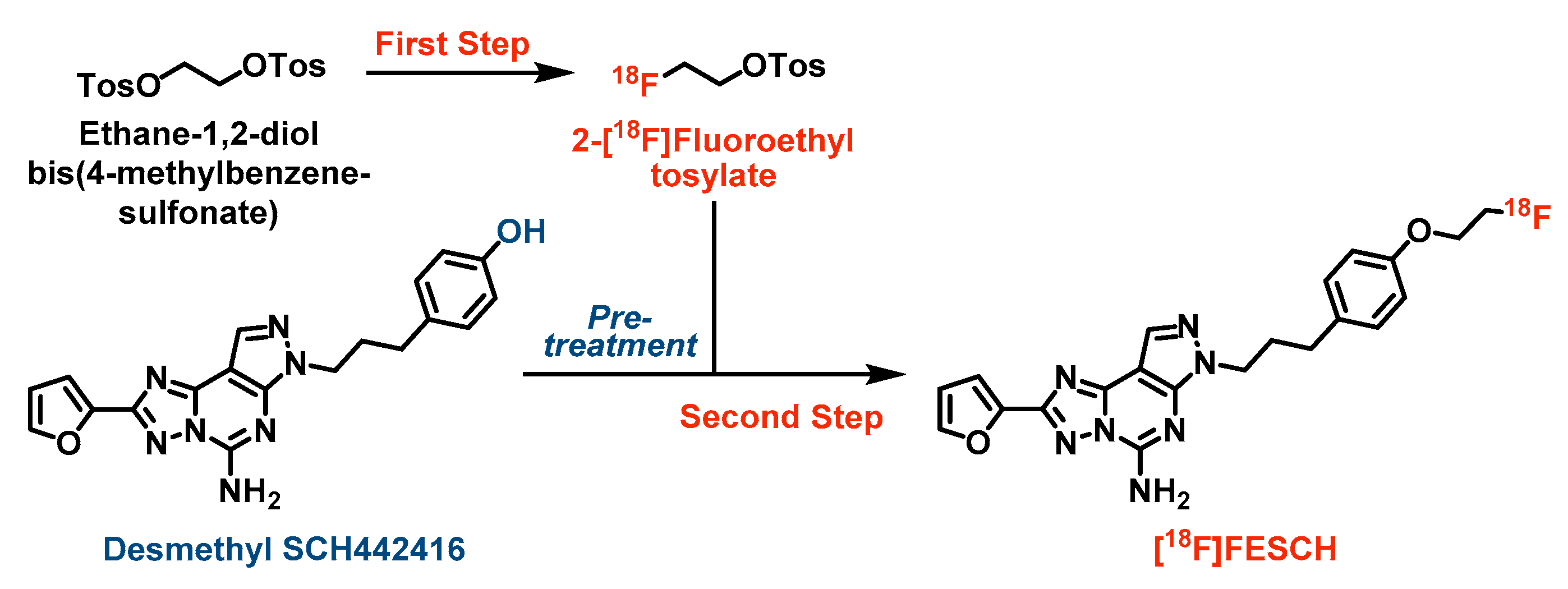
18F]fluoroethyl intermediate, we developed a two-step one-pot strategy for the production of [
18F]
FESCH [
29] (see
Scheme 1). First, ethane-1,2-diol bis(4-methylbenzenesulfonate) was
18F-labeled via nucleophilic substitution of one tosylate group using anhydrous K
+/[
18F]F
-/K
222-carbonate complex in acetonitrile. Notably, desmethyl SCH442416 was pre-treated with aqueous tetrabutylammonium hydroxide (TBAOH) in acetonitrile to generate the activated phenolate which was directly reacted with the non-isolated 2-[
18F]fluoroethyl tosylate. Besides, further bases and solvents were tested for the [
18F]fluoroethylation of desmethyl SCH442416 resulting in decreased yields of [
18F]
FESCH mainly due to partial decomposition of the phenol precursor under these conditions. Preliminary experiments using potassium carbonate or cesium carbonate (1.5–2.5 eq.) for activation of desmethyl SCH442416 (1–2 mg) and either acetonitrile or a 1:1 mixture of acetonitrile and
N,
N-dimethylformamide or
tert-butanol as solvents revealed labeling yields of 4–14% based on TLC analysis of the reaction mixtures after 10 min at 120 °C, respectively. Replacement of the base by TBAOH (2.5 eq.) and the use of acetonitrile only resulted in a significantly increased labeling yield of 25%. Final optimization was achieved by increasing the amount of the phenol precursor to 2.5 mg leading to 46.4 ± 8.5% (n = 9) of non-isolated [
18F]
FESCH.
[
18F]
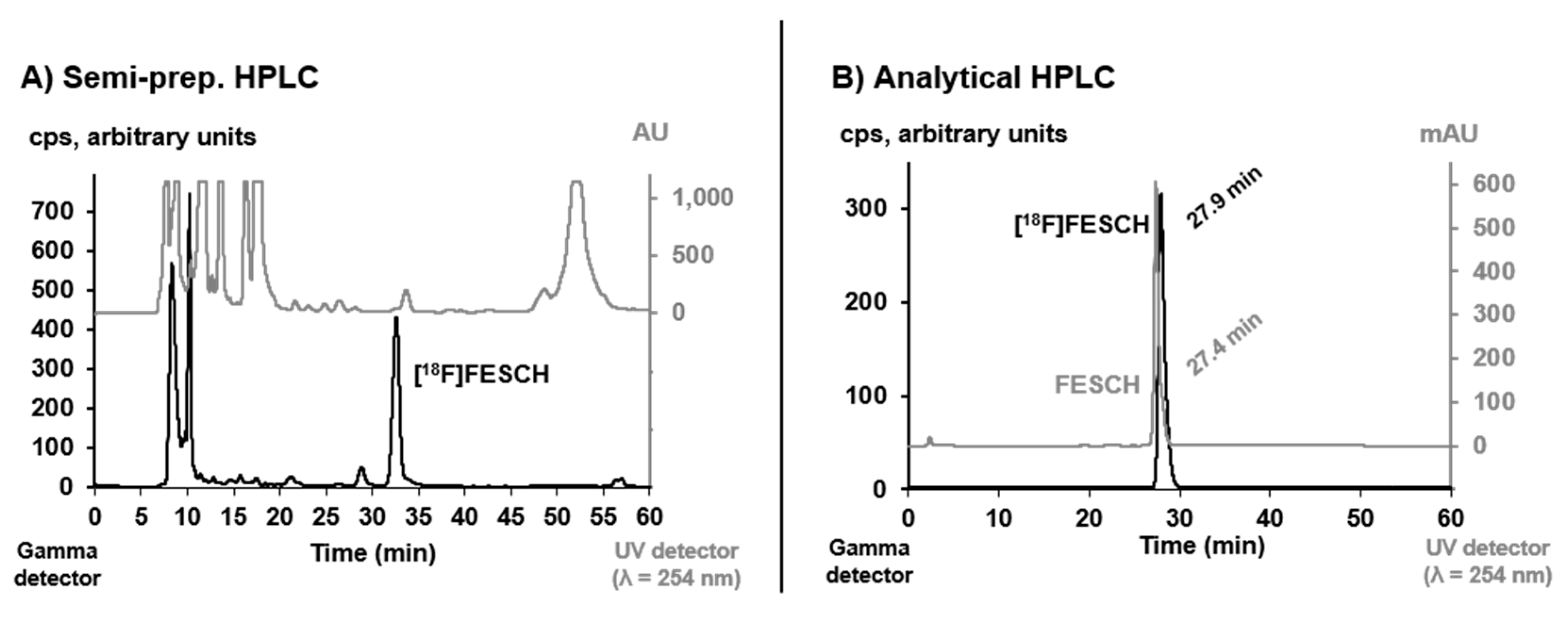
FESCH was purified by semi-preparative HPLC (see
Figure 2) and concentrated via solid-phase extraction on a pre-conditioned reversed-phase cartridge followed by elution with absolute ethanol. After evaporation of the solvent, the radiotracer was finally formulated in sterile isotonic saline with a maximum ethanol content of 10% for better solubility. The identity of [
18F]
FESCH was confirmed by analytical HPLC using an aliquot of the final product spiked with the non-radioactive reference compound (
Figure 2).
The herein described one-pot radiolabeling strategy provided [
18F]
FESCH with a high molar activity of 116 ± 18.5 GBq/μmol (n = 7, end of synthesis) and an overall radiochemical yield of 16.1 ± 1.5% (n = 9, end of bombardment), which is a significant improvement compared to the published two-pot procedure (7 ± 2% [
20]). The required total synthesis time of 114 ± 6 min and the achieved radiochemical purity of ≥ 98% were very similar for both methods.
In vitro stability of [
18F]
FESCH was examined in isotonic saline and pig plasma. Samples of each medium were analyzed by radio-HPLC after 60 min incubation at 37 °C and no degradation or defluorination of the radiotracer was observed. This result is contradictory to the published stability of [
18F]
FESCH especially in saline where only 85–90% of intact radiotracer have been detected by radio-TLC while multiple spots were observed [
20]. Thus, Khanapur et al. [
20] formulated [
18F]
FESCH in phosphate-buffered saline, but Bhattacharjee et al. [
19] used a saline solution of [
18F]
FESCH for biological investigations. At present, we have no explanation for that and as we detected only the intact radiotracer in isotonic saline (see
Figure 2), formulation of [
18F]
FESCH in another medium was not required.
The distribution coefficient of [
18F]
FESCH was determined by partitioning between
n-octanol and phosphate-buffered saline (PBS, pH = 7.4) at ambient temperature using the conventional shake-flask method. The obtained logD
7.4 value of 1.97 ± 0.17 (n = 4) emphasizes the lipophilic character of the radiotracer which allows a passive diffusion through the blood–brain barrier [
30,
31,
32]. However, there is a discrepancy compared to the published logD value of 3.16 ± 0.03 [
20] which might be caused by different experimental setups.
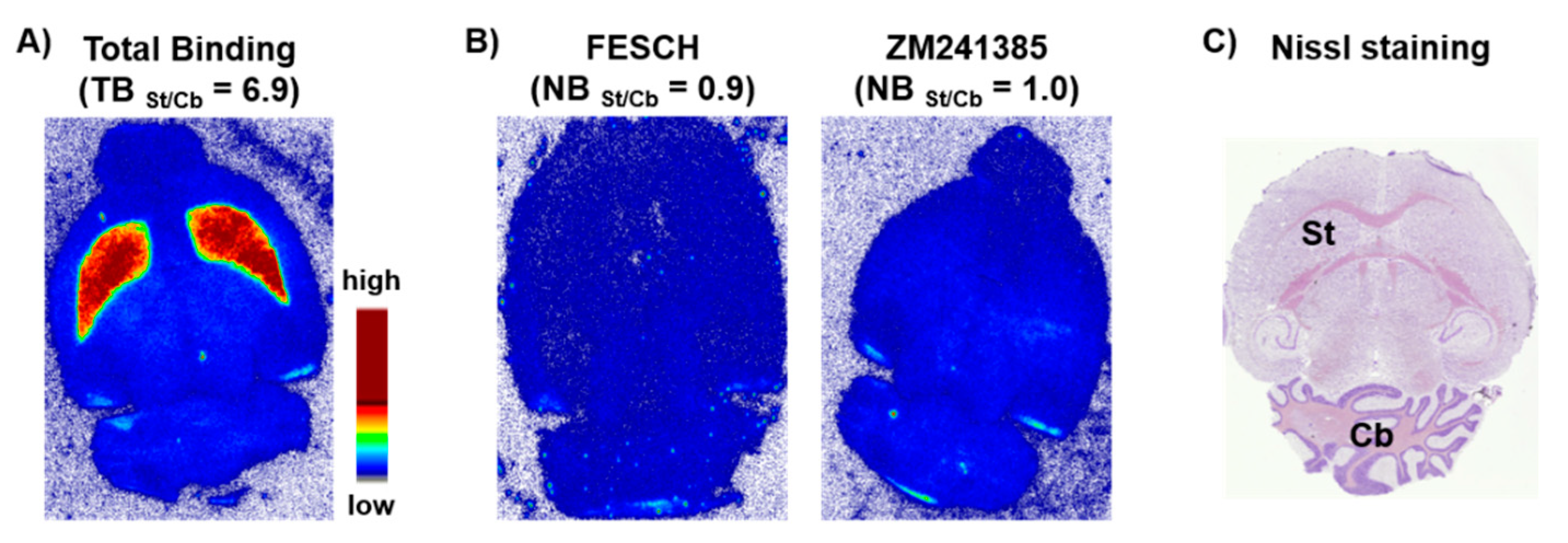
In vitro autoradiography studies were accomplished by incubating sections of mouse brain with [
18F]
FESCH. Non-specific binding was assessed by co-incubation with an excess of either
FESCH or
ZM241385, respectively. The images demonstrated an A
2AR-specific accumulation of [
18F]
FESCH in the striatum (
Figure 3), which is characterized by the binding parameters
KD = 4.69 ± 1.17 nM and
Bmax = 497 ± 97 fmol/mg wet weight consistent with the literature [
33].
For in vivo imaging investigations in healthy CD-1 mice (n = 12, 10 weeks, 30–35 g), [
18F]
FESCH was injected intravenously (baseline: 2.8 ± 2 MBq, vehicle: 5.8 ± 2 MBq, blocking: 6.4 ± 2.5 MBq with n = 4, respectively) and whole body scans were performed for 60 min in listmode with a Mediso nanoScan
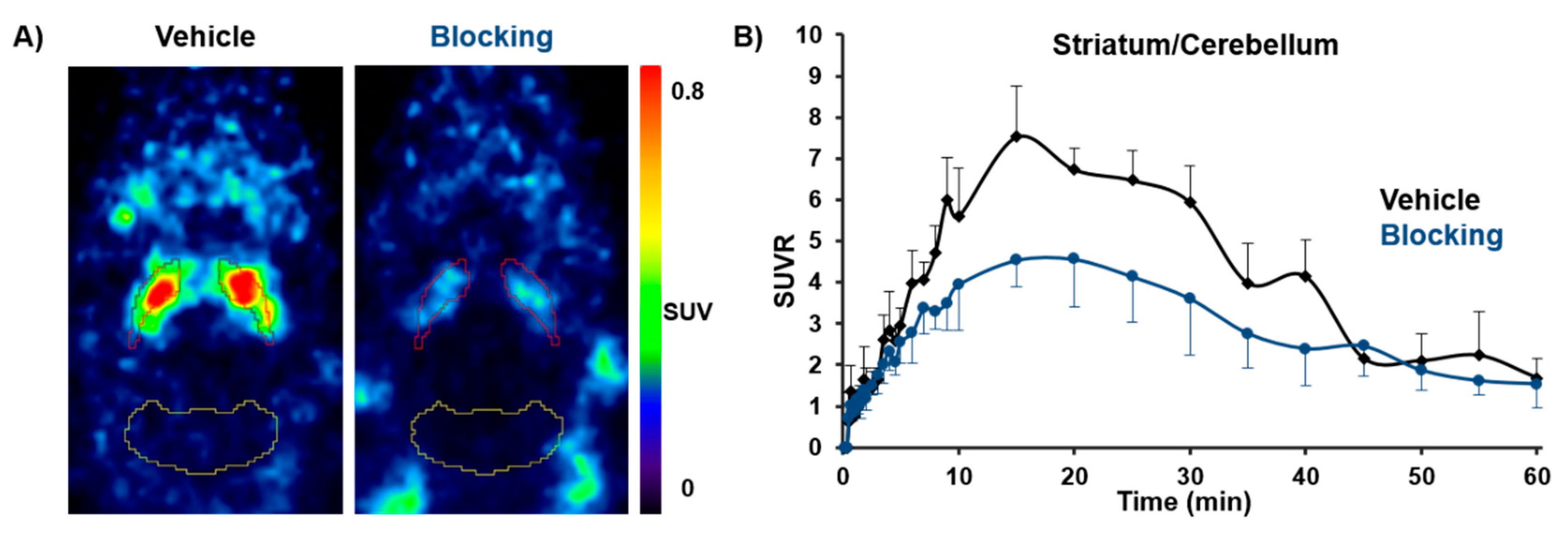
® PET/MR scanner followed by dynamic reconstruction. Time-activity curves (TACs) were generated for regions of interest such as striatum and cerebellum as reference region. The obtained PET images showed a high striatal uptake of [
18F]
FESCH with a standardized uptake value ratio (SUVR) for striatum over cerebellum of > 5 (9–30 min post injection (p.i.)). In vivo selectivity for the A
2AR was highlighted by the significant reduction of the SUVR by 29–42% (9–30 min p.i. with
p < 0.05) after pre-injection of
tozadenant as blocking agent (
Figure 4). The highest achievable dose of
tozadenant was 2.5 mg/kg with respect to solubility and volume of injection as well as the concentrations of DMSO and Kolliphor
® EL suitable for in vivo application in mice. The herein observed blocking effect is in accordance with the estimated A
2AR occupancy for
tozadenant in rhesus monkey of about 72% at 5 mg/kg [
14]. These results indicate that [
18F]
FESCH is a promising radiotracer for molecular imaging of the A
2AR in the brain.
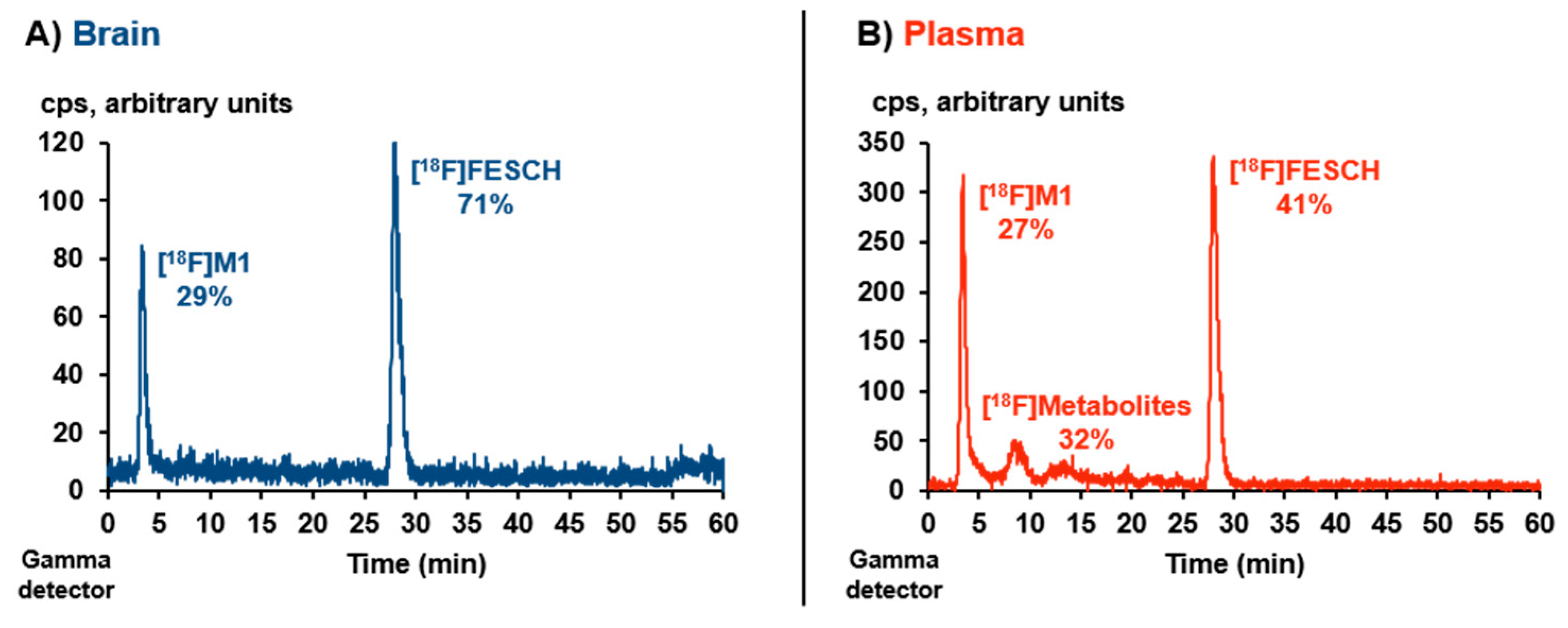
However, a representative metabolism study revealed only moderate in vivo stability of [
18F]
FESCH. Analytical radio-HPLC (
Figure 5) of the extracted mouse plasma sample showed 41% of intact radiotracer at 15 min p.i. (recovery of total activity = 84%). In the analyzed brain sample, one polar radiometabolite ([
18F]
M1) was detected accounting for 29% of the total extracted activity at 15 min p.i. (recovery = 98%).
Compared to the published study in Wistar-Unilever rats (46% intact radiotracer in plasma at 60 min p.i. [
20]) the in vivo degradation of [
18F]
FESCH appears to be somewhat faster in mice. Notably and to the best of our knowledge, the formation of brain-penetrating radiometabolites of [
18F]
FESCH has not been regarded before. Based on our experiences with radiotracers bearing a [
18F]fluoroethoxy moiety [
34,
35], the herein observed radiometabolite [
18F]
M1 is proposed to be 2-[
18F]fluoroethanol or the oxidized 2-[
18F]fluoroacetaldehyde and 2-[
18F]fluoroacetate, resulting from a cytochrome P450 enzyme-induced metabolic degradation [
36,
37], which are able to cross the blood–brain barrier [
38,
39,
40,
41].
For PET/MR studies with [
18F]
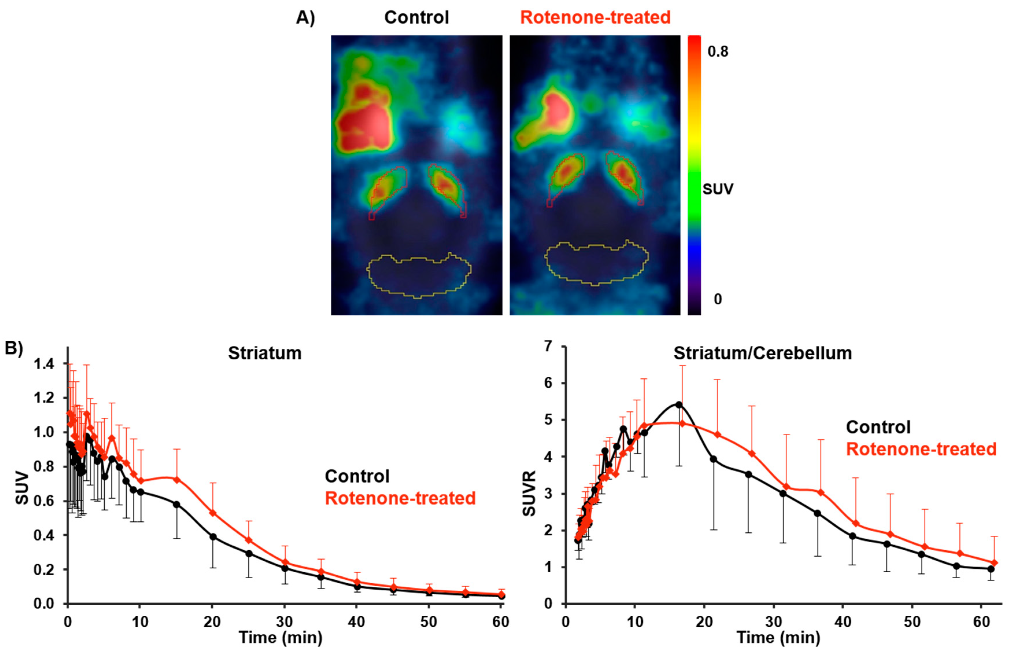
FESCH in the rotenone-based mouse model of PD, the radiotracer (9.7 ± 1.3 MBq) was administrated to C57BL/6JRj mice (control: n = 5; rotenone-treated: n = 7; 16 months, 28–35 g) followed by the same imaging protocol used for the baseline and blocking studies in CD-1 mice. Although statistically not significant, the averaged TACs between 2 and 61 min p.i. revealed a slightly higher uptake of [
18F]
FESCH in the striatum of rotenone-treated mice compared to controls. An increase of the SUVR for striatum over cerebellum by 15–33% was observed, which was caused by the elevated SUV for striatum of 11–27% (21–61 min p.i., respectively;
Figure 6).
These results are in accordance with the determined A
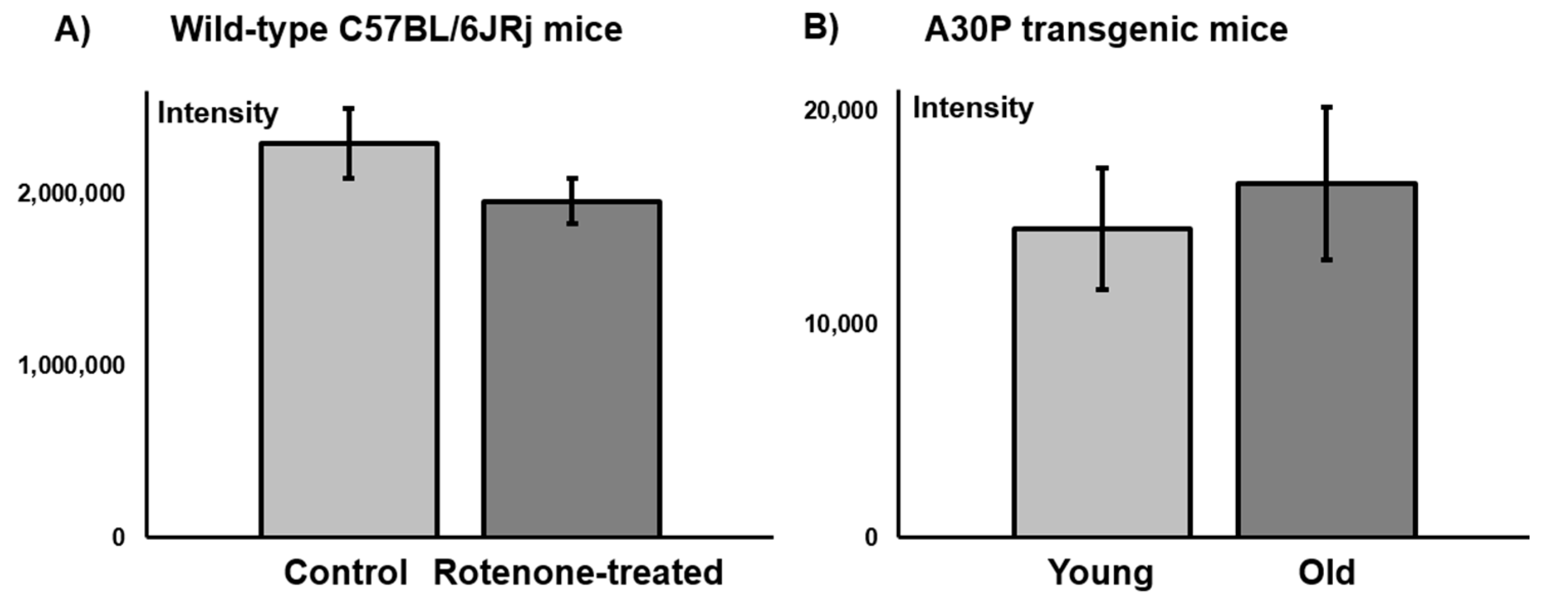
2AR levels on C57BL/6JRj mouse brain sections from a comparative in vitro immunofluorescence study. No significant difference in the fluorescence signal of the A
2AR between control and rotenone-treated mice was observed (
Figure 7).
The increased A
2AR availability in the striatum of PD patients appears to be related to L-DOPA-induced dyskinesia. In the rotenone-treated mice, we did not identify any signs of dyskinesia and, thus, it is not surprising that in this PD mouse model no significant increase in the A
2AR density was detected either by PET imaging or by immunofluorescence staining. With regard to dyskinesia, the rotenone model is comparable to another neurotoxin-based PD mouse model using 1-methyl-4-phenyl-1,2,3,6-tetrahydropyridine (MPTP), where only weak signs of dyskinesia were observed and this exclusively under L-DOPA treatment [
42]. To date, unilateral injection of 6-hydroxydopamine (6-OHDA) is the only effective neurotoxin-related approach to replicate evidently some forms of dyskinesia in rats and mice. In the 6-OHDA mouse model, dyskinesia is detected as abnormal involuntary movements (AIMs) with a more simplified range than in rats, presenting more prominent rotational locomotion with less dystonic features, i.e., axial AIMs [
42]. None of the above-mentioned AIMs were detected in the rotenone-treated mice. Consequently, it remains to be examined whether treatment with L-DOPA would lead to dyskinesia in the rotenone model as shown for the MPTP model and if this would cause a quantifiable increase in striatal A
2AR levels.
In contrast to the above-discussed neurotoxin-based PD animal models, the A30P mouse line represents a transgenic model of PD. This mouse line overexpresses the human A30P mutation in the α-synuclein gene, where the 30th amino acid residue alanine is replaced by proline, in all neurons. The A30P mutation is associated with rare familial cases of PD. Since α-synuclein is a main constituent of Lewy bodies, the A30P transgenic mice are characterized by Lewy body formation in neuronal cell bodies and neurites throughout the brain [
43]. The α-synuclein pathology and PD-associated symptoms become prominent at about 69 weeks of age [
43,
44]. In an ongoing study, we detected slightly, but due to the rather small and highly variable data set statistically non-significant, higher striatal mean fluorescence intensities of the A
2AR signal in older symptomatic A30P transgenic mice compared to younger ones without symptoms (see
Figure 7). We will further investigate this PD mouse model by small animal PET/MR imaging studies using a novel derivative of [
18F]
FESCH currently under development.
4. Materials and Methods
4.1. General Information
Chemicals and solvents were purchased from standard commercial sources in analytical grade and were used without further purification. Radio-TLCs were performed on pre-coated silica gel plates (Alugram® Xtra SIL G/UV254, Polygram® SIL G/UV254; Carl Roth, Karlsruhe, Germany). The compounds were localized at 254 nm (UV lamp). Radio-TLC was recorded using a bioimaging analyzer system (BAS-1800 II, Fuji Photo Film, Co. Ltd., Tokyo, Japan) and images were evaluated with Aida 2.31 software (raytest Isotopenmessgeräte GmbH, Straubenhardt, Germany). Column chromatography was conducted on silica gel (0.06–0.20 mm; Carl Roth, Karlsruhe, Germany). HPLC separations were performed on JASCO systems equipped with UV detectors from JASCO Deutschland GmbH (Pfungstadt, Germany) and activity detectors from raytest Isotopenmessgeräte GmbH (GABI Star, Straubenhardt, Germany).
Semi-preparative HPLC conditions were: Column: Reprosil-Pur C18-AQ, 250 × 10 mm; particle size: 10 µm; eluent: 50% MeCN/20 mM NH4OAcaq.; flow: 7 mL/min; ambient temperature; UV detection at 254 nm.
Analytical HPLC conditions were: Column: Reprosil-Pur C18-AQ, 250 × 4.6 mm; particle size: 5 µm; gradient: 0–10 min: 26% MeCN, 10–35 min: 26% → 90% MeCN, 35–45 min: 90% MeCN, 45–50 min: 90% → 26% MeCN, 50–60 min: 26% MeCN/20 mM NH4OAcaq.; isocratic: 40% MeCN/20 mM NH4OAcaq.; flow: 1 mL/min; ambient temperature; UV detection at 254 nm. Molar activity was determined on the base of a calibration curve (0.05–2.0 µg FESCH) carried out under isocratic HPLC conditions (36% MeCN/20 mM NH4OAcaq.) using chromatograms obtained at 264 nm as the maximum of UV absorbance.
No-carrier-added (n.c.a.) [18F]fluoride (t1/2 = 109.8 min) was produced via the [18O(p,n)18F] nuclear reaction by irradiation of [18O]H2O (Hyox 18 enriched water, Rotem Industries Ltd, Arava, Israel) on a Cyclone®18/9 (IBA RadioPharma Solutions, Louvain-la-Neuve, Belgium) with fixed energy proton beam using Nirta® [18F]fluoride XL target.
4.2. Radiosynthesis
The aqueous solution of no-carrier-added [18F]fluoride (~ 3 GBq) was trapped on a Chromafix® 30 PS-HCO3− cartridge (MACHEREY-NAGEL GmbH & Co. KG, Düren, Germany). The activity was eluted with 300 µL of an aqueous K2CO3-solution (1.78 mg, 12.9 µmol) into a 4-mL V-vial containing Kryptofix 2.2.2 (K2.2.2, 5.6 mg, 14.9 µmol) in 1 mL MeCN. The K+/[18F]F−/K2.2.2-carbonate complex was azeotropically dried under vacuum and argon flow within 7–10 min using a Discover PETwave Microwave CEM® (75 W, 50–60 °C, power cycling mode, CEM GmbH, Kamp-Lintfort, Germany). Two aliquots of MeCN (2 × 1 mL) were added during the drying procedure and the final complex was dissolved in 400 µL MeCN ready for radiolabeling.
The aliphatic radiolabeling of ethane-1,2-diol bis(4-methylbenzenesulfonate) (Sigma-Aldrich, Munich, Germany; 2 mg in 100 µL MeCN) was performed under conventional heating at 90 °C for 10 min. After pre-heating of the phenol precursor desmethyl SCH442416 (2.5 mg in 490 µL MeCN) with 10 µL TBAOHaq. (40%) at 90 °C for 10 min, this solution was directly added to the crude 2-[18F]fluoroethyl tosylate and the reaction mixture was stirred for 10 min at 120 °C. Aliquots of the reaction mixtures from both steps were analyzed by radio-TLC (EtOAc/petroleum ether; first step = 1:1; second step = 6:1) or radio-HPLC (isocratic mode, see General Information) to determine the radiochemical yields of crude 2-[18F]fluoroethyl tosylate (Rf = 0.7, 87.0 ± 2.3%; n = 9) and [18F]FESCH (Rf = 0.5, 46.4 ± 8.5%; tR = 29.4 min, 34.4 ± 4.1%, decay corrected for tR = 0 min; n = 9), respectively.
After dilution with water (1:1), the crude reaction mixture was applied to an isocratic semi-preparative HPLC (see General Information) for isolation of the desired radiotracer [18F]FESCH (tR = 32–33 min). The collected fractions were diluted with water (total volume = 40 mL), passed through a Sep-Pak® C18 Plus cartridge (Waters, Milford, MA, USA; pre-conditioned with 5 mL of absolute EtOH and 60 mL water), and eluted with 1.5 mL of absolute EtOH. Evaporation of the solvent at 70 °C under a gentle argon stream and subsequent formulation of the radiotracer in sterile isotonic saline containing 10% EtOH afforded a [18F]FESCH solution usable for biological investigations.
The identity of the radiotracer was proved by analytical radio-HPLC (see General Information) of samples of [18F]FESCH spiked with the non-radioactive reference compound FESCH using a gradient and an isocratic mode.
4.3. Investigation of In Vitro Stability and Lipophilicity (logD7.4)
The in vitro stability of [18F]FESCH (4–6 MBq) was studied by incubation in isotonic saline and pig plasma (500 µL each) at 37 °C. Samples were taken at 60 min and analyzed by radio-HPLC (see General Information).
The lipophilicity of [18F]FESCH was examined by partitioning between n-octanol and phosphate-buffered saline (PBS, pH = 7.4) at ambient temperature using the conventional shake-flask method. The radiotracer (65 µL, ~ 8 MBq) was added to a tube containing the n-octanol/PBS-mixture (6 mL, 1:1, four-fold determination). The tubes were shaken for 20 min using a mechanical shaker (HS250 basic, IKA Labortechnik GmbH & Co. KG, Staufen, Germany) followed by centrifugation (5500 rpm for 5 min) and separation of the phases. Aliquots were taken from the organic and the aqueous phase (1 mL each) and activity was measured with an automated gamma counter (1480 WIZARD, Fa. Perkin Elmer, Waltham, MA, USA). The distribution coefficient (D7.4) was calculated as [activity (cpm/mL) in n-octanol]/[activity (cpm/mL) in PBS, pH = 7.4] stated as the decade logarithm (logD7.4).
4.4. Binding Assay
Membrane homogenates prepared from stably transfected CHO-K1 cells (Chinese hamster ovary cells, clone K1) with the human A1R or A2AR, provided by Prof. Klotz, University of Würzburg, Würzburg, Germany, were used for the binding experiments. The following radioligands were employed: [3H]ZM241385 for the A2AR and [3H]DPCPX for the A1R. For the determination of A2AR and A1R binding affinity of reference compound, frozen cell suspensions were thawed, homogenized by a 27-gauge needle, and diluted with 50 mM TRIS-HCl buffer (pH = 7.4, 100 mM NaCl, 5 mM MgCl2, 1 mM EDTA) containing 1 µU/mL adenosine deaminase (ADA). Membrane suspension was incubated with 1.5 nM [3H]ZM241385 or 1 nM [3H]DPCPX and various concentrations of the test compound. Non-specific binding was determined by co-incubation with 10 μM ZM241385 or 1 µM DPCPX. The incubation was performed at room temperature for 90 min and terminated by rapid filtration using Whatman GF/B glass-fiber filters, pre-soaked in 0.3% polyethyleneimine and a 48-channel harvester (Biomedical Research and Development Laboratories, Gaithersburg, MD, USA) followed by washing four times with ice-cold TRIS-HCl buffer. Filter-bound radioactivity was quantified by liquid scintillation counting. At least two separate experiments were performed for determination of Ki values. All data were analyzed with GraphPad Prism, Version 4.1 (GraphPad Inc., La Jolla, CA, USA), according to the Cheng–Prusoff equation.
4.5. Animal Experiments
All experimental work including animals was conducted in accordance with the national legislation on the use of animals for research (Tierschutzgesetz (TierSchG), Tierschutz-Versuchstierverordnung (TierSchVersV)) and was approved by the responsible animal care committees of the Free State of Bavaria and the Free State of Saxony (TVV 08/13, 24-9168.11/18/8, June 12th, 2013 and TVV 18/18, DD24.1-5131/446/19, June 20th, 2018; Landesdirektion Sachsen). Female CD-1 mice, 10–12 weeks, were obtained from the Medizinisch-Experimentelles Zentrum at University of Leipzig, Leipzig, Germany.
4.5.1. In Vitro Autoradiography
Cryosections of brains obtained from female CD-1 mice (10–12 weeks) were thawed, dried in a stream of cold air, and pre-incubated in 50 mM TRIS-HCl buffer (pH = 7.4, 100 mM NaCl, 5 mM MgCl2, 1 mM EDTA) containing 1 µU/mL adenosine deaminase (ADA) for 15 min at ambient temperature. Afterwards, brain sections were incubated with 0.1–0.2 MBq/mL [18F]FESCH in buffer for 90 min at room temperature. Non-specific binding was determined in the presence of 1 µM of FESCH or ZM241385, respectively. Subsequently, the sections were washed twice for 5 min in ice-cold TRIS-HCl buffer, and dipped for 5 s in ice-cold deionized water. The sections were rapidly dried in a stream of cold air before being exposed overnight on an imaging plate (Fujifilm Corporation, Tokyo, Japan). Developed autoradiographs were analyzed in a phosphor imager (HD-CR 35, Duerr NDT GmbH, Bietigheim Bissingen, Germany). The quantification was performed by using 2D-densitometric analysis (AIDA 2.31 software, raytest Isotopenmessgeräte GmbH, Straubenhardt, Germany). Further data analysis was performed with GraphPad Prism, Version 4.1 (GraphPad Inc., La Jolla, CA, USA).
4.5.2. Rotenone-Based and A30P Transgenic Mouse Models of Parkinson’s Disease
Animal Housing
Male wild-type C57BL/6JRj mice (Janvier Labs, Le Genest-Saint-Isle, France) and male (Thy1)-
h[A30P]α-syn transgenic C57BL/6JRj mice (generated according to the protocol by Kahle et al. [
43], hereafter “A30P transgenic mice”) were housed at room temperature under a 12:12 h dark:light cycle. Food and water was provided ad libidum.
Oral Rotenone Administration
Wild-type C57BL/6JRj mice (12 months) were divided into two groups and treated 5 days a week for 4 months. A 1.2 mm × 60 mm gavage (Unimed, Lausanne, Switzerland) was used to administer 0.01 mL/g animal weight of rotenone (Sigma-Aldrich, Munich, Germany) solution corresponding to a 5 mg/kg dose. Controls were treated only with the vehicle solution (2% carboxymethyl cellulose (Sigma-Aldrich, Munich, Germany) and 1.25% chloroform (Carl Roth, Karlsruhe, Germany)).
4.5.3. Immunofluorescence Staining and Imaging
Immunostaining
Cryosections of brains obtained from wild-type C57BL/6JRj mice (16 months, vehicle and rotenone-treated) or A30P transgenic mice (young group = 39 weeks; old group = 71–76 weeks) were immunostained using the free-floating technique and all procedures took place in a well of a 12-well plate. Briefly, brain sections were washed with PBS (pH = 7.4) for 10 min, then blocked with blocking solution (PBS, 0.02% Triton-X and 5% donkey serum) for 1 h, and incubated in the first antibody (1:100, rabbit anti-adenosine receptor A2A, ab3461, Abcam, Cambridge, UK) in blocking solution at 4 °C overnight. On the next day, sections were washed in PBS (4 × 15 min) and incubated with the secondary antibody (1:500, donkey Alexa® 555 anti-rabbit, Invitrogen/Thermo Fisher Scientific Inc., Waltham, MA, USA) for three hours at room temperature. Sections were washed in PBS (4 × 15 min), transferred to the slide, and mounted with Mowiol mounting medium (50% PBS, 25% ethylenglycol and 25% glycerol).
Microscopy and Image Analysis
Images were obtained with an Apotome.2 fluorescence microscope (Carl Zeiss Microscopy GmbH, Jena, Germany). Mean fluorescence intensity (MFI) in the striatum was measured using Fiji-ImageJ, a free-software for image analysis (ImageJ v1.52p, National Institutes of Health (NIH), Bethesda, MD, USA). MFI values were normalized to background signal.
4.5.4. Small Animal PET/MR Studies
For the time of the experiments, female CD-1 mice (n = 12; age = 10 weeks; weight = 30–35 g) and male C57BL/6JRj mice (control with n = 5; rotenone-treated with n = 7; age = 16 months; weight = 28–35 g) were kept in a dedicated climatic chamber with free access to water and food under a 12:12 h dark:light cycle at a constant temperature of 24 °C. The animals received an injection of [18F]FESCH into the tail vein (5.0 ± 2.5 MBq for CD-1 mice) or retro-orbital (9.7 ± 1.3 MBq for C57BL/6JRj mice) followed by a 60-min PET/MR scan (Mediso nanoScan®, Budapest, Hungary). Each PET image was corrected for random coincidences, dead time, scatter, and attenuation (AC), based on a whole body (WB) MR scan. The reconstruction parameters for the list mode data were the following: 3D-ordered subset expectation maximization (OSEM), 4 iterations, 6 subsets, energy window = 400–600 keV, coincidence mode = 1–5, ring difference = 81. The mice were positioned prone in a special mouse bed (heated up to 37 °C), with the head fixed to a mouth piece for the anesthetic gas supply with isoflurane in 40% air and 60% oxygen (anesthesia unit: U-410, Agnthos, Lidingö, Sweden; gas blender: MCQ, Rome, Italy). The PET data were collected by a continuous WB scan during the entire investigation. Following the 60-min PET scan, a T1-weighted WB gradient echo sequence (GRE, repetition time = 20 ms, echo time = 6.4 ms) was performed for AC and anatomical orientation. Image registration and evaluation of the region of interest (ROI) were done with PMOD (PMOD Technologies LLC, v. 3.9, Zurich, Switzerland).
The respective brain regions were identified using the mouse brain atlas template Ma–Benveniste–Mirrione–FDG. The activity data are expressed as mean standardized uptake value (SUV) of the overall ROI.
4.5.5. In Vivo Metabolism Study
The radiotracer [18F]FESCH (~ 17 MBq in 150 µL isotonic saline) was injected into a female CD-1 mouse via the tail vein. Brain and blood samples were obtained at 15 min p.i., plasma separated by centrifugation (14,000 × g, 1 min), and brain homogenized in ~ 1 mL isotonic saline on ice (10 strokes of a polytetrafluoroethylene (Teflon®) plunger at 1000 rpm in a borosilicate glass cylinder; Potter S Homogenizer, B. Braun Melsungen AG, Melsungen, Germany).
Two consecutive extractions were performed as duplicate (plasma) or triplicate (brain) determinations. Plasma (50 µL) and brain samples (250 µL) were added to an ice-cold acetone/water mixture (4:1; plasma or brain sample/organic solvent = 1:4). The samples were vortexed for 1 min, incubated on ice for 10 min (first extraction) or 5 min (second extraction) and centrifuged at 10,000 rpm for 5 min. Supernatants were collected and the precipitates were re-dissolved in 100 µL of the ice-cold acetone/water mixture (4:1) for the second extraction. Activity of aliquots from supernatants of each extraction step and of the precipitates was quantified using an automated gamma counter (1480 WIZARD, Fa. Perkin Elmer). The supernatants from both extractions were combined, concentrated at 70 °C under argon stream, and analyzed by radio-HPLC (gradient mode, see General Information). By this protocol, [18F]FESCH was quantitatively extracted from the biological material as proven by in vitro incubation of the radiotracer in pig plasma.
