Recent Bio-Advances in Metal-Organic Frameworks


Abstract
1. Introduction
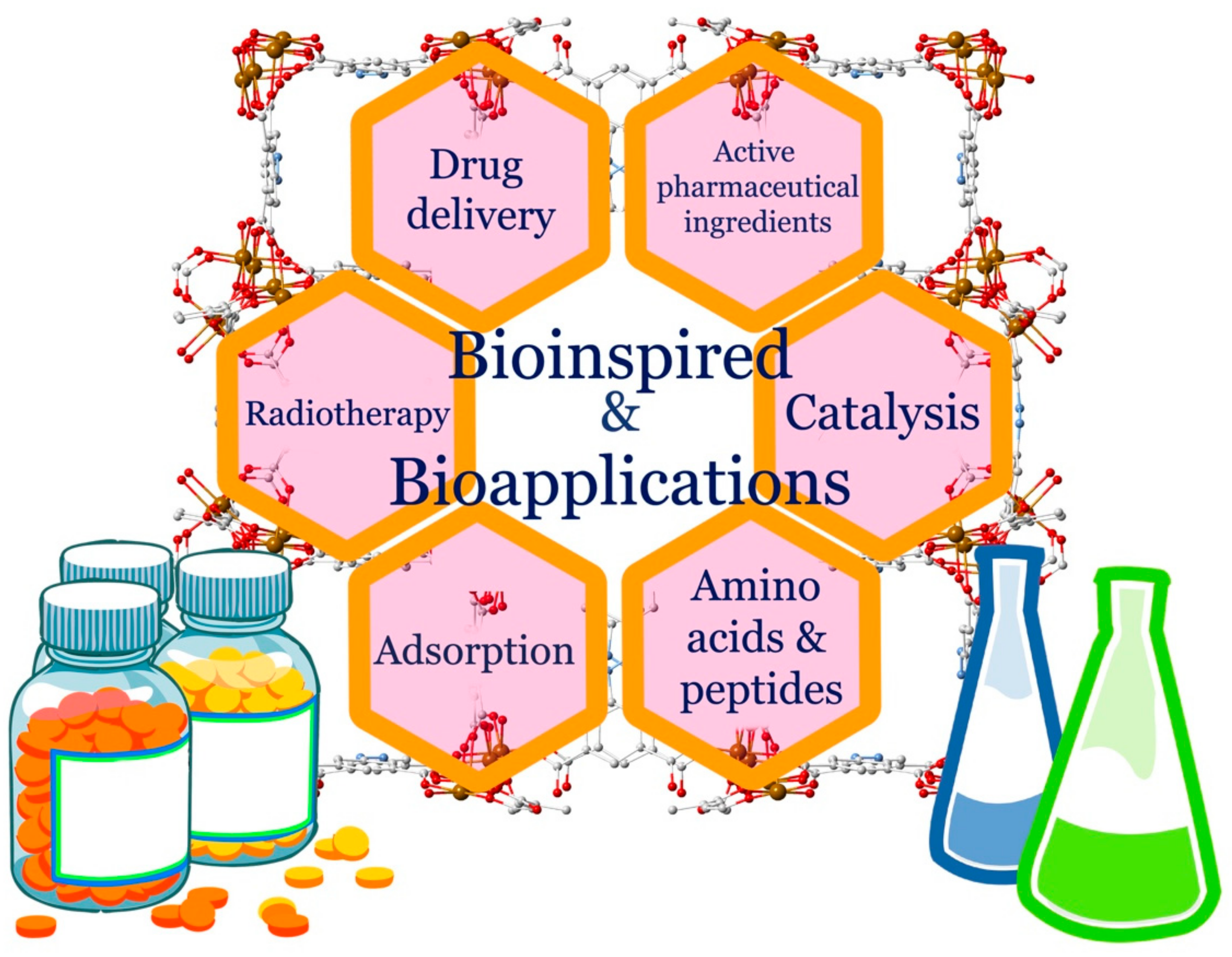
2. Metal-Organic Frameworks (MOFs) Bioapplications
2.1. MOFs and Drug Delivery
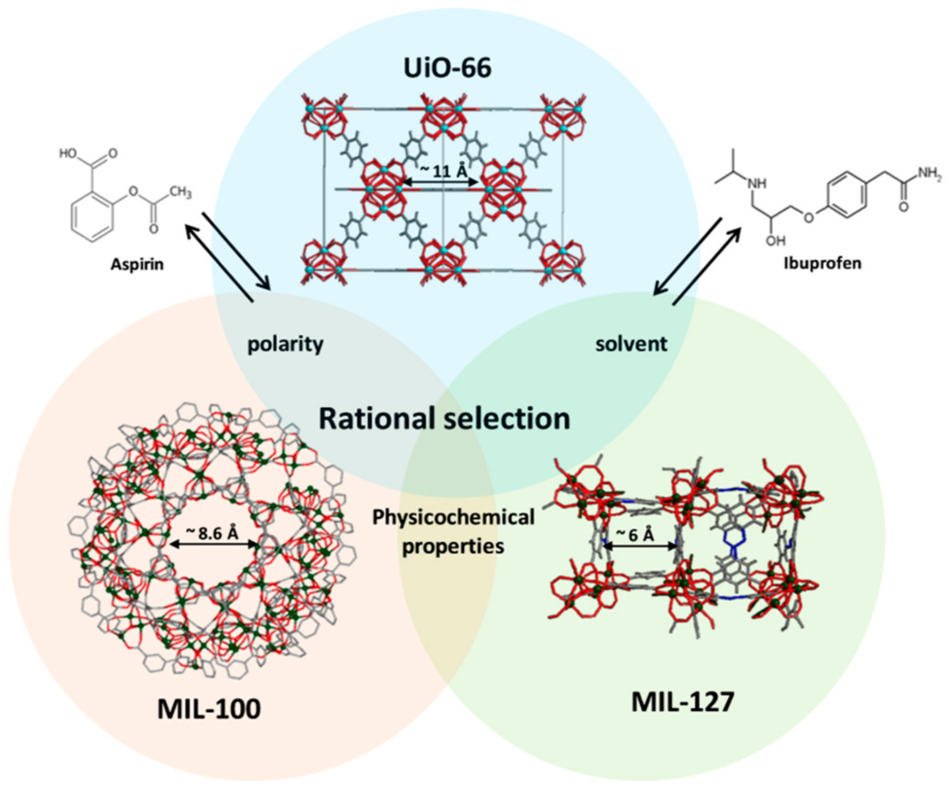
2.1.1. Structure-Activity Relationship Between MOFs and Their Cargo: The Effect of Hydrophilicity/Hydrophobicity and Pore Size
2.1.2. Sugar-Based MOFs: The Benefits of a Renewable, Soluble Material
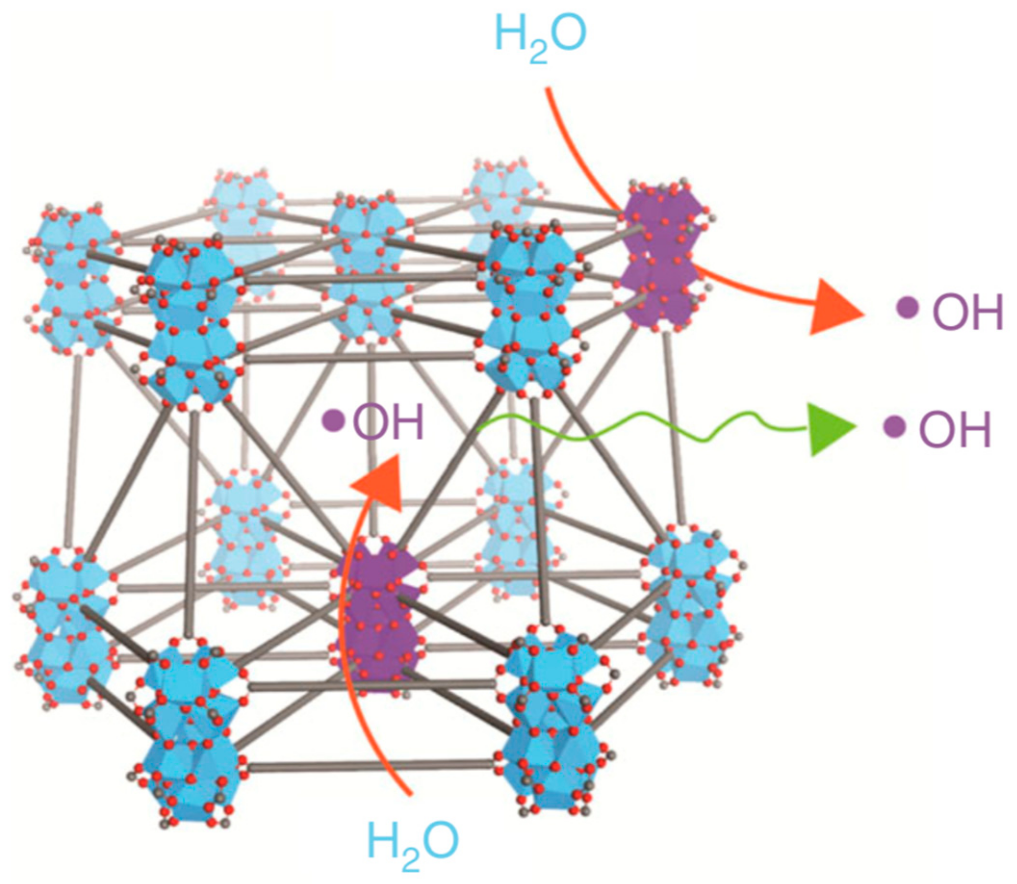
2.1.3. MOFs as Cancer Treatments: Radiotherapy and Transport of Reactive Oxygen Species
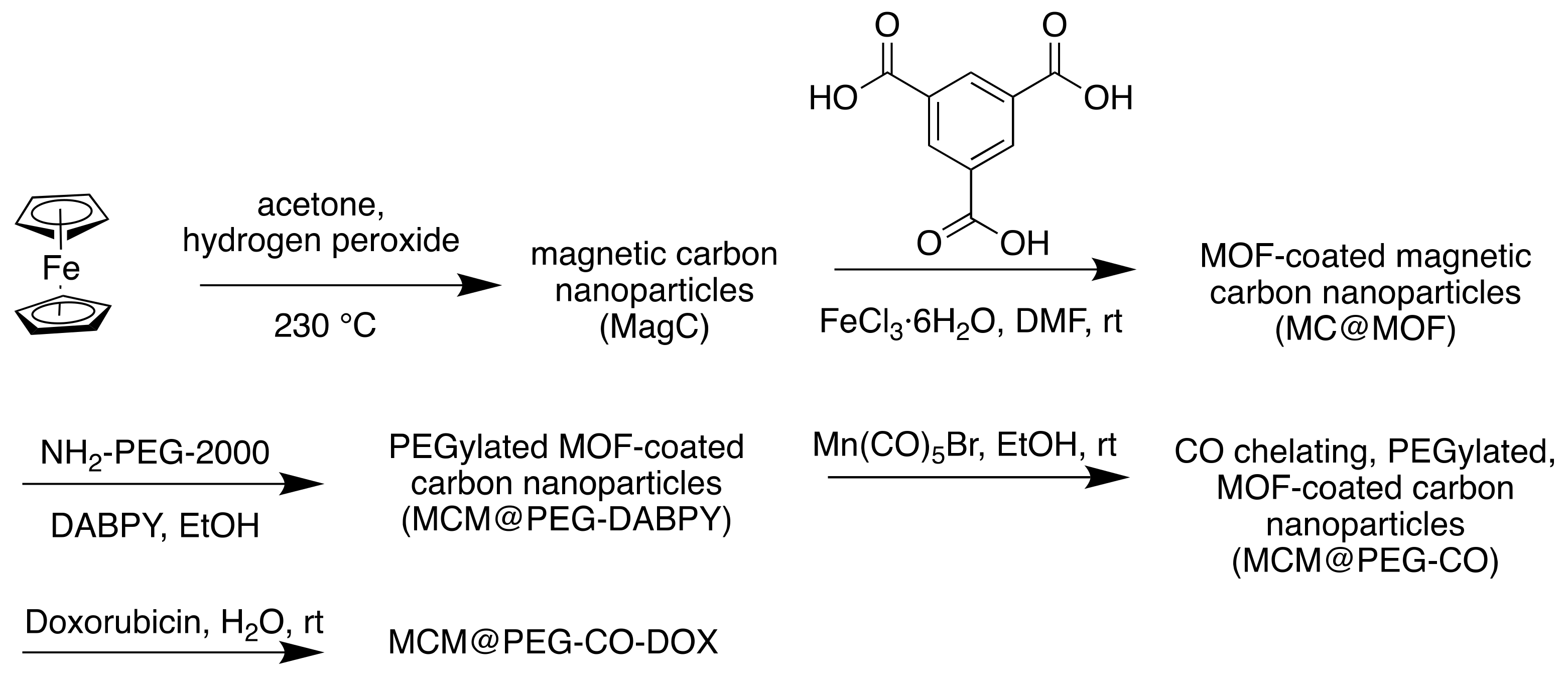
2.1.4. MOFs as Cancer Treatments: Carbon Monoxide Therapy and Magnetic Nuclear Resonance Imaging Through MOF Modification
2.2. MOFs as Detoxifying Agents: Adsorption and Removal of Drugs
2.3. MOFs as Catalysts for the Degradation of Pharmaceutical Contaminants in Wastewater
3. Bioinspired MOFs
3.1. APIs as Linkers
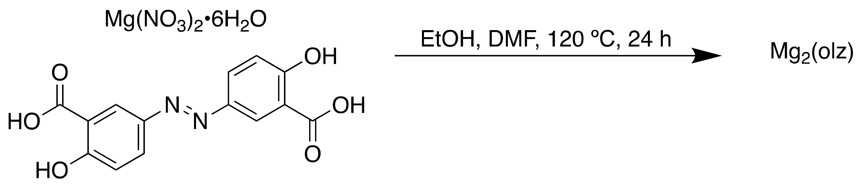
3.1.1. APIs as Linkers: Olsalazine and M2(olz)
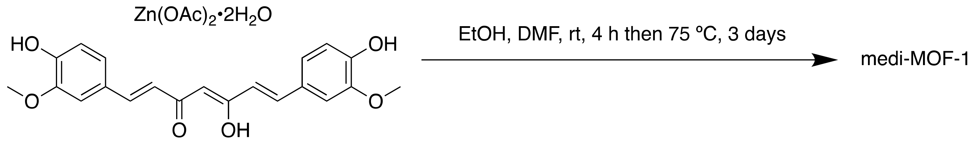
3.1.2. APIs as Linkers: Curcumin and Medi-MOF-1
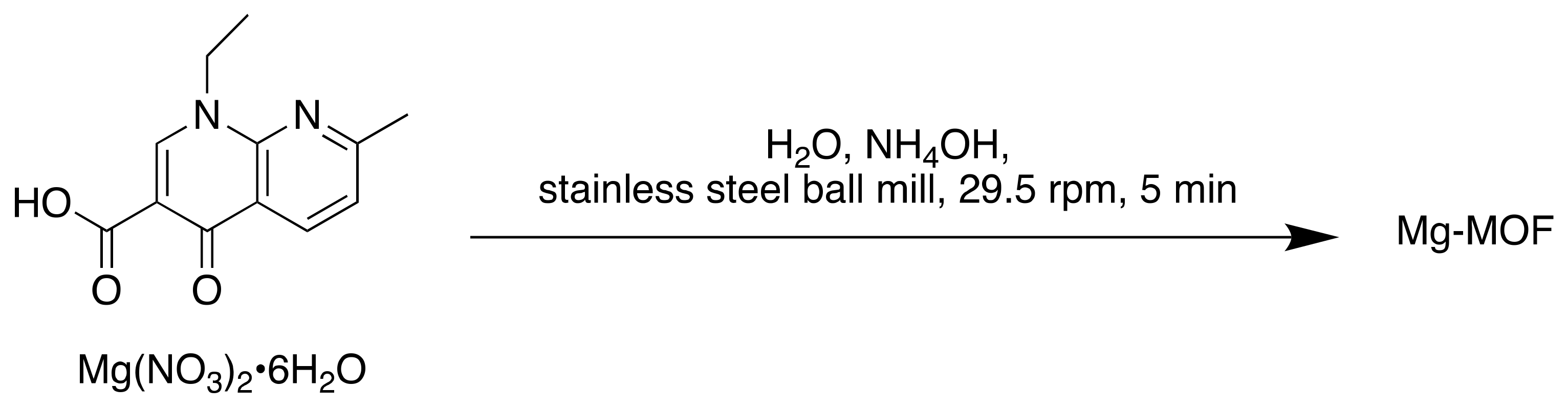
3.1.3. APIs as Linkers: Nalidixic Acid and Mg-MOF
3.2. Amino Acids and Endogenous Small Molecules as Linkers
3.3. The Krebs Cycle
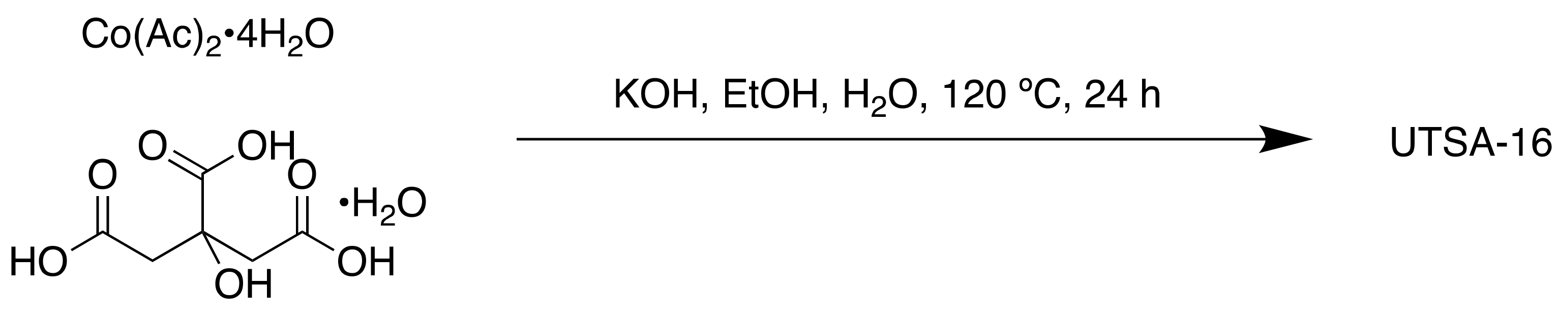
3.3.1. Citric Acid
3.3.2. Lactic Acid
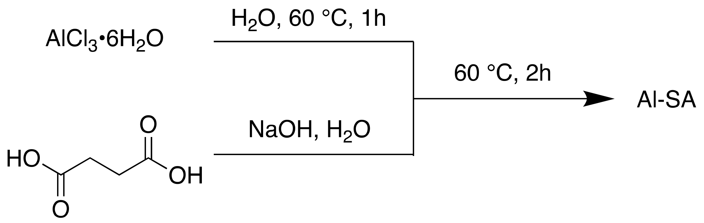
3.3.3. Succinic Acid
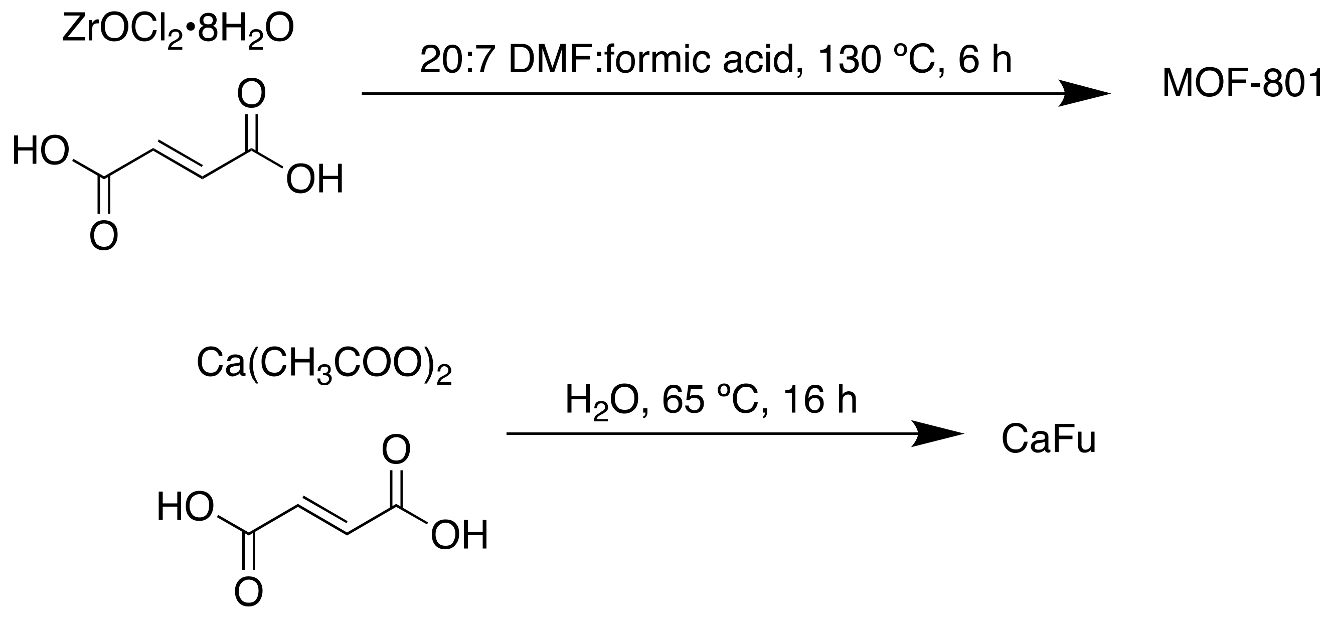
3.3.4. Fumaric Acid
3.3.5. L-Malic Acid
3.4. Peptides as Linkers: Biorelated Structures
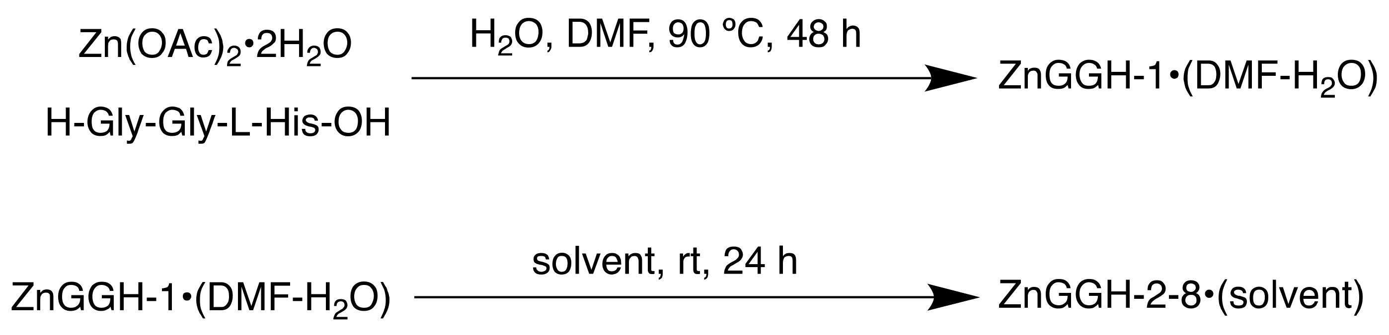
3.4.1. A Conformationally Active MOF Made of and Mimicking Peptides
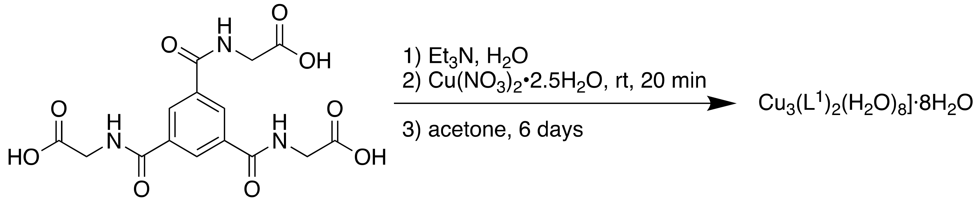
3.4.2. Pseudopeptides as Ligands, for Use in Catalysis
4. Conclusions
Author Contributions
Funding
Conflicts of Interest
References
- García, H.; Navalón, S. Metal-Organic Frameworks: Applications in Separations and Catalysis; Garcia, H., Navalón, S., Eds.; Wiley-VCH: Weinheim, Germany, 2018. [Google Scholar]
- Hoskins, B.F.; Robson, R. Infinite Polymeric Frameworks Consisting of Three Dimensionally Linked Rod-Like Segments. J. Am. Chem. Soc. 1989, 111, 5962–5964. [Google Scholar] [CrossRef]
- Yaghi, O.M.; Li, H. Hydrothermal Synthesis of a Metal-Organic Framework Containing Large Rectangular Channels. J. Am. Chem. Soc. 1995, 117, 10401–10402. [Google Scholar] [CrossRef]
- Chui, S.S.-Y.; Lo, S.M.-F.; Charmant, J.P.H.; Orpen, A.G.; Williams, I.D. A chemically functionalizable nanoporous material. Science 1999, 283, 1148–1150. [Google Scholar] [CrossRef]
- Li, H.; Eddaoudi, M.; O’Keeffe, M.; Yaghi, O.M. Design and synthesis of an exceptionally stable and highly porous metal-organic framework. Nature 1999, 402, 276–279. [Google Scholar] [CrossRef]
- McGuire, C.V.; Forgan, R.S. The surface chemistry of metal–organic frameworks. Chem. Commun. 2015, 51, 5199–5217. [Google Scholar] [CrossRef]
- Furukawa, H.; Ko, N.; Go, Y.B.; Aratani, N.; Choi, S.B.; Choi, E.; Yazaydin, A.Ö.; Snurr, R.Q.; O’Keeffe, M.; Kim, J.; et al. Ultrahigh porosity in metal-organic frameworks. Science 2010, 329, 424–428. [Google Scholar] [CrossRef]
- Klimakow, M.; Klobes, P.; Thünemann, A.F.; Rademann, K.; Emmerling, F. Mechanochemical Synthesis of Metal−Organic Frameworks: A Fast and Facile Approach toward Quantitative Yields and High Specific Surface Areas. Chem. Mater. 2010, 22, 5216–5221. [Google Scholar] [CrossRef]
- Klinowski, J.; Paz, F.A.A.; Silva, P.; Rocha, J. Microwave-Assisted Synthesis of Metal–Organic Frameworks. Dalton Trans. 2011, 40, 321–330. [Google Scholar] [CrossRef]
- Batten, M.P.; Rubio-Martinez, M.; Hadley, T.; Carey, K.-C.; Lim, K.-S.; Polyzos, A.; Hill, M.R. Continuous flow production of metal-organic frameworks. Curr. Opin. Chem. Eng. 2015, 8, 55–59. [Google Scholar] [CrossRef]
- Tansell, A.J.; Jones, C.L.; Easun, T.L. MOF the beaten track: Unusual structures and uncommon applications of metal-organic frameworks. Chem. Cent. J. 2017, 11, 100. [Google Scholar] [CrossRef]
- Sholl, D.S.; Lively, R.P. Defects in Metal-Organic Frameworks: Challenge or Opportunity? J. Phys. Chem. Lett. 2015, 6, 3437–3444. [Google Scholar] [CrossRef]
- Dhakshinamoorthy, A.; Asiri, A.M.; Garcia, H. Tuneable nature of metal organic frameworks as heterogeneous solid catalysts for alcohol oxidation. Chem. Commun. 2017, 53, 10851–10869. [Google Scholar] [CrossRef]
- Sajid, M. Toxicity of nanoscale metal organic frameworks: A perspective. Environ. Sci. Pollut. R. 2016, 23, 14805–14807. [Google Scholar] [CrossRef]
- Furukawa, H.; Cordova, K.E.; O’Keeffe, M.; Yaghi, O.M. The chemistry and applications of metal-organic frameworks. Science 2013, 341, 1230444. [Google Scholar] [CrossRef]
- Horcajada, P.; Serre, C.; Vallet-Regí, M.; Sebban, M.; Taulelle, F.; Férey, G. Metal-organic frameworks as efficient materials for drug delivery. Angew. Chem. Int. Ed. 2006, 45, 5974–5978. [Google Scholar] [CrossRef]
- Wang, H.; Lashkari, E.; Lim, H.; Zheng, C.; Emge, T.J.; Gong, Q.; Yam, K.; Li, J. The moisture-triggered controlled release of a natural food preservative from a microporous metal-organic framework. Chem. Commun. 2016, 52, 2129–2132. [Google Scholar] [CrossRef]
- MOFgen: Biomedical Innovation. Available online: http://www.mofgen.com/, (accessed on 17 January 2019).
- Hinks, N.J.; McKinlay, A.C.; Xiao, B.; Wheatley, P.S.; Morris, R.E. Metal organic frameworks as NO delivery materials for biological applications. Micropor. Mesopor. Mater. 2010, 129, 330–334. [Google Scholar] [CrossRef]
- U.S. National Library of Medicine, Clinical Trials. Phase I Study of RiMO-301 with Radiation in Advanced Tumours. Primary ID RiMO-CL17-001. ClinicalTrials.gov Identifier: NCT03444714. Available online: www.clinicaltrials.gov/ct2/show/NCT03444714, (accessed on 18 March 2019).
- Rojas, S.; Devic, T.; Horcajada, P. Metal organic frameworks based on bioactive components. J. Mater. Chem. B 2017, 5, 2560–2573. [Google Scholar] [CrossRef]
- Yang, J.; Yang, Y.-W. Metal–Organic Frameworks for Biomedical Applications. Small 2020. [Google Scholar] [CrossRef]
- Chedid, G.; Yassin, A. Recent Trends in Covalent and Metal Organic Frameworks for Biomedical Applications. Nanomaterials 2018, 8, 916. [Google Scholar] [CrossRef]
- Zhan, X.; Chen, Z.; Zhang, Q. Recent progress in two-dimensional COFs for energy-related applications. J. Mater. Chem. A 2017, 5, 14463–14479. [Google Scholar] [CrossRef]
- Sharma, R.K.; Yadav, P.; Yadav, M.; Gupta, R.; Rana, P.; Srivastava, A.; Zbořil, R.; Varma, R.S.; Antonietti, M.; Gawande, M.B. Recent development of covalent organic frameworks (COFs): Synthesis and catalytic (organic-electro-photo) applications. Mater. Horiz. 2020, 7, 411–454. [Google Scholar] [CrossRef]
- Zhao, F.; Liu, H.; Mathe, S.D.R.; Dong, A.; Zhang, J. Covalent Organic Frameworks: From Materials Design to Biomedical Application. Nanomaterials 2017, 8, 15. [Google Scholar] [CrossRef] [PubMed]
- Latroche, M.; Surblé, S.; Serre, C.; Mellot-Draznieks, C.; Llewellyn, P.L.; Lee, J.-H.; Chang, J.-S.; Jhung, S.H.; Férey, G. Hydrogen storage in the giant-pore metal-organic frameworks MIL-100 and MIL-101. Angew. Chem. Int. Ed. 2006, 45, 8227–8231. [Google Scholar] [CrossRef] [PubMed]
- Wuttke, S.; Braig, S.; Preiß, T.; Zimpel, A.; Sicklinger, J.; Bellomo, C.; Rädler, J.O.; Vollmar, A.M.; Bein, T. MOF nanoparticles coated by lipid bilayers and their uptake by cancer cells. Chem. Commun. 2015, 51, 15752–15755. [Google Scholar] [CrossRef] [PubMed]
- Márquez, A.G.; Hidalgo, T.; Lana, H.; Cunha, D.; Blanco-Prieto, M.J.; Álvarez-Lorenzo, C.; Boissière, C.; Sánchez, C.; Serre, C.; Horcajada, P. Biocompatible polymer–metal–organic framework composite patches for cutaneous administration of cosmetic molecules. J. Mater. Chem. B. 2016, 4, 7031–7040. [Google Scholar] [CrossRef]
- Zhong, G.; Liu, D.; Zhang, J. Applications of Porous Metal–Organic Framework MIL-100(M) (M = Cr, Fe, Sc, Al, V). Cryst. Growth Des. 2018, 18, 7730–7744. [Google Scholar] [CrossRef]
- Chen, Y.; Li, P.; Modica, J.A.; Drout, R.J.; Farha, O.K. Acid-Resistant Mesoporous Metal-Organic Framework toward Oral Insulin Delivery: Protein Encapsulation, Protection, and Release. J. Am. Chem. Soc. 2018, 140, 5678–5681. [Google Scholar] [CrossRef]
- Xu, J.; Wu, L.; Guo, T.; Zhang, G.; Wang, C.; Li, H.; Li, X.; Singh, V.; Chen, W.; Gref, R.; et al. A “Ship-in-a-Bottle” strategy to create folic acid nanoclusters inside the nanocages of γ-cyclodextrin metal-organic frameworks. Int. J. Pharm. 2019, 556, 89–96. [Google Scholar] [CrossRef]
- Rojas, S.; Colinet, I.; Cunha, D.; Hidalgo, T.; Salles, F.; Serre, C.; Guillou, N.; Horcajada, P. Toward Understanding Drug Incorporation and Delivery from Biocompatible Metal-Organic Frameworks in View of Cutaneous Administration. ACS Omega 2018, 3, 2994–3003. [Google Scholar] [CrossRef]
- Tamames-Tabar, C.; Imbuluzqueta, E.; Guillou, N.; Serre, C.; Miller, S.R.; Elkaïm, E.; Horcajada, P.; Blanco-Prieto, M.J. A Zn azelate MOF: Combining antibacterial effect. CrystEngComm 2015, 17, 456–462. [Google Scholar] [CrossRef]
- Cunha, D.; Yahia, M.B.; Hall, S.; Miller, S.R.; Chevreau, H.; Elkaïm, E.; Maurin, G.; Horcajada, P.; Serre, C. Rationale of Drug Encapsulation and Release from Biocompatible Porous Metal–Organic Frameworks. Chem. Mater. 2013, 25, 2767–2776. [Google Scholar] [CrossRef]
- Gaudin, C.; Cunha, D.; Ivanoff, E.; Horcajada, P.; Chevé, G.; Yasri, A.; Loget, O.; Serre, C.; Maurin, G. A quantitative structure activity relationship approach to probe the influence of the functionalization on the drug encapsulation of porous metal-organic frameworks. Micropor. Mesopor. Mater. 2012, 157, 124–130. [Google Scholar] [CrossRef]
- Cunha, D.; Gaudin, C.; Colinet, I.; Horcajada, P.; Maurin, G.; Serre, C. Rationalization of the entrapping of bioactive molecules into a series of functionalized porous zirconium terephthalate MOFs. J. Mater. Chem. B 2013, 1, 1101–1108. [Google Scholar] [CrossRef]
- Smaldone, R.A.; Forgan, R.S.; Furukawa, H.; Gassensmith, J.J.; Slawin, A.M.Z.; Yaghi, O.M.; Stoddart, J.F. Metal-organic frameworks from edible natural products. Angew. Chem. Int. Ed. 2010, 49, 8630–8634. [Google Scholar] [CrossRef]
- Hartlieb, K.J.; Holcroft, J.M.; Moghadam, P.Z.; Vermeulen, N.A.; Algaradah, M.M.; Nassar, M.S.; Botros, Y.Y.; Snurr, R.Q.; Stoddart, J.F. CD-MOF: A Versatile Separation Medium. J. Am. Chem. Soc. 2016, 138, 2292–2301. [Google Scholar] [CrossRef]
- Hartlieb, K.J.; Ferris, D.P.; Holcroft, J.M.; Kandela, I.; Stern, C.L.; Nassar, M.S.; Botros, Y.Y.; Stoddart, J.F. Encapsulation of Ibuprofen in CD-MOF and Related Bioavailability Studies. Mol. Pharma. 2017, 14, 1831–1839. [Google Scholar] [CrossRef]
- Tiwari, G.; Tiwari, R.; Rai, A.K. Cyclodextrins in delivery systems: Applications. J. Pharm. Bioall. Sci. 2010, 2, 72–79. [Google Scholar] [CrossRef]
- del Valle, E.M.M. Cyclodextrins and their uses: A review. Process Biochem. 2004, 39, 1033–1046. [Google Scholar] [CrossRef]
- Connell, P.P.; Hellman, S. Advances in radiotherapy and implications for the next century: A historical perspective. Cancer Res. 2009, 69, 383–392. [Google Scholar] [CrossRef]
- Gianfaldoni, S.; Gianfaldoni, R.; Wollina, U.; Lotti, J.; Tchernev, G.; Lotti, T. An Overview on Radiotherapy: From Its History to Its Current Applications in Dermatology. Open Access Maced. J. Med. Sci. 2017, 5, 521–525. [Google Scholar] [CrossRef]
- Liu, Y.; Zhang, P.; Li, F.; Jin, X.; Li, J.; Chen, W.; Li, Q. Metal-based NanoEnhancers for Future Radiotherapy: Radiosensitizing and Synergistic Effects on Tumor Cells. Theranostics 2018, 8, 1824–1849. [Google Scholar] [CrossRef]
- Grzmil, M.; Meisel, A.; Behé, M.; Schibli, R. Radiopharmaceutical Chemistry; Lewis, J.S., Windhorst, A.D., Zeglis, B.M., Eds.; Springer: Berlin/Heidelberg, Germany, 2019. [Google Scholar]
- Pottier, A.; Borghi, E.; Levy, L. New Use of Metals as Nanosized Radioenhancers. Anticancer Res. 2014, 34, 443–453. [Google Scholar]
- Ni, K.; Lan, G.; Chan, C.; Quigley, B.; Lu, K.; Aung, T.; Guo, N.; la Riviere, P.; Weichselbaum, R.R.; Lin, W. Nanoscale metal-organic frameworks enhance radiotherapy to potentiate checkpoint blockade immunotherapy. Nat. Comms. 2018, 9, 2351. [Google Scholar] [CrossRef]
- Notman, N. Core Concept: Holey synthetic materials open their pores to medical applications. Proc. Natl. Acad. Sci. USA 2019, 116, 6513–6516. [Google Scholar] [CrossRef]
- Penney, D.G. Carbon Monoxide Toxicity; Penney, D.G., Ed.; CRC Press: Boca Raton, FL, USA, 2000. [Google Scholar]
- Wegiel, B.; Gallo, D.; Csizmadia, E.; Harris, C.; Belcher, J.; Vercellotti, G.M.; Penacho, N.; Seth, P.; Sukhatme, V.; Ahmed, A.; et al. Carbon monoxide expedites metabolic exhaustion to inhibit tumor growth. Cancer Res. 2013, 73, 7009–7021. [Google Scholar] [CrossRef]
- He, Q.; Kiesewetter, D.O.; Qu, Y.; Fu, X.; Fan, J.; Huang, P.; Liu, Y.; Zhu, G.; Liu, Y.; Qian, Z.; et al. NIR-Responsive On-Demand Release of CO from Metal Carbonyl-Caged Graphene Oxide Nanomedicine. Adv. Mater. 2015, 27, 6741–6746. [Google Scholar] [CrossRef]
- Li, W.-P.; Su, C.-H.; Tsao, L.-C.; Chang, C.-T.; Hsu, Y.-P.; Yeh, C.-S. Controllable CO Release Following Near-Infrared Light-Induced Cleavage of Iron Carbonyl Derivatized Prussian Blue Nanoparticles for CO-Assisted Synergistic Treatment. ACS Nano 2016, 10, 11027–11036. [Google Scholar] [CrossRef]
- Yao, J.; Liu, Y.; Wang, J.; Jiang, Q.; She, D.; Guo, H.; Sun, N.; Pang, Z.; Deng, C.; Yang, W.; et al. On-demand CO release for amplification of chemotherapy by MOF functionalized magnetic carbon nanoparticles with NIR irradiation. Biomaterials 2019, 195, 51–62. [Google Scholar] [CrossRef]
- Horcajada, P.; Surblé, S.; Serre, C.; Hong, D.-Y.; Seo, Y.-K.; Chang, J.-S.; Grenèche, J.-M.; Margiolaki, I.; Férey, G. Synthesis and catalytic properties of MIL-100(Fe), an iron(III) carboxylate with large pores. Chem. Commun. 2007, 27, 2820–2822. [Google Scholar] [CrossRef]
- Rojas, S.; Baati, T.; Njim, L.; Manchego, L.; Neffati, F.; Abdeljelil, N.; Saguem, S.; Serre, C.; Najjar, M.F.; Zakhama, A.; et al. Metal-Organic Frameworks as Efficient Oral Detoxifying Agents. J. Am. Chem. Soc. 2018, 140, 9581–9586. [Google Scholar] [CrossRef] [PubMed]
- Vale, J.A.; Kulig, K. American Academy of Clinical Toxicology and European Association of Poisons Centres and Clinical Toxicologists. Position paper: Gastric lavage. J. Toxicol. Clin. Toxicol. 2004, 42, 933–943. [Google Scholar]
- Chyka, P.; Seger, D.; Krenzelok, E.; Vale, J. American Academy of Clinical Toxicology and European Association of Poisons Centres and Clinical Toxicologists. Position paper: Single-dose activated charcoal. Clin. Toxicol. 2005, 43, 61–87. [Google Scholar]
- Betten, D.P.; Vohra, R.B.; Cook, M.D.; Matteucci, M.J.; Clark, R.F. Antidote use in the critically ill poisoned patient. J. Intensive Care Med. 2006, 21, 255–277. [Google Scholar] [CrossRef]
- Graham, L.M.; Nguyen, T.M.; Lee, S.B. Nanodetoxification: Emerging role of nanomaterials in drug intoxication treatment. Nanomedicine 2011, 6, 921–928. [Google Scholar] [CrossRef]
- Liu, Y.; Eubank, J.F.; Cairns, A.J.; Eckert, J.; Kravtsov, V.C.; Luebke, R.; Eddaoudi, M. Assembly of metal-organic frameworks (MOFs) based on indium-trimer building blocks: A porous MOF with soc topology and high hydrogen storage. Angew. Chem. Int. Ed. 2007, 46, 3278–3283. [Google Scholar] [CrossRef]
- Eubank, J.F.; Wheatley, P.S.; Lebars, G.; McKinlay, A.C.; Leclerc, H.; Horcajada, P.; Daturi, M.; Vimont, A.; Morris, R.E.; Serre, C. Porous, rigid-metal(III)-carboxylate metal-organic frameworks for the delivery of nitric oxide. APL Mater. 2014, 2, 124112. [Google Scholar] [CrossRef]
- Gummin, D.D.; Mowry, J.B.; Spyker, D.A.; Brooks, D.E.; Osterthaler, K.M.; Banner, W. 2017 Annual Report of the American Association of Poison Control Centers’ National Poison Data System (NPDS): 35th Annual Report. Clin. Toxicol. 2018, 56, 1213–1415. [Google Scholar] [CrossRef]
- Chevreau, H.; Permyakova, A.; Nouar, F.; Fabry, P.; Livage, C.; Ragon, F.; Garcia-Marquez, A.; Devic, T.; Steunou, N.; Serre, C.; et al. Synthesis of the biocompatible and highly stable MIL-127(Fe): From large scale synthesis to particle size control. CrystEngComm 2016, 18, 4094–4101. [Google Scholar] [CrossRef]
- Decker, W.; Corby, D.; Ibanez, J. Aspirin adsorption with activated charcoal. Lancet 1968, 291, 754–755. [Google Scholar] [CrossRef]
- Bu, Q.; Wang, B.; Huang, J.; Deng, S.; Yu, G. Pharmaceuticals and personal care products in the aquatic environment in China: A review. J. Haz. Mater. 2013, 262, 189–211. [Google Scholar] [CrossRef] [PubMed]
- Wang, W.; Kannan, K. Fate of Parabens and Their Metabolites in Two Wastewater Treatment Plants in New York State, United States. Environ. Sci. Technol. 2016, 50, 1174–1181. [Google Scholar] [CrossRef] [PubMed]
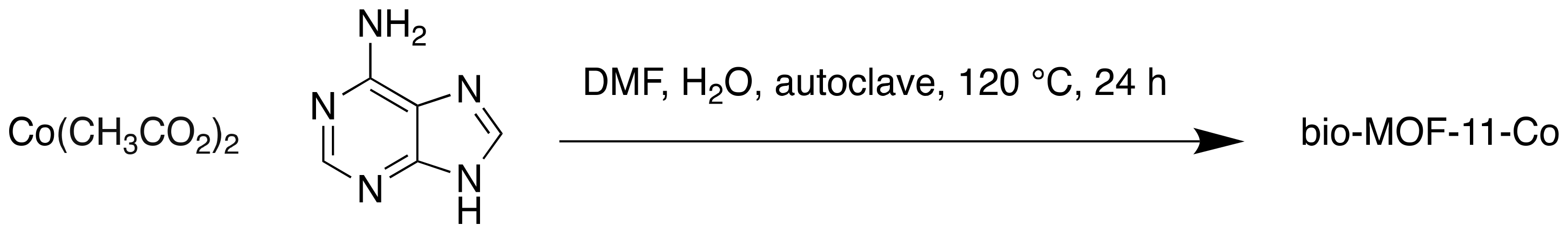
- Azhar, M.R.; Vijay, P.; Tadé, M.O.; Sun, H.; Wang, S. Submicron sized water-stable metal organic framework (bio-MOF-11) for catalytic degradation of pharmaceuticals and personal care products. Chemosphere 2018, 196, 105–114. [Google Scholar] [CrossRef] [PubMed]
- An, J.; Geib, S.J.; Rosi, N.L. High and selective CO2 uptake in a cobalt adeninate metal-organic framework exhibiting pyrimidine- and amino-decorated pores. J. Am. Chem. Soc. 2010, 132, 38–39. [Google Scholar] [CrossRef]
- Yamada, K. Interrelations between Essential Metal Ions and Human Diseases; Sigel, A., Sigel, H., Sigel, R.K.O., Eds.; Springer: Berlin/Heidelberg, Germany, 2013. [Google Scholar]
- 7The National Institute for Occupational Safety and Health (NIOSH), CAS No. 7440-48-4. Available online: https://www.cdc.gov/niosh/npg/npgd0146.html, (accessed on 16 October 2019).
- Kryszak, D.; Stawicka, K.; Trejda, M.; Calvino-Casilda, V.; Martin-Aranda, R.; Ziolek, M. Development of basicity in mesoporous silicas and metallosilicates. Catal. Sci. Technol. 2017, 7, 5236–5248. [Google Scholar] [CrossRef]
- Corma, A. From Microporous to Mesoporous Molecular Sieve Materials and Their Use in Catalysis. Chem. Rev. 1997, 97, 2373–2420. [Google Scholar] [CrossRef]
- Miller, S.R.; Heurtaux, D.; Baati, T.; Horcajada, P.; Grenèche, J.-M.; Serre, C. Biodegradable therapeutic MOFs for the delivery of bioactive molecules. Chem. Commun. 2010, 46, 4526–4528. [Google Scholar] [CrossRef]
- Burrows, A.D.; Jurcic, M.; Keenan, L.L.; Lane, R.A.; Mahon, M.F.; Warren, M.R.; Nowell, H.; Paradowski, M.; Spencer, J. Incorporation by coordination and release of the iron chelator drug deferiprone from zinc-based metal–organic frameworks. Chem. Commun. 2013, 49, 11260–11262. [Google Scholar] [CrossRef]
- Burrows, A.D.; Jurcic, M.; Mahon, M.F.; Pierrat, S.; Roffe, G.W.; Windle, H.J.; Spencer, J. Bismuth coordination networks containing deferiprone: Synthesis, characterisation, stability and antibacterial activity. Dalton Trans. 2015, 44, 13814–13817. [Google Scholar] [CrossRef]
- Selby, W.S.; Barr, G.D.; Ireland, A.; Mason, C.H.; Jewell, D.P. Olsalazine in active ulcerative colitis. Brit. Med. J. 1985, 291, 1373–1375. [Google Scholar] [CrossRef]
- Brown, W.A.; Farmer, K.C.; Skinner, S.A.; Malcontenti-Wilson, C.; Misajon, A.; O’Brien, P.E. 5-aminosalicyclic acid and olsalazine inhibit tumor growth in a rodent model of colorectal cancer. Dig. Dis. Sci. 2000, 45, 1578–1584. [Google Scholar] [CrossRef]
- Xiao, D.-R.; Sun, D.-Z.; Liu, J.-L.; Zhang, G.-J.; Chen, H.-Y.; He, J.-H.; Yan, S.-W.; Yuan, R.; Wang, E.-B. Two Unprecedented Entangled Metal–Olsalazine Complexes with Coexistence of 2D → 3D Polycatenation and meso-Helix. Eur. J. Inorg. Chem. 2011, 2011, 4656–4663. [Google Scholar] [CrossRef]
- Chen, H.-Y.; Xiao, D.-R.; Yan, S.-W.; He, J.-H.; Yang, J.; Wang, X.; Yuan, R.; Wang, E.-B. Two three-dimensional pillared metal–olsalazine complexes based on infinite rod-shaped secondary building units. Inorg. Chim. Acta 2012, 387, 283–288. [Google Scholar] [CrossRef]
- Levine, D.J.; Runčevski, T.; Kapelewski, M.T.; Keitz, B.K.; Oktawiec, J.; Reed, D.A.; Mason, J.A.; Jiang, H.Z.H.; Colwell, K.A.; Legendre, C.M.; et al. Olsalazine-Based Metal-Organic Frameworks as Biocompatible Platforms for H2 Adsorption and Drug Delivery. J. Am. Chem. Soc. 2016, 138, 10143–10150. [Google Scholar] [CrossRef]
- Su, H.; Sun, F.; Jia, J.; He, H.; Wang, A.; Zhu, G. A highly porous medical metal–organic framework constructed from bioactive curcumin. Chem. Commun. 2015, 51, 5774–5777. [Google Scholar] [CrossRef]
- Maheshwari, R.K.; Singh, A.K.; Gaddipati, J.; Srimal, R.C. Multiple biological activities of curcumin: A short review. Life Sci. 2006, 78, 2081–2087. [Google Scholar] [CrossRef]
- Lao, C.D.; Ruffin, M.T.; Normolle, D.; Heath, D.D.; Murray, S.I.; Bailey, J.M.; Boggs, M.E.; Crowell, J.; Rock, C.L.; Brenner, D.E. Dose escalation of a curcuminoid formulation. BMC Complement. Altern. Med. 2006, 6, 10. [Google Scholar] [CrossRef]
- Bowen, P.K.; Guillory, R.J.; Shearier, E.R.; Seitz, J.-M.; Drelich, J.; Bocks, M.; Zhao, F.; Goldman, J. Metallic zinc exibits optimal biocompatibility for bioabsorbable endovascular stents. Mater. Sci. Eng. C 2015, 56, 467–472. [Google Scholar] [CrossRef]
- Chen, Y.; Huang, P.; Chen, H.; Wang, S.; Wang, H.; Guo, J.; Zhang, X.; Zhang, S.; Yan, J.; Xia, J.; et al. Assessment of the Biocompatibility and Biological Effects of Biodegradable Pure Zinc Material in the Colorectum. ACS Biomater. Sci. Eng. 2018, 4, 4095–4103. [Google Scholar] [CrossRef]
- Kunnumakkara, A.B.; Guha, S.; Krishnan, S.; Diagaradjane, P.; Gelovani, J.; Aggarwal, B.B. Curcumin potentiates antitumor activity of gemcitabine in an orthotopic model of pancreatic cancer through suppression of proliferation, angiogenesis, and inhibition of nuclear factor-kappaB-regulated gene products. Cancer Res. 2007, 67, 3853–3861. [Google Scholar] [CrossRef]
- Dandawate, P.R.; Vyas, A.; Ahmad, A.; Banerjee, S.; Deshpande, J.; Swamy, K.V.; Jamadar, A.; Dumhe-Klaire, A.C.; Padhye, S.; Sarkar, F.H. Inclusion complex of novel curcumin analogue CDF and β-cyclodextrin (1:2) and its enhanced in vivo anticancer activity against pancreatic cancer. Pharm. Res. 2012, 29, 1775–1786. [Google Scholar] [CrossRef] [PubMed]
- André, V.; da Silva, A.R.F.; Fernandes, A.; Frade, R.; Garcia, C.; Rijo, P.; Antunes, A.M.M.; Rocha, J.; Duarte, M.T. Mg- and Mn-MOFs Boost the Antibiotic Activity of Nalidixic Acid. ACS Appl. Bio Mater. 2019, 2, 2347–2354. [Google Scholar] [CrossRef]
- Barry, A.L.; Jones, R.N.; Thornsberry, C.; Ayers, L.W.; Gerlach, E.H.; Sommers, H.M. Antibacterial activities of ciprofloxacin, norfloxacin, oxolinic acid, cinoxacin, and nalidixic acid. Antimicrob. Agents Chemother. 1984, 25, 633–637. [Google Scholar] [CrossRef] [PubMed]
- Mallick, S.; Sahu, A.; Pal, K. Dissolution behaviour of nalidixic acid solid dispersions using water soluble dispersion carriers. Acta Pol. Pharm. 2004, 61, 21–30. [Google Scholar]
- Psomas, G.; Kessissoglou, D.P. Quinolones and non-steroidal anti-inflammatory drugs interacting with copper(ii), nickel(ii), cobalt(ii) and zinc(ii): Structural features, biological evaluation and perspectives. Dalton Trans. 2013, 42, 6252–6276. [Google Scholar] [CrossRef]
- Seetharaj, R.; Vandana, P.V.; Arya, P.; Mathew, S. Dependence of solvents, pH, molar ratio and temperature in tuning metal organic framework architecture. Arab. J. Chem. 2019, 12, 295–315. [Google Scholar] [CrossRef]
- Sigma Aldrich: Dorset, UK, 28.06.2019, N,N-Dimethylformamide; SDS No. D4551 [Online]. Available online: https://www.sigmaaldrich.com/MSDS/MSDS/DisplayMSDSPage.do?country=GB&language=en&productNumber=D4551&brand=SIGMA&PageToGoToURL=https%3A%2F%2Fwww.sigmaaldrich.com%2Fcatalog%2Fproduct%2Fsigma%2Fd4551%3Flang%3Den (accessed on 8 January 2019).
- Lovering, F.; Bikker, J.; Humblet, C. Escape from Flatland: Increasing Saturation as an Approach to Improving Clinical Success. J. Med. Chem. 2009, 52, 6752–6756. [Google Scholar] [CrossRef]
- Zhang, J.-H.; Nong, R.-Y.; Xie, S.-M.; Wang, B.-J.; Ai, P.; Yuan, L.-M. Homochiral metal-organic frameworks based on amino acid ligands for HPLC separation of enantiomers. Electrophoresis 2017, 38, 2513–2520. [Google Scholar] [CrossRef]
- Mailloux, R.J.; Bériault, R.; Lemire, J.; Singh, R.; Chénier, D.R.; Hamel, R.D.; Appanna, V.D. The tricarboxylic acid cycle, an ancient metabolic network with a novel twist. PLoS ONE 2007, 2, e690. [Google Scholar] [CrossRef]
- Čelič, T.B.; Jagličič, Z.; Lazar, K.; Logar, N.Z. Structure and magnetic properties of a new iron(II) citrate coordination polymer. Acta Cryst. B 2013, 69, 490–495. [Google Scholar] [CrossRef]
- Abbaspour, N.; Hurrell, R.; Kelishadi, R. Review on iron and its importance for human health. J. Res. Med. Sci. 2014, 19, 164–174. [Google Scholar] [PubMed]
- Jiang, J.; Huang, L.; Liu, X.; Ai, L. Bioinspired Cobalt–Citrate Metal–Organic Framework as an Efficient Electrocatalyst for Water Oxidation. ACS Appl. Mater. Interfaces 2017, 9, 7193–7201. [Google Scholar] [CrossRef] [PubMed]
- Suen, N.-T.; Hung, S.-F.; Quan, Q.; Zhang, N.; Xu, Y.-J.; Chen, H.M. Electrocatalysis for the oxygen evolution reaction: Recent development and future perspectives. Chem. Soc. Rev. 2017, 46, 337–365. [Google Scholar] [CrossRef] [PubMed]
- Farid, S.; Ren, S.; Hao, C. MOF-derived metal/carbon materials as oxygen evolution reaction catalysts. Inorg. Chem. Commun. 2018, 94, 57–74. [Google Scholar] [CrossRef]
- Evangelisti, F.; Güttinger, R.; Moré, R.; Luber, S.; Patzke, G.R. 3d–4f {CoII3Ln(OR)4} Cubanes as Bio-Inspired Water Oxidation Catalysts. J. Am. Chem. Soc. 2013, 135, 18734–18737. [Google Scholar] [CrossRef]
- Hui, S.; Ghergurovich, J.M.; Morscher, R.J.; Jang, C.; Teng, X.; Lu, W.; Esparza, L.A.; Reya, T.; Zhan, L.E.; Guo, J.Y.; et al. Glucose feeds the TCA cycle via circulating lactate. Nature 2017, 551, 115–118. [Google Scholar] [CrossRef]
- Yang, J.; Trickett, C.A.; Alahmadi, S.B.; Alshammari, A.S.; Yaghi, O.M. Calcium l-Lactate Frameworks as Naturally Degradable Carriers for Pesticides. J. Am. Chem. Soc. 2017, 139, 8118–8121. [Google Scholar] [CrossRef]
- Ajwa, H.A.; Trout, T.; Mueller, J.; Wilhelm, S.; Nelson, S.D.; Soppe, R.; Shatley, D. Application of alternative fumigants through drip irrigation systems. Phytopathology 2002, 92, 1349–1355. [Google Scholar] [CrossRef]
- Livage, C.; Egger, C.; Férey, G. Hydrothermal versus Nonhydrothermal Synthesis for the Preparation of Organic−Inorganic Solids: The Example of Cobalt(II) Succinate. Chem. Mater. 2001, 13, 410–414. [Google Scholar] [CrossRef]
- Zhang, B.; Chen, L.; Yang, X.; Xu, T.; Sun, T.; Wang, L.; Zhang, Q. A luminescent Terbium-Succinate MOF fabricated by co-precipitation for sensing of Fe3+ in aqueous environment. J. Mater. Sci. Mater. Electron. 2017, 28, 7326–7332. [Google Scholar] [CrossRef]
- Jung, K.-W.; Choi, B.H.; Lee, S.Y.; Ahn, K.-H.; Lee, Y.J. Green synthesis of aluminum-based metal organic framework for the removal of azo dye Acid Black 1 from aqueous media. J. Ind. Eng. Chem. 2018, 67, 316–325. [Google Scholar] [CrossRef]
- Yaseen, D.A.; Scholz, M. Textile dye wastewater characteristics and constituents of synthetic effluents: A critical review. Int. J. Environ. Sci. Technol. 2019, 16, 1193–1226. [Google Scholar] [CrossRef]
- Puvaneswari, N.; Muthukrishnan, J.; Gunasekaran, P. Toxicity assessment and microbial degradation of azo dyes. Indian, J. Exp. Biol. 2006, 44, 618–626. [Google Scholar]
- Bu, R.; Chen, F.; Li, J.; Li, W.; Yang, F. Adsorption capability for anionic dyes on 2-hydroxyethylammonium acetate-intercalated layered double hydroxide. Colloids Surf. A 2016, 511, 312–319. [Google Scholar] [CrossRef]
- Blanco, S.P.D.M.; Scheufele, F.B.; Módenes, A.N.; Espinoza-Quiñones, F.R.; Marin, P.; Kroumov, A.D.; Borba, C.E. Kinetic, equilibrium and thermodynamic phenomenological modeling of reactive dye adsorption onto polymeric adsorbent. Chem. Eng. J. 2017, 307, 466–475. [Google Scholar] [CrossRef]
- Peng, X.; Hu, X.; Fu, D.; Lam, F.L.Y. Adsorption removal of acid black 1 from aqueous solution using ordered mesoporous carbon. Appl. Surf. Sci. 2014, 294, 71–80. [Google Scholar] [CrossRef]
- Jung, K.-W.; Choi, B.H.; Dao, C.M.; Lee, Y.J.; Choi, J.-W.; Ahn, K.-H.; Lee, S.-H. Aluminum carboxylate-based metal organic frameworks for effective adsorption of anionic azo dyes from aqueous media. J. Ind. Eng. Chem. 2018, 59, 149–159. [Google Scholar] [CrossRef]
- Sepulveda, L.A.; Santana, C.C. Effect of solution temperature, pH and ionic strengthon dye adsorption onto Magellanic peat. Environ. Technol. 2013, 34, 967–977. [Google Scholar] [CrossRef]
- He, C.; Hu, X. Anionic Dye Adsorption on Chemically Modified Ordered Mesoporous Carbons. Ind. Eng. Chem. Res. 2011, 50, 14070–14083. [Google Scholar] [CrossRef]
- Hsu, T.-C. Adsorption of an acid dye onto coal fly ash. Fuel 2008, 87, 3040–3045. [Google Scholar] [CrossRef]
- Samarghandi, M.R.; Zarrabi, M.; Amrane, A.; Soori, M.M.; Sepehr, M.N. Removal of Acid Black Dye by Pumice Stone as a low cost adsorbent; kinetic, thermodynamic and equilibrium studies. Environ. Eng. Manag. J. 2012, 12, 2137–2147. [Google Scholar]
- EU Approved Additives and E Numbers. Available online: https://www.food.gov.uk/business-guidance/eu-approved-additives-and-e-numbers, (accessed on 8 January 2019).
- Ke, F.; Peng, C.; Zhang, T.; Zhang, M.; Zhou, C.; Cai, H.; Zhu, J.; Wan, X. Fumarate-based metal-organic frameworks as a new platform for highly selective removal of fluoride from brick tea. Sci. Rep. 2018, 8, 939. [Google Scholar] [CrossRef] [PubMed]
- Cao, J.; Zhao, Y.; Liu, J. Brick tea consumption as the cause of dental fluorosis among children from Mongol, Kazak and Yugu populations in China. Food Chem. Toxicol. 1997, 35, 827–833. [Google Scholar] [CrossRef]
- Zhang, R.; Cheng, L.; Zhang, T.; Xu, T.; Li, M.; Yin, W.; Jiang, Q.; Yang, Y.; Hu, T. Brick tea consumption is a risk factor for dental caries and dental fluorosis among 12-year-old Tibetan children in Ganzi. Environ. Geochem. Health 2018, 41, 1405–1417. [Google Scholar] [CrossRef]
- Zhang, X.; Gao, H.; Yang, T.; Wu, H.; Wang, Y.; Wan, X. Al3+-promoted fluoride accumulation in tea plants (Camellia sinensis) was inhibited by an anion channel inhibitor DIDS. J. Sci. Food Agric. 2016, 96, 4224–4230. [Google Scholar] [CrossRef]
- Waugh, T.D.; Potter, W.; Limeback, H.; Godfrey, M. Risk Assessment of Fluoride Intake from Tea in the Republic of Ireland and its Implications for Public Health and Water Fluoridation. Int. J. Environ. Res. Public Health 2016, 13, 259. [Google Scholar] [CrossRef]
- Zingiryan, A.; Zhang, J.; Bu, X. Cooperative Self-Assembly of Chiral L-Malate and Achiral Succinate in the Formation of Three-Dimensional Homochiral Framework. Inorg. Chem. 2008, 47, 8607–8609. [Google Scholar] [CrossRef]
- Nagaraja, C.M.; Haldar, R.; Maji, T.K.; Rao, C.N.R. Chiral Porous Metal–Organic Frameworks of Co(II) and Ni(II): Synthesis, Structure, Magnetic Properties, and CO2 Uptake. Cryst. Growth Des. 2012, 12, 975–981. [Google Scholar] [CrossRef]
- Raja, D.S.; Luo, J.-H.; Chang, T.-G.; Lo, S.-H.; Wu, C.-Y.; Lin, C.-H. Synthesis, Crystal Structure, and Luminescence Properties of a New Calcium(II) Coordination Polymer Based on L-Malic Acid. J. Chem. 2013, 2013, 7. [Google Scholar]
- Yutkin, P.; Zavakhina, M.S.; Samsonenko, D.G.; Dybtsev, D.N.; Fedin, V.P. Synthesis and characterization of expected and unexpected topologies of homochiral porous metal(II) malate frameworks. Inorganica Chim. Acta 2013, 394, 367–372. [Google Scholar] [CrossRef]
- Zavakhina, M.S.; Samsonenko, D.G.; Virovets, A.V.; Dybtsev, D.N.; Fedin, V.P. Homochiral Cu(II) and Ni(II) malates with tunable structural features. J. Solid State Chem. 2014, 210, 125–129. [Google Scholar] [CrossRef]
- Xie, S.; Hu, C.; Li, L.; Zhang, J.; Fu, N.; Wang, B.; Yuan, L. Homochiral metal-organic framework for HPLC separation of enantiomers. Microchem. J. 2018, 139, 487–491. [Google Scholar] [CrossRef]
- Nair, L.P.; Bijini, B.R.; Prasanna, S.; Eapen, S.M.; Nair, C.M.K.; Deepa, M.; RajendraBabu, K. Growth and characterisation of a new polymorph of barium maleate: A metal organic framework. Spectrochim. Acta A 2015, 137, 778–784. [Google Scholar] [CrossRef] [PubMed]
- Purves, D.; Augustine, G.J.; Fitzpatrick, D.; Katz, L.C.; LaMantia, A.-S.; McNamara, J.O.; Williams, S.M. Neuroscience, 2nd ed.; Purves, D., Augustine, G.J., Fitzpatrick, D., Katz, L.C., LaMantia, A.-S., McNamara, J.O., Williams, S.M., Eds.; Sinauer Associates: Sunderland, MA, USA, 2001. [Google Scholar]
- Katsoulidis, A.P.; Antypov, D.; Whitehead, G.F.S.; Carrington, E.J.; Adams, D.J.; Berry, N.G.; Darling, G.R.; Dyer, M.S.; Rosseinsky, M.J. Chemical control of structure and guest uptake by a conformationally mobile porous material. Nature 2019, 565, 213–217. [Google Scholar] [CrossRef]
- Loukopoulos, E.; Michail, A.; Kostakis, E.G. A 12-fold ths interpenetrated network utilizing a glycine-based pseudopeptidic ligand. Crystals 2018, 8, 47. [Google Scholar] [CrossRef]
- Lauder, K.; Toscani, A.; Scalacci, N.; Castagnolo, D. Synthesis and Reactivity of Propargylamines in Organic Chemistry. Chem. Rev. 2017, 117, 14091–14200. [Google Scholar] [CrossRef]
- Ricco, R.; Pfeiffer, C.; Sumida, K.; Sumby, C.J.; Falcaro, P.; Furukawa, S.; Champness, N.R.; Doonan, C.J. Emerging applications of metal–organic frameworks. CrystEngComm 2016, 18, 6532–6542. [Google Scholar] [CrossRef]
- Lipinski, C.A.; Lombardo, F.; Dominy, B.W.; Feeney, P.J. Experimental and computational approaches to estimate solubility and permeability in drug discovery and development settings. Adv. Drug Deliv. Rev. 1997, 23, 3–25. [Google Scholar] [CrossRef]
- DiMasi, J.A.; Grabowski, H.G.; Hansen, R.W.; Cao, J.; Zhao, Y.; Liu, J. Food Chem. Toxicol. 1997, 35, 827–833. J. Health Econ. 2016, 47, 20–33. [Google Scholar] [CrossRef]
- Gu, S.; Cui, D.; Chen, X.; Xiong, X.; Zhao, Y. PROTACs: An Emerging Targeting Technique for Protein Degradation in Drug Discovery. BioEssays 2018, 40, e1700247. [Google Scholar] [CrossRef]
- Ryther, R.C.C.; Flynt, A.S.; Phillips, J.A.; Patton, J.G. siRNA therapeutics: Big potential from small RNAs. Gene Ther. 2005, 12, 5–11. [Google Scholar] [CrossRef] [PubMed]
- Hu, B.; Weng, Y.; Xia, X.-H.; Liang, X.; Huang, Y. Clinical advances of siRNA therapeutics. J. Gene Med. 2019, 21, e3097. [Google Scholar] [CrossRef] [PubMed]


| MOF | Cage Size (Å) | Window Size (Å) | Hydrophobic or hydrophilic? | Notes |
|---|---|---|---|---|
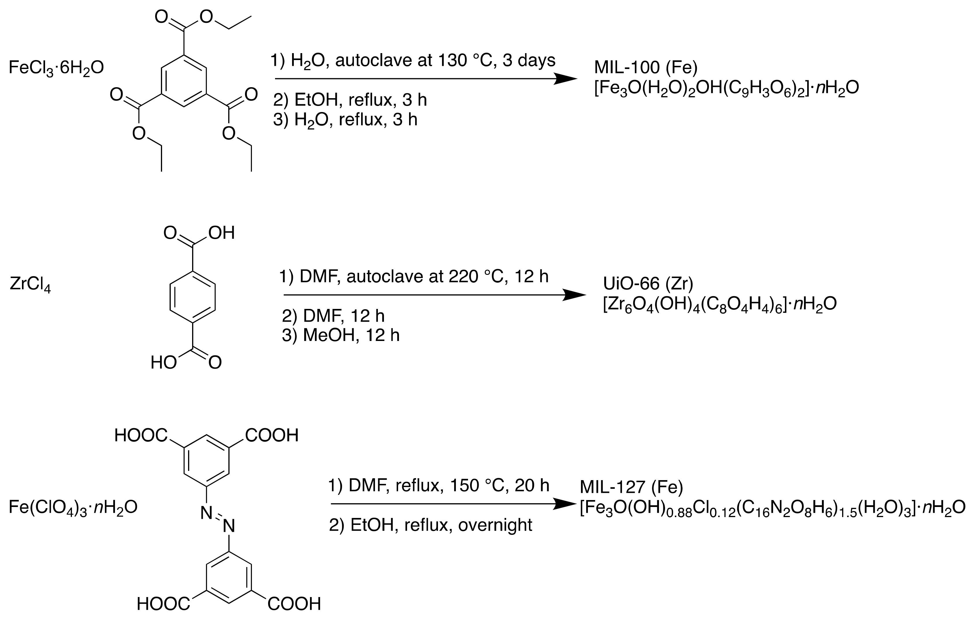
| MIL-100 (Fe) | 25 and 29 | 4.8-5.8 and 8.6 | Hydrophilic | Mesoporous cages accessed through microporous windows |
| MIL-127 (Fe) | 6 (channel system) 10 (cages) | 6 3 | Hydrophobic Hydrophilic | Two pore types; an accessible one-dimensional channel system, and hydrophilic cages accessed through narrow apertures |
| UiO-66 (Zr) | 11 and 8 | 5–7 | Slightly hydrophobic | Octahedral and tetrahedral cavities accessed through triangular micropores |
| M | BET Surface Area (m2 g-1) | Langmuir Surface Area (m2 g-1) |
|---|---|---|
| Mg | 2545 | 4593 |
| Fe | 1485 | 2618 |
| Co | 2060 | 3838 |
| Ni | 2067 | 3813 |
| Zn | 636 | 770 |
| Adsorbent | Temperature | Adsorption Capacity (mg g−1) |
|---|---|---|
| Ammonia-treated CMK-3 | 25 | 769.2 [114] |
| Aluminium (Al2(SO4)3•18H2O)-succinic acid MOFs | 25 | 332.5 [115] |
| Magellanic peat | 10 | 168.9 [116] |
| Ammonia-tailored CMK-3 | 25 | 509.0 [117] |
| Coal fly ash | 60 | 103.1 [118] |
| Pumice stone | 25 | 72.5 [119] |
| Al-SA MOF | 25 | 739.3 [109] |
| Adsorbents | Langmuir | ||
|---|---|---|---|
| Qmax | kL | R2 | |
| Al-SA MOF | 689.38 | 0.0621 | 0.9896 |
| Commercial PAC | 168.42 | 0.9326 | 0.9622 |
| Entry | Aldehyde | Amine | Alkyne | Yield a (%) |
|---|---|---|---|---|
| 1 | Cyclohexane carboxaldehyde | Pyrrolidine | Phenylacetylene | 99 |
| 2 | Cyclohexane carboxaldehyde | Piperidine | Phenylacetylene | 99 |
| 3 | Cyclohexane carboxaldehyde | Diethylamine | Phenylacetylene | 77 |
| 4 | Benzaldehyde | Pyrrolidine | Phenylacetylene | 67 |
| 5 | Cyclohexane carboxaldehyde | Pyrrolidine | 1-hexyne | 95 |
| a NMR yields based on aldehyde | ||||
| Entry | Compound Name | Formula or Formulation | Applications |
|---|---|---|---|
| MOFs with bioapplications | |||
| 1 | MIL-100 (Fe) | [Fe3O(H2O)2OH(C9H3O6)2]·nH2O | Drug delivery (aspirin and ibuprofen) [33] |
| 2 | MIL-127 (Fe) | [Fe3O(OH)0.88Cl0.12(C16N2O8H6)1.5(H2O)3]·nH2O | |
| 3 | UiO-66 (Zr) | [Zr6O4(OH)4(C8O4H4)6]·nH2O | |
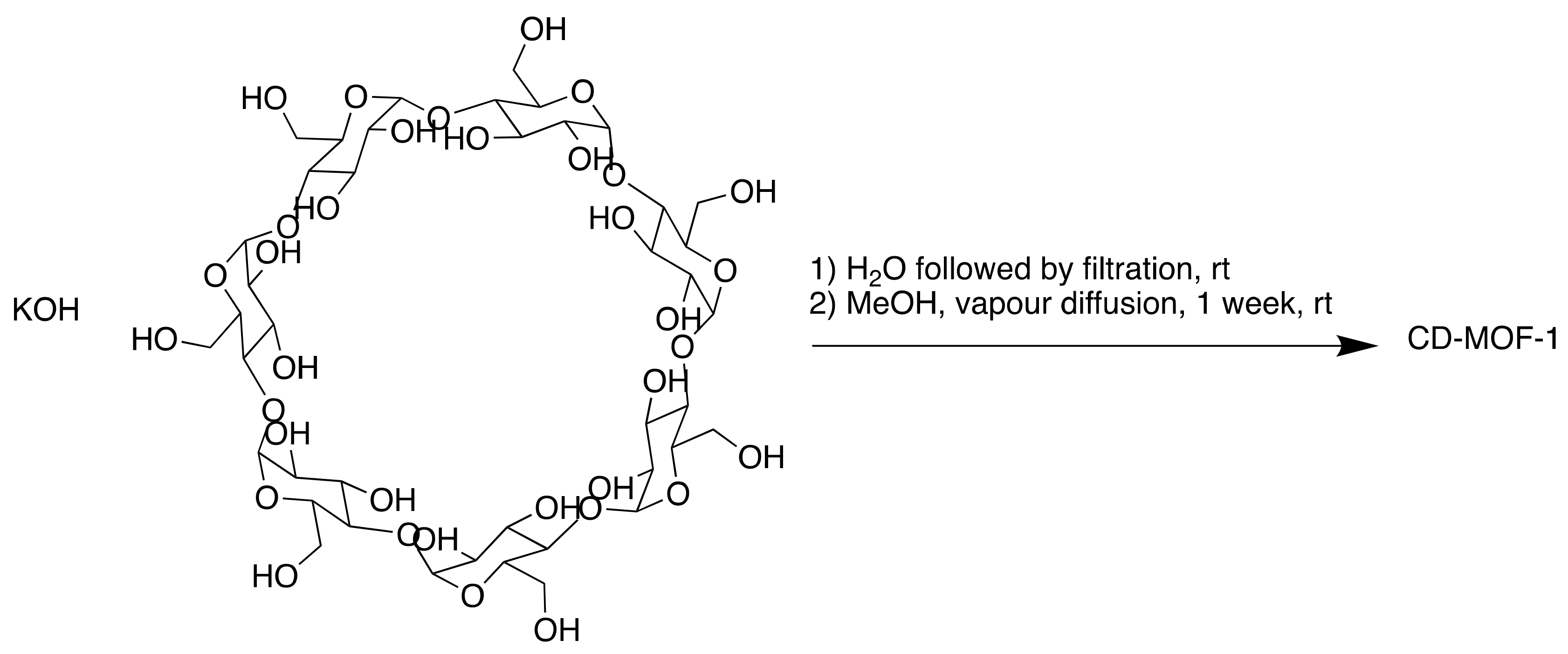
| 4 | CD-MOF-1 | [(C48H80O40)(KOH)2]n | Drug delivery (ibuprofen) [40] |
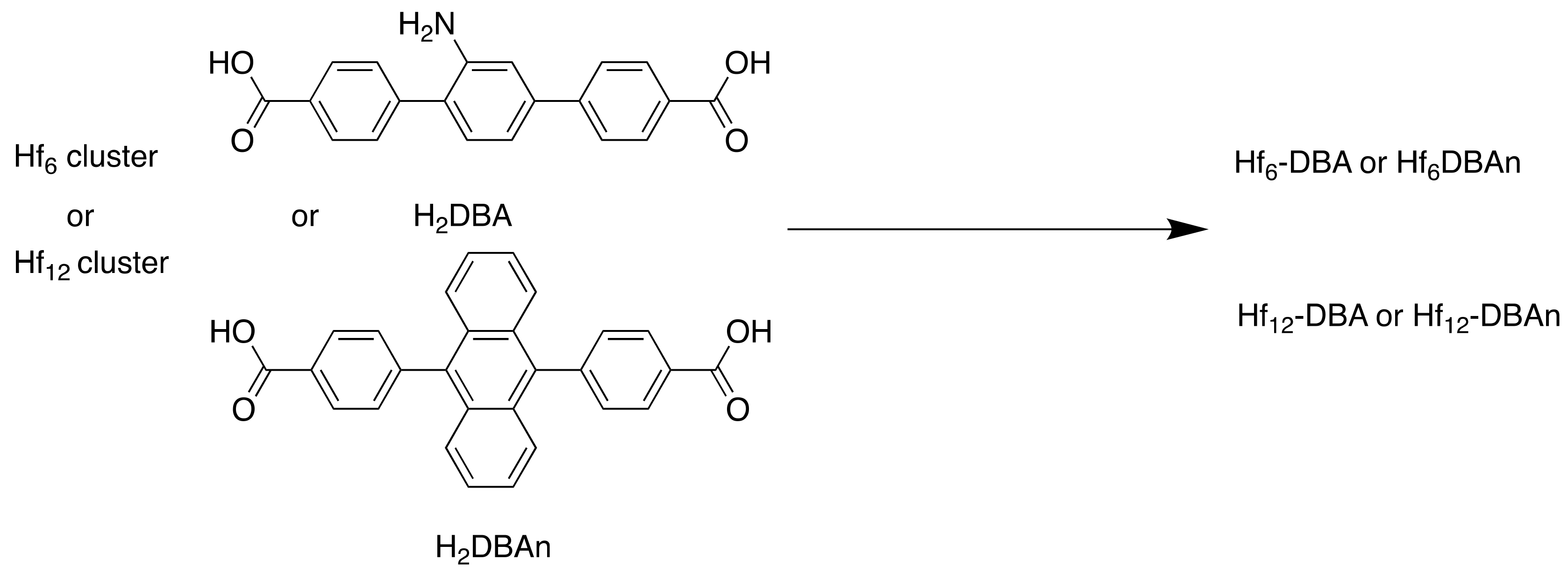
| 5 | Hf6-DBA | Hf6(μ3-O)4(DBA)6 | Radioenhancers [48] |
| 6 | Hf12-DBA | Hf12(μ3-O)8(μ3-OH)8(μ2-O)6(DBA)9 | |
| 7 | MIL-100 (Fe) | MCM@PEG-CO-DOX | CO therapy, drug delivery (Doxorubicin) [54] |
| 8 | MIL-127 (Fe) | Fe3(OH)0.66Cl0.33(C16N2O8H6)1.5(H2O)3·nH2O | Detoxifying agent (aspirin) [56] |
| 9 | bio-MOF-11-Co | Co2(adenine)2(CO2CH3)2·2DMF·0.5H2O | Paraben degradation catalyst [68] |
| Bioinspired MOFs | |||
| 10 | Mg2(olz) | Mg2(C14H10N2O6) | Drug delivery/co-delivery (PEA) [81] |
| 11 | medi-MOF-1 | [Zn3(C21H20O6)2·7(DMA)·3(EtOH)] | Drug delivery/co-delivery (ibuprofen) [82] |
| 12 | Mg-nalidixic acid | C12H17N3O9Mg | Antibacterial agents [89] |
| 13 | UTSA-16 | [KCo3(C6H4O7)(C6H5O7)(H2O)2]·8H2O | Oxygen evolution reaction (OER) electrocatalyst [100] |
| 14 | MOF-1201 | Ca14(L-lactate)20(acetate)8(C2H5OH)(H2O) | Fumigant sorption formulation [105] |
| 15 | MOF-1203 | Ca6(L-lactate)3(acetate)9(H2O) | |
| 16 | Al-SA | - | Azo dye adsorption material [109] |
| 17 | MOF-801 | Zr6O4(OH)4(fumarate)6 | Fluoride adsorption material to treat brick tea [121] |
| 18 | CaFu | - | |
| 19 | BM | C24H14O24Ba5⋅7H2O | Gas storage and separation, optoelectronics [132] |
| 20 | ZnGGH-1-9 | Zn(glycine–glycine–L-histidine)-n•(solvent) | Guest molecule sorption [134] |
| 21 | ThSi2 CP | [Cu3(L1)2(H2O)8]·8H2O | A3 coupling catalyst [135] |
© 2020 by the authors. Licensee MDPI, Basel, Switzerland. This article is an open access article distributed under the terms and conditions of the Creative Commons Attribution (CC BY) license (http://creativecommons.org/licenses/by/4.0/).
Share and Cite
Tibbetts, I.; Kostakis, G.E. Recent Bio-Advances in Metal-Organic Frameworks. Molecules 2020, 25, 1291. https://doi.org/10.3390/molecules25061291
Tibbetts I, Kostakis GE. Recent Bio-Advances in Metal-Organic Frameworks. Molecules. 2020; 25(6):1291. https://doi.org/10.3390/molecules25061291
Chicago/Turabian StyleTibbetts, Isobel, and George E. Kostakis. 2020. "Recent Bio-Advances in Metal-Organic Frameworks" Molecules 25, no. 6: 1291. https://doi.org/10.3390/molecules25061291
APA StyleTibbetts, I., & Kostakis, G. E. (2020). Recent Bio-Advances in Metal-Organic Frameworks. Molecules, 25(6), 1291. https://doi.org/10.3390/molecules25061291