Article

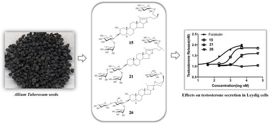
Steroidal Glycosides from Allium tuberosum Seeds and Their Roles in Promoting Testosterone Production of Rat Leydig Cells

1
Key Laboratory of Coal Processing and Efficient Utilization, Ministry of Education, China University of Mining & Technology, Xuzhou 221116, Jiangsu, China
2
Jiangsu Hanbon Science & Technology Co., Ltd., Huaian 223005, Jiangsu, China
3
State Key Laboratory of High-efficiency Coal Utilization and Green Chemical Engineering, Ningxia University, Yinchuan 750021, Ningxia, China
*
Author to whom correspondence should be addressed.
Molecules 2020, 25(22), 5464; https://doi.org/10.3390/molecules25225464
Submission received: 22 October 2020 / Revised: 13 November 2020 / Accepted: 14 November 2020 / Published: 22 November 2020
(This article belongs to the Section Natural Products Chemistry)

Abstract

:
A systematic phytochemical study on the components in the seeds of Allium tuberosum was performed, leading to the isolation of 27 steroidal glycosides (SGs 127). The structures of SGs were identified mainly by nuclear magnetic resonance and mass spectrometries as well as the necessary chemical evidence. In the SGs, 110 and 2226 are new steroidal saponin analogues. An in vitro bioassay indicates that 1, 2, 7, 8, 10, 1315, 20, 23, and 26 display promotional roles in testosterone production of rat Leydig cells with the EC50 values of 1.0 to 4.5 μM, respectively.

Graphical Abstract

1. Introduction

Steroidal saponins (SSs) are the important class of secondary metabolites in many medicinal plants. Structurally, SSs are mostly in the form of glycosides which are composed of one or more hydrophilic sugar residues and hydrophobic steroidal part [1]. All the time, they have been a kind of constituents concerned by scholars owing to the wide range of their biological actions. For example, SSs from Dioscorea zingiberensis are widely used for preventing cardiovascular diseases [2], SSs of Ophiopogon japonicus displayed multiple biofunctions of anticancer, immunomodulation, anti-oxidation, anti-inflammation, and anti-diabetes [3]; SSs in Paris species are used to treat cancer and bleeding [4], and timosaponin AIII obtained from Anemarrhena asphodeloides exhibits inhibitory activity against tumor cells [5].
SSs have been reported in more than 40 different Allium species [6]. Allium tuberosum is a type of Allium plant widely cultivated as food in China, and the mature seeds of this plant are used as a traditional herb medicine treating both impotence and nocturnal emissions [7]. The seeds of this plant are famous for their sulfur-containing biologically active natural products [8,9,10], and they also contain amounts of SSs as the main constituents [11,12,13,14,15,16,17]. Previous studies reported many SSs from A. tuberosum seeds (ATSs), while a systematic phytochemical investigation for clarifying the bioactive SSs is still necessary. Recently, a systematic phytochemical study on the components in ATSs finally let us to obtain total 27 SSs (127) (Figure 1). According to the traditional pharmacological action of ATSs, all isolated compounds were tested for their effects on testosterone production of rat Leydig cells.

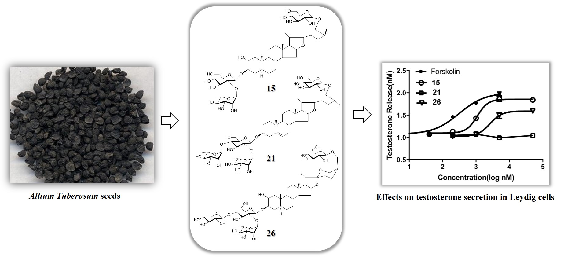
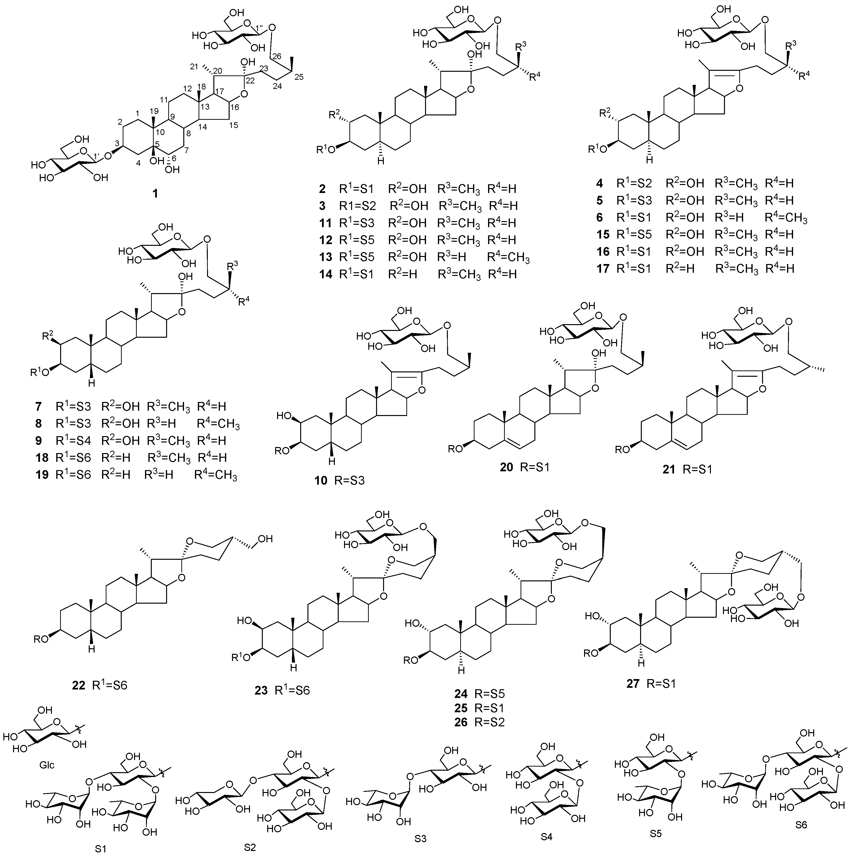
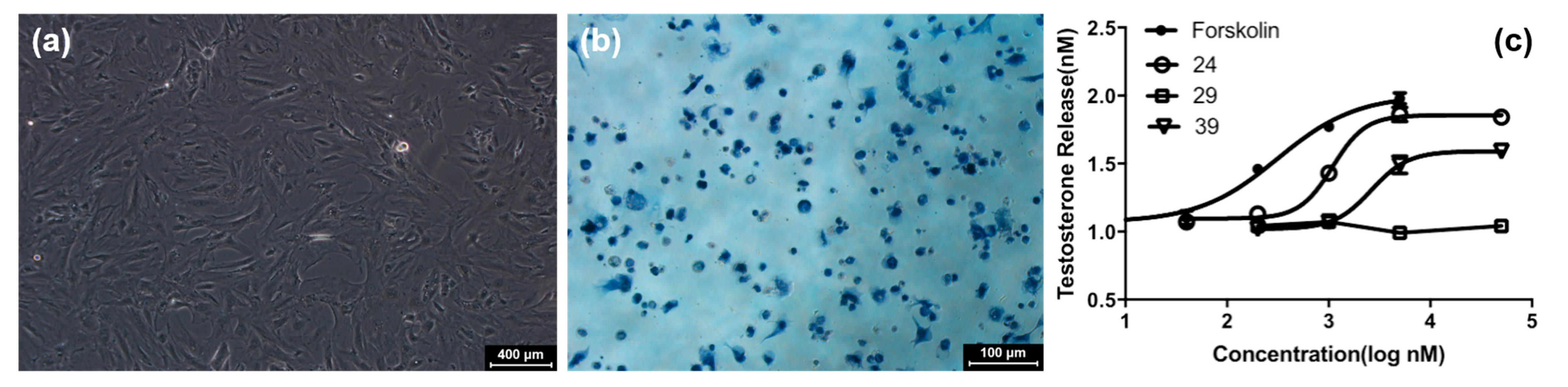
2. Results and Discussion

By comparing the nuclear magnetic resonance (NMR) data with the reported values, the known compounds 1118, 20, 21, and 27 are identified as trigofoenoside B (11) [18], trigoneoside Xa (12) [19], trigoneoside Xb (13) [19], nicotianoside F (14) [20], tuberoside A (15) [11], tuberoside B (16) [11], 26-O-β-d-glucopyranosyl-(25S)-5α-furostan-△20(22)-ene-3β,26-glycol-3-O-α-l-rhamnopyranosyl-(1→4)-[α-l-rhamnopyranosyl(1→2)]-β-d-glucopyranoside (17) [21], shatavarin I (18) [22], protoneodioscin (20) [23], pseudoprotodioscin (21) [24], tuberoside L (27) [13], respectively. The structure of 19 has been already registered in CAS with the numbers of 1493828-40-2 and also reported as 3-O-α-l-rhamnopyranosyl(1→4)-[β-d-glucopyranosyl(1→2)]-β-d-glucopyranosyl-26-O-β-d-glucopyranosyl-(25R)-5β-furostane-3β,22α,26-triol in the literature [25], but no full NMR data are available for them. By systematic spectroscopic data analysis, its NMR data are assigned (see Supplementary Materials). The remaining new analogues are identified mainly by analysis of NMR and MS spectra as well as the necessary chemical evidence such as acid hydrolysis experiments for determining absolute configuration of the sugar units in the structures.
Compound 1 is in the form of white amorphous powders with a molecular formula of C39H66O16 as determined by HR-ESI-MS at m/z 789.4272 [M − H] (calcd for C39H65O16, 789.4273) together with its 13C NMR data (Table 1). In the 1H NMR spectrum, four typical methyl proton signals at δ 0.88 (3H, s), 1.17 (3H, s), 1.33 (3H, d, J = 6.8 Hz), 1.05 (3H, d, J = 7.0 Hz), and two anomeric proton signals at δ 4.83 (1H, d, J = 7.8 Hz) and 5.11 (1H, d, J = 7.8 Hz) are observed. Its 13C NMR spectrum exhibits total 39 carbon resonances including 27 ones due to the aglycone part and twelve ones attributed to the two hexoses. Comparison of NMR data suggests that 1 has the identical planar structure of aglycone skeleton with 26-O-β-d-glucopyranosyl-(25S)-3β,5β,6α,22,26-pentahydroxyl-5β-furostane 3-O-α-l-rhamnopyranosyl-(1→4)-β-d-glucopyranoside [17], which is further confirmed by detailed analyses of 1H-1H COSY, HSQC, and HMBC spectra of 1. The chemical shifts of H-26a (δ 4.09) and H-26b (δ 3.50) (Δab > 0.57) further confirm the C-25S configuration of 1 [26]. Starting from the anomeric protons at δ 4.83 (H-1′) and 5.11 (H-1′′), the proton signals of sugars are delineated relying on the 1H-1H COSY correlations, contributing to establish the structures of the two sugar unites. The large coupling constants of J1′, 2′ (7.8 Hz) and the 13C NMR data facilitate defining the sugars as β-d-glucopyranoses, which is also supported by the result of the acid hydrolysis experiment. The connectivity of the glucopyranose is identified by the HMBC correlations of δ 4.83 (H-1′)/79.2 (C-3), and δ 5.11 (H-1′′)/75.4 (C-26), respectively. Consequently, the structure of 1 is elucidated as 26-O-β-d-glucopyranosyl-(25S)-furost-3β,5β,6α,22α,26-pentanol 3-O-β-d-glucopyranoside, named allituberoside A.
Compound 2 has a molecular formula of C51H86O23 as determined by HR-ESI-MS. Its 1H NMR spectrum obviously shows four typical methyl proton signals at δ 0.87 (3H, s), 0.90 (3H, s), 1.05 (3H, d, J = 7.0 Hz), and 1.31 (3H, d, J = 6.8 Hz), and four anomeric proton signals at δ 4.82 (1H, d, J = 7.9 Hz), 5.03 (1H, d, J = 7.1 Hz), 5.86 (1H, br s), and 6.40 (1H, br s). Its 13C NMR spectrum exhibits total 45 carbon resonances including 27 ones due to the aglycone part and 24 ones attributed to the sugar moieties consisting of four hexoses. The 13C NMR data of 2 suggests that it had the identical aglycone skeleton with 11 and the same sugar moieties with 14, which is supported by its 1H-1H COSY, HSQC, and HMBC spectral data. The chemical shifts of H-26a (δ 4.10) and H-26b (δ 3.49) (Δab > 0.57) further confirm the C-25S configuration of 2. Thus, the structure of 2 is elucidated as 26-O-β-d-glucopyranosyl-(25S)-5α-furost-2α,3β,22α,26-tetrol 3-O-α-l-rhamnopyranosyl -(1→4)-[α-l-rhamnopyranosyl-(1→2)]-β-D-glucopyranoside, named allituberoside B.
Compound 3 displays a molecular formula of C50H84O23 as confirmed by HR-ESI-MS. Its NMR data suggests that 3 has the identical structure with 2 except for the terminal sugar unit at C-4′. The neutral missing fragment of 136 Da presented on the mass spectrum of 3 suggests that 3 has a xylose moiety, and detailed analyses of 1H-1H COSY, HSQC, and HMBC spectra finally identified the structure of the C-3 sugar chain of 3 to be 3-O-β-d-xylopyranosyl-(1→4)-[α-l-rhamnopyranosyl-(1→2)]-β-d-glucopyranoside. The chemical shifts of H-26a (δ 4.11) and H-26b (δ 3.49) (Δab > 0.57) further confirm the C-25S configuration of 3. Therefore, the structure of 3 is elucidated as 26-O-β-d-glucopyranosyl-(25S)-5α-furost-2α,3β,22α,26-tetrol 3-O-β-d-xylopyranosyl-(1→4)-[α-l-rhamnopyranosyl-(1→2)]-β-d-glucopyranoside, named allituberoside C.
Compound 4 has a molecular formula of C50H82O22 as determined by HR-ESI-MS. Its NMR data suggests that the structure of 4 is closely similar to that of 3 except for the different substructure surrounding C-20 and C-22 positions. In its 13C NMR spectrum, the characteristic carbon signals of δ 103.6 and 152.4 which are the same as those of 1517 indicate the existence of the Δ20(22)-ene substructure in 4, which is supported by the HMBC correlations of δ 1.62 (H-21)/103.6 (C-20) and 152.4 (C-22). By detailed analysis of 1H-1H COSY, HSQC, and HMBC spectra, the structure of 4 is further confirmed, and its C-25S configuration is deduced according to the chemical shifts of H-26a (δ 4.10) and H-26b (δ 3.49) (Δab > 0.57). Subsequently, the structure of 4 is elucidated as 26-O-β-d-glucopyranosyl-(25S)-5α-furost-Δ20(22)-ene-2α,3β,26-triol 3-O-β-d-xylopyranosyl-(1→4)-[α-l-rhamnopyranosyl-(1→2)]-β-d-glucopyranoside, named allituberoside D.
Compound 5 has a molecular formula of C45H74O18 as established by HR-ESI-MS. Its NMR data suggests that except for the C-3 sugar chain, 5 and 4 have the same structure. Analyses of the 1H-1H COSY spectrum give the structures of the glucose and rhamnose comprising the C-3 sugar chain, and the HMBC correlations between δ 5.92 (H-1′′ of Rha) and 78.3 (C-4′ of 3-O-Glc) confirm their connectivity. By detailed analyses of 1H-1H COSY, HSQC, and HMBC spectra, the structure of 5 is further confirmed. The C-25S configuration is deduced according to the chemical shifts of H-26a (δ 4.10) and H-26b (δ 3.49) (Δab > 0.57). Thus, the structure of 5 is elucidated as 26-O-β-d-glucopyranosyl-(25S)-5α-furost-Δ20(22)-ene-2α,3β,26-triol 3-O-α-l-rhamnopyranosyl-(1→2)-β-d-glucopyranoside, named allituberoside E.
Compound 6 has a molecular formula of C51H84O22 as identified by HR-ESI-MS. Its NMR data suggest that 6 has the identical aglycone structure with 5 and has the same C-3 sugar chain as 2. According to its 1H-1H COSY, HSQC and HMBC spectra, the structure of 6 is confirmed. The chemical shifts of H-26a (δ 3.96) and H-26b (δ 3.63) (Δab < 0.48) further deduce its C-25R configuration [26]. Therefore, the structure of 6 is elucidated as 26-O-β-d-glucopyranosyl-(25R)-5α-furost-Δ20(22)-ene-2α,3β,26-triol 3-O-α-l-rhamnopyranosyl-(1→4)-[α-l-rhamnopyranosyl-(1→2)]-β-D-glucopyranoside, named allituberoside F.
Compound 7 with a molecular formula of C45H76O19 confirmed by HR-ESI-MS has the same sugar moieties as 5 by detailed comparison of the NMR data. Further comparison of the NMR data of 7 and 18 deduces that they share the same aglycone skeleton. According to its 1H-1H COSY, HSQC, and HMBC spectra, the structure of 7 is confirmed. The chemical shifts of H-26a (δ 4.10) and H-26b (δ 3.49) (Δab > 0.57) further confirm the C-25S configuration of 7. Finally, the structure of 7 is elucidated as 26-O-β-d-glucopyranosyl-(25S)-5β-furost-2β,3β,26-triol 3-O-α-l-rhamnopyranosyl-(1→4)-β-d-glucopyranoside, named allituberoside J.
Compound 8 has the same molecular formula of C45H76O19 as 7 determined by HR-ESI-MS together with its 13C NMR data. The same 13C NMR data suggests that 8 and 7 are C-25R/S isomers of each other. The chemical shifts of H-26a (δ 3.95) and H-26b (δ 3.63) (Δab < 0.48) exhibit that the C-25 configuration is R. Consequently, the structure of 8 is elucidated as 26-O-β-d-glucopyranosyl-(25R)-5β-furost-2β,3β,26-triol 3-O-α-l-rhamnopyranosyl-(1→4)-β-d-glucopyranoside, named allituberoside H.
Compound 9 has a molecular formula of C45H76O20 as confirmed by HR-ESI-MS along with its 13C NMR data (Table 2). The NMR data suggests that 9 has the identical aglycone structure but has different C-3 sugar from 7. Analysis of the 1H-1H COSY spectrum allows to establish the structures of the two glucoses comprising of the C-3 sugar chain, and the HMBC correlation between δ 5.39 (H-1′′) and 83.1 (C-2′) confirm their connectivity. By detailed analyses of 1H-1H COSY, HSQC, and HMBC spectra, the structure of 9 is further confirmed. The chemical shifts of H-26a (δ 4.10) and H-26b (δ 3.50) (Δab > 0.57) exhibit that the C-25S configuration. Consequently, the structure of 9 is elucidated as 26-O-β-d-glucopyranosyl-(25R)-5β-furost-2β,3β,26-triol 3-O-β-d-glucopyranosyl-(1→2)-β-d-glucopyranoside, named allituberoside I.
Compound 10 has a molecular formula of C45H74O18 as assigned by HR-ESI-MS. Its NMR data suggest that it shares the identical structure with 7 except for the differences surrounding the C-20 and C-22 positions. The characteristic carbon signals of δ 103.6 and 152.4 indicate the existence of the Δ20(22)-ene substructures in the molecule. Detailed analysis of 1H-1H COSY, HSQC, and HMBC spectra allows the structure of 10 to be further confirmed. The chemical shifts of H-26a (δ 4.09) and H-26b (δ 3.49) (Δab > 0.57) exhibit that the C-25S configuration of 10. Thus, the structure of 10 is elucidated as 26-O-β-d-glucopyranosyl-(25S)-5β-furost-Δ20(22)-ene-2α, 3β, 26-diol 3-O-α-l-rhamnopyranosyl-(1→4)-β-d-glucopyranoside, named allituberoside J.
Compound 22 is isolated in the form of white powders with a molecular formula of C45H74O18 based on HR-ESI-MS and 13C NMR data. In its 1H NMR spectrum, the anomeric proton signals at δ 4.87 (1H, d, J = 7.2 Hz), 5.93 (1H, br s), and 5.47 (1H, d, J = 7.6 Hz) suggest that 22 has three sugar units. Comparing the NMR data of 22 and 18 reveals that they share the same C-3 sugar chain together with the substructure of A-E rings. Only three typical methyl proton signals at δ 0.85 (3H, s), 0.97 (3H, s), 1.18 (3H, d, J = 6.8 Hz) are observed in the 1H NMR spectrum of 22, and its 13C NMR spectrum shows that the chemical shift of C-27 (δ 64.1) shifted to a lower field, suggesting the linkage of hydroxyl group to the C-27. By further comparing the NMR data, it is deduced that 22 had the same F-ring structure as (25S)-27-hydroxypenogenin-3-O-α-l-rhamnopyranosyl-(1→2)-O-β-d-glucopyranoside [15]. Thus, the structure of 22 is elucidated as (25S)-5β-spirost-3β, 27-diol 3-O-α-l-rhamnopyranosyl-(1→4)-[β-d-glucopyranosyl-(1→2)]-β-d-glucopyranoside, named allituberoside K.
Compound 23 has a molecular formula of C51H84O24 as measured by HR-ESI-MS and 13C NMR data. In the 1H NMR spectrum, the anomeric proton signals at δ 4.94 (2H, d, J = 7.8 Hz), 5.45 (1H, d, J = 7.6 Hz), and 5.90 (1H, br s) suggest that it has four sugar units. According to its NMR data, 23 has the same C-3 sugar chain as 22, and shares the identical A-E rings substructure with 79. The 1H NMR spectrum of 23 shows only three typical methyl proton signals at δ 0.78 (3H, s), 0.98 (3H, s), 1.09 (3H, d, J = 6.8 Hz), suggesting that its C-27 is substituted by an additional glucose unit, and that is also supported by the HMBC correlations of δ 4.37 (H-27a), 3.94 (H-27b)/21.4 (C-24), 33.5 (C-25), 61.0 (C-26), and of δ 4.94 (H-1′′′′)/69.5 (C-27). While the different NMR data of F-ring (C-22~C-27) between 23 and 27 suggest that they might have different C-25 configurations. The key carbon signals of δ 27.1 (C-23), 21.4 (C-24), and 61.0 (C-26) which are in accordance with those of trikamsteroside A [27] suggest that the C-25 configuration of 23 to be R. Thus, the structure of 23 is elucidated as 27-O-β-d-glucopyranoside-(25R)-5β-spirost-2β,26,27-triol 3-O-α-l-rhamnopyranosyl-(1→4)-[β-d-glucopyranosyl-(1→2)]-β-d-glucopyranoside, named allituberoside L.
Compound 24 with a molecular formula of C45H74O19 as deduced by HR-ESI-MS and 13C NMR data has the identical sugar chain at C-3 position and A-E rings substructure with 1113 and shares the same F-ring including the 27-O-β-d-glucopyranose with 23 by comparing their NMR data. A detailed analysis of 1H-1H COSY, HSQC, and HMBC spectra allows the structure of 24 to be further confirmed as 27-O-β-d-glucopyranoside-(25R)-5α-spirost-2α,26,27-triol 3-O-α-l-rhamnopyranosyl-(1→4)-[β-d-glucopyranosyl-(1→2)]-β-d-glucopyranoside, named allituberoside M.
Compound 25 has a molecular formula of C51H84O23 as determined by HR-ESI-MS and 13C NMR data. The NMR data suggests that, except for the different sugar chain at C-3 position, it has the identical structure with 24. Comparison of NMR data further reveals that it has the same C-3 sugar chain as 27. Detailed analysis of 1H-1H COSY, HSQC, and HMBC spectra finally confirms the structure of 25 to be 27-O-β-d-glucopyranoside-(25R)-5α-spirost-2α,26,27-triol 3-O-α-l-rhamnopyranosyl-(1→4)-[α-l-rhamnopyranosyl-(1→2)]-β-d-glucopyranoside, named allituberoside N.
Compound 26 has a molecular formula of C50H82O23 as revealed according to HR-ESI-MS. Its NMR data suggests that 26 shares the identical structure with 24 except for the difference in structure of sugar chain at C-3. Further comparison of the NMR data reveals that the C-3 sugar chain of 26 is same as that of 3. According to its 1H-1H COSY, HSQC, and HMBC spectra data, the whole structure of 26 is finally confirmed. Therefore, the structure of 26 is confirmed to be 27-O-β-d-glucopyranoside-(25R)-5α-spirost-2α,26,27-triol 3-O-β-d-xylopyranosyl-(1→4)-[β-d-glucopyranosyl-(1→2)]-β-d-glucopyranoside, named allituberoside O.
An in vitro bioassay is carried out on compounds 127 for evaluating their promotional roles in testosterone production of rat Leydig cells. The results of 3β-HSD staining show that Leydig cells are successfully isolated from testes (Figure 2), with an approximate purity of ca. 90%. After treatment with these compounds at 50 μM, the cell survival rates are higher than 80%, suggesting that no compound exhibits the noticeable cytotoxic effect on the rat Leydig cells.
Exposure to forskolin results in a significant increase in levels of testosterone production in Leydig cells. Similarly, 1, 2, 7, 8, 10, 1315, 20, 23, and 26 display a good role in increasing testosterone secretion with the EC50 values of 1.0 to 4.5 μM, respectively, and other compounds exhibit no significant activities (Each EC50 > 50 μM) (Table 3 and Figure 2).

3. Materials and Methods

3.1. Experimental Procedures for Phytochmistry Study

3.1.1. General Experimental Procedures

Optical rotations were recorded on a Rudolph Autopol® IV polarimeter. HR-ESI-MS was recorded on a Synapt MS (Waters Corporation, Milford, MA, USA). The NMR experiments were performed on Varian UNITY INOVA 600 spectrometer (600 MHz for 1H NMR and 150 MHz for 13C NMR). The optical rotations were measured with a JASCO J-810 polarimeter. HPLC analysis is performed on an Agilent 1100 system equipped with an Alltech 2000 evaporative light scattering detector. Semi-preparative HPLC is performed on an NP7000 module (Hanbon Co. Ltd., Huaian, China) equipped with a Shodex RID 102 detector (Showa Denko Group, Tokyo, Japan). Silgreen HPLC C18 columns (4.6/10.0 × 250 mm, 5 μm, Silgreen Co. Ltd., Beijing, China) were used for HPLC and Semi-preparative HPLC. Silica gel H (Qingdao Marine Chemical, Qingdao, China), AB-8 macroporous adsorption resin (Solarbio, Beijing, China), SP825 macroporous adsorption resin (Mitsubishi Chemicals, Tokyo, Japan), MCI gel (Mitsubishi Chemicals, Tokyo, Japan), and ODS silica-gel (50 μm, YMC, Kyoto, Japan) were applied for column chromatography.

3.1.2. Plant Material

The dried ATSs were purchased from Shoguang City of Shandong Province in Oct 2018, and identified by Professor Baolin Guo (Institute of Medicinal Plant Development, Chinese Academy of Medical Sciences, Beijing, China). A voucher specimen was deposited in the authors’ lab.

3.1.3. Extraction and Isolation

Allium tuberosum seeds (50 kg) were crushed and extracted with 70% aq. CH3CH2OH (400 L) at reflux three times (1 h for each time). The filtered solution was concentrated in vacuo to get the supernatants and sediments. The supernatants were subjected to an AB-8 macroporous adsorption resin column eluted with CH3CH2OH-H2O (v/v, 15:85→45:55→75:25→90:10) to yield five fractions (Fr.A~Fr.D). Fr.B were further subjected to a SP825 macroporous adsorption resin column eluted with CH3CH2OH-H2O (v/v, 0:100→20:80→75:25) to yield three fractions (Fr.B1~Fr.B3). Fr.B3 was suspended with water and partitioned with ethyl acetate and butanol in turn to yield Fr.B3-A and Fr.B3-B. Fr.B3-B was subjected to silica-gel column chromatography eluted with a gradient mixture of CHCl3-CH3OH (v/v, 5:1→4:1→3:1→0:100) to yield 66 subfractions (Fr.B3-B-S1~Fr.B3-B-S66). Fr.B3-B-S36–40 was subjected to ODS column chromatography eluted with CH3OH--H2O (v/v, 60:40) to afford 35 subfractions (Fr.B3-B-S36–40-O1~Fr.B3-B-S36–40-O35). Among them, Fr.B3-B-S36–40-O16–17 was separated by semi-preparative HPLC with CH3CN-H2O (v/v, 23:77, flowrate 4.0 mL/min) to afford 1 (12.5 mg); Fr.B3-B-S36–40-O20–23 was separated by semi-preparative HPLC with CH3CN-H2O (v/v, 24:76, flowrate 4.0 mL/min) to afford 7 (225.7 mg), 8 (13.5 mg), and 11 (125.6 mg); Fr.B3-B-S36–40-O30–34 was separated by semi-preparative HPLC with CH3CN-H2O (v/v, 27:73, flowrate 4.0 mL/min) to afford 10 (78.5 mg); Fr.B3-B-S36–40-O35–36 was separated by semi-preparative HPLC with CH3CN-H2O (v/v, 30:70, flowrate 4.0 mL/min) to afford 5 (18.7 mg) and 22 (4.5 mg). Fr.B3-B-S48–54 was subjected to ODS column chromatography eluted with methanol-H2O (v/v, 25:75→28:72→30:70) to afford 30 subfractions (Fr.B3-B-S48–54-O1~Fr.B3-B-S48–54-O30). Among them, Fr.B3-B-S48–54-O6–8 was separated by semi-preparative HPLC with CH3CN-H2O (v/v, 30:70, flowrate 4.0 mL/min) to afford 2 (18.3 mg), 12 (122.6 mg), 13 (20.7 mg) and 24 (20.8 mg); Fr.B3-B-S48–54-O18–20 was separated by semi-preparative HPLC with CH3CN-H2O (v/v, 26:74, flowrate 4.0 mL/min) to afford 14 (87.8 mg) and 20 (18.4 mg); Fr.B3-B-S48–54-O26–29 was separated by semi-preparative HPLC with CH3CN-H2O (v/v, 30:70, flowrate 4.0 mL/min) to afford 15 (48.5 mg); Fr.B3-B-S48–54-O34–38 was separated by semi-preparative HPLC with CH3CN-H2O (v/v, 30:70, flowrate 4.0 mL/min) to afford 17 (84.2 mg) and 21 (13.0 mg). Fr.B3-B-S55–65 was subjected to ODS column chromatography eluted with CH3OH-H2O (v/v, 23:77→30:70) to afford 39 subfractions (Fr.B3-B-S55–65-O1~Fr.B3-B-S55–65-O39). Among them, Fr.B3-B-S55–65-O7-9 was separated by semi-preparative HPLC with CH3CN-H2O (v/v, 22:78, flowrate 4.0 mL/min) to afford 9 (45.8 mg) and 18 (19.2 mg); Fr.B3-B-S55–65-O14–15 was separated by semi-preparative HPLC with CH3CN-H2O (v/v, 23:77, flowrate 4.0 mL/min) to afford 2 (53.3 mg), 3 (18.1 mg), 25 (25.6 mg), and 26 (49.6 mg); Fr.B3-B-S55–65-O22–25 was separated by semi-preparative HPLC with CH3CN-H2O (v/v, 24:76, flowrate 4.0 mL/min) to afford 18 (134.5 mg), 19 (16.5 mg), and 27 (18.3 mg); Fr.B3-B-S55–65-O33 was separated by semi-preparative HPLC with CH3CN -H2O (v/v, 25:75, flowrate 4.0 mL/min) to afford 14 (25.6 mg); Fr.B3-B-S55–65-O34–37 was separated by semi-preparative HPLC with CH3OH-H2O (v/v, 60:40, flowrate 4.0 mL/min) to afford 4 (9.4 mg), 6 (7.2 mg), and 16 (36.5 mg). Fr.B3-B-S66 was subjected to ODS column chromatography eluted with CH3OH-H2O (v/v, 40:60→60:40) to afford 7 subfractions (Fr.B3-B-S66-O1~Fr.B3-B-S66-O7). Among them, Fr.B3-B-S66-O4 was separated by semi-preparative HPLC with CH3CN-H2O (v/v, 22:78, flowrate 4.0 mL/min) to afford 23 (14.0 mg).
Allituberoside A (1): C39H66O16; white amorphous powder; [α] D 25 −44.4 (c 0.036, CH3OH); 1H NMR (600 MHz, pyridine-d5) δ 4.71 (1H, m, H-3), 0.88 (3H, s, H-18), 1.17 (3H, s, H-19), 1.33 (3H, d, J = 6.8 Hz, H-21), 4.09 (1H, overlap, H-26-Ha), 3.50 (1H, dd, J = 8.9, 7.3 Hz, H-26-Hb), 1.05 (3H, d, J = 7.0 Hz, H-27), 5.11 (1H, d, J = 7.8 Hz, H-1′ of 3-O-Glc), 4.83 (1H, d, J = 7.8 Hz, H-1′′ of 26-O-Glc); 13C NMR (150 MHz, pyridine-d5) spectroscopic data see Table 1; HR-ESI-MS m/z 789.4272 [M − H] (calcd. for C39H65O16, 789.4273).
Allituberoside B (2): C51H86O23; white amorphous powder; [α] D 25 −78.5 (c 0.028, CH3OH); 1H NMR (600 MHz, pyridine-d5): δ 3.89 (1H, m, H-3), 0.87 (3H, s, H-18), 0.90 (3H, s, H-19), 1.31 (3H, d, J = 6.7 Hz, H-21), 4.10 (1H, overlap, H-26-Ha), 3.49 (1H, dd, J = 9.0, 7.2 Hz, H-26-Hb), 1.03 (3H, d, J = 7.0 Hz, H-27), 5.03 (1H, d, J = 7.1 Hz, H-1′ of 3-O-Glc), 6.40 (1H,br s, H-1′′ of 2′-O-Rha), 5.86 (1H, br s, H-1′′′ of 4′-O-Rha), 4.82 (1H, d, J = 7.8 Hz, H-1′′′′ of 26-O-Glc); 13C NMR (150 MHz, pyridine-d5) spectroscopic data see Table 1; HR-ESI-MS: m/z 1065.5466 [M − H] (calcd. for C51H85O23, 1065.5482).
Allituberoside C (3): C50H84O23; white amorphous powder; [α] D 25 −70.3 (c 0.037, CH3OH); 1H NMR (600 MHz, pyridine-d5) δ 3.88 (1H, m, H-3), 0.87 (3H, s, H-18), 0.91 (3H, s, H-19), 1.31 (3H, d, J = 6.8 Hz, H-21), 4.11 (1H, overlap, H-26-Ha), 3.49 (1H, dd, J = 9.2, 7.1 Hz, H-26-Hb), 1.03 (3H, d, J = 6.6 Hz, H-27), 5.03 (1H, d, J = 7.9 Hz, H-1′ of 3-O-Glc), 6.26 (1H, br s, H-1′′ of 2′-O-Rha), 5.04 (1H, d, J = 7.8 Hz, H-1′′′ of 4′-O-Xyl), 4.82 (1H, d, J = 8.0 Hz, H-1′′′′ of 26-O-Glc); 13C NMR (150 MHz, pyridine-d5) spectroscopic data see Table 1; HR-ESI-MS m/z 1051.5316 [M − H] (calcd. for C50H83O23, 1051.5325).
Allituberoside D (4): C50H82O22; white amorphous powder; [α] D 25 −75.2 (c 0.028, CH3OH); 1H NMR (600 MHz, pyridine-d5) δ 3.88 (1H, m, H-3), 0.68 (3H, s, H-18), 0.92 (3H, s, H-19), 1.62 (3H, s, H-21), 4.10 (1H, overlap, H-26-Ha), 3.49 (1H, dd, J = 9.1, 7.1 Hz, H-26-Hb), 1.04 (3H, d, J = 6.7 Hz, H-27), 5.04 (1H, d, J = 7.4 Hz, H-1′ of 3-O-Glc), 6.27 (1H, br s, H-1′′ of 2′-O-Rha), 5.05 (1H, d, J = 7.8 Hz, H-1′’’ of 4′-O-Xyl), 4.85 (1H, d, J = 7.7 Hz, H-1′′′′ of 26-O-Glc); 13C NMR (150 MHz, pyridine-d5) spectroscopic data see Table 1; HR-ESI-MS m/z 1033.5208 [M − H] (calcd. for C50H81O22, 1033.5219).
Allituberoside E (5): C45H74O18; white amorphous powder; [α] D 25 −66.7 (c 0.024, CH3OH); 1H NMR (600 MHz, pyridine-d5) δ 3.86 (1H, m, H-3), 0.69 (3H, s, H-18), 0.75 (3H, s, H-19), 1.62 (3H, s, H-21), 4.10 (1H, overlap, H-26-Ha), 3.49 (1H, dd, J = 9.1, 7.1 Hz, H-26-Hb), 1.04 (3H, d, J = 6.7 Hz, H-27), 5.04 (1H, d, J = 7.9 Hz, H-1′ of 3-O-Glc), 5.92 (1H, br s, H-1′′ of 2′-O-Rha), 4.85 (1H, d, J = 7.7 Hz, H-1′′′ of 26-O-Glc); 13C NMR (150 MHz, pyridine-d5) spectroscopic data see Table 1; HR-ESI-MS m/z 901.4807 [M − H] (calcd. for C45H73O18,901.4797).
Allituberoside F (6): C51H84O22; white amorphous powder; [α] D 25 −70.8 (c 0.030, CH3OH); 1H NMR (600 MHz, pyridine-d5) δ 3.89 (1H, m, H-3), 0.70 (3H, s, H-18), 0.91 (3H, s, H-19), 1.63 (3H, d, J = 6.7 Hz, H-21), 3.96 (1H, dd, J = 9.1, 7.9 Hz, H-26-Ha), 3.63 (1H, dd, J = 9.4, 5.8 Hz, H-26-Hb), 1.03 (3H, d, J = 7.0 Hz, H-27), 5.03 (1H, d, J = 6.8 Hz, H-1′ of 3-O-Glc), 6.41 (1H,br s, H-1′′ of 2′-O-Rha), 5.87 (1H, br s, H-1′′′ of 4′-O-Rha), 4.86 (1H, d, J = 8.0 Hz, H-1′′′′ of 26-O-Glc); 13C NMR (150 MHz, pyridine-d5) spectroscopic data see Table 1; HR-ESI-MS m/z 1047.5400 [M − H] (calcd. for C51H83O22, 1047.5376).
Allituberoside G (7): C45H76O19; white amorphous powder; [α] D 25 −69.0 (c 0.029, CH3OH); 1H NMR (600 MHz, pyridine-d5): δ 3.90 (1H, m, H-3), 0.87 (3H, s, H-18), 0.85 (3H, s, H-19), 1.33 (3H, d, J = 6.8 Hz, H-21), 4.10 (1H, dd, J = 9.4, 5.8 Hz, H-26-Ha), 3.49 (1H, dd, J = 9.2, 7.1 Hz, H-26-Hb), 1.04 (3H, d, J = 6.7 Hz, H-27), 4.94 (1H, d, J = 7.9 Hz, H-1′ of 3-O-Glc), 5.92 (1H, br s, H-1′′ of 4′-O-Rha), 4.83 (1H, d, J = 7.8 Hz, H-1′′ of 26-O-Glc); 13C NMR (150 MHz, pyridine-d5) spectroscopic data see Table 1; HR-ESI-MS m/z 919.4893 [M − H] (calcd. for C45H75O19, 919.4903).
Allituberoside H (8): C45H76O19; white amorphous powder; [α] D 25 −88.0 (c 0.025, CH3OH); 1H NMR (600 MHz, pyridine-d5) δ 3.90 (1H, m, H-3), 0.87 (3H, s, H-18), 0.85 (3H, s, H-19), 1.34 (3H, d, J = 6.4 Hz, H-21), 3.95 (1H, overlap, H-26-Ha), 3.63 (1H, dd, J = 9.4, 6.0 Hz, H-26-Hb), 1.04 (3H, d, J = 6.7 Hz, H-27), 4.94 (1H, d, J = 7.9 Hz, H-1′ of 3-O-Glc), 5.92 (1H, br s, H-1′′ of 4′-O-Rha), 4.84 (1H, d, J = 7.8 Hz, H-1′′′ of 26-O-Glc); 13C NMR (150 MHz, pyridine-d5) spectroscopic data see Table 1; HR-ESI-MS m/z 919.4892 [M − H] (calcd. for C45H75O19, 919.4903).
Allituberoside I (9): C45H76O20; white amorphous powder; [α] D 25 −47.4 (c 0.038, CH3OH); 1H NMR (600 MHz, pyridine-d5) δ 4.26 (1H, m, H-3), 0.87 (3H, s, H-18), 1.00 (3H, s, H-19), 1.32 (3H, d, J = 6.8 Hz, H-21), 4.10 (1H, overlap, H-26-Ha), 3.50 (1H, dd, J = 9.2, 7.1 Hz, H-26-Hb), 1.04 (3H, d, J = 6.6 Hz, H-27), 5.04 (1H, d, J = 7.8 Hz, H-1′ of 3-O-Glc), 5.39 (1H, d, J = 7.6 Hz, H-1′′ of 2′-O-Glc), 4.83 (1H, d, J = 7.8 Hz, H-1′′′ of 26-O-Glc); 13C NMR (150 MHz, pyridine-d5) spectroscopic data see Table 2; HR-ESI-MS m/z 935.4837 [M − H] (calcd. for C45H75O20, 935.4852).
Allituberoside J (10): C45H74O18; white amorphous powder; [α] D 25 −51.6 (c 0.031, CH3OH); 1H NMR (600 MHz, pyridine-d5) δ 4.47 (1H, m, H-3), 0.68 (3H, s, H-18), 0.87 (3H, s, H-19), 1.62 (3H, d, J = 6.8 Hz, H-21), 4.09 (1H, dd, J = 9.3, 5.8 Hz, H-26-Ha), 3.49 (1H, dd, J = 9.2, 7.1 Hz, H-26-Hb), 1.04 (3H, d, J = 6.6 Hz, H-27), 4.96 (1H, d, J = 7.8 Hz, H-1′ of 3-O-Glc), 5.93 (1H, d, J = 7.6 Hz, H-1′′ of 4′-O-Rha), 4.86 (1H, d, J = 7.6 Hz, H-1′′′ of 26-O-Glc); 13C NMR (150 MHz, pyridine-d5) spectroscopic data see Table 2; HR-ESI-MS m/z 901.4787 [M − H] (calcd. for C45H73O18, 901.4797).
Allituberoside K (22): C45H74O18; white amorphous powder; [α] D 25 −99.3 (c 0.025, CH3OH); 1H NMR (600 MHz, pyridine-d5) δ 4.26 (1H, m, H-3), 0.85 (3H, s, H-18), 0.97 (3H, s, H-19), 1.18 (3H, d, J = 6.8 Hz, H-21), 4.17 (1H, dd, J = 10.6, 3.8 Hz, H-27-Ha), 3.93 (1H, t, J = 11.1 Hz, H-27-Hb), 4.87 (1H, d, J = 7.2 Hz, H-1′ of 3-O-Glc), 5.47 (1H, d, J = 7.6 Hz, H-1′′ of 2′-O-Glc), 5.93 (1H, br s, H-1′′′ of 4′-O-Rha); 13C NMR (150 MHz, pyridine-d5) spectroscopic data see Table 2; HR-ESI-MS m/z 901.4800 [M − H] (calcd. for C45H73O18,901.4797).
Allituberoside L (23): C51H84O24; white amorphous powder; [α] D 25 −108.3 (c = 0.024, CH3OH); 1H NMR (600 MHz, pyridine-d5) δ 4.27 (1H, m, H-3), 0.78 (3H, s, H-18), 0.98 (3H, s, H-19), 1.09 (3H, d, J = 6.8 Hz, H-21), 4.37 (1H, overlap, H-27-Ha), 3.94 (1H, t, J = 8.5 Hz, H-27-Hb), 4.94 (1H, d, J = 7.8 Hz, H-1′ of 3-O-Glc), 5.45 (1H, d, J = 7.6 Hz, H-1′′ of 2′-O-Glc), 5.90 (1H, br s, H-1′′′ of 4′-O-Rha), 4.94 (1H, d, J = 7.8 Hz, H-1′′′′ of 27-O-Glc); 13C NMR (150 MHz, pyridine-d5) spectroscopic data see Table 2; HR-ESI-MS m/z 1079.5267 [M − H] (calcd. for C51H83O24, 1079.5274).
Allituberoside M (24): C45H74O19; white amorphous powder; [α] D 25 −87.2 (c 0.039, CH3OH); 1H NMR (600 MHz, pyridine-d5) δ 3.93 (1H, m, H-3), 0.77 (3H, s, H-18), 0.89 (3H, s, H-19), 1.08 (3H, d, J = 6.9 Hz, H-21), 4.36 (1H, overlap, H-27-Ha), 3.92 (1H, overlap, H-27-Hb), 5.10 (1H, d, J = 7.6 Hz, H-1′ of 3-O-Glc), 6.39 (1H, br s, H-1′′ of 2′-O-Glc), 4.93 (1H, d, J = 7.8 Hz, H-1′′′ of 27-O-Glc); 13C NMR (150 MHz, pyridine-d5) spectroscopic data see Table 2; HR-ESI-MS m/z 917.4756 [M − H] (calcd. for C45H73O19,917.4746).
Allituberoside N (25): C51H84O23; white amorphous powder; [α] D 25 −93.8 (c 0.032, CH3OH); 1H NMR (600 MHz, pyridine-d5) δ 3.88 (1H, m, H-3), 0.77 (3H, s, H-18), 0.89 (3H, s, H-19), 1.07 (3H, d, J = 6.3 Hz, H-21), 4.37 (1H, overlap, H-27-Ha), 3.92 (1H, t, J = 8.5 Hz, H-27-Hb), 5.03 (1H, d, J = 7.3 Hz, H-1′ of 3-O-Glc), 6.40 (1H, br s, H-1′′ of 2′-O-Glc), 5.86 (1H, br s, H-1′′′ of 4′-O-Rha), 4.93 (1H, d, J = 7.8 Hz, H-1′′′′ of 27-O-Glc); 13C NMR (150 MHz, pyridine-d5) spectroscopic data see Table 2; HR-ESI-MS m/z 1063.5320 [M − H] (calcd. for C51H83O23, 1063.5325).
Allituberoside O (26): C50H82O23; white amorphous powder; [α] D 25 −84.6 (c 0.026, CH3OH); 1H NMR (600 MHz, pyridine-d5) δ 3.88 (1H, m, H-3), 0.77 (3H, s, H-18), 0.89 (3H, s, H-19), 1.07 (3H, d, J = 6.3 Hz, H-21), 4.36 (1H, overlap, H-27-Ha), 3.93 (1H, overlap, H-27-Hb), 5.03 (1H, d, J = 7.6 Hz, H-1′ of 3-O-Glc), 6.26 (1H, br s, H-1′′ of 2′-O-Glc), 5.04 (1H, d, J = 7.6 Hz, H-1′′′ of 4′-O-Rha), 4.93 (1H, d, J = 7.7 Hz, H-1′′′′ of 27-O-Glc); 13C NMR (150 MHz, pyridine-d5) spectroscopic data see Table 2; HR-ESI-MS m/z 1049.5180 [M − H] (calcd. for C50H81O23, 1049.5169).

3.1.4. Acid Hydrolysis and Absolute Configuration Determination

Compounds 110 and 2026 (each 1.0 mg) were individually hydrolyzed by heating in 1 mL of 6 M TFA at 90 °C for 2 h. After cooling, the reaction mixture was extracted with CHCl3. Then, each aqueous layer was evaporated to dryness, and the residue was dissolved in 1 mL of pyridine containing 1 mg of L-cysteine methyl ester hydrochloride and further heated at 60 °C for 1 h. Following, o-tolyl isothiocyanate (5 μL) was added to each mixture, and heated at 60 °C for 1 h. Standard sugars (each 5 mg) and L-cysteine methyl ester hydrochloride (5 mg) was dissolved in pyridine (5 mL) and heated to 60 °C for 1 h. Then o-tolyl isothiocyanate (10 μL) was added to the mixture and refluxed for 1 h. The reaction mixture was analyzed by HPLC. As a result, the D-configurations for glucoses and xylose, L-configrations for rhamnoses in the corresponding compounds were identified by comparing the retention time with the standards.

3.2. Bioactivity Assay

3.2.1. Preparation of Rat Leydig Cells and Primary Culture

Leydig cells were isolated from 50–70-day-old Sprague Dawley rats followed the procedure described in the literature with some modifications [28]. In a nutshell, the decapsulated testes were minced into 2–3 mm pieces on the icebox and dispersed in the DME/F-12 medium (Hyclone) for 15 min at 34 °C with gentle shaking. The suspension was repeatedly dissociated with a Pasteur pipette to break up large clumps, then dissolved in 0.05% collagenase I (Invitrogen) dissociation medium. Subsequently, the digestion was stopped by DMEM-F12 culture medium containing 9% bovine serum albumin, 1% horse serum, and 0.5% penicillin-streptomycin mixture (GIBCO), and the solution was filtered through a nylon mesh (70 µm). The gradient was centrifuged for 30 min at 800× g at 4 °C, and cells localized between Percoll gradient 70 and 58% were isolated (the second layer). After the repeating wash steps of the medium, the Leydig cells were incubated in the DMEM-F12 culture medium.
The purity of Leydig cells were determined by 3β-hydroxysteroid dehydrogenase (3β-HSD) histochemical staining [29]. Leydig cells were maintained in 24-well plates at 37 °C with 5% CO2. The staining solution contained PBS supplemented with 0.1 mg/mL nitro-blue tetrazolium (Biosharp), 1.0 mg/mL nicotinamide adenine dinucleotide (Sigma-Aldrich), 0.1 mg/mL dehydroepiandrosterone (Sigma-Aldrich, Burlington, MA, USA), and 0.1 mg/mL niacinamide for 90 min. The positive cells were stained a dark blue.
Animal experiments were approved by the Institutional Animal Care and Use Committee and the local experimental Ethics Committee (Laboratory Animal Certificate no. SYXK2017-0067). Male Sprague-Dawley rats were purchased from the Hubei Provincial Center for Disease Control and Prevention (SCXK 2015-0018; Wuhan, China).

3.2.2. Cellular Viability and Testosterone Production

Purified Leydig cells (5 × 10³/mL) were cultured in 96-well plates at 37 °C with 5% CO2 for 48 h. The cells were afterward cultured in serum-free medium containing different doses of compounds, forskolin, HCG (1 IU/mL) for 24 h. Cellular viability was evaluated using the MTT proliferation assay. The MTT (Sigma-Aldrich) solution was maintained for 4 h, then 100 μL DMSO was added. Finally, the absorbance was measured at 570 nm by a microplate reader (Synergy HT). Testosterone secreted into the culture medium was measured using ELISA kits according to the manufacturer’s instructions (Nanjing Jiancheng Biological Technology, Nanjing, China).

4. Conclusions

This phytochemical work presented a study on ATSs leading to the isolation of 27 steroidal saponins, which facilitates understanding the structural composition of steroidal saponins as the main constituents in ATSs. The subsequent activity assay shows that nearly half of the isolated steroidal glycosides can remarkably promote the testosterone production of rat Leydig cells, proving that the steroidal saponin could be considered as the basis of active material of this traditional herb medicine for playing a role in treating both impotence and nocturnal emissions. The result of this work reveals the active substance basis of ASTs to some extent. Meanwhile, this work clarifies the structure of steroidal saponin in ASTs, establishing a foundation for the quality control research of this traditional medicine.

Supplementary Materials

NMR spectra of compounds 127 are available.

Author Contributions

D.-B.Z. performed the whole research study; X.-Y.W. planned, designed and organized the whole research study. All authors have read and agreed to the published version of the manuscript.

Funding

This research received no external funding.

Conflicts of Interest

There are no conflicts of interests of all authors.

References

  1. Williams, J.R.; Gong, H. Biological activities and syntheses of steroidal saponins: the shark-repelling pavoninins. Lipids 2007, 42, 77–86. [Google Scholar] [CrossRef]
  2. Qin, Y.; Wu, X.; Huang, W.; Gong, G.; Li, D.; He, Y.; Zhao, Y. Acute toxicity and sub-chronic toxicity of steroidal saponins from Dioscorea zingiberensis C.H. Wright in rodents. J. Ethnopharmacol. 2009, 126, 543–550. [Google Scholar] [CrossRef]
  3. Chen, M.H.; Chen, X.J.; Wang, M.; Lin, L.G.; Wang, Y.T. Ophiopogon japonicus—A phytochemical, ethnomedicinal and pharmacological review. J. Ethnopharmacol. 2016, 181, 193–213. [Google Scholar] [CrossRef]
  4. Wang, Y.H.; Niu, H.M.; Zhang, Z.Y.; Hu, X.Y.; Li, H. Medicinal values and their chemical bases of Paris. Zhongguo Zhongyao Zazhi 2015, 40, 833–839. (In Chinese) [Google Scholar] [PubMed]
  5. Wang, Y.; Xu, L.; Lou, L.L.; Song, S.J.; Yao, G.D.; Ge, M.Y.; Hayashi, T.; Tashiro, S.I.; Onodera, S.; Ikejima, T. Timosaponin AIII induces apoptosis and autophagy in human melanoma A375-S2 cells. Arch. Pharm. Res. 2016, 40, 69–78. [Google Scholar] [CrossRef] [PubMed]
  6. Sobolewska, D.; Michalska, K.; Podolak, I.; Grabowska, K. Steroidal saponins from the genus Allium. Phytochem. Rev. 2016, 15, 1–35. [Google Scholar] [CrossRef] [PubMed] [Green Version]
  7. Hu, G.H.; Lu, Y.H.; Mao, R.G.; Wei, D.Z.; Ma, Z.Z.; Zhang, H. Aphrodisiac properties of Allium tuberosum seeds extract. J. Ethnopharmacol. 2009, 122, 579–582. [Google Scholar]
  8. Kim, S.Y.; Park, K.W.; Kim, J.Y.; Jeong, I.Y.; Byun, M.W.; Park, J.E.; Yee, S.T.; Kim, K.H.; Rhim, J.S.; Yamada, K.; et al. Thiosulfinates from Allium tuberosum L. induce apoptosis via caspase-dependent and -independent pathways in PC-3 human prostate cancer cells. Bioorg. Med. Chem. Lett. 2008, 18, 199–204. [Google Scholar] [CrossRef]
  9. Park, K.W.; Kim, S.Y.; Jeong, I.Y.; Byun, M.W.; Park, K.H.; Yamada, K.; Seo, K.I. Cytotoxic and antitumor activities of thiosulfinates from Allium tuberosum L. J. Agric. Food Chem. 2007, 55, 7957–7961. [Google Scholar] [CrossRef]
  10. Lee, J.H.; Yang, H.S.; Park, K.W.; Kim, J.Y.; Lee, M.K.; Jeong, I.Y.; Shim, K.H.; Kim, Y.S.; Yamada, K.; Seo, K.I. Mechanisms of thiosulfinates from Allium tuberosum L.-induced apoptosis in HT-29 human colon cancer cells. Toxicol. Lett. 2009, 188, 142–147. [Google Scholar] [CrossRef]
  11. Sang, S.M.; Lao, A.N.; Wang, H.C.; Chen, Z.L. Furostanol saponins from Allium tuberosum. Phytochemistry 1999, 52, 1611–1615. [Google Scholar] [CrossRef]
  12. Sang, S.M.; Lao, A.N.; Wang, H.C.; Chen, Z.L. Two new spirostanol saponins from Allium tuberosum. J. Nat. Prod. 1999, 62, 1028–1029. [Google Scholar] [CrossRef] [PubMed]
  13. Sang, S.M.; Zou, M.L.; Xia, Z.H.; Lao, A.N.; Chen, Z.L.; Ho, C.T. New spirostanol saponins from Chinese chives (Allium tuberosum). J. Agric. Food Chem. 2001, 49, 4780–4783. [Google Scholar] [CrossRef] [PubMed]
  14. Sang, S.M.; Mao, S.L.; Lao, A.N.; Chen, Z.L.; Ho, C.T. Four new steroidal saponins from the seeds of Allium tuberosum. J. Agric. Food Chem. 2001, 49, 1475–1478. [Google Scholar] [CrossRef]
  15. Zou, Z.M.; Yu, D.Q.; Cong, P.Z. A steroidal saponin from the seeds of Allium tuberosum. Phytochemistry 2001, 57, 1219–1222. [Google Scholar] [CrossRef]
  16. Fang, Y.S.; Cai, L.; Li, Y.; Wang, J.P.; Xiao, H.; Ding, Z.T. Spirostanol steroids from the roots of Allium tuberosum. Steroids 2015, 100, 1–4. [Google Scholar] [CrossRef]
  17. Ikeda, T.; Tsumagari, H.; Nohara, T. Steroidal oligoglycosides from the seeds of Allium tuberosum. Chem. Pharm. Bull. 2000, 48, 362–365. [Google Scholar] [CrossRef] [Green Version]
  18. Gupta, R.K.; Jain, D.C.; Thakur, R.S. Two furostanol saponins from Trigonella foenum-graecum. Phytochemistry 1986, 25, 2205–2207. [Google Scholar] [CrossRef]
  19. Murakami, T.; Kishi, A.; Matsuda, H.; Yoshikawa, M. Medicinal foodstuffs. XVII. Fenugreek seed. (3): Structures of new furostanol-type steroid saponins, trigoneosides Xa, Xb, XIb, XIIa, XIIb, and XIIIa, from the seeds of Egyptian trigonella foenum-graecum L. Chem. Pharm. Bull. 2000, 48, 994–1000. [Google Scholar] [CrossRef] [Green Version]
  20. Shvets, S.A.; Gutsu, O.N.; Kintia, P.K. Steroidal glycosides from Nicotiana tabacum L. seeds and their biological activity. Adv. Exp. Med. Biol. 1996, 405, 247–257. [Google Scholar]
  21. Zuo, Y.M.; Xu, Y.L.; Zhang, Z.L.; Liu, D.H.; Cai, M.T. Chemical components in roots and rhizome of Trillium tschonoskii (V). Shizhen Guoyi Guoyao 2016, 27, 260–263. (In Chinese) [Google Scholar]
  22. Hayes, P.Y.; Jahidin, A.H.; Lehmann, R.; Penman, K.; Kitching, W.; De Voss, J.J. Structural revision of shatavarins I and IV, the major components from the roots of Asparagus racemosus. Tetrahedron Lett. 2006, 47, 6965–6969. [Google Scholar] [CrossRef]
  23. Hu, K.; Dong, A.J.; Yao, X.S.; Kobayashi, H.; Iwasaki, S. Antineoplastic agents. Part 2. Four furostanol glycosides from rhizomes of Dioscorea collettii var. hypoglauca. Planta Med. 1997, 63, 161–165. [Google Scholar] [CrossRef] [PubMed]
  24. Huang, H.L.; Liu, R.H.; Shao, F. Structural determination of two new steroidal saponins from Smilax china. Magn. Reson. Chem. 2009, 47, 741–745. [Google Scholar] [CrossRef] [PubMed]
  25. Jian, R.; Zeng, K.W.; Li, J.; Li, N.; Jiang, Y.; Tu, P.F. Anti-neuroinflammatory constituents from Asparagus cochinchinensis. Fitoterapia 2013, 84, 80–84. [Google Scholar] [CrossRef] [PubMed]
  26. Agrawal, P.K. Dependence of 1H-NMR chemical shifts of geminal protons of glycosyloxy methylene (H2-26) on the orientation of the 27-methyl group of furostane-type steroidal saponins. Magn. Reson. Chem. 2004, 42, 990–993. [Google Scholar] [CrossRef] [PubMed]
  27. Ono, M.; Takamura, C.; Sugita, F.; Masuoka, C.; Yoshimitsu, H.; Ikeda, T.; Nohara, T. Two new steroid glycosides and a new sesquiterpenoid glycoside from the underground parts of Trillium kamtschaticum. Chem. Pharm. Bull. 2007, 55, 551–556. [Google Scholar] [CrossRef] [PubMed] [Green Version]
  28. Sharma, R.S.; Pal, P.C.; Rajalakshmi, M. Isolation and culture of Leydig cells from adult rats. Indian J. Clin. Biochem. 2006, 21, 27–33. [Google Scholar] [CrossRef] [Green Version]
  29. Wisniewski, J.R.; Zougman, A.; Nagaraj, N.; Mann, M. Universal sample preparation method for proteome analysis. Nat. Methods 2009, 6, 359–362. [Google Scholar] [CrossRef]
Sample Availability: Not available.
Figure 1. Structures of 127.
Molecules 25 05464 g001
Figure 2. (a) Cell morphology of purified rat Leydig cells. (b) 3β-HSD staining of purified rat Leydig cells. The positive cells were stained in dark blue color. (c) Effects of compounds 15, 21, and 26 on testosterone secretion in Leydig cells.
Molecules 25 05464 g002
Table 1. 13C NMR data for 18 (δ in pyridine-d5).
POS.12345678
136.045.945.946.045.845.940.340.3
229.270.670.670.570.470.666.966.9
379.285.185.185.184.985.180.280.2
435.233.533.533.533.933.531.731.7
573.144.644.744.744.644.736.436.4
666.228.228.228.228.128.226.226.2
735.632.332.332.532.432.426.726.7
834.634.634.634.434.434.435.535.5
944.754.554.554.454.454.441.441.4
1043.136.936.936.936.936.936.936.9
1121.921.521.521.621.621.621.321.4
1240.140.140.239.839.839.840.540.5
1341.041.141.143.743.743.841.241.2
1456.356.356.354.754.654.656.356.3
1532.432.432.434.434.334.432.432.4
1681.181.181.284.584.584.581.281.2
1763.863.963.964.664.664.664.064.0
1816.716.716.714.414.414.416.716.7
1917.613.613.613.613.413.623.823.8
2040.740.740.7103.6103.6103.640.740.7
2116.516.516.511.811.811.816.516.5
22110.7110.6110.6152.4152.4152.4110.6110.6
2337.237.237.231.431.431.537.237.3
2428.428.328.423.723.623.728.428.4
2534.534.534.533.833.733.434.534.3
2675.475.475.475.375.275.075.475.3
2717.517.517.517.217.217.417.517.5
3-O-Glc3-O-Glc3-O-Glc3-O-Glc3-O-Glc3-O-Glc3-O-Glc3-O-Glc
1′102.2100.9100.6100.6103.0100.9104.0104.0
2′74.877.977.377.375.377.975.075.0
3′79.077.976.576.576.677.976.676.6
4′71.678.581.481.478.378.778.178.1
5′78.777.277.877.877.477.277.477.5
6′62.461.161.461.461.361.161.361.3
26-O-Glc2′-O-Rha2′-O-Rha2′-O-Rha4′-O-Rha2′-O-Rha4′-O-Rha4′-O-Rha
1″105.2102.1102.2102.1102.7102.1102.7102.7
2″75.372.572.472.472.672.572.672.7
3″78.672.872.872.872.872.872.872.8
4″71.774.174.174.174.074.174.074.0
5″78.569.569.669.670.569.570.470.4
6″62.818.618.618.618.618.618.618.6
4′-O-Rha4′-O-Xyl4′-O-Xyl26-O-Glc4′-O-Rha26-O-Glc26-O-Glc
1‴ 102.9105.8105.8105.2103.0105.2105.0
2‴ 72.675.075.075.272.675.275.2
3‴ 72.878.478.478.672.878.678.6
4‴ 73.970.870.871.773.971.771.7
5‴ 70.567.467.478.670.578.578.5
6‴ 18.5 62.818.662.862.8
26-O-Glc26-O-Glc26-O-Glc 26-O-Glc
1‴′ 105.2105.2105.2 104.9
2‴′ 75.375.375.3 75.2
3‴′ 78.678.778.7 78.7
4‴′ 71.771.771.7 71.7
5‴′ 78.678.578.6 78.6
6‴′ 62.862.862.9 62.9
Table 2. 13C NMR data for 9, 10 and 2226 (δ in pyridine-d5).
No.9102223242526
140.340.030.940.445.945.945.9
267.267.027.067.170.770.670.6
381.780.175.381.185.485.085.1
431.631.730.731.233.633.533.5
536.536.436.732.144.644.644.6
626.326.226.826.328.228.228.2
726.826.826.826.832.132.132.1
835.635.235.535.534.634.634.6
941.536.940.341.454.454.454.4
1037.136.935.337.036.936.936.9
1121.440.521.221.321.421.521.5
1240.640.540.240.240.040.040.0
1341.243.840.940.840.740.840.8
1456.354.656.556.356.356.356.3
1532.434.432.232.132.332.332.3
1681.284.581.381.481.281.381.3
1764.064.662.963.062.962.962.9
1816.714.416.616.516.516.616.6
1923.823.824.023.913.513.613.6
2040.7103.642.142.442.442.442.4
2116.511.815.114.914.814.814.8
22110.6152.4109.7109.7109.7109.7109.7
2337.331.431.627.127.127.127.1
2428.423.624.121.421.421.521.5
2534.533.739.233.533.533.533.5
2675.475.264.461.060.960.960.9
2717.517.264.169.569.569.569.5
3-O-Glc3-O-Glc3-O-Glc3-O-Glc3-O-Glc3-O-Glc3-O-Glc
1′102.7104.0101.9102.3101.2100.8100.5
2′83.175.482.882.378.177.977.8
3′78.176.677.177.179.678.076.5
4′71.478.177.377.371.978.581.4
5′78.477.576.476.378.477.277.3
6′62.461.361.361.062.561.161.5
2′-O-Glc4′-O-Rha2′-O-Glc2′-O-Glc2′-O-Rha2′-O-Rha2′-O-Rha
1″106.2102.7105.7105.6102.2102.2102.2
2″77.172.777.177.072.572.572.4
3″77.972.877.978.072.872.872.8
4″71.874.071.871.974.274.174.1
5″78.570.478.678.569.569.669.6
6″62.818.663.262.918.618.618.6
26-O-Glc26-O-Glc4′-O-Rha4′-O-Rha27-O-Glc4′-O-Rha4′-O-Xyl
1‴105.2105.2102.4102.4105.0103.0105.8
2‴75.375.072.672.575.372.675.4
3‴78.778.672.872.878.672.878.4
4‴71.771.774.074.071.874.070.8
5‴78.678.670.370.378.670.567.4
6‴62.862.818.518.662.818.5
27-O-Glc 27-O-Glc27-O-Glc
1‴′ 105.0 105.1105.1
2‴′ 75.3 75.475.0
3‴′ 78.6 78.778.6
4‴′ 71.7 71.871.8
5‴′ 78.6 78.778.5
6‴′ 62.9 62.862.8
Table 3. Roles of compounds 1-27 in promoting testosterone production of rat Leydig cells.
CompoundEC50 (μM)CompoundsEC50 (μM)CompoundEC50 (μM)CompoundsEC50 (μM)
11.081.6151.122>50
22.69>5016>50232.1
3>50101.417>5024>50
4>5011>5018>5025>50
5>50121.819>50264.5
6>50131.0202.027>50
74.4141.621>50Forskolin0.3
Publisher’s Note: MDPI stays neutral with regard to jurisdictional claims in published maps and institutional affiliations.

