Article

Dependence of Biocatalysis on D/H Ratio: Possible Fundamental Differences for High-Level Biological Taxons

1
Peoples Friendship University of Russia (RUDN University), 6 Miklukho-Maklaya St, 117198 Moscow, Russia
2
State Institute of Genetic and Regenerative Medicine NAMS of Ukraine, 04114 Kyiv, Ukraine
3
Shemyakin-Ovchinnikov Institute of Bioorganic Chemistry of the Russian Academy of Sciences, 16/10 Miklukho-Maklaya, 199997 Moscow, Russia
4
Federal Research Center of Epidemiology and Microbiology named after N.F. Gamaleya, 117198 Moscow, Russia
5
Mendeleev University of Chemical Technology of Russia, 117198 Moscow, Russia
*
Author to whom correspondence should be addressed.
Molecules 2020, 25(18), 4173; https://doi.org/10.3390/molecules25184173
Submission received: 24 July 2020 / Revised: 3 September 2020 / Accepted: 6 September 2020 / Published: 11 September 2020
(This article belongs to the Special Issue Medicinal Biochemistry of Deuterium Discrimination)

Abstract

:
The kinetics of biological reactions depends on the deuterium/protium (D/H) ratio in water. In this work, we describe the kinetic model of biocatalytic reactions in living organisms depending on the D/H ratio. We show that a change in the lifetime or other characteristics of the vital activity of some organisms in response to a decrease or increase in the content of deuterium in the environment can be a sign of a difference in taxons. For animals—this is a curve with saturation according to the Gauss’s principle, for plants—it is the Poisson dependence, for bacteria a weakly saturated curve with a slight reaction to the deuterium/protium ratio toward increasing deuterium. The biological activity of the aquatic environment with reduced, elevated, and natural concentrations of deuterium is considered. The results of the study are presented in different vital indicators of some taxons: the bacteria kingdom—the colony forming units (CFU) index (Escherichia coli); animals—the activation energy of the death of ciliates (Spirostomum ambiguum), embryogenesis of fish (Brachydanio rerio); plants—germination and accumulation of trace elements Callisia fragrans L., sprouting of gametophores and peptidomics of moss Physcomitrella patens. It was found that many organisms change their metabolism and activity, responding to both high and low concentrations of deuterium in water.

1. Introduction

All living objects contain a large amount of water, which is the habitat for many of them. Nutrient (vital) substances enter the organism with water and metabolic products are removed by water [1,2]. The full absorption of nutrients as well as the complete removal of biotransformation products depend on the intensity and nature of the metabolism. In the process of evolution, animals and plant organisms developed the ability to self-regenerate and reproduce, which is based on the metabolism, which in turn, depends on the properties of water as a habitat or food source (dissolved minerals) [3,4]. Therefore, the task of improving water quality, besides the purification, was added with the idea of changing its physicochemical properties or quality, in order to optimize metabolism processes [5].
Natural water contains hydrogen isotopes (protium, deuterium, and tritium) in different proportions. Evolutionarily, all living organisms are adapted to natural water with a constant ratio of protium and deuterium [6,7]. Some physicochemical properties of water with different hydrogen isotope content presented in Table 1. Therefore, it can be assumed that a change in the isotopic composition of water leads to a modification of the vital indicators of eukaryotes and prokaryotes. Moreover, natural water contains 6400 times more protium than deuterium, so it is likely that living organisms better tolerate the deuterium-depleted water than the deuterated one [8].
It is proven that biological systems are the most sensitive to isotopic effects involving deuterium [9,10]. In organic chemical compounds that are part of living tissues, deuterated water is more stable and inactive. Therefore, it is not included in metabolic processes and inhibits them [11,12]. The presence of deuterium leads to a change in intracellular structures and properties of living systems [13,14,15]. The most important for the macromolecule are hydrogen (deuterium) bonds. They are formed between adjacent atoms of deuterium (hydrogen) and heteroatoms of oxygen, carbon, nitrogen, sulfur, etc. They play one of the main roles in determining the structures of macromolecular chains and intracellular processes. Using different biological and chemical substances as an example, it was studied that different kinetic isotope effects are observed in deuterium-depleted and deuterated waters [16]. This effect is proportional to the concentration of deuterium in the medium and is especially pronounced at lower concentrations compared to natural ones [17,18].
The information on the biological effects of water with a modified isotopic composition on living organisms of different taxonomic levels is insufficient. The research in this direction is incomplete. Therefore, the purpose of this work was to study the effect of D/H ratio in water on the vital indicators of some organisms belonging to different taxons, to identify sensitivity and general mechanisms of action.

2. Results and Discussion

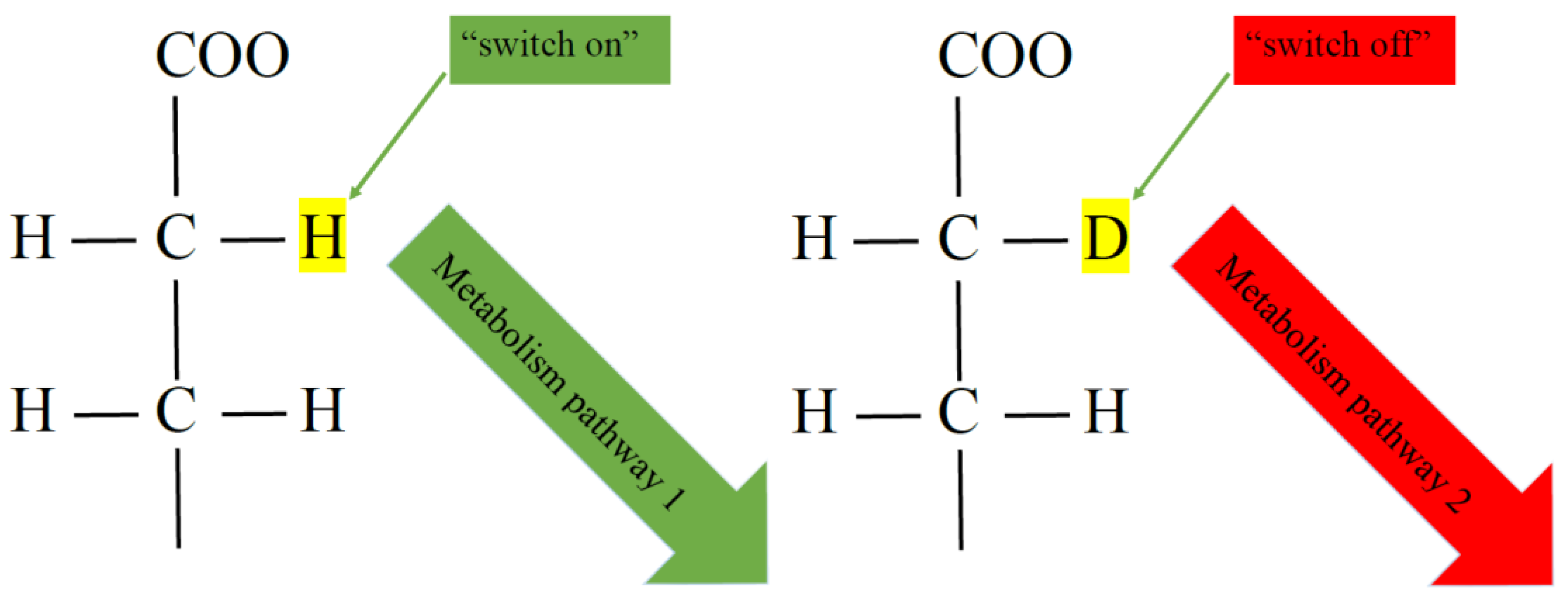
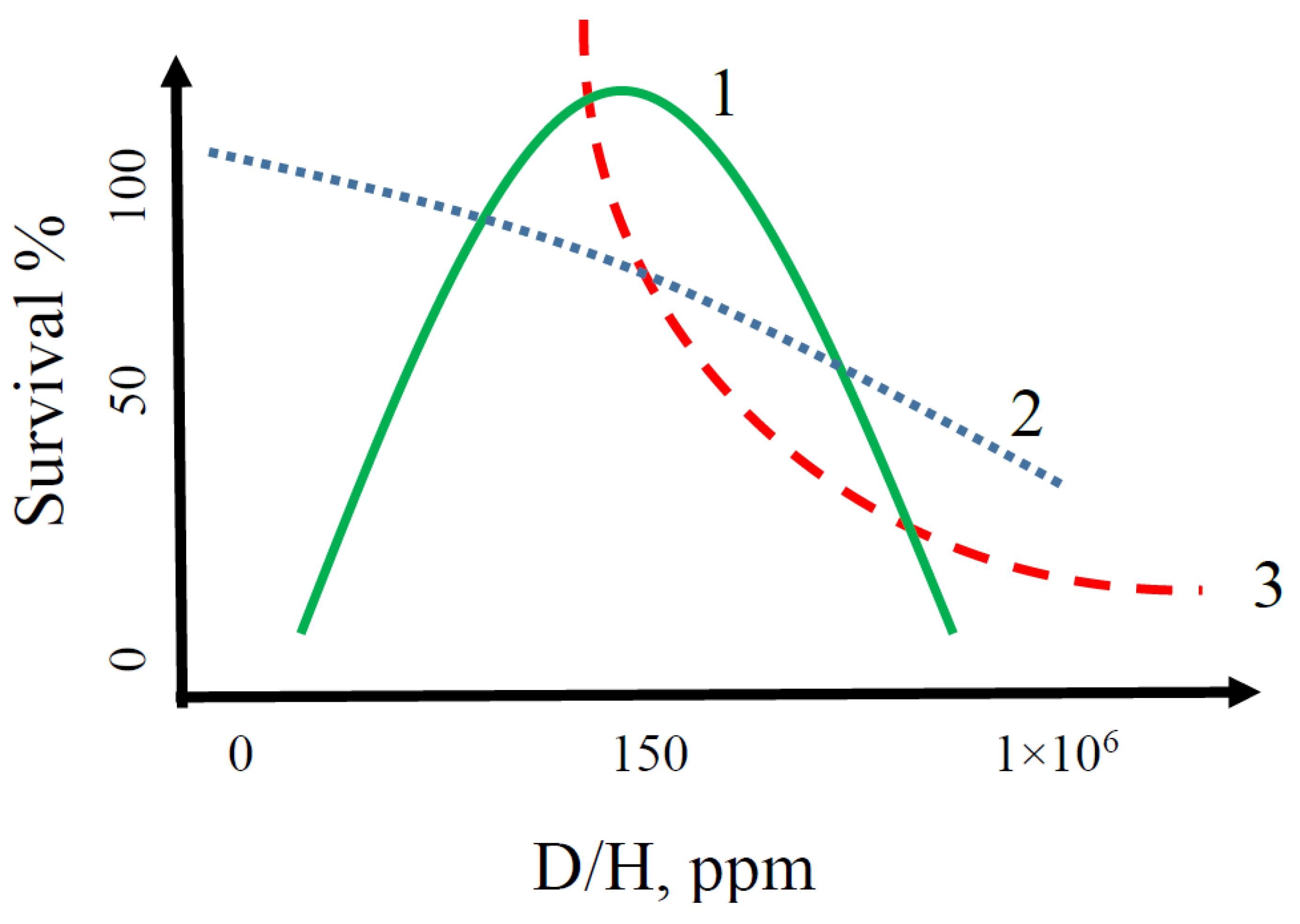
2.1. General Characteristics of Changes in Indicators of Various Taxonomic Groups of Organisms in Water with Different D/H Isotopic Ratios

As a result of research, we determined the effect of different concentrations of deuterium in water on the change in the values of the indices of some groups of taxons. All the values were conventionally given in one system of result assay (in percentage terms, where the results obtained in water with natural isotopic composition were taken as 100%) to unify the presentation of changes in the values of different indicators (growth, survival, embryotoxicity, etc.) (Table 2).
In bacteria, according to the results of changes in the values of the colony forming units (CFU) of E. coli, we noted the inhibition of colony growth in an environment based on the deuterated water. Therefore, this representative of the bacteria kingdom can be evaluated as an indicator of changes in the D/H isotopic ratio in water toward enrichment with deuterium.
Changes in the moss peptidom under the conditions of sprouting in the deuterium-depleted water led to a 30% change compared to sprouting on water with the natural ratio of deuterium. We did not observe moss growth in the deuterated water, which makes the representative of this taxon highly sensitive to the deuterium content in water.
Changes in the accumulation of C. fragrans L. in waters with different D/H isotopic compositions show that the redistribution and assimilation of mineral components in the deuterium-depleted water goes much faster—two or more times faster than in water with the natural isotopic composition. The accumulation and germination on the deuterated water did not give statistically significant results. That is C. fragrans L. can be classified as a highly sensitive representative of the kingdom of higher plants with respect to changes in the deuterium content in water.
The survival values of the unicellular biosensor S.ambiguum show the dependence on the D/H isotopic ratio in water according to the principle of the distribution of the Gaussian curve, taking into account the previously presented data [17,19]. Optimal lifespan values correlated with the natural D/H ratio in water. It is interesting that in this case Ea does not change, as we noted it earlier [19].
When studying embryogenesis of fish, it is worth noting that in the deuterium-depleted environment, high mortality (a chronic level of toxicity) was observed, compared with the control (natural D/H ratio). The acute toxicity of 50% was observed in the deuterated medium. In the deuterated environment, sublethal signs of toxicity were also recorded—a slowdown (delay) in the fish embryogenesis. Therefore, the indicator of embryogenesis of fish can be classified as highly sensitive with respect to changes in the D/H ratio in water.
According to the obtained results [1,20,21], it is noticeable that the deuterated water significantly reduces the activity of all indicators in different groups of organisms. The analysis of changes in the vital indicators of the organisms in the deuterium-depleted water environment does not give a definite clear correlation. Some biological effects of water with a modified isotopic composition was presented earlier [1,17,19,22,23,24,25]. The results of deuterium-depleted water exposure on the lower and higher plants, not previously published considered below.

2.2. Change in CFU of E. Coli in a Medium with Different D/H Isotopic Ratios

As an example of the reaction of a bacterial culture to a change in the isotopic composition of the medium, the effect of the heavy water (D2O) on the E. coli culture was investigated. The physiological state of the cell changes with increasing or decreasing deuterium content in water [1]. We found a rearrangement of the size spectrum of the culture during the growth of E. coli in D2O: the number of single cells decreases sharply, the aggregation of the culture takes place with the formation of associates of 4, 10 and 70 μm in size. The numerical and volume distribution of cells and associates of E. coli culture also varies; in the control culture grown in the environment of the natural D/H isotopic ratio the volume concentration of biomass was 0.024% and in the culture grown in D2O—0.008%.
It is worth noting that with the growth of the E. coli culture in an environment based on the deuterium-depleted water, no statistically significant deviations of the results from the control culture were observed.

2.3. Change in the Accumulation of Minerals during the Germination of C. fragrans L. in Water with Different D/H Isotopic Ratios

The content of zinc in the leaves of C. fragrans L. after saturation with zinc glycinate increased to 1.5–2 μg/g. At the starting points, the content of zinc was 0.01–0.02 μg/g. Furthermore, we observed twofold variability of the initial content of zinc and other trace elements while maintaining the species profile (biogeochemical profile). It is interesting that during the incubation of shoots in the solution of zinc glycinate, zinc was unevenly accumulated in the lamina over the entire length of the lamina. The highest zinc saturation was noted in the lower third (in the petiole) for leaves of 8–10 cm. The control of the topology and the uniform distribution of zinc allowed the X-ray diffraction analysis with intravital focusing on various zones of the lamina.
Table 3 shows how the coefficient of zinc accumulation in the leaves of C. fragrans L. changes during the incubation of shoots in solutions of zinc glycinate (20 mM Gly, 1 mM Zn2+, pH 7.0) in water with the natural D/H ratio and deuterium-depleted water.
It should be noted that a change in the isotopic composition of water made it possible, as expected, to achieve zinc accumulation two orders of magnitude higher than the natural D/H ratio in water. It cannot but emphasizes that similar results of Zn enrichment were obtained for another species—Kalanchoe daigremontianae [26]. It is interesting that iron has a low dynamics of accumulation, while the manganese content (Table 3) either does not increase or (in ddw) decreases slightly. In our opinion, this change in the elemental profile indicates not only the accumulation of zinc, but also a change in the metabolism of macroelements in the deuterium-depleted water [27].
The increase in the zinc content in the plant compared to the biogeochemical norm of the species has natural limitations, which follows from the Nernst equation and is caused by the natural accumulating ability of the plant at the concentration of free dissolved chelating agents and sorption at the phase interface. An increase in the assimilation ability of a plant is possible due to a change in the isotopic composition of the aqueous solution used for irrigation or in hydroponics. Instead of ordinary aqueous solutions, we use water depleted in heavy isotopes, which differs from ordinary water in some physicochemical parameters, including the freezing point, proton spin-spin relaxation time, and self-diffusion coefficient [28,29]. The reaction of living systems to the depletion of an aqueous solution with respect to the deuterium content, oxygen isotopes 17 and 18 is usually described as a change in metabolism [30,31]. One of the most interesting consequences of this change in the isotopic composition of water is a change in the rate constants of absorption/release of the solute. This is the effect that allows us to “circumvent” the thermodynamic limitations in nonequilibrium conditions, increasing the saturation of the medicinal plant with zinc compounds.
Thus, the use of zinc glycinate solutions in the deuterium-depleted water altered the metabolism of C. fragrans. A 200-fold accumulation of zinc in the lower third of the lamina up to 2 mg per 1 g of dry weight was noted. This result will allow offering special metal-modified medicinal and spicy-aromatic plants to compensate for zinc-deficient conditions in the future.
We also noted the inhibition of C. fragrans growth in the deuterated water, which did not allow the elemental analysis.

2.4. Change in the P. patens Moss Peptidom When Sprouting in Water with Different D/H Isotope Ratios

Studies of changes in the peptide composition of living organisms are very promising and interesting from the point of view of identifying intracellular mechanisms in response to stimuli of any kind and order. The increased interest in the most ancient plant, P. patens moss, is associated with the fact that it has become a leading model in the study of molecular mechanisms of plant development [32,33,34]. The relatively large genome of P. patens was sequenced and became available in open databases for comparative genomic evolutionary studies almost immediately after determining the complete genomic sequences of rice and poplar as well as of three types of green algae [35]. P. patens moss, widely used as a model system for molecular biology and ontogenetic studies, has become an important fundamental object of proteomics. To date, two independent projects for proteomic mapping of P. patens moss were implemented in Germany [36] and Korea [37]. As a result, using the MASCOT search system and the BLASTX comparison software, more than 300 proteins isolated from the protonema were processed [36].
We studied the peptide composition of the P. patens moss under sprouting conditions on deuterium-depleted and natural isotopic composition media. It should be noted that we did not observe moss growth with the deuterated water, which makes the representative of this taxon highly sensitive to the increased deuterium content.
Table 4 presents a partial peptidom of moss sprouted on waters with the natural D/H isotopic ratio and deuterium-depleted. It should be noted that 1–47 peptide residues were noted only in the sprouted moss based on water with the natural D/H ratio and were not determined in the medium based on DDW. Peptide residues 48–86 were determined only in the peptidom of moss sprouted based on deuterium-depleted water, and were not defined in the comparison group based on water with the natural D/H isotopic ration.
Thus, it should be noted that a change in the D/H isotopic composition leads to a significant peptide change in the composition of moss, which can lead to changes in the metabolism of the entire organism.

2.5. Kinetic Model of the Vital Activity of Living Organisms Depending on the D/H Ratio

It was previously noted that with a lack of deuterium, the rate of metabolic reactions changes [38,39,40,41,42,43]. Moreover, from the point of view of the classical concepts of the dilution of substances, deuterium concentrations <100 ppm, at which changes in metabolism are observed, should be insignificant, but the observed effects are reliable. Therefore, deuterium acts as an element that is necessary as a regulator of the rate of reactions in complexes and cascades of biochemical reactions. On the other hand, deuterium can be considered to be an element that affects the chirality of the substance [17,44], which can explain the mechanism of many changes as a result of different D/H ratios (Figure 1). In other words, the presence of deuterium or protium in the substance (“switch on/off” mechanism) leads to different ways of further reactions and, respectively, the entire metabolism can go in different directions (at different rates) depending on the presence of deuterium or protium in the original or intermediate substance. If we accept this assumption, the key metabolic regions and reaction pathways, in which the deuterium/protium ratio can act as a trigger, as a regulator of metabolic processes according to the “switch on/off” system, remain unknown.
Thus, there is a dependence on the D/H ratio and vital activity of living organisms! In our opinion, the diagram of the kinetic model of the vital activity of living organisms depending on the D/H ratio in the aquatic environment looks as follows (Figure 2).
The studies allowed to determine the sensitivity of some different taxons to changes in the content of deuterium in the aquatic environment. With some choice of taxons to determine the sensitivity to deuterium, we can give a preliminary assessment of the quality of the aquatic environment in relation to the D/H isotopic composition, in which the organism is located. It should be noted that several other authors independently obtained results similar to our data on the sensitivity of different taxons to deuterium concentrations for various vital indicators [27,38,45,46,47,48].
Since other authors previously presented data on changes in metabolism depending on the ratio of deuterium/protium, we did not focus on this. We tried to combine the data and describe the possible mechanism of metabolic alteration through the chirality of substances. Thus, the studies of bacteria were carried that showed a high sensitivity to the deuterated water [49,50]. Earlier studied the metabolism of the symbiotic organism medusomycetes (tea fungus) and the influence of D2O on its development [51]. Recent studies on the effects of low concentrations of deuterium on rats and mice show that the organism accelerates its metabolism [43,52,53,54]. In this regard showed that one could use ddw water to test inhibitors of de novo lipogenesis (DNL) by performing back-to-back studies in higher species, treat nonhuman primates with platensimycin, an inhibitor of fatty acid synthase [55]. Broad varieties of morphological and physiological changes were observed in deuterium-treated cells and organisms, including changes in fundamental processes such as cell division or energy metabolism [56]. However, it should be noted that we showed before how an excessive deficiency or an excess of deuterium can cause cytotoxic effects at the cell level [23,57]. Therefore, additional research of the use of water with a reduced deuterium content is required.
Taking into account many evidence of the kinetic isotope effect (KIE) for pharmaceutical substances in waters with different D/H ratios, it can be predicted that deuterium becomes a medical and chemical tool. Deuterated water is used as an inhibitor in medicines [58], which allows effective fighting against malignant tumors. This effect was also noted earlier in the deuterium-depleted water [39,40], although drugs based on such water have not yet been registered. The effect of deuterium on the kinetic effects of biotransformation of pharmaceutical substances may have significantly lower rates of metabolism, and hence a longer half-life. More and more deuterated compounds are characterized as promising substances for possible use as drugs. The first patents for deuterated substances were issued in the United States in the middle of the last century [59,60]. Such an example is deutetrabenazine (d6-tetrabenazine) approved by the Food and Drug Administration (FDA) in 2017 [61]. Two independent research groups reported a decrease in the biotransformation rate of d2-tyramine and d3-morphine compared to the parent proton-containing compounds [62,63].
Special attention should be paid to the mechanism of action of deuterium in living organisms. Several authors cite various theories [6,11,18,44,48,50], but they all boil down to the fact that the ratio of hydrogen isotopes modifies the metabolism in living organisms or their cells. Studied and considered different mechanisms of deuterated molecules action in living organisms: deuterium can make stronger chemical bonds than hydrogen and influence on the stretch frequency of bonds. Each C-H bond has a slightly different stretch frequency clustered around approximately 3.000 cm−1. Deuteration reduces the stretch frequencies by approximately the square root of 2.000 to 2.100 to 2.200 cm−1 [64]. Perhaps, in our opinion, the key mechanism is the chirality of substances in methylene groups with deuterium or protium in their structure. Recent data with a single 2H/1H replacement in DNA confirm this mechanism [65]. Therefore, further experimental data will help to solve the mechanism of action of deuterated or deuterium-depleted aqueous media on living organisms.
All of the above leads to the conclusion that the range of natural concentrations of deuterium in water is the most optimal for some living organisms on Earth, which apparently was formed over millions of years of evolution. Many organisms of different taxons are highly sensitive to high or low deuterium content in water. However, some taxons—for example, microorganisms—do not show statistically significant sensitivity to a change in the isotopic composition of water toward a decrease in deuterium compared with natural water. All this allowed us to assume that maybe the D/H ratio is a taxonomic determining factor for animals and plants, which presented in scheme Figure 2.

3. Materials and Methods

3.1. Physico-Chemical Analysis of Water with Different Deuterium Contentss

We used initial water samples with different deuterium contents in our work: deuterium-depleted water (DDW, ddw) with D/H = 5 ppm (Sigma-Aldrich, St. Louis, MO, USA); deuterated water D2O = 99 abs. at.% (Sigma-Aldrich). To study the effect of deuterium on the biological properties of organisms, media with different concentrations of deuterium were prepared by diluting deuterium-depleted and deuterated waters. As a control, water with the D/H ratio of 140 ppm was used. According to the physical indicators [8,17] and microelement composition, deuterium-depleted and deuterated waters had no difference, except for the deuterium content—this excluded the influence of multifactoriality in the system from the point of view of physico-chemical components for all compared groups. The method was described in more detail earlier [17]. The deuterium content was monitored using multipass laser absorption spectroscopy on Isotopic Water Analyzer-912-0032 (Los Gatos Research Inc., San Jose, CA, USA).
The chemical analysis of water with different deuterium contents was carried out by inductively coupled plasma mass spectrometry using an Agilent 7500CE ICP-QMS instrument (Agilent Technologies, Santa Clara, CA, USA); the method was described in more detail earlier [17,22]. To calibrate the instrument, calibration solutions were used in a wide range of element concentrations (from 0.1 μg/dm3 to 100 μg/dm3), they were prepared according to international reference sample (ISS) 2.74473.0100 ICP Multi Element Standard Solution XXI Certi PUR®. ISS contains the following elements: Ag, Al, As, Ba, Be, Bi, Ca, Cd, Co, Cr, Cs, Cu, Fe, Ga, In, K, Li, Mg, Mn, Na, Ni, Pb, Rb, Se, Sr, Tl, U, V, Zn, Hg. In deionized, deuterium-depleted and deuterated waters, the concentration of all elements did not exceed the upper detection limit (detection limit is 0.1–10 ppm).
The contents of Cd, Cu, Ni, Pb, Mn, Zn, Cr, Al, Fe, As, V were determined by atomic absorption spectrometry (AAS) method using an atomic absorption spectrometer Spectr AA-800 (Varian Inc., Palo Alto, CA, USA) with electrothermal atomization and the Zeeman effect according to the manufacturer’s protocol with modifications according to the results of the international intercalibration with the MEL laboratory of the IAEA (Monaco). The method was described in more detail earlier [17,22]. The source of radiation was single element hollow cathode lamps Spectr AA (Varian Inc., Palo Alto, CA, USA). The current of the lamps for elements Ni, Cu, Hg, Cd—4.0 mA; for Al, As—10 mA; Mn, Zn—5.0 mA; for Cr, V, Pb—7.0 mA, for Co, Sn—7 mA. The slit width of the monochromator was 0.5 nm when measuring Al, Ni, Cu, Zn, Hg; 0.2 nm for Cr, Fe, Mn, V, Co, Sn; 1.0 nm for Cd, Pb, As. The baseline correction mode and hot injection were carried out −80 °C. The following wavelengths (resonance lines) and modifiers were used: Al—256.8 nm, Mg(NO3)2; Ni—232.0 nm, Mg(NO3)2; Cr—429.0 nm, Mg(NO3)2; Mn—403.1 nm, Mg(NO3)2; Fe—386.0 nm, Mg(NO3)2; Cu—327.4 nm, Pd(NO3)2; Zn—307.6 nm, Mg(NO3)2, As—193.7 nm, Pd(NO3)2 + Mg(NO3)2; Sn—286.3 nm, Mg(NO3)2 + NH4H2PO4; V—318.5 nm; Co—242.5 nm, Pd(NO3)2; Cd—228.8 nm, Pd(NO3)2 + Mg(NO3)2 + NH4H2PO4; Pb—283.3 nm, Pd(NO3)2 + Mg(NO3)2 + NH4H2PO4. The relative standard deviation in the determination with a confidence level of 0.95 did not exceed 20%. The composition of the reference sample is given in [66].

3.2. Microbiological Methods

The method was previously described in more detail [1]. Escherichia coli 1257 strain was used in microbiological studies, which was obtained from the Research Institute for Standardization and Control of Medical Biological Preparations (Moscow, Russia). Bacteria from nutrient agar were inoculated into meat-peptone broth (MPB) and incubated in a temperature-controlled chamber at 37 °C for 18 h. The suspension was centrifuged at 7000 rpm for 15 min. The sedimental liquid was removed, and the sediment obtained was washed three times with sterile saline (0.9% NaCl) during the centrifugation. After that, the sediment was resuspended in the same physiological saline to a density of 1 × 10⁸ colony forming units (CFU) in 1 cm3. The corresponding aliquot of the initial suspension of E. coli was introduced into the test medium until obtaining the required microorganism density of 1 × 10⁵ CFU/cm3 [1].
The survival of microorganisms was determined by the presence of CFU when plating the selected samples on Endo agar medium. Petri dishes with medium were incubated in a temperature-controlled chamber at 37 °C for 18–24 h with subsequent counting of grown colonies. The result was expressed as the ratio of the logarithm of the test microorganism concentration, which remained in the solution (Nt), to its initial count (N0).

3.3. Peptidomics of Moss Physcomitrella Patens

The method was described in more detail earlier [67].

3.3.1. Sprouting of Moss Gametophores

P. patens moss gamethophores (Gransden strain) were grown on Knop modified agar medium (PPNO3 medium) in Petri dishes (diameter 9 cm) under white light from F96T12/GRO/VHO/WS fluorescent tubes with photon flux of 61 μM/(m2·s) under conditions of a 16-h photoperiod at 26 °C. Media based on water with various concentrations of deuterium were prepared for the study by diluting deuterium-depleted and deuterated waters. The gametophore groups were divided with a tweezer into fragments containing 4 to 5 shoots and transplanted onto fresh nutrient medium. 1 month old gametophores were used for the proteomic analysis.

3.3.2. Isolation of Protein from Moss Tissues

The extraction of proteins from moss gametophores was carried out according to the method adapted for the P. patens moss [37]. Making of protein preparations from the gametophores: 1 month old shoots of moss were cut with a scalpel at a height of 1 mm from the surface of the agar medium and transferred with a tweezer to a porcelain mortar pre-cooled to −70 °C, in which they were immediately frozen with liquid nitrogen. Then, frozen shoots of moss were crushed to fine powder using a pre-cooled pestle (−70 °C). The plant material was poured with a 10% solution of 3-chloroacetic acid (TCA) in acetone with 0.07% dithiothreitol (DTT), cooled to −20 °C, then the material was incubated for 1 h at the same temperature to realize the protein precipitation. The protein precipitate suspension was centrifuged at 30,000× g for 15 min at 4 °C. The protein precipitate was poured with acetone cooled to −20 °C and containing 0.07% DTT, 1 mM PMSF, 2 mM EDTA, and shaken vigorously to remove pigments and lipids from the preparation. Then the protein precipitate suspension was centrifuged under the same conditions and the described procedure of washing the protein preparation with acetone was repeated two more times. Samples of protein preparations in centrifuge plastic tubes (2 mL) were dried in a vacuum centrifuge and stored at −70 °C for 10 days max (until the time of carrying out 2-dimensional gel electrophoresis).

3.3.3. 2D Electrophoresis of Proteins

Two-dimensional separation of proteins was carried out according to the procedure [68] with some minor modifications. Upon completion of electrophoresis, the gels were labeled and stained with silver with thiosulfate [69]. The images of the silver stained gels were obtained using an Epson Perfection 4990 scanner and analyzed using the PDQuest 8.0 software (BioRad, Hercules, CA, USA).

3.3.4. Trypsin Hydrolysis of Proteins

Proteins separated by 2D electrophoresis were hydrolyzed in PAGE pieces according to the technique proposed by Shevchenko and colleagues [70] with minor modifications.

3.3.5. MALDI Mass Spectrometry

Tryptic peptides were extracted from the gel using 0.5% solution of 3-fluoroacetic acid (TFA) in water. The resulting extract in a volume of 0.20 μL was mixed with 0.25 μL of 2% solution of 2,5-dihydroxybenzoic acid in 30% solution of acetonitrile containing 0.5% glacial acetic acid by volume on a steel target, which was further dried in air at 23 °C for 30 min The peptides were analyzed on an Ultraflex-TOF/TOF time-of-flight mass spectrometer (Bruker Daltonics, Hamburg, Germany) equipped with a UV laser (337 nm). Positive ions were detected in the reflectron mode at voltages: at the ion source IS1—25 kV, IS2—21.75, 9.5 kV, reflectron Ref1—26.43 kV, Ref2—13.80 kV. Ions were detected at m/z = 700–4000. The peaks of trypsin autolytic fragments (m/z = 842.508, 1045.563, 2211.093), as well as keratin (m/z = 1475.780) and other impurities, which were excluded from the final lists of detected masses, were used as an internal reference.

3.3.6. Analysis of Mass Spectrometry Results

Mass spectra were processed using the Flex Analysis 2.4 software (Bruker Daltonics). Smoothing by the Savitzky-Golay algorithm [71] (width 0.1 m/z, 1 cycle) and subtraction of the baseline according to the algorithm were applied to the mass spectra, Convex Hull [16]. The following peak detection parameters were used: peak detection algorithm—SNAP, signal-to-noise ratio—6, spectrum quality threshold—100. The search and identification of proteins by the “peptide imprint” method in the database of the nuclear genome of moss P. patens (fasta format, version 1.1, http://genome.jgi’psf.org/Phypa1_1/Phypa1_1.home.html) was performed using the MASCOT software package (local version 2.1.03, Matrix Science, Boston, MA, UK). In this case, the following search parameters were used: mass determination accuracy—100 ppm, possible post-translational modifications—methionine oxidation. Proteins with the probabilistic parameter MOWSE score higher than 58, which was considered the threshold for the base of the P. patens moss genome used, were considered reliably identified (95%, p > 0.05). An additional criterion for the reliability of protein identification was the coincidence of its molecular weight and/or isoelectric point with the corresponding parameters determined experimentally or predicted theoretically for the corresponding protein or its analogues.

3.3.7. Liquid Chromatography-Mass Spectrometry (LC-MS/MS)

Peptide preparations obtained by trypsinolysis of proteins separated by 2D electrophoresis were analyzed on the Esquire 6000 plus mass spectrometer (Bruker Daltonics) equipped with a nanospray ion source with a quadrupole ion trap as a mass analyzer. The instrument is online mated with an Ultimate LC Packings nanochromatograph and Famos LC Packings sampling system (Dionex, Sunnyvale, CA, USA). The chromatographic part of the installation is two series-connected columns, between which there was an electromagnetic valve. Column No. 1 (100 μm × 3 cm) filled with the Poros R2 polymer phase (hydrophobic polymer phase with a large pore diameter, analog of C8) was used for preliminary concentration of the sample and its desalination. The second column (75 μm × 25 cm) filled with a Phenomenex sorbent (C18, grain size 5 μm, pore diameter 300 E) was used directly to separate a desalted mixture of tryptic peptides. The chromatographic separation on the system described above was carried out at a flow rate of 200 μL/min (up to the splitter). In this case, the actual flow rate during desalination was of 900 on average, and 200 μL/min during the separation. Peptides were separated using a linear gradient of “5–60%” solution of 75% acetonitrile, 25% isopropanol in 0.1% formic acid for 48 min. The measurements were carried out at m/z = 300–2500 with the “trap optimization mass” of 700. Tandem experiments were carried out only with ions, the charge number of which was 2, with an intensity above the threshold. The resulting lists of determined masses were sent to the MASCOT search engine. The search was carried out in the base of the nuclear genome of moss JGI (http://genome.jgi’psf.org/Phypa1_1/Phypa1_1.home.html), as well as in database NCBI (http://www.ncbi.nlm.nih.gov//genomes/geblast.cgi?bact=off&gi=5880) with the P. patens taxon sampling. The search results were verified using the Scaffold software package (version 01_07_00) (http://www.pro’teomesoftware.com) to confirm the correct determination of proteins and the identification of identical records when searching in two databases. Proteins identified by two or more peptides with a probability of at least 95% were left in the final list.

3.3.8. 2D Electrophoresis of Proteins with Differential Staining (DiGE)

Total preparations of proteins isolated from moss gametophores were dissolved in a water buffer mixture (8 M urea (Amersham, St. Louis, MO, USA), 2 M thiourea (Amersham, St. Louis, MO, USA), 0.3% Chaps (Amersham), 0.1% Nonidet P_40 (Fluka, Munich, Germany), 10 mM Tris_HCl (Amersham), pH 8.0) for chromatography (Merck, Kenilworth, NJ, USA). Then, the samples were centrifuged at 14,000× g for 15 min at room temperature 25 °C. The protein concentration was measured by the Bradford protein assay using bovine serum albumin (BSA) as a reference (BioRad). Isoelectrofocusing and the second direction of electrophoresis were carried out according to the standard method. Scanning of the obtained gels was performed on a Typhoon instrument (Amersham) in the 600 PTM mode. After the fluorescence analysis, the gels were stained with silver according to the procedure described above.

3.4. Germination and Accumulation of Minerals in Callisia fragrans L.

The method was described in more detail earlier [72,73].

3.4.1. Materials and Method at Germination of C. fragrans L.

To study the accumulation of minerals in waters with different D/H ratios, the leaves of the basket plant were used in the work (Callisia fragrans, fam. Commelinaceae). The plants were grown in climatic chambers (MIR-3, Ukraine, Russia) at 20 °C and relative humidity of 60%. The germination procedure was consistent with the methodology [73] with minor modifications. The following composition of the solution was used to saturate the shoots of the plant with a zinc complex: zinc glycinate (Zn (II) + Gly 1 mM + 20 mM), respectively (the experiment included three groups of plants: 1—germinated in water with natural isotopic composition; 2—in water with natural isotopic composition + Gly complex, 3—in deuterium-depleted water + Gly complex. Shoots were germinated in the prepared solution for 14 days. On the 12th day of the experiment and on the 14th day, the accumulation of elements in the lamina of the plant was observed. The lamina was divided into three parts of 2 cm: lower, medium and upper.
The fragrant callisia leaves were dried under mild (natural) conditions at the temperature of 25 °C for 5–7 days until a constant mass was achieved by weighing the sample once a day. Weighing was carried out on an ATL-80d4 analytical balance (Acculab, Vernon Hills, IL, USA). When fully dried, the leaves should be easy to grind into powder. Weight of dry raw material sample of leaves was 0.1576 g

3.4.2. Dry Raw Material Analysis

The determination of chemical elements in the dried plant material was carried out without destroying the sample using a Shimadzu EDX-7000 X-ray fluorescence dispersion spectrophotometer (Shimadzu, Kyoto, Japan) based on a thermoelectrically cooled silicon drift detector equipped with the PCEDX-Navi software (Kyoto, Japan). The setup measurement conditions: current 100 μA, closed cuvette, 10 mm collimator, air medium, mylar and polypropylene films. The IAEA reference sample NIST SRM 2976 was used to calibrate the results. All results are presented for three repetitions with a confidence level of 95%. Each experimental repetition had three complete reproduction cycles of the entire experiment. The studies were carried out by the quantitative change of elements: Zn, Mn, Fe.

3.5. Determination of Biological Activity Using a Unicellular Biosensor Spirostomum ambiguum

The method was described in more detail earlier [19]. To determine the biological activity of a unicellular free-living eukaryotic organism of S. ambiguum ciliates in water with a modified D/H isotopic composition, the Spirotox method was used [19]. Observation of the biosensor was carried out using a binocular microscope MBR-10 (Altami, Saint Petersburg, Russia). To maintain a constant temperature in the medium, the Lauda A6 thermostat (Lauda-Königshofen, Germany) was used. As a result, of the study, the lifetime was determined and the activation energy (Ea) of the biosensor was found for different deuterium contents in water with a change in temperature conditions.

3.6. Determination of Embryotoxicity of Fish

A group of mature individuals Brachydanio rerio (Hamilton, 1822) was used in the studies. The conditions of keeping, feeding, and spawning of fish were described earlier [74]. Cultivation media were prepared according to the standard protocol based on deuterium-depleted, deuterated water and water. All reagents used were from the Sigma-Aldrich company, USA.
At the embryogenesis stages of blastomeres 4–32, the fertilized and non-fertilized eggs were separated from each other and transferred to the test aqueous solution. Only fertilized eggs were used for the experiments. The embryos were placed in Petri dishes of 7 cm diameter filled with the test solution. Each dilution was tested on at least 10 embryos at the rate of 2 mL of solution per one egg. The experiment was triplicated for each group. The control was in parallel. The results were considered reliable if no more than 10% of the embryos died in the control.
The embryos were observed for 72 h at the temperature of 26 °C in climatic chambers (MIR-3, Ukraine, Russia), the light regime corresponded to the alternation of day and night. At different stages of embryo development, the parameters indicating an embryogenesis developmental disorder were recorded [74].
The embryos were observed through an MBR-10 binocular microscope (Altami, Saint Petersburg, Russia) using a Canon PowerShot G9 digital camera with data and photo processing by MVideo and AxioVision software (White Plains, NY, USA). During the incubation, the parameters of the studied solutions were recorded: concentration of dissolved oxygen—using the Azha-101M oxygen meter (Altami), pH values—using a portable pH-meter pH-150M (Altami). In the studies, the indicators of the aquatic environment corresponded to the optimal parameters of the life of aquatic organisms—the oxygen level corresponded to 5–8 mg O2/cm3, and the pH value was within the range of 6.5–8.5.

3.7. Ethical Standards of Studies in Animals

All the studies were carried out in accordance with international ethics requirements approved by Directive 2010/63/EU for experiments using animals.

3.8. Statistical Data Processing

All statistical data processing was performed using Student’s t-test, as well as using the one-way analysis of variance (ANOVA) in the Origin Pro software. The differences were considered statistically significant at p < 0.05.

4. Conclusion

We determined the reactions of some major taxonomic groups of organisms to changes in the ratio of D/H isotopes in the aquatic environment. It was found that the studied organisms react differently to both high and low concentrations of deuterium in aqueous solutions, reducing or increasing their biological activity, perhaps by variety metabolism (moss peptidom changes). Perhaps ratio of D/H this is a determining taxonomic factor for living organisms. This theory is supported by the developed kinetic model and the presented diagram of biocatalytic reactions of the vital activity of living organisms depending on the D/H ratio in aqueous solutions.

Author Contributions

Conceptualization, I.Z., T.P., A.S. (Alexander Skripnikov), T.M. and A.S. (Anton Syroeshkin); Data curation, I.Z., T.G., O.L., I.S. and A.S. (Anton Syroeshkin); Formal analysis, I.Z., T.P., A.S. (Alexander Skripnikov), T.M., M.M. and I.S.; Funding acquisition, A.S. (Anton Syroeshkin); Investigation, I.S.; Methodology, T.P., A.S. (Alexander Skripnikov), T.G., T.M., N.A., O.L. and M.M.; Project administration, T.P. and A.S. (Anton Syroeshkin); Resources, A.S. (Anton Syroeshkin); Software, N.A.; Supervision, T.G.; Validation, A.S. (Alexander Skripnikov), T.M. and O.L.; Visualization, I.Z., N.A., O.L. and M.M.; Writing—original draft, I.Z., T.M., O.L. and A.S. (Anton Syroeshkin); Writing—review & editing, T.G., N.A. and A.S. (Anton Syroeshkin) All authors have read and agreed to the published version of the manuscript.

Funding

This research received no external funding.

Acknowledgments

The publication has been prepared with the support of the “RUDN University Program 5-100”.

Conflicts of Interest

The authors declare that there are no conflicts of interest regarding the publication of this paper.

References

  1. Goncharuk, V.V.; Pleteneva, T.V.; Grebennikova, T.V.; Syroeshkin, A.V.; Uspenskaya, E.V.; Antipova, N.V.; Kovalenko, V.F.; Saprykina, M.N.; Skil’Skaya, M.D.; Zlatskiy, I.A. Determination of Biological Activity of Water Having a Different Isotope Ratio of Protium and Deuterium. J. Water Chem. Technol. 2018, 40, 27–34. [Google Scholar] [CrossRef]
  2. Lobyshev, V.N.; Kalinichenko, L.P. Isotopic Effects in Biological Systems; Nauka: Moscow, Russia, 1978. (In Russian) [Google Scholar]
  3. McCartney, D.; Desbrow, B.; Irwin, C. Post-exercise Ingestion of Carbohydrate, Protein and Water: A Systematic Review and Meta-analysis for Effects on Subsequent Athletic Performance. Sports Med. 2017, 48, 379–408. [Google Scholar] [CrossRef] [PubMed]
  4. Said, H.M. Intestinal absorption of water-soluble vitamins in health and disease. Biochem. J. 2011, 437, 357–372. [Google Scholar] [CrossRef] [PubMed] [Green Version]
  5. Goncharuk, V.V. Science about Water; Naukova Dumka: Kiev, Ukraine, 2010. (In Russian)
  6. Basov, A.; Fedulova, L.; Baryshev, M.; Dzhimak, S. Deuterium-Depleted Water Influence on the Isotope 2H/1H Regulation in Body and Individual Adaptation. Nutrients 2019, 11, 1903. [Google Scholar] [CrossRef] [PubMed] [Green Version]
  7. Bowen, G.J.; Winter, D.A.; Spero, H.J.; Zierenberg, R.; Reeder, M.D.; Cerling, T.E.; Ehleringer, J.R. Stable hydrogen and oxygen isotope ratios of bottled waters of the world. Rapid Commun. Mass Spectrom. 2005, 19, 3442–3450. [Google Scholar] [CrossRef]
  8. Goncharuk, V.V.; Lapshin, V.B.; Burdeynaya, T.N.; Pleteneva, T.V.; Chernopyatko, A.S.; Atamanenko, I.D. Physico-chemical properties and biological activity of water, lean on heavy isotope. Chem. Technol. Water 2011, 33, 15–25. [Google Scholar]
  9. Lewis, G.N. Biology of heavy water. Nature 1934, 133, 620. [Google Scholar] [CrossRef]
  10. Timmins, G.S. Deuterated drugs: Where are we now? Expert Opin. Ther. Patents 2014, 24, 1067–1075. [Google Scholar] [CrossRef]
  11. Boros, L.G.; D’Agostino, D.P.; Katz, H.E.; Roth, J.P.; Meuillet, E.J.; Somlyai, G. Submolecular regulation of cell transformation by deuterium depleting water exchange reactions in the tricarboxylic acid substrate cycle. Med. Hypotheses 2016, 87, 69–74. [Google Scholar] [CrossRef]
  12. Robins, R.J.; Remaud, G.S.; Billault, I. Natural mechanisms by which deuterium depletion occurs in specific positions in metabolites. Eur. Chem. Bull. 2012, 1, 39–40. [Google Scholar]
  13. Cleland, W.W. The use of isotope effects to determine enzyme mechanisms. J. Biol. Chem. 2003, 278, 51975–51984. [Google Scholar] [CrossRef] [PubMed] [Green Version]
  14. Strekalova, T.; Evans, M.; Chernopiatko, A.; Couch, Y.; Costa-Nunes, J.P.; Cespuglio, R.; Chesson, L.; Vignisse, J.; Steinbusch, H.W.; Anthony, D.C.; et al. Deuterium content of water increases depression susceptibility: The potential role of a serotonin-related mechanism. Behav. Brain Res. 2015, 277, 237–244. [Google Scholar] [CrossRef]
  15. Cărpinişan, L.; Petcu, M.D.; Petrovici, S.; Chiş, C.; Ghişe, A.; Zehan, R. The Influence of deuterium depleted water on the hematocrit and the leukocyte formula in rats intoxicated with chromium. Sci. Pap. Anim. Sci. Biotechnol. 2010, 43, 464–468. [Google Scholar]
  16. Huynh, M.H.V.; Meyer, T.J. Colossal kinetic isotope effects in proton-coupled electron transfer. Proc. Natl. Acad. Sci. USA 2004, 101, 13138–13141. [Google Scholar] [CrossRef] [PubMed] [Green Version]
  17. Syroeshkin, A.; Pleteneva, T.; Uspenskaya, E.; Zlatskiy, I.A.; Antipova, N.; Grebennikova, T.; Levitskaya, O.V. D/H control of chemical kinetics in water solutions under low deuterium concentrations. Chem. Eng. J. 2019, 377, 119827. [Google Scholar] [CrossRef]
  18. Basov, A.; Fedulova, L.; Vasilevskaya, E.R.; Dzhimak, S. Possible Mechanisms of Biological Effects Observed in Living Systems during 2H/1H Isotope Fractionation and Deuterium Interactions with Other Biogenic Isotopes. Molecules 2019, 24, 4101. [Google Scholar] [CrossRef] [Green Version]
  19. Goncharuk, V.V.; Syroeshkin, A.V.; Zlatskiy, I.A.; Uspenskaya, E.V.; Orekhova, A.V.; Levitskaya, O.V.; Dobrovolskiy, V.I.; Pleteneva, T. Quasi-chemical description of the kinetics of cell death Spirostomum ambiguum biosensor for biological activity of aqueous solutions. J. Water Chem. Technol. 2017, 39, 97–102. [Google Scholar] [CrossRef]
  20. Atzrodt, J.; Derdau, V.; Kerr, W.J.; Reid, M. Deuterium- and Tritium-Labelled Compounds: Applications in the Life Sciences. Angew. Chem. Int. Ed. 2018, 57, 1758–1784. [Google Scholar] [CrossRef] [Green Version]
  21. Farthing, D.E.; Buxbaum, N.P.; Lucas, P.J.; Maglakelidze, N.; Oliver, B.; Wang, J.; Hu, K.; Castro, E.; Bare, C.V.; Gress, R.E. Comparing DNA enrichment of proliferating cells following administration of different stable isotopes of heavy water. Sci. Rep. 2017, 7, 4043. [Google Scholar] [CrossRef]
  22. Syroeshkin, A.; Antipova, N.; Zlatska, A.; Zlatskiy, I.A.; Skylska, M.; Grebennikova, T.; Goncharuk, V. The effect of the deuterium depleted water on the biological activity of the eukaryotic cells. J. Trace Elem. Med. Boil. 2018, 50, 629–633. [Google Scholar] [CrossRef]
  23. Zlatska, A.V.; Vasyliev, R.G.; Gordiienko, I.M.; Rodnichenko, A.E.; Morozova, M.A.; Vulf, M.A.; Zubov, D.O.; Novikova, S.N.; Litvinova, L.S.; Grebennikova, T.V.; et al. Effect of the deuterium on efficiency and type of adipogenic differentiation of human adipose-derived stem cells in vitro. Sci. Rep. 2020, 10, 1–16. [Google Scholar] [CrossRef] [PubMed] [Green Version]
  24. Makarova, M.; Syroeshkin, A.; Maksimova, T.; Pleteneva, T.; Zlatskiy, I.; Antipova, N.; Skripnikov, A.; Dzhavakhyan, M. D/H modification of plant peptides and microelements metabolome. FEBS Open Bio 2019, 9, 287. [Google Scholar]
  25. Tsisanova, E.S.; Uspenskaya, E.V.; Pleteneva, T.V.; Syroeshkin, A.V. Study of biological activity and D/H ratio of water with the aid of cellular biosensor spirostomum ambiguum. Trace Elem. Med. 2010, 11, 85. [Google Scholar]
  26. Chuparina, E.V.; Gunicheva, T.N. State and issues of X-ray fluorescence analysis of herbal raw materials. Anal. Control 2004, 8, 211–226. [Google Scholar]
  27. McCluney, K.E.; Sabo, J.L. Tracing Water Sources of Terrestrial Animal Populations with Stable Isotopes: Laboratory Tests with Crickets and Spiders. PLoS ONE 2010, 5, e15696. [Google Scholar] [CrossRef]
  28. Goncharuk, V.V.; Taranov, V.V.; Kurlyantseva, A.Y.; Syroeshkin, A.V. Phase transition in waters with different content of deuterium. J. Water Chem. Technol. 2015, 37, 219–223. [Google Scholar] [CrossRef]
  29. Rodin, S.; Rebellato, P.; Lundin, A.; Zubarev, R.A. Isotopic resonance at 370 ppm deuterium negatively affects kinetics of luciferin oxidation by luciferase. Sci. Rep. 2018, 8, 16249. [Google Scholar] [CrossRef] [Green Version]
  30. Dzhimak, S.; Basov, A.A.; Baryshev, M.G. Content of deuterium in biological fluids and organs: Influence of deuterium depleted water on D/H gradient and the process of adaptation. Dokl. Biochem. Biophys. 2015, 465, 370–373. [Google Scholar] [CrossRef]
  31. Luo, A.L.; Zheng, Y.L.; Cong, F.S. Research progress of biological effects of deuterium-depleted water. J. Shanghai Jiaotong Univ. (Med. Sci.) 2018, 38, 467–471. [Google Scholar]
  32. Trouiller, B.; Schaefer, D.G.; Charlot, F.; Nogué, F. MSH2 is essential for the preservation of genome integrity and prevents homeologous recombination in the moss Physcomitrella patens. Nucleic Acids Res. 2006, 34, 232–242. [Google Scholar] [CrossRef]
  33. Schaefer, D.G. Gene targeting in Physcomitrella patens. Curr. Opin. Plant Biol. 2001, 4, 143–150. [Google Scholar] [CrossRef] [Green Version]
  34. Mittmann, F.; Brücker, G.; Zeidler, M.; Repp, A.; Abts, T.; Hartmann, E.; Hughes, J. Targeted knockout in Physcomitrella reveals direct actions of phytochrome in the cytoplasm. Proc. Natl. Acad. Sci. USA 2004, 101, 13939–13944. [Google Scholar] [CrossRef] [PubMed] [Green Version]
  35. Rensing, S.A.; Lang, D.; Zimmer, A.; Terry, A.; Salamov, A.; Shapiro, H.; Nishiyama, T.; Perroud, P.-F.; Lindquist, E.; Kamisugi, Y.; et al. The Physcomitrella Genome Reveals Evolutionary Insights into the Conquest of Land by Plants. Science 2007, 319, 64–69. [Google Scholar] [CrossRef] [PubMed] [Green Version]
  36. Sarnighausen, E.; Wurtz, V.; Heintz, D.; Van Dorsselaer, A.; Reski, R. Mapping of the Physcomitrella patens proteome. Phytochemisty 2004, 65, 1589–1607. [Google Scholar] [CrossRef]
  37. Cho, S.H.; Hoang, Q.T.; Kim, Y.Y.; Shin, H.Y.; Ok, S.H.; Bae, J.M.; Shin, J.S. Proteome analysis of gametophores identified a metallothionein involved in various abiotic stress responses in Physcomitrella patens. Plant Cell Rep. 2006, 25, 475–488. [Google Scholar] [CrossRef]
  38. Avila, D.S.; Somlyai, G.; Somlyai, I.; Aschner, M. Anti-aging effects of deuterium depletion on Mn-induced toxicity in a C. elegans model. Toxicol. Lett. 2012, 211, 319–324. [Google Scholar] [CrossRef] [PubMed] [Green Version]
  39. Somlyai, G. Defeating Cancer. The Biological Effects of Deuterium Depletion; Author House: Bloomington, Indiana, 2002. [Google Scholar]
  40. Somlyai, G.; Gyöngyi, Z.; Somlyai, I.; Boros, L.G. Pre-clinical and clinical data confirm the anticancer effect of Deuterium depletion. Eur. J. Integr. Med. 2016, 8, 1–7. [Google Scholar] [CrossRef]
  41. Zhang, K.; Toki, H.; Fujita, Y.; Ma, M.; Chang, L.; Qu, Y.; Harada, S.; Nemoto, T.; Mizuno-Yasuhira, A.; Yamaguchi, J.-I.; et al. Lack of deuterium isotope effects in the antidepressant effects of (R)-ketamine in a chronic social defeat stress model. Psychopharmacology 2018, 235, 3177–3185. [Google Scholar] [CrossRef]
  42. Yavari, K.; Kooshesh, L. Deuterium Depleted Water Inhibits the Proliferation of Human MCF7 Breast Cancer Cell Lines by Inducing Cell Cycle Arrest. Nutr. Cancer 2019, 71, 1019–1029. [Google Scholar] [CrossRef]
  43. Halenova, T.; Zlatskiy, I.A.; Syroeshkin, A.V.; Maximova, T.; Pleteneva, T. Deuterium-Depleted Water as Adjuvant Therapeutic Agent for Treatment of Diet-Induced Obesity in Rats. Molecules 2019, 25, 23. [Google Scholar] [CrossRef] [Green Version]
  44. Goncharuk, V.V.; Syroeshkin, A.V.; Pleteneva, T.V.; Uspenskaya, E.V.; Levitskaya, O.V.; Tverdislov, V.A. On the possibility of chiral structure-density submillimeter inhomogeneities existing in water. J. Water Chem. Technol. 2017, 39, 319–324. [Google Scholar] [CrossRef]
  45. Basov, A.A.; Elkina, A.A.; Samkov, A.A.; Volchenko, N.N.; Moiseev, A.V.; Fedulova, L.V.; Baryshev, M.G.; Dzhimak, S.S. Influence of Deuterium-Depleted Water on the Isotope D/H Composition of Liver Tissue and Morphological Development of Rats at Different Periods of Ontogenesis. Iran. Biomed. J. 2019, 23, 129–141. [Google Scholar]
  46. Rasooli, A.; Fatemi, F.; Hajihosseini, R.; Vaziri, A.; Akbarzadeh, K.; Malayeri, M.R.M.; Dini, S.; Foroutanrad, M. Synergistic effects of deuterium depleted water and Mentha longifolia L. essential oils on sepsis-induced liver injuries through regulation of cyclooxygenase-2. Pharm. Boil. 2019, 57, 125–132. [Google Scholar] [CrossRef] [PubMed] [Green Version]
  47. Vasilevskaya, E.R.; Akhremko, A. Proteomic study of pig’s spleen. Potravin. Slovak J. Food Sci. 2019, 13, 314–317. [Google Scholar] [CrossRef]
  48. Pedersen, L.G.; Bartolotti, L.; Li, L. Deuterium and its role in the machinery of evolution. J. Theor. Boil. 2006, 238, 914–918. [Google Scholar] [CrossRef]
  49. Xie, X.; Zubarev, R.A. On the Effect of Planetary Stable Isotope Compositions on Growth and Survival of Terrestrial Organisms. PLoS ONE 2017, 12, e0169296. [Google Scholar] [CrossRef]
  50. Zubarev, R.A. Role of stable isotopes in life—Testing isotopic resonance hypothesis. Genom. Proteom. Bioinform. 2011, 9, 15–20. [Google Scholar] [CrossRef] [Green Version]
  51. Kutyshenko, V.P.; Iurkevich, D.I. Effect of heavy water on metabolism of a symbiotic organism. Biofizika 2003, 48, 690–700. [Google Scholar]
  52. Gyöngyi, Z.; Budán, F.; Szabó, I.; Ember, I.; Kiss, I.; Krempels, K.; Somlyai, I.; Somlyai, G. Deuterium Depleted Water Effects on Survival of Lung Cancer Patients and Expression of Kras, Bcl2, and Myc Genes in Mouse Lung. Nutr. Cancer 2013, 65, 240–246. [Google Scholar] [CrossRef] [Green Version]
  53. Kravtsov, A.A.; Kozin, S.V.; Vasilevskaya, E.R.; Elkina, A.A.; Fedulova, L.V.; Popov, K.; Malyshko, V.V.; Moiseev, A.; Shashkov, D.I.; Baryshev, M.G.; et al. Effect of Drinking Ration with Reduced Deuterium Content on Brain Tissue Prooxidant-Antioxidant Balance in Rats with Acute Hypoxia Model. J. Pharm. Nutr. Sci. 2018, 8, 42–51. [Google Scholar] [CrossRef]
  54. Basov, A.A.; Kozin, S.V.; Bikov, I.M.; Popov, K.A.; Moiseev, A.V.; Elkina, A.A.; Dzhimak, S.S. Changes in Prooxidant-Antioxidant System Indices in the Blood and Brain of Rats with Modelled Acute Hypoxia which Consumed a Deuterium-Depleted Drinking Diet. Biol. Bull. 2019, 46, 531–535. [Google Scholar] [CrossRef]
  55. Previs, S.F.; Herath, K.; Nawrocki, A.R.; Rodriguez, C.G.; Slipetz, D.; Singh, S.B.; Kang, L.; Bhat, G.; Roddy, T.P.; Conarello, S.; et al. Using [2H]water to quantify the contribution of de novo palmitate synthesis in plasma: Enabling back-to-back studies. Am. J. Physiol. Metab. 2018, 315, E63–E71. [Google Scholar] [CrossRef] [PubMed] [Green Version]
  56. Kselíková, V.; Vítová, M.; Bišová, K. Deuterium and its impact on living organisms. Folia Microbiol. 2019, 64, 673–681. [Google Scholar] [CrossRef] [PubMed]
  57. Zlatska, A.; Gordiienko, I.; Vasyliev, R.; Zubov, D.O.; Gubar, O.; Rodnichenko, A.E.; Syroeshkin, A.; Zlatskiy, I.A. In Vitro Study of Deuterium Effect on Biological Properties of Human Cultured Adipose-Derived Stem Cells. Sci. World J. 2018, 2018, 1–10. [Google Scholar] [CrossRef] [PubMed] [Green Version]
  58. Pirali, T.; Serafini, M.; Cargnin, S.; Genazzani, A.A. Applications of Deuterium in Medicinal Chemistry. J. Med. Chem. 2019, 62, 5276–5297. [Google Scholar] [CrossRef]
  59. Tung, R.D. Deuterium medicinal chemistry comes of age. Future Med. Chem. 2016, 8, 491–494. [Google Scholar] [CrossRef] [Green Version]
  60. Timmins, G.S. Deuterated drugs; updates and obviousness analysis. Expert Opin. Ther. Patents 2017, 27, 1353–1361. [Google Scholar] [CrossRef]
  61. Russak, E.; Bednarczyk, E.M. Impact of Deuterium Substitution on the Pharmacokinetics of Pharmaceuticals. Ann. Pharmacother. 2018, 53, 211–216. [Google Scholar] [CrossRef]
  62. Elison, C.; Rapoport, H.; Laursen, R.; Elliott, H.W.; De Vries, H.; Dreimanis, A. Effect of Deuteration of N—CH3 Group on Potency and Enzymatic N-Demethylation of Morphine. Science 1961, 134, 1078–1079. [Google Scholar] [CrossRef]
  63. Sipes, I.G.; Gandolfi, A.J.; Pohl, L.R.; Krishna, G.; Brown, B.R. Comparison of the biotransformation and hepatotoxicity of halothane and deuterated halothane. J. Pharmacol. Exp. Ther. 1980, 214, 716–720. [Google Scholar]
  64. Franco, M.I.; Turin, L.; Mershin, A.; Skoulakis, E.M. Molecular vibration-sensing component in Drosophila melanogaster olfaction. Proc. Natl. Acad. Sci. USA 2011, 108, 3797–3802. [Google Scholar] [CrossRef] [PubMed] [Green Version]
  65. Basov, A.; Drobotenko, M.; Svidlov, A.; Gerasimenko, E.; Malyshko, V.; Elkina, A.; Baryshev, M.; Dzhimak, S. Inequality in the Frequency of the Open States Occurrence Depends on Single 2H/1H Replacement in DNA. Molecules 2020, 25, 3753. [Google Scholar] [CrossRef] [PubMed]
  66. Coquery, M.; Villeneuve, J.P. Final Report on the Split Sampling Exercises and Quality Assurance Activities; EU Project Number ENVRUS9602; ICWS: Amsterdam, The Netherlands, 2001; 51p. [Google Scholar]
  67. Skripnikov, A.Y.; Polyakov, N.B.; Tolcheva, E.V.; Velikodvorskaya, V.V.; Dolgov, S.V.; Demina, I.A.; Rogova, M.A.; Govorun, V.M. Proteome Analysis of the Moss Physcomitrella patens (Hedw.) B.S.G.A. Biochemistry (Moscow) 2009, 74, 480–490. [Google Scholar] [CrossRef] [PubMed]
  68. O’Farrell, P.H. High resolution two-dimensional electrophoresis of proteins. J. Boil. Chem. 1975, 250, 4007–4021. [Google Scholar]
  69. Blum, H.; Beier, H.; Gross, H.J. Improved silver staining of plant proteins, RNA and DNA in polyacrylamide gels. Electrophoresis 1987, 8, 93–99. [Google Scholar] [CrossRef]
  70. Shevchenko, A.; Tomas, H.; Havli, J.; Olsen, J.V.; Mann, M. In-gel digestion for mass spectrometric characterization of proteins and proteomes. Nat. Protoc. 2006, 1, 2856–2860. [Google Scholar] [CrossRef]
  71. Savitzky, A.; Golay, M.J.E. Smoothing and Differentiation of Data by Simplified Least Squares Procedures. Anal. Chem. 1964, 36, 1627–1639. [Google Scholar] [CrossRef]
  72. Makarova, M.P.; Syroeshkin, A.V.; Maksimova, T.V.; Matveeva, I.S.; Pleteneva, T.V. Features of the rapid determination of trace elements in medicinal and unofficial plants. Dev. Regist. Med. 2019, 8, 93–97. (In Russian) [Google Scholar]
  73. Syroeshkin, A.; Uspenskaya, E.; Pleteneva, T.; Morozova, M.; Maksimova, T.; Koldina, A.; Makarova, M.; Levitskaya, O.; Zlatskiy, I. Mechanochemical activation of pharmaceutical substances as a factor for modification of their physical, chemical and biological properties. Int. J. Appl. Pharm. 2019, 11, 118–123. [Google Scholar] [CrossRef]
  74. Kovalenko, V.F.; Zlatskiy, I.A. Toxicity of chlorophenols in embryogenesis of fishes. Hydrobiol. J. 2019, 55, 87–92. [Google Scholar] [CrossRef]
Sample Availability: The experimental data used to support the findings of this study are available from the corresponding author upon request.
Figure 1. Scheme of changes in deuterium metabolism depending on chirality according to the “switch on/off” mechanism.
Molecules 25 04173 g001
Figure 2. Possible kinetic model diagram of the vital activity of high-level biological taxons depending on the D/H ratio in the aquatic environment (1—animal; 2—bacteria; 3—plants).
Molecules 25 04173 g002
Table 1. Some physico-chemical properties of water with different heavy isotope contents H 1 2 ( D ) (T = 20.00 ± 0.04 °C) [8]. (*—according to the LALLS method data).
No.Physico-Chemical ParameterWater D/H = 4 ± 0.9 ppm (ddw)High-Resistance Water of Natural Isotopic Composition
D/H = 140 ± 0.9 ppm
D2O
99.9%
1Surface Tension σ, mN/m75.17272.86067.800
2Kinematic Viscosity, mm2/s0.9871.0121.274
3Density *, g/cm3
* O18/O16 = 757 ppm; t° = 25 ± 0.05 °C
0.99690.99821.1042
4Freezing Point, T °C−1.50+3.8
5Dl—self-diffusion coefficient,
109, m2s−1
0.630.460.52
6Spin-spin Relaxation Time of the Water Proton t, s0.3472.000
7Volume concentration * of density inhomogeneities, vc (%)0.201.000.18
8Obscuration * due to laser light scattering, laserobscuration (λ = 633 nm)0.0030.0200.005
Table 2. The influence of the changed D/H isotopic composition in water on the values of vital indicators of different taxonomic groups of organisms. (Control—indicators in water with the natural ratio of deuterium/protium (140 ppm); *—death).
D/H, ppmOrganisms, Indicators
BacteriaPlantsAnimals
BryopsidaMagnoliophytaProtozoaMetazoa (Fish)
E.coli,
CFU
P. patens,
growth
C. fragrans L., accumulation of ZnS. ambiguum, survivalB. rerio, embryo toxicity (survival)
10100%70%4 × 10³%5%80%
140100%100%100%100%100%
D2O35%0% *0% *1%50%
Table 3. Zinc accumulation factor (k) in C. fragrans leaves after incubation with zinc glycinate solutions in ordinary MilliQ water (D/H = 142 ppm) and (ddw) (D/H = 12 ppm). k = [Zn] after incubation with Zn (II) glycinate/[Zn] before incubation.(where [Zn] is total concentration of the element). Measurements were carried out for the lower (in the petiole) part of the leaf blade. Data for a seven-day incubation are presented. *—the value corresponds to the absolute content of the element Zn—1.5 mg per 1 g for dry weight.
Elementk
Zn-GlyZn-Gly in ddw
Zn40 ± 10160 ± 10 *
Mn1 ± 0.30.7 ± 0.2
Fe3 ± 110 ± 2 *
Table 4. Changes in the moss peptidom during the sprouting on DDW (peptide names are given according to international nomenclature [36]). “+”—present; “−”—absent.
Item
No.
Peptide Code and Characterization of the ProteinsSprouting on MilliQSprouting on DDW
1A9RAW3_PHYPA Uncharacterized protein OS = Physcomitrella patens subsp. patens GN = PHYPADRAFT_158431 PE = 3; SV = 1+
2A9REY5_PHYPA Predicted protein OS = Physcomitrella patens subsp. patens GN = PHYPADRAFT_113064 PE = 3; SV = 1+
3A9RFC9_PHYPA Predicted protein OS = Physcomitrella patens subsp. patens GN = PHYPADRAFT_113309 PE = 3; SV = 1; D2XNF3+
4A9RJM3_PHYPA NADH-cytochrome b5 reductase OS = Physcomitrella patens subsp. patens GN = PHYPADRAFT_115361 PE = 3; SV = 1+
5A9RKT5_PHYPA Predicted protein OS = Physcomitrella patens subsp. patens GN = PHYPADRAFT_175733 PE = 4; SV = 1; A9S7I6+
6A9RLI4_PHYPA Predicted protein OS=Physcomitrella patens subsp. patens GN = PHYPADRAFT_203665 PE = 4; SV = 1+
7A9RQ53_PHYPA Predicted protein OS = Physcomitrella patens subsp. patens GN = PHYPADRAFT_62373 PE = 4; SV = 1+
8A9SNF4_PHYPA Serine/threonine-protein phosphatase OS = Physcomitrella patens subsp. patens GN = PHYPADRAFT_186751 PE = 3; SV = 1; A9S099+
9A9RR01_PHYPA Predicted protein OS = Physcomitrella patens subsp. patens GN = PHYPADRAFT_160535 PE = 4; SV = 1+
10A9RWR3_PHYPA Predicted protein OS = Physcomitrella patens subsp. patens GN = PHYPADRAFT_206327 PE = 4; SV = 1+
11A9RXH5_PHYPA Predicted protein (Fragment) OS = Physcomitrella patens subsp. patens GN = PHYPADRAFT_20993 PE = 4; SV = 1; A9RXH0+
12A9RXS6_PHYPA ATPase ASNA1 homolog OS = Physcomitrella patens subsp. patens GN = PHYPADRAFT_206681 PE = 3; SV = 1+
13A9RY14_PHYPA Predicted protein OS = Physcomitrella patens subsp. patens GN = PHYPADRAFT_161687 PE = 3; SV = 1+
14A9RZM5_PHYPA Malic enzyme OS = Physcomitrella patens subsp. patens GN = PHYPADRAFT_179808 PE = 3; SV = 1+
15A9S3P3_PHYPA Predicted protein OS = Physcomitrella patens subsp. patens GN = PHYPADRAFT_123533 PE = 4; SV = 1; Q05KM6+
16A9S3W0_PHYPA Predicted protein (Fragment) OS = Physcomitrella patens subsp. patens GN = PHYPADRAFT_5967 PE = 3; SV = 1+
17A9SVS7_PHYPA Predicted protein (Fragment) OS = Physcomitrella patens subsp. patens GN = PHYPADRAFT_19427 PE = 4; SV = 1; A9S6D6+
18A9S6F0_PHYPA Predicted protein OS = Physcomitrella patens subsp. patens GN = PHYPADRAFT_163089 PE = 4; SV = 1+
19A9SDC7_PHYPA Predicted protein OS = Physcomitrella patens subsp. patens GN = PHYPADRAFT_183678 PE = 4; SV = 1+
20A9SE00_PHYPA Predicted protein OS = Physcomitrella patens subsp. patens GN = PHYPADRAFT_164448 PE = 4; SV = 1+
21A9SE49_PHYPA Predicted protein OS = Physcomitrella patens subsp. patens GN = PHYPADRAFT_211358 PE = 4; SV = 1+
22A9SJI0_PHYPA Predicted protein OS = Physcomitrella patens subsp. patens GN = PHYPADRAFT_213039 PE = 3; SV = 1+
23A9SNN0_PHYPA Predicted protein OS = Physcomitrella patens subsp. patens GN = PHYPADRAFT_186902 PE = 4; SV = 1+
24A9SPQ9_PHYPA Acyl carrier protein OS = Physcomitrella patens subsp. patens GN = PHYPADRAFT_110052 PE = 3; SV = 1+
25A9SRJ7_PHYPA Predicted protein OS = Physcomitrella patens subsp. patens GN = PHYPADRAFT_134103 PE = 4; SV = 1+
26A9SUV8_PHYPA Beta-amylase OS = Physcomitrella patens subsp. patens GN = PHYPADRAFT_107034 PE = 3; SV = 1+
27A9SZ47_PHYPA Predicted protein OS = Physcomitrella patens subsp. patens GN = PHYPADRAFT_217333 PE = 3; SV = 1+
28A9SZE9_PHYPA Xyloglucan endotransglucosylase/hydrolase OS = Physcomitrella patens subsp. patens GN = PHYPADRAFT_137834 PE = 3; SV = 1+
29A9SZY0_PHYPA Predicted protein OS = Physcomitrella patens subsp. patens GN = PHYPADRAFT_167354 PE = 4; SV = 1+
30A9T1V0_PHYPA Predicted protein (Fragment) OS = Physcomitrella patens subsp. patens GN = PHYPADRAFT_138980 PE = 4; SV = 1+
31A9T469_PHYPA 40S ribosomal protein S8 OS = Physcomitrella patens subsp. patens GN = PHYPADRAFT_191463 PE = 3; SV = 1+
32A9T7K4_PHYPA Predicted protein OS = Physcomitrella patens subsp. patens GN = PHYPADRAFT_110253 PE = 3; SV = 1+
33A9TE26_PHYPA Cysteine proteinase inhibitor OS = Physcomitrella patens subsp. patens GN = PHYPADRAFT_144213 PE = 3; SV = 1+
34A9TF90_PHYPA Predicted protein (Fragment) OS = Physcomitrella patens subsp. patens GN = PHYPADRAFT_144707 PE = 4; SV = 1+
35A9THA6_PHYPA Predicted protein OS = Physcomitrella patens subsp. patens GN = PHYPADRAFT_222325 PE = 4; SV = 1+
36A9THL7_PHYPA Predicted protein OS = Physcomitrella patens subsp. patens GN = PHYPADRAFT_170119 PE = 4; SV = 1+
37A9THY4_PHYPA Predicted protein (Fragment) OS = Physcomitrella patens subsp. patens GN = PHYPADRAFT_6064 PE = 4; SV = 1+
38A9TJ89_PHYPA Predicted protein OS = Physcomitrella patens subsp. patens GN = PHYPADRAFT_146390 PE = 3; SV = 1+
39A9TLJ0_PHYPA Carboxypeptidase OS = Physcomitrella patens subsp. patens GN = PHYPADRAFT_147350 PE = 3; SV = 1+
40A9TMY7_PHYPA Predicted protein OS = Physcomitrella patens subsp. patens GN = PHYPADRAFT_223748 PE = 3; SV = 1+
41A9TPH1_PHYPA Predicted protein OS = Physcomitrella patens subsp. patens GN = PHYPADRAFT_224194 PE = 4; SV = 1; A9U2L4+
42A9TQB7_PHYPA Predicted protein OS = Physcomitrella patens subsp. patens GN = PHYPADRAFT_148985 PE = 3; SV = 1+
43A9TUQ5_PHYPA Predicted protein OS = Physcomitrella patens subsp. patens GN = PHYPADRAFT_108826 PE = 4; SV = 1; A9T4Q4+
44Q8GU37_PHYPA Putative phosphatidylcholine-sterol acetyltransferase (Fragment) OS = Physcomitrella patens subsp. patens PE = 2; SV = 1; A9TVL8+
45A9TY79_PHYPA Predicted protein OS = Physcomitrella patens subsp. patens GN = PHYPADRAFT_199405 PE = 3; SV = 1+
46A9U536_PHYPA Predicted protein (Fragment) OS = Physcomitrella patens subsp. patens GN = PHYPADRAFT_23950 PE = 4; SV = 1; A9U5D9+
47Q1XGA6_PHYPA Cytochrome b OS = Physcomitrella patens subsp. patens GN = cob PE = 3; SV = 1+
48RR19_PHYPA 30S ribosomal protein S19, chloroplastic OS = Physcomitrella patens subsp. patens GN = rps19; PE = 3; SV = 1+
49A9SDA4_PHYPA Predicted protein OS = Physcomitrella patens subsp. patens GN = PHYPADRAFT_211044 PE = 3; SV = 1; A9TQF6+
50A9SE53_PHYPA Predicted protein OS = Physcomitrella patens subsp. patens GN = PHYPADRAFT_78151 PE = 4 SV = 1; A9SUI0+
51A9RXH3_PHYPA Predicted protein OS = Physcomitrella patens subsp. patens GN = PHYPADRAFT_121056 PE = 4; SV = 1+
52A9RQE4_PHYPA 40S ribosomal protein S30 (Fragment) OS = Physcomitrella patens subsp. patens GN = PHYPADRAFT_118030 PE = 3; SV = 1; A9RQD4+
53A9SBL7_PHYPA Predicted protein OS = Physcomitrella patens subsp. patens GN = PHYPADRAFT_183147 PE = 4; SV = 1+
54A9SF90_PHYPA Predicted protein (Fragment) OS = Physcomitrella patens subsp. patens GN = PHYPADRAFT_128900 PE = 4; SV = 1+
55A9T230_PHYPA Predicted protein OS = Physcomitrella patens subsp. patens GN = PHYPADRAFT_233069 PE = 1; SV = 1+
56A9SWB0_PHYPA Predicted protein OS = Physcomitrella patens subsp. patens GN = PHYPADRAFT_136325 PE = 4; SV = 1+
57A9SUQ5_PHYPA Predicted protein OS = Physcomitrella patens subsp. patens GN = PHYPADRAFT_84227 PE = 4; SV = 1+
58A9T6P0_PHYPA Predicted protein OS = Physcomitrella patens subsp. patens GN = PHYPADRAFT_168383 PE = 4; SV = 1+
59Q1L642_PHYPA Plasma membrane aquaporin OS = Physcomitrella patens subsp. patens GN = PIP1;1 PE = 3; SV = 1; A9RBK8+
60A9SKG1_PHYPA Predicted protein OS = Physcomitrella patens subsp. patens GN = PHYPADRAFT_131352 PE = 4; SV = 1+
61A9RGM6_PHYPA Predicted protein OS = Physcomitrella patens subsp. patens GN = PHYPADRAFT_114028 PE = 3; SV = 1+
62RK14_PHYPA 50S ribosomal protein L14, chloroplastic OS = Physcomitrella patens subsp. patens GN = rpl14; PE = 3; SV = 1+
63A9SG27_PHYPA Predicted protein OS = Physcomitrella patens subsp. patens GN = PHYPADRAFT_129247 PE = 3; SV = 1; A9RZ43+
64A9TEN6_PHYPA Predicted protein OS = Physcomitrella patens subsp. patens GN = PHYPADRAFT_169641 PE = 4; SV = 1+
65A9SZZ7_PHYPA Predicted protein OS = Physcomitrella patens subsp. patens GN = PHYPADRAFT_217573 PE = 4; SV = 1+
66A9RS97_PHYPA Predicted protein (Fragment) OS = Physcomitrella patens subsp. patens GN = PHYPADRAFT_118507 PE = 4; SV = 1; A9SZB8+
67A9SKW2_PHYPA Peptidylprolyl isomerase (Fragment) OS = Physcomitrella patens subsp. patens GN = PHYPADRAFT_49268 PE = 4; SV = 1; A9TGA1+
68A9U1T5_PHYPA Predicted protein (Fragment) OS = Physcomitrella patens subsp. patens GN = PHYPADRAFT_5105 PE = 4; SV = 1+
69A9U1Y5_PHYPA Predicted protein OS = Physcomitrella patens subsp. patens GN = PHYPADRAFT_173229 PE = 4; SV = 1+
70A9U341_PHYPA Predicted protein (Fragment) OS = Physcomitrella patens subsp. patens GN = PHYPADRAFT_8489 PE = 4; SV = 1+
71A9T9M8_PHYPA Predicted protein OS = Physcomitrella patens subsp. patens GN = PHYPADRAFT_142289 PE = 3; SV = 1+
72A9U096_PHYPA Predicted protein OS = Physcomitrella patens subsp. patens GN = PHYPADRAFT_153718 PE = 4; SV = 1; A9SU96+
73A9T027_PHYPA Predicted protein OS = Physcomitrella patens subsp. patens GN = PHYPADRAFT_86316 PE = 3; SV = 1+
74A9TA00_PHYPA Predicted protein OS = Physcomitrella patens subsp. patens GN = PHYPADRAFT_220250 PE = 3; SV = 1+
75Q5KSB5_PHYPA Germin-like protein OS = Physcomitrella patens subsp. patens GN = PpGLP6 PE = 2; SV = 1+
76A9S3L2_PHYPA Predicted protein OS = Physcomitrella patens subsp. patens GN = PHYPADRAFT_180874 PE = 3; SV = 1+
77A9RGB1_PHYPA Uncharacterized protein OS = Physcomitrella patens subsp. patens GN = PHYPADRAFT_104506 PE = 3; SV = 1+
78A9S1H8_PHYPA Predicted protein (Fragment) OS = Physcomitrella patens subsp. patens GN = PHYPADRAFT_44384 PE = 4; SV = 1; A9SQA7+
79A9TNC3_PHYPA Predicted protein OS = Physcomitrella patens subsp. patens GN = PHYPADRAFT_196700 PE = 4; SV = 1+
80A9RY38_PHYPA Purple acid phosphatase OS = Physcomitrella patens subsp. patens GN = PHYPADRAFT_121352 PE = 3; SV = 1+
81A9TJF9_PHYPA Predicted protein (Fragment) OS = Physcomitrella patens subsp. patens GN = PHYPADRAFT_33800 PE = 4; SV = 1+
82A9SKS5_PHYPA Predicted protein OS = Physcomitrella patens subsp. patens GN = PHYPADRAFT_186065 PE = 4; SV = 1; A9RPK4+
83A9SIT1_PHYPA Predicted protein OS = Physcomitrella patens subsp. patens GN = PHYPADRAFT_165118 PE = 4; SV = 1+
84A9U068_PHYPA Predicted protein OS = Physcomitrella patens subsp. patens GN = PHYPADRAFT_227113 PE = 4; SV = 1+
85A9T8V9_PHYPA Predicted protein OS = Physcomitrella patens subsp. patens GN = PHYPADRAFT_89614 PE = 4; SV = 1; B7SB99+
86A9TJZ6_PHYPA Predicted protein OS = Physcomitrella patens subsp. patens GN = PHYPADRAFT_146721 PE = 4; SV = 1+

