Article

Cytotoxic Phenanthrene, Dihydrophenanthrene, and Dihydrostilbene Derivatives and Other Aromatic Compounds from Combretum laxum

by
Eder Bisoli
1,
Talita Vilalva Freire
1,
Nídia Cristiane Yoshida
1,
Walmir Silva Garcez
1,
Lyara Meira Marinho Queiróz
2,
Maria de Fátima Cepa Matos
2,
Renata Trentin Perdomo
2 and
Fernanda Rodrigues Garcez
1,*
1
Institute of Chemistry, Universidade Federal de Mato Grosso do Sul, Campo Grande 79074-460, MS, Brazil
2
Laboratory of Molecular Biology and Cell Culture, School of Pharmaceutical Sciences, Food Technology, and Nutrition, Universidade Federal de Mato Grosso do Sul, Campo Grande 79070-900, MS, Brazil
*
Author to whom correspondence should be addressed.
Molecules 2020, 25(14), 3154; https://doi.org/10.3390/molecules25143154
Submission received: 7 May 2020 / Revised: 22 May 2020 / Accepted: 26 May 2020 / Published: 10 July 2020
(This article belongs to the Section Natural Products Chemistry)

Abstract

:
The chemical investigation of the roots and stems of Combretum laxum yielded a new dihydrostilbene derivative, 4′-hydroxy-3,3′,4-trimethoxy-5-(3,4,5-trimethoxyphenoxy)-bibenzyl (1), two phenanthrenes (23), and three dihydrophenanthrenes (46), along with one lignan, three triterpenoids, one aurone, one flavone, one naphthoquinone, and two benzoic acid derivatives. Their structures were determined by 1D and 2D nuclear magnetic resonance (NMR) spectroscopic techniques and/or mass spectrometry data. The occurrence of dihydrostilbenoid, phenanthrene and dihydrophenanthrene derivatives is unprecedented in a Combretum species native to the American continent. 2,7-Dihydroxy-4,6-dimethoxyphenanthrene, 2,6-dihydroxy-4,7-dimethoxy-9,10-dihydrophenanthrene and 5-O-methyl apigenin are novel findings in the Combretaceae, as is the isolation of compounds belonging to the chemical classes of aurones and naphthoquinones, while (+)-syringaresinol is reported for the first time in the genus Combretum. Compounds 16 were also evaluated for their in vitro cytotoxicity against five human cancer cell lines, and radical-scavenging ability against 1,1-diphenyl-2-picryl-hydrazyl (DPPH). 6-Methoxycoelonin (4) was the most cytotoxic against melanoma cells (IC50 2.59 ± 0.11 µM), with a high selectivity index compared with its toxicity against nontumor mammalian cells (SI 25.1). Callosin (6), despite exhibiting the strongest DPPH-scavenging activity (IC50 17.7 ± 0.3 µM), proved marginally inhibitory to the five cancer cell lines tested, indicating that, at least for these cells, antioxidant potential is unrelated to antiproliferative activity.

Graphical Abstract

1. Introduction

The genus Combretum, found in tropical and subtropical areas, is the largest within the Combretaceae and most of its species are extensively used in the folk medicine of African and Asian countries for the treatment of a wide variety of health disorders [1,2,3]. Amongst the approximately 20 genera comprising the Combretaceae, the genus Combretum is notable for providing a number of classes of biologically active chemical constituents. Among these, typical examples are the combretastatins and their analogues—stilbenoid derivatives which are included in the group of the most potent antineoplastic agents of natural origin [4,5,6,7].
Combretum laxum Jacq., popularly known as “pombeiro branco”, is a woody shrub distributed in different habitats throughout Brazil, and in Mato Grosso do Sul state (Midwest Brazil) is part of the Pantanal biome flora. In the course of the chemical investigation on Brazilian plants from the Pantanal, we previously reported the isolation of 11 triterpenoid derivatives from the stems of C. laxum [8]. In the present study, we describe the isolation of the new dihydrostilbenoid 4′-hydroxy-3,3′,4-trimethoxy-5-(3,4,5-trimethoxyphenoxy)-bibenzyl (1), the phenanthrenes 2,7-dihydroxy-4,6-dimethoxyphenanthrene (2) and 2,6-dihydroxy-3,4,7-trimethoxyphenanthrene (3), the dihydrophenanthrenes 6-methoxycoelonin (4), 2,6-dihydroxy-3,4,7-trimethoxy-9,10-dihydrophenanthrene (5), and callosin (6), and the lignan syringaresinol (7) from its roots, in addition to the triterpenoids arjunolic (13), betulinic (14) and maslinic (15) acids—two of which were already described in our former study on the stems of C. laxum. Further investigation of the stems afforded the aurone sulfuretin (8), the flavone 5-O-methyl apigenin (9), the naphthoquinone lapachol (10), and the benzoic acid derivatives 3,4-dimethoxybenzoic (11) and 3-hydroxy-4-methoxybenzoic acids (12). Compounds 2, 6 and 9 are being reported for the first time in the Combretaceae, as well as 7 in the genus Combretum, while this is the second reported occurrence of 3 and 4 in this genus. This is also the first time that compounds belonging to the chemical classes of aurones and naphthoquinones are being described in a member from the Combretaceae. The cytotoxic potential of compounds 16 against five human cancer cell lines, namely MCF-7, 786-0, UACC-62, NCI/ADR-RES, and Hep2, as well as their free-radical scavenging ability were also evaluated in this work.

2. Results and Discussion

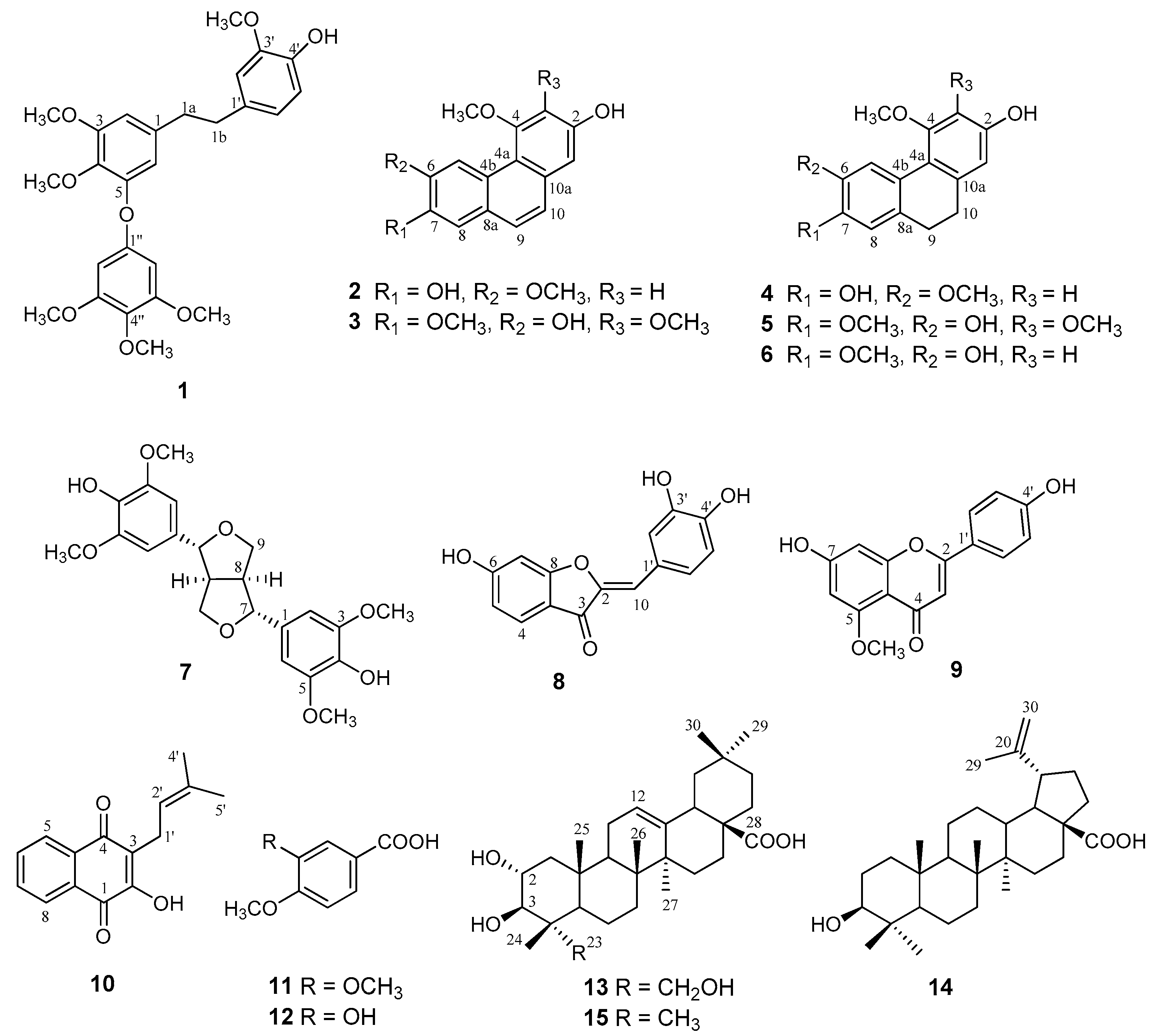
2.1. Extraction, Isolation, and NMR Spectroscopic Data

After a combination of column chromatography on silica gel, gel filtration on Sephadex LH-20 and reversed-phase HPLC separations of the CH2Cl2 phase resulting from partitioning of the EtOH extract from the roots of C. laxum, compounds 17, and 1315, comprising phenanthrenes, dihydrophenanthrenes, dihydrostilbene, lignan, and triterpenes (Figure 1), were obtained.
Compound 1 was isolated as a yellow powder. Its molecular formula was determined to be C26H30O8, as revealed from its HRESIMS (high resolution electrospray ionization mass spectrometry) data (m/z 509.1588 [M + K]+) data (Figure S1). The aromatic nature of 1 was deduced by the presence of 16 carbon signals observed in the 13C NMR spectrum as seven methines, ranging from δC 93 to 122, and 11 quaternary carbons (including eight oxygen-bearing carbons) found between δC 132 to 135, as well as by proton resonances in the region of δH 6.08 to 6.68 (Table 1) [9]. These spectra also showed signals for six aromatic methoxy groups, wherein two were shown to be sterically hindered, as revealed by their chemical shifts at δC 61.0 and 61.3 [9]. In the 1H nuclear magnetic resonance (NMR) spectrum, two multiplets assignable to two pairs of methylene benzylic protons were observed at δH 2.74 and 2.76 [9,10], which in turn showed correlations in the HSQC (heteronuclear single quantum coherence) spectrum with the carbon signals at δC 39.4 and 38.6, respectively. These data, together with long-range HMBC (heteronuclear multiple bond correlation) correlations between the foregoing protons and benzene ring carbons at δC 134.7 and 139.3, respectively, were in accordance with the presence of a dihydrostilbene moiety in the structure of 1 [10,11]. Further information given by the chemical shifts and splitting patterns of the signals of the aromatic protons indicated that one benzene ring of the bibenzyl unit (ring A) was 3,4,5-trioxygenated, as revealed by a pair of meta-coupled protons at δH 6.25 and 6.30 (J = 3.0 Hz each) [9,10], which, in turn, showed cross-peak correlations in the HSQC spectrum with the carbon signals at δC 105.0 and 110.3, respectively. Three protons displayed as an ABC set at δH 6.64 (d, J = 2.0 Hz), 6.68 (d, J = 9.0 Hz), and 6.60 (dd, J = 9.0, 2.0 Hz) supported a 3,4-dioxygenated substitution pattern for the other benzene ring (ring B) in the structure of 1 [9,10]. The remaining signals observed in the 1H and 13C NMR spectra of 1 were ascribable to a 1,3,4,5-tetraoxygenated benzene ring, as shown by the two-proton singlet at δH 6.08 and carbon resonances at δC 155.4 (C), 94.0 (2 × CH), 155.0 (2 × C). Compound 1 was thus assumed to be an oxygenated dihydrostilbene derivative, bearing an additional 3,4,5-trioxygenated phenoxy substituent. The HMBC experiments allowed the positions of the oxygenated functions in the benzene rings A and B of the dihydrostilbene moiety, as well as in the trioxygenated phenoxy substituent to be ascertained. Accordingly, the signal at δH 3.77 related to one of the aromatic methoxy groups showed a three-bond proton-carbon correlation with the carbon signal at δC 148.7 in the HMBC spectrum. A prominent cross-peak between the latter and the doublet at δH 6.68 (H-5′, 3J), as well as a two-bond correlation with the doublet at δH 6.64, established the location of the OCH3 group in the 3,4-dioxygenated B ring at C-3′. No correlations were found between the other methoxy protons and the carbon resonances assigned to the B ring, thus supporting the placement of a hydroxy group at C-4′ (δC 145.6). Long-range connectivities between C-4′ and H-2′, H-5′ and H-6′ corroborated these assignments. Other correlations discernible in the HSQC and HMBC spectra allowed the positions of the methoxy groups at δH 3.75/δC 56.3 and δH 3.73/δC 61.0 to be established at C-3 and C-4 of the A ring, respectively, as well as the linkage site of the trimethoxylated phenoxy substituent at C-5. Particularly, the signal at δH 6.25 related to H-2 showed two- and three-bond correlations with the carbon signals at δC 154.2 and 135.8, which were thus assigned to C-3 and C-4, respectively. The latter also displayed a three-bond-correlation with H-6 (δH 6.30). The HMBC spectrum also exhibited a two-bond correlation between H-6 and the carbon signal at δC 151.2 attributed to C-5, whose chemical shift was in accordance with that of an aromatic carbon linked to a phenoxy substituent. From the foregoing data, the structure of compound 1 was deduced to be 4′-hydroxy-3,3′,4-trimethoxy-5-(3,4,5-trimethoxyphenoxy)-bibenzyl, hitherto unreported in the literature.
The molecular formula of compound 2 was deduced as C16H14O4 based on the [M + H]+ ion at m/z 271.0963 in the HRESIMS (Figure S8), indicating 10 degrees of unsaturation. The 1D-NMR data of 2 clearly revealed its aromatic nature and supported the presence of eight fully substituted carbons (four of which oxygenated), and six methine carbons, in addition to two aromatic methoxy groups, evidenced at δH 4.02/δC 56.2 and δH 4.12/δC 56.0 (Table 2). The 1H NMR spectrum of 2 showed a pair of meta-coupled protons in a 1,2,3,5-tetrasubstituted benzene ring, at δH 6.79 and 6.89 (J = 3.0 Hz each), two isolated protons at δH 7.24 (s) and 9.11 (s), and a pair of ortho-coupled protons at δH 7.44 and 7.56 (d, J = 9.0 Hz each). The chemical shifts and splitting patterns of these last two protons were shown to be characteristic of H-9 and H-10 of a phenanthrene derivative [12], which in turn showed cross-peak correlations in the HSQC spectrum with carbon resonances at δC 127.9 and 125.4, respectively. Therefore, resonances of the doublets at δH 6.79 and 6.89 corresponding to the meta-coupled protons, and that of the one-proton singlet at δH 9.11, together with their respective 1H-13C connectivities detectable in the HSQC spectrum, implied that the structure of 2 comprised a 2,4,6,7-tetraoxygenated phenanthrene skeleton. Considering that only two methoxy groups were identified in the NMR spectra, the remaining oxygenated functions in the structure of 2 must be attributed to the presence of two hydroxyls. The signals at δH 7.24 and 9.11 were thus ascribed to H-8 and the anisotropically deshielded H-5, respectively [13], which depicted correlations with the corresponding carbon resonances at δC 112.2 and 109.6 in the HSQC spectrum. HMBC long-range connectivities from H-5 and H-8 to C-6 (δC 148.3, 2J and 3J, respectively), C-7 (δC 145.8, 3J and 2J, respectively), and C-4b (δC 125.5, 2J and 3J, respectively), in addition to 3J couplings between H-5 and C-4a (δC 115.5) and C-8a (δC 128.2), and between H-8 and C-9 (δC 127.9) were consistent with the foregoing assignments. The relative positions of the hydroxy and methoxy functionalities in 2 were ascertained from key nuclear Overhauser effect (NOE) relationships found between the methoxy at δH 4.12 and H-3 (d, δH 6.79), and between the methoxy at δH 4.02 and H-5 (δH 9.11), thus indicating the location of these OCH3 groups at C-4 and C-6, respectively. The linkage sites of the hydroxy functions were therefore determined at C-2 and C-7. Compound 2 was thus shown to be 2,7-dihydroxy-4,6-dimethoxyphenanthrene, whose spectroscopic data agreed with those reported for this phenanthrene obtained from Bulbophyllum vaginatum (Orchidaceae) [14], which is thus being described for the first time in the Combretaceae.
The molecular formula of 3 was established as C17H16O5, as deduced by an [M + H]+ ion at m/z 301.1076 in the HRESIMS (Figure S15). This data, together with analysis of the 1H and 13C NMR spectra of 3, revealed their high similarity to those of 2 (Table 2). However, the spectrometric data of 3 indicated the presence of five oxygenated substituents (three of which as methoxy groups), instead of four as in compound 2, located at carbons C-2, C-3, C-4, C-6, and C-7 in the phenanthrene skeleton. The foregoing information were supported by a singlet at δH 7.04 ascribable to H-1, which showed a connectivity with the carbon signal at δC 109.8 in the HSQC spectrum, which in turn displayed a three-bond correlation with H-10 (δH 7.33, d, J = 9.0 Hz) in the HMBC spectrum. Likewise, long-range connectivities were observed between H-1 and C-2 (δC 150.4, 2J), C-3 (δC 142.9, 3J), C-10 (δC 124.9, 3J), and C-4a (δC 118.9, 3J). Since the chemical shifts of two of the methoxy carbons (δC 60.5 and 61.5) revealed their sterically hindered nature, they were placed at C-3 and C-4 positions. Three-bond correlations between the methoxyl protons at δH 4.00, 3.98, and 3.99 and C-3, C-4 (δC 152.8), and C-7 (δC 148.4), respectively, determined the attachment of the methoxy groups to these corresponding aromatic carbons, therefore establishing the location of the hydroxy functions at C-2 (δC 150.4) and C-6 (δC 147.4). Further correlations discernible in the HMBC spectrum between H-5 (δH 8.90) and both C-6 (δC 147.4, 2J) and C-7 (δC 148.4, 3J), H-8 (δh 7.27) and both C-6 (3J) and C-7 (2J), and H-9 (δH 7.50) and C-8 (δC 109.6, 3J), together with NOE-cross peaks observed between H-8 and OCH3 at δH 3.99, and between H-5 and OCH3 at δH 3.98, reinforced these assignments. The foregoing data could be satisfactorily assembled to establish the structure of 3 as 2,6-dihydroxy-3,4,7-trimethoxyphenanthrene, formerly obtained as a plant constituent only from Combretum apiculatum, but with no full description of its 1H and 13C NMR data [15], which are being reported herein for the first time.
Compound 4 had the molecular formula of C16H16O4 (nine degrees of unsaturation), as determined by HRESIMS (m/z 273.1118, [M + H]+) [Figure S21]. Its 1H and 13C NMR data closely resembled those of 2 (Table 2), except for the lack of signals at δH 7.56 and 7.44 assigned to H-9 and H-10, respectively, in the phenanthrene skeleton of 2, and the presence instead of two-proton multiplets at δH 2.58 and 2.60 attributable to benzylic methylene groups of a 9,10-dihydrophenanthrene derivative [16]. Likewise, the signals at δC 127.9 (C-9) and 125.4 (C-10) in the 13C NMR spectrum of 2 were replaced by δC 32.1 and 30.2, respectively, in the spectrum of 4, therefore establishing the structure of 4 as 2,7-dihydroxy-4,6-dimethoxy-9,10-dihydrophenanthrene. Additional evidence for structure 4 was provided by correlations observed in the HSQC, HMBC and NOESY spectra. The spectrometric data of 4 were in accordance with those of 6-methoxycoelonin [17], previously isolated from the orchid Cymbidium aloifolium [18] and further obtained from other Orchidaceae [17,19,20], in addition to Dioscorea nipponica (Dioscoriaceae) [21], but scarcely reported in the Combretaceae, e.g., in Combretum hereroense [22].
Compound 5 was assigned the molecular formula C17H18O5 on the basis of its HRESIMS (m/z 303.1235, [M + H]+) [Figure S29], with nine degrees of unsaturation. 1D- and 2D-NMR spectroscopic analysis of 5 revealed, as occurred with compounds 2 and 4, that the structure of 5 differed from that of 3 only for the presence in the former of methylene sp3 carbons at C-9 and C-10 (Table 2). This assumption was confirmed by the signals at 30.4 and 31.5 in the 13C NMR spectrum, which showed correlations with the four-proton singlet at 2.60 in the HSQC spectrum, in addition to further information given by the 1D- and 2D-NMR spectra, including those provided by a NOESY experiment. Thus, compound 5 was shown to be 2,6-dihydroxy-3,4,7-trimethoxy-9,10-dihydrophenanthrene, a previously reported dihydrophenanthrene isolated from Combretum molle and C. apiculatum [15,23]. Its 1H and 13C NMR data are being reported for the first time.
Compound 6 had a molecular formula of C16H16O4 as determined from the [M + H]+ ion at m/z 273.1121 in the HRESIMS (Figure S37). Comparison of the 1D- and 2D-NMR spectra of 6 with those of 5 indicated their close relationship (Table 2), except for the absence of a methoxy group at C-3 in the former, as revealed by the signals of two meta-coupling protons at δH 6.30 (J = 3.0 Hz, H-1) and 6.39 (J = 3.0 Hz, H-3). Correlations in the HSQC spectrum between H-1/C-1 (δC 108.5) and H-3/C-3 (δC 99.3), as well as further correlations observed in the HSQC, HMBC and nuclear Overhauser effect spectroscopy (NOESY) spectra, lent support to these assignments. Compound 6 was thus identified as 2,6-dihydroxy-4,7-dimethoxy-9,10-dihydrophenanthrene, whose NMR data agreed with those reported for callosin, previously isolated only from two species of the Orchidaceae (Agrostophyllum callosum and Coelogyne flaccida [17,24]. Therefore, this is the first reported occurrence of callosin from a member of the Combretaceae.
The signals in the 1H NMR spectrum of compound 7 assignable to a symmetric molecule bearing two 3,5-dimethoxy-4-hydroxy substituted aromatic rings at δH 6.65 (s, 4H) and 3.84 (s, 12H), together with those belonging to a spin system at δH 4.70 (d, J = 3.0 Hz, 2H), 3.13 (brs, 2H), 3.70–3.80 (m, 2H), and 4.18–4.24 (m, 2H), suggested that 7 was a bistetrahydrofuran lignan. This assumption was supported by the eight signals observed in the 13C NMR, in which four of them were attributed to the symmetrically substituted aromatic rings, namely four methines (δC 104.5), six oxygenated carbons (δC 136.2 and 149.3), and two carbons linked to the bistetrahydrofuran moiety (δC 133.1). The remaining four signals were ascribed to the four methoxy groups at δC 56.8, and to the methine (δC 55.5 and 72.5) and methylene (δC 87.6) carbons of the foregoing bistetrahydrofuran residue. These assignments were further corroborated by HSQC and HMBC correlations, while the carbon resonances of the tetrahydrofuran rings were indicative of the pseudoequatorial orientation of the aromatic rings in the structure of 7, as well as established their linkage to C-7/C-7′ [25]. The optical rotation value and NMR spectroscopic characteristics of 7 agreed with those of the lignan (+)-syringaresinol [26], which is being reported for the first time in the genus Combretum. Despite their wide distribution in plants, lignans have been scarcely found in the Combretaceae, particularly within the genus Combretum, with only two records in C. fruticosum and C. alfredi [27,28].
The identities of the pentacyclic triterpenes arjunolic (13), betulinic (14), and maslinic (15) acids were verified by comparing their NMR spectroscopic data with those of authentic samples [8,29]. Triterpenes 13 and 14 have already been isolated in our previous study on the stems of C. laxum [8], while maslinic acid is of common occurrence in species of the Combretaceae, including those belonging to the genus Combretum [30].
After partitioning of the EtOH extract from the stems of C. laxum, the resulting CH2Cl2 phase afforded compounds 812—comprising an aurone, a flavone, a naphthoquinone, and two benzoic acid derivatives (Figure 1)—after fractionation procedures by silicagel and Sephadex LH-20 column chromatography, and reversed-phase high performance liquid chromatography (HPLC).
Analysis of the 1H NMR spectrum of compound 8 revealed the presence of two sets of signals for a total of six aromatic protons, and a vinylic singlet at δH 6.58. These data, together with 15 signals in the range of δC 99–169 in the 13C NMR spectrum, indicated the flavonoid nature of 8. The oxygenation pattern of rings A and C was defined by the characteristic signals of a 6,3′,4′-trihydroxylated flavonoid [31]. The signal at δH 6.58 was assigned to a methine proton attributable to H-10 of an aurone. This assumption was confirmed by the olefinic carbon resonances at δC 112.4 (CH) and 147.2 (C), thus assigned to C-10 and C-2, respectively. These data, along with additional information provided by 1H-1H correlation spectroscopy (COSY), HSQC, and HMBC experiments, led to the identification of 8 as the aurone sulfuretin, whose spectroscopic data were comparable to those obtained for 8 [32]. Although the 1H and 13C NMR data of 8 agreed with those published for sulfuretin, the previously reported resonance values for H-4 (δ 6.84) and H-5′ (δ 7.59) should be interchanged. This assumption was substantiated by correlations observed in the 1H-1H COSY spectrum of 8 between H-4 (δ 7.56) and H-5 (δ 6.72) and between H-5′ (δ 6.89) and H-6′ (δ 7.27), together with long-range connectivities observed in the HMBC spectrum from H-5′ to C-3′ (146.4) and C-6′(125.4), thus allowing unambiguous assignments of H-4 and H-5′ resonances as shown. Despite being present in various plant sources, the isolation of sulfuretin from C. laxum is noteworthy, because not only is it being reported for the first time in the Combretaceae, but also it is the first occurrence of aurones in this family.
Compound 9 exhibited in its 1H NMR spectrum characteristic signals of a 5,7,4′-trioxygenated flavone, viz. a typical proton singlet at δH 6.48 ascribed to H-3, along with a pair of broad singlets at δH 6.34 and 6.46 of meta-coupled protons at ring A (H-6 and H-8, respectively), and a pair of doublets of a para-oxygenated ring B at δH 7.78 (2H, J = 9.0 Hz, H-2′/H-6′) and 6.89 (2H, J = 9.0 Hz, H-3′/H-5′) [31]. This spectrum also showed a three-proton singlet at δH 3.85 ascribed to a methoxy group. Characteristic signals of ring C carbons were observed at δC 161.6 (C-2), 106.2 (C-3), and 180.2 (C-4) [33]. The linkage of the methoxyl group to C-5 was established by HMBC coupling of the methoxyl hydrogens to C-5 (δC 162.4), which was corroborated by a correlation discernible in the NOESY spectrum between H-6 and the methoxyl hydrogens. Therefore, compound 9 was shown to be 5-methoxy-7,4′-dihydroxyflavone, also known as 5-O-methyl apigenin, whose spectroscopic data were comparable to those of 9 [34]. This flavone derivative, with restricted distribution in plant species, is being reported for the first time in the Combretaceae.
The 1H NMR spectrum of 10 showed a pair of doublets at δH 8.05 and 8.10 (J = 6.0 Hz) and a pair of triplets of doublets at δH 7.73 and 7.66 (J = 7.0 and 1.5), which, together with the chemical shifts observed in the 13C NMR spectrum in the range of δC 126–135, were attributed to an ortho-substituted aromatic ring. Evidence of a 2-hydroxy-1,4-naphthoquinone skeleton bearing a side chain at C-3 was given by the presence of two carbonyl resonances at δC 181.7 and 184.5, as well as the signals of two substituted sp2 carbons at δC 123.5 and 152.7. The nature of the side chain was promptly established as a prenyl group, based on its characteristic methyl singlets at δHc 1.67/25.7 and 1.77/17.9, as well as the signals ascribed to a trisubstituted double bond linked to methylene group, evidenced by the one-proton broad triplet at δH 5.19 (J = 6.0 Hz) and a two-proton doublet at δH 3.29 (J = 6.0 Hz), respectively. Accordingly, the remaining carbon resonances of the prenyl group were observed at δC 119.6 (C-2′), 133.8 (C-3′) and 22.6 (C-1′). Connectivities discernible from HSQC and HMBC experiments provided further evidence for the structure of compound 10, which was identified as shown. Its 1H and 13C NMR data were in accordance with those reported in the literature for the 1,4-naphthoquinone known as lapachol [35]. Lapachol, which occurs in a number of plants belonging to several families, has long been recognized for its wide array of biological activities, particularly significant antitumor-promoting effects [36,37]. The isolation of lapachol from C. laxum is remarkable, since, to our knowledge, no reports on the occurrence of this or any other naphthoquinone representatives in the Combretaceae have hitherto been found in the literature.
Compounds 11 and 12 were readily identified as the benzoic acid derivatives 3,4-dimethoxybenzoic and 3-hydroxy-4-methoxybenzoic acids, respectively, whose NMR data were in full agreement with those reported in the literature [38].

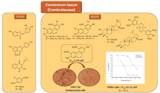
2.2. In Vitro Cytotoxic Evaluations

Given the known antineoplastic potentialities of phenanthrene, dihydrophenanthrene and dihydrostilbenoid derivatives, particularly those obtained from members of the Combretaceae and Orchidaceae [7,39,40], compounds 1-6 were further assessed for their in vitro antiproliferative effects against five human neoplastic cell lines (except for 1 and 3, which were tested against four cell lines due to insufficient material), based on the SRB (sulforhodamine B) assay and using cisplatin as a positive control.
As depicted in Table 3, all compounds showed inhibitory activities against at least one of the five cell lines tested, with 6-methoxycoelonin (4) displaying a remarkable effect against UACC-62 cells (IC50 2.59 ± 0.11 µM). This dihydrophenanthrene derivative proved not only seven times more active against this cell line than cisplatin, but also at least 86 times more potent than the other compounds tested. This result led us to assess the effect of 4 on nontumor mammalian VERO cells in order to determine its selectivity index. The obtained IC50 value, 65.12 ± 4.51 µM, revealed that 4 is roughly 25 times more selective for UACC-62 cells than for nontumor cells (SI = 25.1). In addition, 4 inhibited the proliferation of UACC-62 and VERO cells in a dose-dependent manner. As shown in Figure 2, the points above zero in the curves indicate that 6-methoxycoelonin had a cytostatic (growth inhibition) effect on UACC-62 and VERO cells at the concentrations of 0.25, 2.5 and 25 µg mL−1. In addition, as revealed by the points below zero in the growth curves, while 4 had a cytocidal (cell death) effect on UACC-62 cells from the concentration of 2.5 µg mL−1, the viability of nonneoplastic VERO cells at this same concentration remained close to 100% (p < 0.5).
A literature survey on the cytotoxic potentialities of the known compounds 26 revealed that the antiproliferative effects of 6-methoxycoelonin (4) against UACC-62, 786-0, Hep-2, and NCI/ADR RES cells are being described for the first time, while, to our knowledge, 2,7-dihydroxy-4,6-dimethoxyphenanthrene (2), 2,6-dihydroxy-3,4,7-trimethoxyphenanthrene (3), 2,6-dihydroxy-3,4,7-trimethoxy-9,10-dihydrophenanthrene (5), and callosin (6) have not yet been screened for their in vitro cytotoxic properties against any neoplastic cell line. Regarding the earlier reported effects of 4 against MCF-7 cells, two different IC50 values, namely 9.58 and 37.9 µM, were described for 6-methoxycoelonin [19,41], the latter being closer to that obtained in the present study.
Based on the IC50 values obtained for phenanthrene 2 compared with those of its corresponding 9,10-dihydro derivative 4, cytotoxicity is significantly enhanced in the latter by reduction of carbons C-9 and C-10, particularly against UACC-62 cells, wherein 4 was at least 96 times more potent than 2. Nevertheless, the assumption that cytotoxicity of the phenanthrenes and dihydrophenanthrenes might be directly related to the lack of aromaticity in ring B does not apply to phenanthrene 3 and its corresponding 9,10-dihydro derivative 5, since their cytotoxic effects do not follow the same uniform pattern against the cells tested as that of 2 and 4. Another significant feature observed for the tested dihydrophenanthrenes is that minimal structural differences, as that found between 4 and 6, may account for expressive effects on their cytotoxic potentials. Accordingly, as demonstrated by their IC50 values, activity of 6—which only differ from 4 by the change in position of the methoxyl and hydroxyl functionalities in ring A, at C-6 and C-7—is remarkably reduced against all cell lines when compared with that of 4. Likewise, by analyzing the effects of compounds 2, 4 and 6, the inversion between the substituents at C-6 and C-7 (as in 4 and 6) leads to a greater reduction of cytotoxicity than that caused by the maintenance of the aromaticity of ring in 2 when compared to its dihydro derivative 4. On the other hand, regarding the cytotoxic effects of 6 and 5, the presence of an additional methoxy substituent at C-3 renders 5 more potent against all cell lines tested. Therefore, at least with respect to the effects of the foregoing compounds against UACC-62 cells, the presence of methoxy and hydroxy groups at C-6 and C-7, respectively, as well as the lack of aromaticity of ring B can be considered as important structural features for cytotoxicity. Although no extensive studies on structure/activity relationships for natural phenanthrenes and dihydrophenanthrenes have been reported in the literature, some results from previous works suggest the relevance of the numbers and the substituted positions of methoxy and hydroxy groups in the phenanthrene/dihydrophenanthrene skeleton for the cytotoxic activity of these classes of natural compounds [19,20,42,43,44,45].

2.3. DPPH-Radical-Scavenging Assay

Antioxidants are known by their effects in the prevention of several oxidative stress associated diseases, such as cancer, given their ability to inhibit the oxidative damage to DNA caused by scavenging free radicals [46,47]. In this sense, the antioxidant potential of compounds 16 were further evaluated using the DPPH-radical-scavenging assay, in order to find whether the radical-scavenging ability of the structurally related compounds 26 correlated with their anticancer potential. As depicted in Table 4, compounds 2, 5, and 6 showed radical-scavenging capacity of similar potencies to the positive control caffeic acid, with IC50 values ranging from 17.7 ± 0.25 to 32.9 ± 0.25 µM, while 1, 3, and 4 had lower activities (IC50 values between 45.6 ± 0.35 and 56.5 ± 0.29 µM). Based on the results obtained for 2 and 4, it can be inferred that unsaturation at C-9/C-10 had a positive influence on the antioxidant capacity of 2 in this assay. In contrast, however, a decrease in the antioxidant ability of 3 was observed when compared with that of 5, thus suggesting that other structural features, such as the presence and/or nature of oxygenated substituents, might play key roles in the radical-scavenging capacity of phenanthrenes and their corresponding dihydro derivatives. Accordingly, significant differences in the DPPH-scavenging properties were observed for 4 and 6, although their structures only differ for the position of the hydroxy and methoxy groups at C-6 and C-7. Likewise, the introduction of a methoxy group at C-3, as in 5, lowers its scavenging capacity when compared with that of its structural related compound 6. The foregoing results also revealed that callosin (6), despite exhibiting the strongest DPPH-scavenging activity, was the least cytotoxic compound, indicating that, at least for the cell lines tested, antioxidant potential is unrelated to antiproliferative activity. Several phenanthrene and dihydrophenathrene derivatives are known for their DPPH scavenging properties [39]. However, literature data reveal that, as observed for compounds 26, no relevant structure-activity relationships could clearly be established within these chemical classes, being postulated that the antioxidant capacity seemed to be related with the number of phenolic hydroxyl groups, either alone or together with methoxy groups, among other particular structural characteristics [45,48,49]. Despite a number of reports on the cytotoxic potential of plant extracts together with their radical scavenging activity against DPPH, particularly from the Orchidaceae, which is recognized as a rich source of these phenanthrene- and dihydrophenathrene-type compounds [37,38,50,51,52], no comprehensive studies on possible relationships between structure and DPPH-scavenging/cytotoxic properties for these classes of compounds have, to our knowledge, been previously described.

3. Materials and Methods

3.1. General Experimental Procedures

HRESIMS data were acquired with electrospray ionization in negative ion mode on an UltrOTOF-Q instrument (Bruker Daltonics, Billerica, MA, USA). NMR spectroscopic data were recorded at room temperature in CDCl3, acetone-d6, CD3OD, and/or pyridine-d5 (Cambridge Isotope Laboratories, Andover, MA, USA) on a Bruker DPX-300 spectrometer (Bruker, Karlhue, Germany) operating at 300.13 MHz (1H)/75.47 MHz (13C). Standard pulse sequences were used for homo- and heteronuclear correlation experiments. Optical rotation was determined on a Perkin Elmer 341 polarimeter (λ = 589 nm, PerkinElmer Inc., Waltham, MA, USA). Column chromatography procedures were performed on silica gel 60 (70–230 mesh, Merck, Darmstadt, Germany), silica gel 60 RP-18 (230–400 mesh, Merck, Darmstadt, Germany) and Sephadex LH-20 (Amersham Biosciences, Buckinghamshire, UK). Reversed-phase semipreparative HPLC separations were carried out with a Shimadzu (Shimadzu, Kyoto, Japan) LC-6AD pump using a Phenomenex Luna RP-18 column (5 µm, 21.6 × 250 mm) at flow rates of 12 or 14 mL/min, with monitoring at 210, 230 or 254 nm.

3.2. Plant Material

Roots and stems of C. laxum were collected from Corumbá, Mato Grosso do Sul, Brazil, in July 2016. The plant material was identified by Prof. Arnildo Pott (Institute of Biosciences, Universidade Federal de Mato Grosso do Sul). A voucher specimen (no. 39343) has been deposited at the CGMS Herbarium of the Universidade Federal de Mato Grosso do Sul. License for research on Brazil’s biodiversity, #A5DBC20.

3.3. Extraction and Isolation

Air-dried and powdered roots of C. laxum (1422 g) were extracted at room temperature with EtOH. After concentration in vacuo, the residue obtained from the EtOH extract was partitioned between n-butanol and H2O 1:1. The resulting syrupy n-butanol phase was subsequently partitioned between MeOH–H2O 9:1 and hexane, and between MeOH–H2O 1:1 and CH2Cl2 to give the corresponding hexane (2.24 g) and CH2Cl2 (4.52 g) phases. The CH2Cl2 phase was chromatographed on a silica gel 70–230 mesh column, using step gradient elution with hexane, hexane–CH2Cl2 (1:1), CH2Cl2, CH2Cl2–EtOAc (1:1), and EtOAc to give 12 fractions (A → L). Fraction F (CH2Cl2 100%, 232.2 mg) was further separated by CC on Sephadex LH-20 (MeOH) to furnish six subfractions (F.1 → F.6). Compounds 3 (3.2 mg) and 4 (4.5 mg) were obtained from subfraction F.4 (22.0 mg), after reversed-phase semipreparative HPLC (MeO–H2O 40:60, 4.0 mL/min., 254 nm), while 2 (33.1 mg) was obtained from subfraction F.5. Fraction H (CH2Cl2–EtOAc 1:1, 1159.0 g) was chromatographed on RP-18 silica gel 230–400 mesh column by elution with a MeOH–H2O gradient (4:6, 6:4, 8:2) and MeOH, to afford four subfractions (H.1 → H.4). Compounds 1 (3.1 mg), 5 (14.2 mg), 6 (6.9 mg), 7 (6.9 mg), and further amounts of 4 (4.5 mg) were isolated from subfraction H.1 (MeOH–H2O 4:6, 152.3 mg), after reversed-phase semipreparative HPLC (MeCN–H2O 28:72, 14 mL/min., 254 nm). Reversed-phase semipreparative HPLC (MeCN–H2O 48:52, 14 mL/min., 254 nm) of subfraction H.3 (MeOH–H2O 8:2, 358.4 mg) yielded 13 (40.8 mg). Compounds 14 (8.5 mg) and 15 (5.3 mg) were obtained from subfraction H.4 (MeOH, 350.3 mg), after column chromatography on RP-18 silica gel 230–400 mesh, using step gradient elution with MeCN–H2O 6:4, 8:2, and MeOH 100%, followed by reversed-phase semipreparative HPLC (MeCN–H2O 60:40, 14 mL/min., 210 nm).
Air-dried and powdered stems of C. laxum (2760 g) were extracted at room temperature with EtOH. After concentration in vacuo, the residue obtained from the EtOH extract was subsequently partitioned between MeOH/H2O 9:1 and hexane, and between MeOH/H2O 1:1 and CH2Cl2 to give the corresponding hexane (1.50 g) and CH2Cl2 (1.10 g) phases. The CH2Cl2 phase was chromatographed on a silica gel 70–230 mesh column, using hexane, hexane–CH2Cl2 (3:1, 1:1, 1:3), CH2Cl2, CH2Cl2-EtOAc (3:1, 1:1, 1:3), EtOAc, and EtOAc–MeOH (9:1, 3:1, 1:1) as eluents, to furnish 12 fractions (A → L). Fraction D (hexane–CH2Cl2 1:3) gave 10 (8.1 mg), while fraction H (CH2Cl2 1:3, 104.9 mg) yielded 8 (3.0 mg), 11 (6.5 mg), and 12 (2.3 mg), after reversed-phase semipreparative HPLC (MeCN–H2O 28:72, 12 mL/min., 210 nm). Fraction J (EtOAc–MeOH 9:1, 150.6 mg) was re-chromatographed on Sephadex LH-20 (MeOH) to afford five subfractions (J.1 → J.5). Subfraction J.2 gave compound 13 (33.8 mg), while compound 9 (2.5 mg) was obtained from subfraction J.4 (11.3 mg), after separation by reversed-phase semipreparative HPLC (MeOH–H2O 62:38, 12 mL/min., 254 nm).
4′-Hydroxy-3,3′,4-trimethoxy-5-(3,4,5-trimethoxyphenoxy)-bibenzyl (1): amorphous solid; HRESIMS m/z 509.1588 [M + K]+ (calcd for C26H30O8K, 509.1572); 1H and 13C NMR data (Table 1).
2,7-Dihydroxy-4,6-dimethoxyphenanthrene (2): amorphous solid; HRESIMS m/z 271.0963 [M + H]+ (calcd for C16H15O4, 271.0970); 1H and 13C NMR data (Table 2).
2,6-Dihydroxy-3,4,7-trimethoxyphenanthrene (3): amorphous solid; HRESIMS m/z 301.1076 [M + H]+ (calcd for C17H17O5, 301.1076); 1H and 13C NMR data (Table 2).
6-Methoxycoelonin (4): amorphous solid; HRESIMS m/z 273.1118 [M + H]+ (calcd for C16H17O4, 273.1126); 1H and 13C NMR data (Table 2).
2,6-Dihydroxy-3,4,7-trimethoxy-9,10-dihydrophenanthrene (5): amorphous solid; HRESIMS m/z 303.1235 [M + H]+ (calcd for C17H19O5, 303.1233); 1H and 13C NMR data (Table 2).
Callosin (6): amorphous solid; HRESIMS m/z 273.1121 [M + H]+ (calcd for C16H17O4, 273.1126); 1H and 13C NMR data (Table 2).
(+)-Syringaresinol (7): amorphous solid; [α]D 20 + 11.1 (c 0.23, CH3OH); 1H-NMR (CD3OD): δ 3.13 (2H, brs, H-8, H-8′); 3.70-3.80 (2H, m; H-9a, H-9′a); 3.84 (12H, s, OCH3-3, 3′, 5, 5′); 4.18-4.24 (2H, m, H-9b, H-9′b); 4.70 (2H, d, J = 3.0 Hz, H-7, H-7′); 6.65 (4H, s, H-2, H-2′, H-6, H-6′). 13C-NMR (CD3OD): δ 133.1 (C-1, C-1′); 104.5 (C-2, C-2′, C-6, C-6′); 149.3 (C-3, C-3′, C-5, C-5′); 136.2 (C-4, C-4′); 87.6 (C-7, C-7′); 55.5 (C-8, C-8′); 72.5 (C-9a, C-9′a); 56.8 (OCH3-3, 3′, 5, 5′).
Sulfuretin (8): amorphous solid; 1H-NMR (acetone-d6): δ 6.58 (1H, s, H-10); 6.72 (1H, dd, J = 8.5 and 1.5 Hz, H-5); 6.79 (1H, d, J = 1.5 Hz, H-7); 6.89 (1H, d, J = 8.3 Hz, H-5′); 7.27 (1H, dd, J = 8.3 and 2.2 Hz, H-6′); 7.56 (1H, d, J = 8.5 Hz, H-4). 13C-NMR (acetone-d6): δ 147.2 (C-2), 182.5 (C-3); 126.3 (C-4); 113.8 (C-5); 167.7 (C-6); 99.4 (C-7); 169.0 (C-8); 114.4 (C-9); 112.4 (C-10); 125.2 (C-1′); 118.7 (C-2′); 146.4 (C-3′); 148.6 (C-4′); 116.6 (C-5′); 125.4 (C-6′).
5-O-Methyl Apigenin (9): amorphous solid; 1H-NMR (CD3OD): δ 3.85 (3H, s, OCH3-5); 6.34 (1H, brs, H-6); 6.46 (1H, brs, H-8); 6.48 (1H, s, H-3); 6.89 (2H, d, J = 9.0 Hz, H-3, H-5′′); 7.78 (2H, d, J = 9.0 Hz, H-2′, H-6′). 13C-NMR (CD3OD): δ 161.6 (C-2); 106.2 (C-3); 180.2 (C-4); 162.4 (C-5); 98.7 (C-6); 163.5 (C-7); 97.2 (C-8); 123.4 (C-1′); 129.0 (C-2′, C-6′); 117.0 (C-3′, C-5′); 162.3 (C-4′); 56.3 (OCH3-5).
Lapachol (10): amorphous solid; 1H-NMR (CDCl3): δ 1.67 (3H, s, H-4′); 1.77 (3H, s, H-5′); 3.29 (2H, d, J = 6.0 Hz, H-1′); 5.19 (1H, brt, J = 6.0 Hz, H-2′); 7.66 (1H, td, J = 7.0 and 1.5 Hz, H-7); 7.73 (1H, td, J = 7.0 and 1.5 Hz, H-6); 8.05 (1H, d, J = 6.0 Hz, H-8); 8.10 (1H, d, J = 6.0 Hz, H-5). 13C-NMR (CD3OD): δ 181.7 (C-1); 152.7 (C-2); 123.5 (C-3); 184.5 (C-4); 126.8 (C-5); 134.8 (C-6); 132.8 (C-7); 126.0 (C-8); 129.4 (C-9); 132.9 (C-10); 22.6 (C-1′); 119.6 (C-2′); 133.8 (C-3′); 25.7 (C-4′); 17.9 (C-5′).
3,4-Dimethoxybenzoic Acid (11): amorphous solid; 1H-NMR (CD3OD): δ 3.85 * (3H, s, OCH3-3); 3.87 * (3H, s, OCH3-4); 6.98 (1H, d, J = 9.0 Hz, H-5); 7.54 (1H, brs, H-2); 7.64 (1H, brd, J = 9.0 Hz, H-6). 13C-NMR (CD3OD): δ 124.3 (C-1); 113.6 (C-2); 150.0 (C-3); 154.6 (C-4); 111.8 (C-5); 125.0 (C-6); 169.9 (C-7); 56.4 * (OCH3-3 and OCH3-4) * interchangeable signals.
3-Hydroxy-4-methoxybenzoic Acid (12): amorphous solid; 1H-NMR (CD3OD): δ 3.89 (3H, s, OCH3-4); 6.83 (1H, brd, J = 9.0 Hz, H-5); 7.55 (1H, d, J = 9.0 Hz, H-6); 7.56 (1H, brs, H-2). 13C-NMR (CD3OD): δ 123.6 (C-1); 113.8 (C-2); 152.4 (C-3); 148.6 (C-4); 115.8 (C-5); 125.2 (C-6); 169.8 (C-7); 56.4 (OCH3-4).
Arjunolic Acid (13): amorphous solid; 1H-NMR (CD3OD): δ 0.68 (3H, s, H-24); 0.80 (3H, s, H-26); 0.91 (3H, s, H-29); 0.93 (3H, s, H-30); 1.01 (3H, s, H-25); 1.15 (3H, s, H-27); 2.84 (1H, dd, J = 12.8 and 3.4 Hz, H-18); 3.30 (1H, d, J = 9.0 Hz, H-3); 3.33 (1H, d, J = 12.0 Hz, H-23a); 3.49 (1H, d, J = 12.0 Hz, H-23b); 3.68 (1H, m, H-2); 5.24 (1H, brs, H-5); 13C-NMR (CD3OD): δ 47.6 (C-1); 69.7 (C-2); 78.2 (C-3); 44.1 (C-4); 48.2 (C-5); 19.1 (C-6); 33.8 (C-7); 40.5 (C-8); 47.9 (C-9); 39.0 (C-10); 24.0 * (C-11); 123.4 (C-12); 145.4 (C-13); 43.0 (C-14); 28.8 (C-15); 24.6 * (C-16); 47.6 (C-17); 42.7 (C-18); 47.2 (C-19); 31.6 (C-20); 34.9 (C-21); 33.3 (C-22); 66.4 (C-23); 13.9 (C-24); 17.8 (C-25); 17.6 (C-26); 26.5 (C-27); 181.9 (C-28); 33.6 (C-29); 24.0 (C-30) * interchangeable signals.
Betulinic Acid (14): amorphous powder; 1H- and 13C-NMR data in accordance with those of an authentic sample and with literature [8].
Maslinic Acid (15): amorphous solid; 1H-NMR (pyridine-d5): δ 0.93 (3H, s, H-30); 0.97 (3H, s, H-29); 0.98 (3H, s, H-25); 1.00 (3H, s, H-23); 1.06 (3H, s, H-26); 1.25 (3H, s, H-27); 1.26 (3H, s, H-24); 3.30 (1H, m, H-18); 3.38 (1H, d, J = 10.0 Hz, H-3); 4.11 (1H, ddd, J = 13.0, 9.0 and 3.0 Hz); 5.45 (1H, brs; H-12). 13C-NMR (pyridine-d5): δ 46.4 (C-1); 68.6 (C-2); 83.8 (C-3); 39.8 (C-4); 55.9 (C-5); 18.8 (C-6); 33.2 (C-7); 39.8 (C-8); 48.1 (C-9); 38.5 (C-10); 23.7 (C-11); 122.4 (C-12); 144.8 (C-13); 42.2 (C-14); 28.2 (C-15); 23.7 (C-16); 46.6 (C-17); 42.0 (C-18); 46.4 (C-19); 30.9 (C-20); 34.2 (C-21); 33.2 (C-22); 29.3 (C-23); 17.6 (C-24); 16.8 (C-25); 17.4 (C-26); 26.1 (C-27); 180.2 (C-28); 33.3 (C-29); 23.7 (C-30).

3.4. In Vitro Cytotoxic Assay

Cytotoxicity of compounds 16 was evaluated against five human neoplastic cell lines—namely, MCF-7 (breast), 786-0 (kidney), UACC-62 (melanoma), NCI/ADR-RES (ovary, multidrug-resistant phenotype), and Hep2 (larynx), all of which were kindly provided by Prof. João Ernesto de Carvalho, of the School of Pharmaceutical Sciences, CPQBA, Universidade Estadual de Campinas, Campinas, Brazil. VERO (monkey kidney) nonneoplastic cells were obtained from the Rio de Janeiro cell bank. To this end, a sulforhodamine B (SRB; purity ≥ 97%; Sigma, St. Louis, MO, USA) assay was performed, as described elsewhere [53,54]. Cisplatin (purity ≥ 99.9%; Sigma, St. Louis, MO, USA) was used as the positive control. Each sample was tested in triplicate at four different concentrations (0.25, 2.5, 25, and 250 μg mL−1). IC50 values were calculated from the differences in absorbance readings at 540 nm in untreated (negative control) and treated cells on a SpectraMax 190 microplate reader (Molecular Devices, San Jose, CA, USA) [53] through nonlinear regression analysis, using Origin 6.0 software (OriginLab, Northampton, MA, USA), and growth percentages were calculated as described elsewhere [53]. The data presented are the mean ± standard deviation of at least three independent cell preparations made in triplicate. Statistical analysis was performed with OriginPro 9.55 (OriginLab, Northampton, MA, USA) applying t-test for pairwise comparison (threshold value p < 0.05).

3.5. DPPH-Radical-Scavenging Assay

The radical-scavenging activities of compounds 16 were determined using DPPH (a stable free radical), employing the method of microdilution in 96-well microplates described by Zhang et al. and Yamaguchi et al. [49,55], with some modifications. The assays were performed in triplicate, using caffeic acid as a standard compound and a DPPH solution in EtOH (200 µM) as a negative control. Solutions of samples in EtOH at 200 µM were serially diluted to 100, 50, 25, 12.5, and 6.25 µM. Each solution (100 µL) was mixed with 100 µL of DPPH solution. The samples were allowed to stand at room temperature in the dark for 30 min, after which their absorbances were recorded at 515 nm. The ability of test materials to scavenge DPPH radicals was calculated as follows: DPPH scavenging effect (%) = 100 (Acontrol − Asample)/Acontrol. Radical-scavenging activities were assessed on the basis of their IC50 values determined by linear regression.

4. Conclusions

The foregoing results constitute new information on the chemical composition of a specimen of C. laxum from the Brazilian Pantanal. Among the one new and 14 known compounds comprising nine different classes of secondary metabolites, the isolation of dihydrostilbenoid, phenanthrene and dihydrophenanthrene derivatives is unprecedented in a Combretum species native to the American continent. Also remarkable is the presence of aurone and naphthoquinone representatives, since these chemical classes are being reported for the first time in the Combretaceae, as are the isolation of phenanthrene 2, dihydrophenanthrene 6, and flavone 9, and the first reported occurrence of lignan 7 in the genus Combretum. The results of the present study also revealed that, at least with respect to the effects of compounds 26 against melanoma (UACC-62) cells, the presence of methoxy and hydroxy groups at C-6 and C-7, respectively, as well as the lack of aromaticity of ring B can be considered as important structural features for cytotoxicity. On the other hand, when compared with their radical-scavenging ability against DPPH, cytotoxicity of 16 is unrelated to their antioxidant potential, at least for the five cancer cell lines tested.
The anticarcinogenic, antimetastatic, and chemopreventive potentialities of plant-derived compounds either isolated or in combination with chemotherapy drugs have been the subject of an increasing number of recent preclinical and clinical studies aiming at the development of new antineoplastic agents. These studies reveal that combination of synthetic chemotherapy drugs with selected plant constituents not only may improve pharmacological activity and simultaneously minimize toxic side effects of synthetic chemical drugs, but also delay or even overcome the development of drug resistance [56,57,58,59,60,61,62]. The IC50 and selectivity index values presented by 4 (6-methoxycoelonin) against melanoma (UACC-62) cells thus indicate that this dihydrophenanthrene derivative can be considered as a promising candidate for further investigation of its mechanism of action. Future research on association of 6-methoxycoelonin with current anticancer drugs aiming at the development of potential new drug combination therapies within clinical oncology is also strongly encouraged.

Supplementary Materials

Supplementary Materials (Figures S1–S82) are available online.

Author Contributions

Conceptualization, F.R.G.; Formal analysis, E.B., T.V.F., N.C.Y., W.S.G. and F.R.G.; Funding acquisition, W.S.G. and F.R.G.; Investigation, E.B. and L.M..M.Q.; Methodology, E.B., M.d.F.C.M., R.T.P. and F.R.G.; Project administration, M.d.F.C.M., R.T.P. and F.R.G.; Resources, W.S.G., M.d.F.C.M., R.T.P. and F.R.G.; Supervision, W.S.G. and F.R.G.; Validation, E.B., L.M.M.Q., M.d.F.C.M. and R.T.P.; Visualization, T.V.F., N.C.Y., M.d.F.C.M., R.T.P. and F.R.G.; Writing–original draft, E.B., T.V.F., N.C.Y. and F.R.G.; Writing–review & editing, T.V.F., N.C.Y. and F.R.G. All authors have read and agreed to the published version of the manuscript.

Funding

This research was supported by Fundação de Apoio ao Desenvolvimento do Ensino, Ciência e Tecnologia do Estado de Mato Grosso do Sul (FUNDECT-MS), grant number 020793, and Coordenação de Aperfeiçoamento de Pessoal de Nível Superior (CAPES), grant number 001.

Acknowledgments

The authors are grateful to Fundação de Apoio ao Desenvolvimento do Ensino, Ciência e Tecnologia do Estado de Mato Grosso do Sul - FUNDECT-MS (grant 020793), Coordenação de Aperfeiçoamento de Pessoal de Nível Superior-CAPES (Finance Code 001), and CPq-PROPP-UFMS for their financial support. Thanks are also given to Conselho Nacional de Ciência e Tecnologia-CNPq (EB) for the grants awarded, and to Norberto P. Lopes (Faculdade de Ciências Farmacêuticas, USP, Ribeirão Preto, Brazil) for the HRESIMS. Arnildo Pott (CGMS Herbarium, Universidade Federal de Mato Grosso do Sul, Campo Grande, MS, Brazil) is acknowledged for his assistance in the identification of the plant material.

Conflicts of Interest

The authors declare no conflict of interest.

References

  1. Pettit, G.R.; Singh, S.B.; Niven, M.L.; Hamel, E.; Schmidt, J.M. Antineoplastic agents.124. Isolation, structure, and synthesis of Combretastatins A-1 and B-1, potent new inhibitors of microtubule assembly, derived from Combretum caffrum. J. Nat. Prod. 1987, 50, 119–131. [Google Scholar] [CrossRef] [PubMed]
  2. Fyhrquist, P.; Mwasumbi, L.; Haeggstrom, C.A.; Vuorela, H.; Hiltunen, R.; Vuorela, P. Ethnobotanical and antimicrobial investigation on some species of Terminalia and Combretum (Combretaceae) growing in Tanzania. J. Ethnopharmacol. 2002, 79, 169–177. [Google Scholar] [CrossRef]
  3. de Morais Lima, G.R.; Praxedes de Sales, I.R.; Dutra Caldas Filho, M.R.; Taveira de Jesus, N.Z.; Falcão, H.D.S.; Barbosa-Filho, J.M.; Silveira Cabral, A.G.; Souto, A.L.; Tavares, J.F.; Batista, L.M. Bioactivities of the genus Combretum (Combretaceae): A review. Molecules 2012, 17, 9142–9206. [Google Scholar] [CrossRef] [Green Version]
  4. Arora, S.; Gonzalez, A.F.; Solanki, K. Combretastatin A-4 and its analogs in cancer therapy. Int. J. Pharm. Sci. Rev. Res. 2013, 22, 168–174. [Google Scholar]
  5. Fraga, A.G.M. Combretastatins and their analogues: Nature as an alternative source for the therapy of cancer. Rev. Virtual Quim. 2015, 7, 765–790. [Google Scholar] [CrossRef]
  6. Jaroch, K.; Karolak, M.; Gorski, P.; Jaroch, A.; Krajewski, A.; Ilnicka, A.; Sloderbach, A.; Stefanski, T.; Sobiak, S. Combretastatins: In vitro structure-activity relationship, mode of action and current clinical status. Pharmacol. Rep. 2016, 68, 1266–1275. [Google Scholar] [CrossRef] [PubMed]
  7. Faustino, C.; Francisco, A.P.; Isca, V.M.S.; Duarte, N. Cytotoxic stilbenes and derivatives as promising antimitotic leads for cancer therapy. Curr. Pharm. Des. 2018, 24, 4270–4311. [Google Scholar] [CrossRef]
  8. Bisoli, E.; Garcez, W.S.; Hamerski, L.; Tieppo, C.; Garcez, F.R. Bioactive pentacyclic triterpenes from the stems of Combretum laxum. Molecules 2008, 13, 2717–2728. [Google Scholar] [CrossRef]
  9. Silverstein, R.M.; Webster, F.X.; Kiemle, D.J. Spectrometric Identification of Organic Compounds, 7th ed.; Wiley: New York, NY, USA, 2005; pp. 1–502. [Google Scholar]
  10. Biondi, D.M.; Rocco, C.; Ruberto, G. Dihydrostilbene derivatives from Glycyrrhiza glabra leaves. J. Nat. Prod. 2005, 68, 1099–1102. [Google Scholar] [CrossRef]
  11. Majumder, P.L.; Guha, S.; Sen, S. Bibenzyl derivatives from the orchid Dendrobium amoenum. Phytochemistry 1999, 52, 1365–1369. [Google Scholar] [CrossRef]
  12. Majumder, P.L.; Kar, A. Confusarin and confusaridin two phenanthrene derivatives of the orchid Eria confusa. Phytochemistry 1987, 26, 1127–1129. [Google Scholar] [CrossRef]
  13. Malan, E.; Swinny, E. Substituted bibenzyls, phenanthrenes and 9,10-dihydrophenanthrenes from the heartwood of Combretum apiculatum. Phytochemistry 1993, 34, 1139–1142. [Google Scholar] [CrossRef]
  14. Leong, Y.W.; Kang, C.C.; Harrison, L.J.; Powell, A.D. Phenanthrenes, dihydrophenanthrenes and bibenzyls from the orchid Bulbophyllum vaginatum. Phytochemistry 1997, 44, 157–165. [Google Scholar] [CrossRef]
  15. Letcher, R.M.; Nhamo, L.R.M. Chemical constituents of Combretaceae. 1. Substituted phenanthrenes and 9,10-dihydrophenanthrenes from heartwood of Combretum apiculatum. J. Chem. Soc. C 1971, 3070–3076. [Google Scholar] [CrossRef]
  16. Majumder, P.L.; Sen, S.; Majumder, S. Phenanthrene derivatives from the orchid Coelogyne cristata. Phytochemistry 2001, 58, 581–586. [Google Scholar] [CrossRef]
  17. Majumder, P.L.; Banerjee, S.; Sen, S. Three stilbenoids from the orchid Agrostophyllum callosum. Phytochemistry 1996, 42, 847–852. [Google Scholar] [CrossRef]
  18. Juneja, R.K.; Sharma, S.C.; Tandon, J.S. Two substituted bibenzyls and a dihydrophenanthrene from Cymbidium aloifolium. Phytochem 1987, 26, 1123–1125. [Google Scholar] [CrossRef]
  19. Lee, C.-L.; Chang, F.-R.; Yen, M.-H.; Yu, D.; Liu, Y.-N.; Bastow, K.F.; Morris-Natschke, S.L.; Wu, Y.-C.; Lee, K.-H. Cytotoxic phenanthrenequinones and 9,10-dihydrophenanthrenes from Calanthe arisanensis. J. Nat. Prod. 2009, 72, 210–213. [Google Scholar] [CrossRef] [PubMed] [Green Version]
  20. Apel, C.; Dumontet, V.; Lozach, O.; Meijer, L.; Gueritte, F.; Litaudon, M. Phenanthrene derivatives from Appendicula reflexa as new CDK1/cyclin B inhibitors. Phytochem. Lett. 2012, 5, 814–818. [Google Scholar] [CrossRef]
  21. Lu, D.; Liu, J.-P.; Li, H.-J.; Li, P.-Y. Phenanthrene derivatives from the stems and leaves of Dioscorea nipponica Makino. J. Asian Nat. Prod. Res. 2010, 12, 1–6. [Google Scholar] [CrossRef]
  22. Katerere, D.R.; Gray, A.I.; Nash, R.J.; Waigh, R.D. Phytochemical and antimicrobial investigations of stilbenoids and flavonoids isolated from three species of Combretaceae. Fitoterapia 2012, 83, 932–940. [Google Scholar] [CrossRef] [PubMed]
  23. Letcher, R.M.; Nhamo, L.R.M.; Gumiro, I.T. Chemical constituents of Combretaceae. 2. Substituted phenanthrenes and 9,10-dihydrophenanthrenes and a substituted bibenzyl from heartwood of Combretum molle. J. Chem. Soc. Perkin 1 1972, 206–210. [Google Scholar] [CrossRef]
  24. Majumder, P.L.; Banerjee, S.; Maiti, D.C.; Sen, S. Stilbenoids from the orchids Agrostophyllum callosum and Coelogyne flaccida. Phytochemistry 1995, 39, 649–653. [Google Scholar] [CrossRef]
  25. Agrawal, P.K.; Thakur, R.S. C-13 NMR spectral investigations, part 9. C-13 NMR-spectroscopy of lignan and neolignan derivatives. Magn. Reson. Chem. 1985, 23, 389–418. [Google Scholar] [CrossRef]
  26. Park, J.A.; Kim, H.J.; Jin, C.B.; Lee, K.T.; Lee, Y.S. A new pterocarpan, (-)-maackiain sulfate, from the roots of Sophora subprostrata. Arch. Pharm. Res. 2003, 26, 1009–1013. [Google Scholar] [CrossRef] [PubMed]
  27. Bai, M.; Wu, L.J.; Cai, Y.; Wu, S.Y.; Song, X.P.; Chen, G.Y.; Zheng, C.J.; Han, C.R. One new lignan derivative from the Combretum alfredii Hance. Nat. Prod. Res. 2017, 31, 1022–1027. [Google Scholar] [CrossRef] [PubMed]
  28. Moura, A.F.; Lima, K.S.B.; Sousa, T.S.; Marinho, J.D.B.; Pessoa, C.; Silveira, E.R.; Pessoa, O.D.L.; Costa-Lotufo, L.V.; Moraes, M.O.; Araujo, A.J. In vitro antitumor effect of a lignan isolated from Combretum fruticosum, trachelogenin, in HCT-116 human colon cancer cells. Toxicol. In Vitro 2018, 47, 129–136. [Google Scholar] [CrossRef]
  29. Garcez, F.R.; Garcez, W.S.; Miguel, D.L.S.; Serea, A.A.T.; Prado, F.C. Chemical constituents from Terminalia glabrescens. J. Brazil Chem. Soc. 2003, 14, 461–465. [Google Scholar] [CrossRef] [Green Version]
  30. Wang, L.Q.; Wu, M.M.; Wang, J.H.; Jiang, J.H.; Chen, Y.G.; Li, T. A new chalcone glycoside from Combretum griffithii. Chem. Nat. Compd. 2014, 50, 258–260. [Google Scholar] [CrossRef]
  31. Mabry, T.J.; Markham, K.R.; Thomas, M.B. The determination and interpretation of NMR spectra of flavonoids. In The Systematic Identification of Flavonoids; Mabry, T.J., Markham, K.R., Thomas, M.B., Eds.; Springer: Berlin, Germany, 1970; pp. 253–273. [Google Scholar]
  32. Li, Y.L.; Li, J.; Wang, N.L.; Yao, X.S. Flavonoids and a new polyacetylene from Bidens parviflora Willd. Molecules 2008, 13, 1931–1941. [Google Scholar] [CrossRef]
  33. Agrawal, P.K.; Thakur, R.S.; Bansal, M.C. Flavonoids. In Carbon-13 NMR of Flavonoids, 1st ed.; Agrawal, P.K., Ed.; Elsevier: Amsterdam, Holland, 1989; pp. 95–182. [Google Scholar]
  34. Wagner, H.; Chari, V.M.; Sonnenbichler, J. 13C-NMR-spektren natürlich vorkommender flavonoide. Tetrahedron Lett. 1976, 21, 1799–1802. [Google Scholar] [CrossRef]
  35. Park, B.S.; Lee, H.K.; Lee, S.E.; Piao, X.L.; Takeoka, G.R.; Wong, R.Y.; Ahn, Y.J.; Kim, J.H. Antibacterial activity of Tabebuia impetiginosa Martius ex DC (Taheebo) against Helicobacter pylori. J. Ethnopharmacol. 2006, 105, 255–262. [Google Scholar] [CrossRef]
  36. Epifano, F.; Genovese, S.; Fiorito, S.; Mathieu, V.; Kiss, R. Lapachol and its congeners as anticancer agents: A review. Phytochem. Rev. 2014, 13, 37–49. [Google Scholar] [CrossRef]
  37. Rao, K.V.; McBride, T.J.; Oleson, J.J. Recognition and evaluation of lapachol as an antitumor agent. Cancer Res. 1968, 28, 1952–1954. [Google Scholar]
  38. Pouchert, C.J.; Behnke, J. The Aldrich Library of 13C and 1H NMR Spectra, 1st ed.; Aldrich Chemical Co.: St. Louis, MO, USA, 1993; pp. 1–4300. [Google Scholar]
  39. Toth, B.; Hohmann, J.; Vasas, A. Phenanthrenes: A promising group of plant secondary metabolites. J. Nat. Prod. 2018, 81, 661–678. [Google Scholar] [CrossRef]
  40. Kovacs, A.; Vasas, A.; Hohmann, J. Natural phenanthrenes and their biological activity. Phytochemistry 2008, 69, 1084–1110. [Google Scholar] [CrossRef] [PubMed]
  41. Lee, C.-L.; Lin, Y.-T.; Chang, F.-R.; Chen, G.-Y.; Backlund, A.; Yang, J.-C.; Chen, S.-L.; Wu, Y.-C. Synthesis and biological evaluation of phenanthrenes as cytotoxic agents with pharmacophore modeling and ChemGPS-NP prediction as topo II inhibitors. PLoS ONE 2012, 7, e37897. [Google Scholar] [CrossRef] [PubMed] [Green Version]
  42. Rethy, B.; Kovacs, A.; Zupko, I.; Forgo, P.; Vasas, A.; Falkay, G.; Hohmann, J. Cytotoxic phenanthrenes from the rhizomes of Tamus communis. Planta Med. 2006, 72, 767–770. [Google Scholar] [CrossRef] [Green Version]
  43. Lee, Y.H.; Park, J.D.; Baek, N.I.; Kim, S.I.; Ahn, B.Z. In vitro and in vivo antitumoral phenanthrenes from the aerial parts of Dendrobium nobile. Planta Med. 1995, 61, 178–180. [Google Scholar] [CrossRef] [PubMed]
  44. Zhang, G.-N.; Zhong, L.-Y.; Bligh, S.W.A.; Guo, Y.-L.; Zhang, C.-F.; Zhang, M.; Wang, Z.-T.; Xu, L.-S. Bi-bicyclic and bi-tricyclic compounds from Dendrobium thyrsiflorum. Phytoochemistry 2005, 66, 1113–1120. [Google Scholar] [CrossRef]
  45. Ma, W.; Zhang, Y.; Ding, Y.-Y.; Liu, F.; Li, N. Cytotoxic and anti-inflammatory activities of phenanthrenes from the medullae of Juncus effusus L. Arch. Pharm. Res. 2016, 39, 154–160. [Google Scholar] [CrossRef]
  46. Mena, S.; Ortega, A.; Estrela, J.M. Oxidative stress in environmental-induced carcinogenesis. Mutat. Res. Genet. Toxicol. Environ. Mutagen. 2009, 674, 36–44. [Google Scholar] [CrossRef] [PubMed]
  47. Dai, J.; Mumper, R.J. Plant phenolics: Extraction, analysis and their antioxidant and anticancer properties. Molecules 2010, 15, 7313–7352. [Google Scholar] [CrossRef]
  48. Zhang, X.; Xu, J.-K.; Wang, N.-L.; Kurihara, H.; Yao, X.-S. Antioxidant phenanthrenes and lignans from Dendrobium nobile. J. Chin. Pharm. Sci. 2008, 17, 314–318. [Google Scholar]
  49. Zhang, X.; Xu, J.-K.; Wang, J.; Wang, N.-L.; Kurihara, H.; Kinataka, S.; Yao, X.-S. Bioactive bibenzyl derivatives and fluorenones from Dendrobium nobile. J. Nat. Prod. 2007, 70, 24–28. [Google Scholar] [CrossRef] [PubMed]
  50. Araujo-Lima, C.F.; Oliveira, J.P.S.; Coscarella, I.L.; Aiub, C.A.F.; Felzenszwalb, I.; Caprini, E.; Geisa, P.; Macedo, A.F. Metabolomic analysis of Cyrtopodium glutiniferum extract by UHPLC-MS/MS and in vitro antiproliferative and genotoxicity assessment. J. Ethnopharmacol. 2020, 253, 112607. [Google Scholar] [CrossRef]
  51. Paudel, M.R.; Chand, M.B.; Pant, B.; Pant, B. Antioxidant and cytotoxic activities of Dendrobium moniliforme extracts and the detection of related compounds by GC-MS. BMC Complem. Altern. Med. 2018, 18, 134. [Google Scholar] [CrossRef] [Green Version]
  52. Yang, M.; Cai, L.; Tai, Z.; Zeng, X.; Ding, Z. Four new phenanthrenes from Monomeria barbata Lindl. Fitoterapia 2010, 81, 992–997. [Google Scholar] [CrossRef]
  53. Monks, A.; Scudiero, D.; Skehan, P.; Shoemaker, R.; Paull, K.; Vistika, D. Feasibility of a high-flux anticancer drug screen using a diverse panel of cultured human tumor cell lines. J. Natl. Cancer Inst. 1991, 83, 757–766. [Google Scholar] [CrossRef] [PubMed]
  54. Houghton, P.; Fang, R.; Techatanawat, I.; Steventon, G.; Hylands, P.J.; Lee, C.C. The sulphorhodamine (SRB) assay and other approaches to testing plant extracts and derived compounds for activities related to reputed anticancer activity. Methods 2007, 42, 377–387. [Google Scholar] [CrossRef] [PubMed]
  55. Yamaguchi, T.; Takamura, H.; Matoba, T.; Terao, J. HPLC method for evaluation of the free radical-scavenging activity of foods by using 1,1-diphenyl-2-picrylhydrazyl. Biosci. Biotechnol. Biochem. 1998, 62, 1201–1204. [Google Scholar] [CrossRef] [PubMed]
  56. Kapinova, A.; Kubatka, P.; Liskova, A.; Baranenko, D.; Kruzliak, P.; Matta, M.; Büsselberg, D.; Malicherova, B.; Zulli, A.; Kwon, T.K.; et al. Controlling metastatic cancer: The role of phytochemicals in cell signaling. J. Cancer Res. Clin. Oncol. 2019, 145, 1087–1109. [Google Scholar] [CrossRef] [PubMed]
  57. Samec, M.; Liskova, A.; · Kubatka, P.; Uramova, S.; Zubor, P.; Samuel, S.M.; Zulli, A.; Pec, M.; Bielik, T.; Biringer, K.; et al. The role of dietary phytochemicals in the carcinogenesis via the modulation of miRNA expression. J. Cancer Res. Clin. Oncol. 2019, 145, 1665–1679. [Google Scholar] [CrossRef] [PubMed]
  58. Zhang, Y.; Li, H.; Zhang, J.; Zhao, C.; Lu, S.; Qiao, J.; Han, M. The combinatory effects of natural products and chemotherapy drugs and their mechanisms in breast cancer treatment. Phytochem. Rev. 2019. [Google Scholar] [CrossRef]
  59. Abotaleb, M.; Kubatka, P.; Caprnda, M.; Varghese, E.; Zolakova, B.; Zubor, P.; Opatrilova, R.; Kruzliak, P.; Stefanicka, P.; Busselberg, D. Chemotherapeutic agents for the treatment of metastatic breast cancer: An update. Biomed. Pharmacother. 2018, 101, 458–477. [Google Scholar] [CrossRef] [PubMed]
  60. Kapinova, A.; Kubatka, P.; Golubnitschaja, O.; Kello, M.; Zubor, P.; Solar, P.; Pec, M. Dietary phytochemicals in breast cancer research: Anticancer effects and potential utility for effective chemoprevention. Environ. Health Prev. Med. 2018, 23, 1–18. [Google Scholar] [CrossRef]
  61. Nagaprashantha, L.D.; Adhikari, R.; Singhal, J.; Chikara, S.; Awasthi, S.; Horne, D.; Singha, S.S. Translational opportunities for broad-spectrum natural phytochemicals and targeted agent combinations in breast cancer. Int. J. Cancer 2018, 142, 658–670. [Google Scholar] [CrossRef] [Green Version]
  62. Wagner, H. Synergy research: Approaching a new generation of phytopharmaceuticals. Fitoterapia 2011, 82, 34–37. [Google Scholar] [CrossRef]
Sample Availability: Samples of compounds 1315 are available from the authors.
Figure 1. Structures of compounds (115) isolated from the roots and stems ofCombretum laxum.
Molecules 25 03154 g001
Figure 2. Effect of 6-methoxycoelonin (4) [0.25, 2.5, 25, and 250 µg mL−1] on cell viability in UACC-62 human melanoma and VERO nonneoplastic cell lines.
Molecules 25 03154 g002
Table 1. 1H (300 MHz) and 13C (75 MHz) nuclear magnetic resonance (NMR) data for compound 1 (CD3OD).
PositionδHδCHMBC (H → C)
2J3J
1-139.3
26.25 (d, 3.0)105.5C-3C-4, C-6, C-1a
3-154.2
4-135.8
5-151.2
66.30 (d, 3.0)110.3C-5C-2, C-4, C-1a
1a2.74 (m)39.4C-1, C-1bC-2, C-6, C-1′
1b2.76 (m)38.6C-1a, C-1′C-1, C-2′, C-6′
1′-134.7
2′6.64 (d, 2.0)113.5C-1′, C-3′C-1b, C-4′, C-6′
3′-148.7
4′-145.6
5′6.68 (d, 9.0)116.0C-4′C-1′, C-3′
6′6.60 (dd, 9.0, 2.0)122.0-C-1b, C-2′, C-4′
OCH3-43.74 (s)61.0-C-4
OCH3-33.75 (s)56.3-C-3
OCH3-3′3.77 (s)56.3-C-3′
1″-155.4 -
2″, 6″6.09 (s)94.0C-1″, C-3″,5″C-4″
3″, 5″-155.0
4″-132.2-
OCH3-3″, 5″3.77 (s)56.3-C-3″,5″
OCH3-4″3.67 (s)61.3-C-4″
Table 2. 1H (300 MHz) and 13C (75 MHz) NMR data for compounds 2 (acetone-d6) and 36 (CD3OD).
Position23456
δHδCδHδCδHδCδHδCδHδC
16.89 (d, 3.0)105.47.04 (s)109.86.30 (d, 3.0)108.66.50 (s)112.26.30 (d, 3.0)108.5
2-156.1-150.4-157.6-150.2-157.8
36.79 (d, 3.0)100.0-142.96.40 (d, 3.0)99.4-141.36.39 (d, 3.0)99.3
4-160.2-152.8-158.9-152.7-159.2
4a-115.5-118.9-116.9-120.9-116.5
4b-125.5-126.1-125.5-126.9-127.3
59.11 (s)109.68.90 (s)112.47.83 (s)113.77.78 (s)115.67.75 (s)116.5
6-148.3-147.4-146.6-145.5-144.9
7-145.8-148.4-145.2-147.2-146.6
87.24 (s)112.27.27 (s)109.66.62 (s)115.36.76 (s)112.36.75 (s)112.0
8a-128.2-128.1-132.2-130.9-130.8
97.56 (d, 9.0)127.97.50 (d, 9.0)127.32.58 (m)32.12.60 (s)30.42.62 (s)32.2
107.44 (d, 9.0)125.47.33 (d, 9.0)124.92.60 (m)30.22.60 (s)31.52.62 (s)30.4
10a-135.7-131.5-141.9-136.1-142.2
OCH3-3--4.00 (s)61.5--3.85 (s)61.3--
OCH3-44.12 (s)56.03.98 (s)60.53.84 (s)56.13.70 (s)60.63.84 (s)56.4
OCH3-64.02 (s)56.2--3.84 (s)56.7----
OCH3-7--3.99 (s)56.2--3.84 (s)56.43.83 (s)55.9
Table 3. Cytotoxicity of compounds 16 against human cancer cell lines (IC50, µM).
Compound786-0MCF-7Hep2UACC-62NCI/ADR-RES
1112.86 ± 2.8972.69 ± 4.87218.27 ± 2.52NT32.09 ± 4.31
273.26 ± 7.70118.40 ± 9.29> 250> 25083.99 ± 5.40
364.27 ± 9.62226.10 ± 5.09NT246.75 ± 10.32116.88 ± 2.66
456.98 ± 9.2946.99 ± 5.55 207.93 ± 17.092.59 ± 0.1158.83 ± 2.33
5199.46 ± 6.75 42.01 ± 9.33222.61 ± 2.81221.62 ± 3.04212.03 ± 14.06
6257.14 ± 6.51160.20 ± 8.21547.58 ± 0.11268.24 ± 13.8303.02 ± 12.58
Cisplatin *20.66 ± 2.6722.00 ± 2.935.00 ± 0.2318.66 ± 3.7325.32 ± 1.43
Values represent means ± SD from three independent experiments. *—Positive control. NT: not tested. 786-0: kidney carcinoma; MCF-7: breast carcinoma; HEP-2: larynx carcinoma; UACC-62: human melanoma; NCI/ADR-RES: ovary carcinoma, multidrug-resistant phenotype.
Table 4. Radical-scavenging activity (assessed against DPPH) of compounds 16.
CompoundIC50 (µM)
156.5 ± 0.3
220.4 ± 0.3
345.6 ± 0.3
455.6 ± 0.4
532.9 ± 0.3
617.7 ± 0.3
Caffeic acid (positive control)10.9 ± 0.1
Values represent means ± SD from three independent experiments.

