Article

Evaluation of Bronopol and Disulfiram as Potential Candidatus Liberibacter asiaticus Inosine 5′-Monophosphate Dehydrogenase Inhibitors by Using Molecular Docking and Enzyme Kinetic

1
College of Horticulture and Forestry, Key Laboratory of Horticultural Plant Biology of Ministry of Education, Huazhong Agricultural University, Wuhan 430070, China
2
State Key Laboratory of Agricultural Microbiology, Huazhong Agricultural University, Wuhan 430070, China
*
Author to whom correspondence should be addressed.
Molecules 2020, 25(10), 2313; https://doi.org/10.3390/molecules25102313
Submission received: 10 April 2020 / Revised: 30 April 2020 / Accepted: 11 May 2020 / Published: 14 May 2020

Abstract

:
Citrus huanglongbing (HLB) is a destructive disease that causes significant damage to many citrus producing areas worldwide. To date, no strategy against this disease has been established. Inosine 5′-monophosphate dehydrogenase (IMPDH) plays crucial roles in the de novo synthesis of guanine nucleotides. This enzyme is used as a potential target to treat bacterial infection. In this study, the crystal structure of a deletion mutant of CLas IMPDHΔ98-201 in the apo form was determined. Eight known bioactive compounds were used as ligands for molecular docking. The results showed that bronopol and disulfiram bound to CLas IMPDHΔ98-201 with high affinity. These compounds were tested for their inhibition against CLas IMPDHΔ98-201 activity. Bronopol and disulfiram showed high inhibition at nanomolar concentrations, and bronopol was found to be the most potent molecule (Ki = 234 nM). The Ki value of disulfiram was 616 nM. These results suggest that bronopol and disulfiram can be considered potential candidate agents for the development of CLas inhibitors.

1. Introduction

Huanglongbing (HLB) is one of the most destructive citrus diseases; it affects the citrus industry worldwide. HLB is a phloem-restricted, Gram-negative bacterium and caused by Candidatus Liberibacter asiaticus (CLas), Candidatus Liberibacter africanus, and Candidatus Liberibacter americanus. The pathogen is transmitted by the citrus psyllid [1]. CLas is highly virulent and distributed worldwide. HLB can infect all commercial varieties of cultivated citrus, and has caused enormous economic losses in the past [2]. Although some developments on CLas and plant-Liberibacter interaction have been achieved, no effective management method is presently available to control this disease once the trees are infected [3,4,5,6,7,8,9]. In September 2019, Ha and Beyenal from Washington State University were part of a research team that determined the process of culturing in a laboratory the bacteria that cause citrus greening. However, the culture technology of HLB has not been used in the development of drugs to control HLB [10]. Chemical control is considered to be an effective method to control citrus HLB. Controlling the transmit vector is a critical component which can slow down the spread, but it is not sufficient to eliminate this disease. Additional attempts have focused on the pathogen, and some broad-spectrum antimicrobials have been used against Ca. Liberibacter spp [11,12]. Streptomycin, penicillin G, and oxytetracycline reduce the titer of CLas in infected trees, but they also affect the native microbiota [13,14,15]. Compounds specifically targeting CLas have also been confirmed [16,17,18,19]. However, to date, no agents have been used commercially to combat this disease in the field. Targeting small-molecule inhibitors of pathogenic proteins is a new concept to control HLB. This process is potentially valuable to identify a new target to treat HLB infection.
Purine metabolism is critical for the growth and virulence of many bacterial pathogens [20,21,22,23]. Inosine 5′-monophosphate dehydrogenase (IMPDH) is the first and rate-limiting step in guanine nucleotide biosynthesis, controlling the gateway to guanine nucleotides. IMPDH catalyzes the oxidation of inosine 5′-monophosphate to xanthosine 5′-monophosphate (XMP) with a concomitant reduction of NAD+ to NADH. Guanosine 5′-monophosphate synthetase (GMPS) subsequently converts XMP to guanosine 5′-monophosphate (GMP). Almost every organism, except Giardia lamblia and Trichomonas vaginalis, has the IMPDH/GMPS pathway [24,25]. Bacteria can also acquire guanine nucleotides through the salvage pathways. However, in microbial infection, rapid proliferation places the demand upon the guanine nucleotide that the purine salvage pathway be insufficient for bacteria survival. The inhibition of IMPDH results in the depletion of guanine nucleotides. In recent decade, numerous IMPDH inhibitors have been used as anticancer, antiviral, and immunosuppressive agents [26,27,28]. IMPDH is also a promising target for antibacterial drug discovery [29,30,31,32].
The crystal structure of IMPDH in bacteria such as Tritrichomonas foetus, Bacillus anthracis, and Pseudomonas aeruginosa exists as a tetramer with a D4 square planar symmetry [33,34,35]. A monomer consists of two domains, namely, the catalytic domain, which is an eight-fold β/α barrel, and a subdomain, including two tandem cystathione-synthetase motifs (CBS domain or Bateman domain), which protrudes from the corners of the homotetramer [36]. The function of the CBS domain remains unclear. Deletion of the CBS subdomain by mutagenesis has little or no effect on enzymatic activity, but improves stabilization and crystallization [37,38,39].
To date, over 100 crystal structures of IMPDH have been added to the Protein Data Bank (PDB). Information on the binding sites between the protein and substrate, cofactor, or inhibitors is revealed from these crystal structures. Although eukaryotic and prokaryotic IMPDHs have similar overall structures, their kinetic properties and sensitivities to inhibitors are significantly different [40]. Structural comparisons revealed that the IMP binding site is well defined and highly conserved. By contrast, among IMPDHs, the cofactor site is more diverse, and species-specific inhibitors targeting this site have been developed [41]. One of the earliest reports discovered pathogenic IMPDH inhibitors in a high-throughput screening of small molecules against Cryptosporidum parvum IMPDH (CpIMPDH) [42]. Significant success has been achieved in the development of inhibitors of bacterial IMPDH, such as benzimidazoles, benzoxazoles, indazoles, triazoles, isobenzofurans, arylurea derivatives, and indoles [43,44,45,46,47,48,49,50,51,52,53,54,55,56,57]. Although bacterial IMPDHs have high sequence similarities and many species have the IMSM motif, the structure–activity relationships of each inhibitor are different [39,43]. Accordingly, simple predictions regarding compounds targeting a specific IMPDH are not sufficient, and experimental validation is necessary.
In this study, a CLas IMPDH variant without the CBS domain (CLas IMPDHΔ98-201) was designed. The recombinant CLas IMPDHΔ98-201 protein was expressed in the Escherichia coli system and purified using a Ni–NTA resin affinity chromatograph and high-resolution gel filtration column. The crystal structure of CLas IMPDHΔ98-201 was determined in the apo form. Repurposing drugs for a new target is increasingly used to find novel compounds. Eight known bioactive compounds were selected for molecular docking analysis, and the binding affinities were assessed. The inhibitions of these compounds against CLas IMPDHΔ98-201 activity were tested in vitro.

2. Results

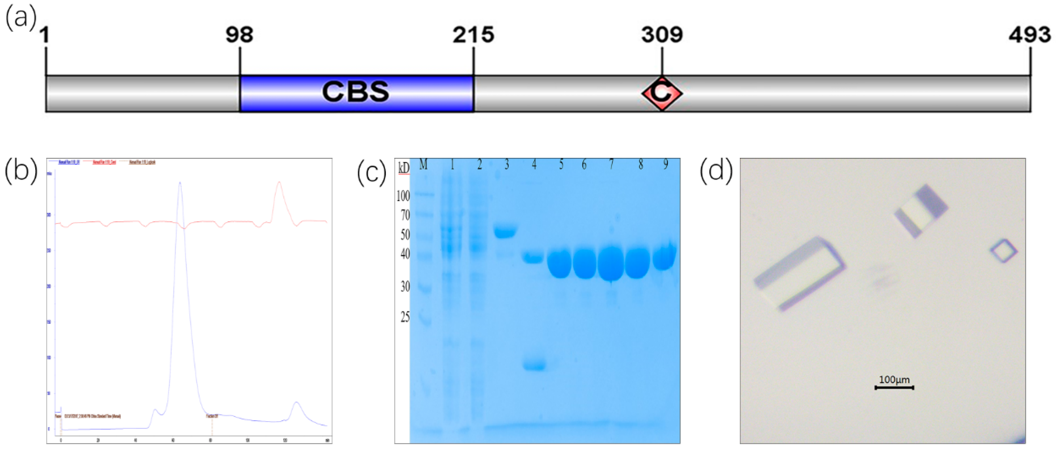
2.1. Protein Purification of CLas IMPDHΔ98-201 and Crystallization Screening

To get the CBS deletion construct, 104 residues (98-201aa) were replaced with a G amino acid, and the catalytic residue Cys309 was completely conserved (Figure 1a). In the E. coli express system, recombinant CLas IMPDHΔ98-201 was soluble and stable. This protein was purified using a Ni-NTA resin affinity chromatograph and a high-resolution gel filtration column (Superdex 200), which showed a main peak (Figure 1b). CLas IMPDHΔ98-201 consisted of 390 amino acids with a theoretical molecular mass of 41 kDa. CLas IMPDHΔ98-201 appeared as a single band at approximately 40 kDa (Figure 1c).
The initial crystallization conditions that were tested from Index, SaltRx, PEG/Ion Screen, Crystal Screen kits (Hampton Research, Aliso Viejo, CA, USA), and the Wizard kit (Emerald BioSystems, Bainbridge Island, WA, USA). After the initial screening, crystals formed under two conditions only. After further optimization, diffraction-quality crystals were obtained by mixing 1 µL of protein solution at 8 mg/mL with 1 µL of reservoir solution (consisting of 30% PEG400, 200 mM sodium chloride, and 100 mM HEPES, pH 7.0) at 20 °C. Long rectangular crystals of approximately 0.2 × 0.1 × 0.05 mm formed (Figure 1d).

2.2. Crystal Structure and Loop Refinement of CLas IMPDHΔ98-201

Crystals of CLas IMPDHΔ98-201 appeared after 3 days at 293 K. The resolution of the diffracting crystal was 2.55 Å. Data collection and refinement parameters are summarized in Table 1.
The structure of CLas IMPDHΔ98-201 was determined through molecular replacement using IMPDH from Campylobacter jejuni (PDB entry 4R7J) as a template. Finally, the structure was refined to 2.55 Å resolution by using the PHENIX software. This crystal protein existed as a homotetramer (Figure 2a), which is well conserved in other IMPDHs. The space group of CLas IMPDHΔ98-201 was C121, and the unit-cell parameters for CLas IMPDHΔ98-201 were a = 143.13, b = 134.86, and c = 85.62 Å.
The structure of CLas IMPDHΔ98-201 was very well defined, and the refinement parameters were Rwork = 22.2% and Rfree = 26.5%. A comparison of the structures of CLas IMPDHΔ98-201 and BaIMPDHΔ95-200 (Figure 2b) showed that the structure of CLas IMPDHΔ98-201 was highly similar to that of BaIMPDHΔ95-200 in its apo form. The RMSD was 0.929 Å.
The flap loop and a C-terminal loop are not visible in the electron density. Thus, the loop refinement of CLas IMPDHΔ98-201 (PDB ID: 6KCF) was performed using Modeller 9.23 (Figure 2c). The nonterminal missing structure was refined (Figure 2d). Verification of the 3D results showed that 88.27% of the amino acid residues had an average 3D–1D score ≥ 0.2 (Figure S2). The Ramachandran plot analysis indicated that 82.4% of the residues were in the core region, 13.4% of the residues were in the allowed region, 2.9% of the residues were in the generously allowed region, and 1.3% of the residues were in the disallowed region (Figure S3).

2.3. Molecular Docking

The eight candidate compounds and the refined structure were selected to perform molecular docking. CDOCKER was used to perform a docking study of the selected molecule; the molecular docking binding affinities are shown in Table 2. Three molecules, namely, bronopol, mercaptopurine, and disulfiram, showed the -CDOCKER_ENERGY ≥ 10. Because mercaptopurine is an analog of IMP, it was hypothesized that bronopol and disulfiram would exhibit the best inhibitory effect for CLas IMPDH. The pose with the lowest binding energy was recognized as the most stable conformation for further structural analysis.
The 3D and 2D structures of the CLas IMPDHΔ98-201 with bronopol and disulfiram are displayed in Figure 3. Nine hydrogen bonds formed between bronopol and the residues ILE189, Gly190, Gly192, ASP228, Gly229, Gly230, Gly251, and Ser252 of CLas IMPDHΔ98-201 (Figure 3a,b). Disulfiram formed two hydrogen bonds with CLas IMPDHΔ98-201, namely, Ala41 and Ala42; four alkyl hydrophobic interaction with Met43, Pro190, and Met249; and one sulfur-x interaction with Met43 (Figure 3c,d). The 3D and 2D structures of the CLas IMPDHΔ98-201 with the rest of molecules are displayed in Figure S4.

2.4. Kinetic Characterization of CLas IMPDHΔ98-201

According to the standard assay conditions, the kinetic properties of CLas IMPDHΔ98-201 were as follows: Kcat = 7.2 ± 0.2 s−1; K M I M P = 181 ± 19 µM (Figure 4a); and K M N A D + = 318 ± 24 µM (Figure 4b). Similar to other IMPDHs, substrate inhibition was also observed at high NAD+ levels, K i i N A D + = 7.3 ± 1.1 mM.
The steady-state parameters from other bacterial species are listed in Table S2. All IMPDHs had similar Km values for the substrate, but for CLas IMPDHΔ98-201, K M I M P was the largest, and K M N A D + was the smallest. These results indicate that CLas IMPDHΔ98-201 bound to IMP with the lowest affinity, but was the highest affinity binding NAD+ among the tested IMPDHs. The Kcat value may be due to the fact that the results described here were measured at 30 °C, whereas the other IMPDHs were measured at the lower temperature of 25 °C.

2.5. Inhibitory Assay against CLas IMPDHΔ98-201 Enzyme Activity

Extending the measurement time, no exponential enzyme decay against CLas IMPDHΔ98-201 was observed. Hence, the inhibition of bronopol, disulfiram, and ebselen was treated as a reversible mode (Figure S5). As shown in Figure S6a, the Vmax was found to be reduced with an increase in the inhibitor concentration, suggesting that bronopol inhibited CLas IMPDHΔ98-201 in a noncompetitive manner against IMP. Disulfiram also inhibited CLas IMPDH in a noncompetitive manner against IMP, where regression lines meet on the X-axis (Figure S6b). The various types of inhibition by other small molecule inhibitors are summarized in Figure S6.
To study the mechanism of enzyme inhibition, the inhibition constant Ki with respect to the IMP substrates was measured at a fixed NAD+ concentration. The Ki values of these eight compounds are summarized in Table 3.
All values ranged from 0.234 µM to 3500 µM. Although the percentage of DMSO and the high concentration of the compound affected the stability of the target protein, the values for mizoribine and ribavirin may have been inaccurate (Figure S7c,f). Ribavirin is a guanosine analog with broad-spectrum activity against RNA virus [58], and has almost no effect on the CLas IMPDHΔ98-201 enzyme activity. Mizoribine is an imidazole nucleoside which is used as an immunosuppressive agent [59]. Mizoribine was a potent inhibitor of IMPDHs, with Ki = 307.7 µM for CLas IMPDHΔ98-210, whereas the Ki value of E. coli IMPDH was 0.5 µM. Mercaptopurine yielded uncompetitive inhibition with Ki = 165 µM (Figure S7b). Mycophenolic acid was shown to be a potent inhibitor of mammalian IMPDHs with Ki = 2.43 µM (Figure S7d). Mycophenolate mofetil is a prodrug of mycophenolic acid [60], yielding Ki = 24.42 µM (Figure S7e). Three compounds, namely, disulfiram, bronopol, and ebselen, have been repurposed as IMPDH inhibitors [61]. Bronopol had the best inhibitory effect with Ki = 234 nM (Figure 5a). The Ki value of disulfiram was 616 nM (Figure 5b). The Ki values of ebselen was 4.13 µM (Figure S7a).

3. Discussion

CLas causes HLB and affects citrus. Although HLB has become a global problem, no effective HLB management strategy is available [11]. IMPDH is a validated target for the design of potent antibacterial agents, and the inhibition of this enzyme depletes cellular guanine nucleotides [36]. The development of inhibitors against bacterial IMPDHs has attracted increasing attention [62]. This study focused on the development of CLas IMPDH inhibitors. The first structure of CLas IMPDHΔ98-201 was determined. On the basis of its crystal structure, the refined structure was constructed, and molecular docking was performed to predict the binding energy. Then, we used an inhibition assay against CLas IMPDHΔ98-201 to validate the molecular docking predictions.

3.1. Purification and Crystallization of CLas IMPDHΔ98-201

To overcome the instability of CLas IMPDH, CLas IMPDH mutation was designed and purified. In the solution, recombinant CLas IMPDHΔ98-201 was more stable than the wild type. MtbIMPDH2 without the CBS domain displayed higher solubility [51]. The steady-state kinetics parameters of CLas IMPDHΔ98-201 were similar to those of other IMPDHs (Table S2), suggesting that deleting the CBS domain would not affect the CLas IMPDHΔ98-201 catalytic properties [7]. Crystals of the apo form of CLas IMPDHΔ98-201 were obtained in 100 mM HEPES (pH 7.5) and 200 mM NaCl, with 30% (w/v) PEG 4000 as the precipitant. The RMSD of CLas IMPDHΔ98-201 and BaIMPDHΔ95-200 was 0.929 Å.

3.2. Docking Interaction Analysis of CLas IMPDHΔ98-201 with Molecules

To find inhibitors of CLas IMPDHΔ98-201, molecular docking was performed using Discovery Studio 2018. The docking scores of bronopol and disulfiram binding to CLas IMPDHΔ98-201 were −11.19 and −25.03 kcal/mol, respectively. Bronopol was stabilized by nine hydrogen bond interactions with residues ILE189, Gly190, Gly192, ASP228, Gly229, Gly230, Gly251, and Ser252. Additionally, disulfiram was stabilized by hydrophobic and sulfur-x interactions. Given that a flap loop and a C-terminal loop were not visible in the apo form structure of CLas IMPDHΔ98-201, the nonterminal missing structure of CLas IMPDHΔ98-201 was refined by Modeller. The Ramachandran plot and Verify 3D analysis suggested that the refined CLas IMPDHΔ98-201 structure was reliable. Bacterial IMPDHs were similar in sequence and structure (Figure S1). Homology modeling and in silico docking were performed to study the structure–activity relationship of indole derivatives against Helicobacter pylori IMPDH [63]. The crystal structure of IMPDH from Cricetulus griseus was prepared by using Discovery Studio 2.5 to build a pharmacophore model of IMPDH inhibitors and for the in silico docking analysis [64]. These studies supported the feasibility of molecular docking.

3.3. Inhibitory Assay against CLas IMPDHΔ98-201 Activity

To explore the inhibition of the eight compounds, an inhibitory assay against CLas IMPDHΔ98-201 activity was measured by monitoring the production of NADH. The inhibitions of BaIMPDH92-220, CjIMPDHΔ92-195, and ClpIMPDHΔ89-215 to a given compound showed significant differences, although the same residues interacted with the inhibitor [7]. A previous study found that a single residue showed mycophenolic acid resistance, although the binding sites were identical [65]. The kinetic mechanism was controlled for the mycophenolic acid resistance of PbIMPDH-A and PbIMPDH-B [66]. These studies showed that virtual screening by simple prediction is fast and low cost, although experimental verification is needed. Many other compounds against HLB have been reported. Five compounds, namely, C16, C17, C18, C19, and C20, were identified against CLas SecA, with IC50 values of 0.25, 0.92, 0.48, 0.64, and 0.44 µM, respectively [16]. ZINC05491830 is one of the most potent inhibitors of CLas Esbp, with an IC50 value of 2.59 µM [19]. ChemDiv C549-0604 is an inhibitors of CLas VisNR, with an IC50 value of 0.7 µM [18]. The inhibition assay suggested that the Ki values of bronopol and disulfiram were 234 and 616 nM, respectively. The inhibition of CLas IMPDHΔ98-201 suggested that bronopol and disulfiram, unlike the aforementioned other compounds, could be used as CLas IMPDHΔ98-201 inhibitors against other CLas genes.

4. Materials and Methods

4.1. Cloning and Mutant Construction of CLas IMPDH Gene

The coding sequence of IMPDH was amplified by PCR from the chromosomal DNA of CLas (strain psy62). The PCR product was cloned into the pET28at-plus expression vector.
The CBS domain deletion mutant (CLas IMPDHΔ98-201) was constructed via splicing overlapping extension polymerase chain reaction (PCR). The ΔS construct involved the deletion of 104 residues from M98 to T201. The CLas IMPDH gene in vector pET28at-plus was used as a template. The F1 and R1 primers were applied to amplify a region of CLas IMPDH ranging from residue M1 to residue M98. The F2 and R2 primers were used to amplify a region of CLas IMPDH ranging from residue T201 to residue I493. Codons for residues M98–T201 were replaced with codons for G. I1 and I2 were used as templates. PCR was performed to amplify the CLas IMPDH CBS domain deletion mutant gene by using the F1 and R2 primers. The CLas IMPDHΔ98-201 gene was digested by Bam HI and Xho I and inserted into a pET28a-SUMO vector. Then, pET28a-SUMO-CLas IMPDHΔ98-201 was transformed into E. coli BL21(DE3) cells.

4.2. Protein Purification and Crystallization of CLas IMPDHΔ98-201

Cells carrying pET28a-SUMO-CLas IMPDHΔ98-201 plasmid were cultured in LB media supplemented with 50 µg/mL of kanamycin at 37 °C. The culture was induced by adding 0.3 mM of isopropyl-β-D-thiogalactopyranoside when its OD600 reached 0.8–1.0. After 20 h of incubation at 16 °C, the cells were harvested by centrifugation at 6000 rpm for 6 min at 4 °C, resuspended in lysis buffer [20 mM Tris-HCl (pH 8.0), 500 mM KCl, 40 mM imidazole, 1 mM PMSF, and 10% glycerol], and then sonicated. The lysate was clarified by centrifugation at 16,000 rpm for 50 min at 4 °C. Clarified lysate was subsequently purified on a Ni–NTA agarose column, and the protein was eluted with the same buffer containing 500 mM imidazole. The SUMO tag was subsequently removed with the Ulp1 protease at 16 °C for 1 h. The target protein was additionally purified using a Ni affinity chromatograph to remove the released tag and uncut protein, followed by a size exclusion chromatography step on a SuperdexTM 200 (GE Healthcare) column equilibrated with buffer [20 mM Tris-HCl (pH 8.0), 100 mM KCl, and 10% glycerol]. All proteins were purified according to this protocol.
Crystallization screening was set up using the sitting drop vapor diffusion method in 96-well plates. Crystals of the protein appeared after 3 days at 293 K. The best crystals of CLas IMPDHΔ98-201 were obtained by mixing 1 µL of protein solution at 8 mg/mL with 1 µL of reservoir solution consisting of 30% (v/v) PEG 400, 100 mM HEPES (pH 7.5), and 200 mM sodium chloride.

4.3. Data Collection and Processing

Crystals were mounted on nylon loops and flash-cooled in liquid nitrogen. Diffraction data were collected at 100 K by using the Q315r CCD detector at the BL17U beamline of the Shanghai Synchrotron Radiation Facility. Single wavelength data at 0.9792 Å were obtained, and all data were processed and scaled with HKL3000 [67]. The structure of the CLas IMPDHΔ98-201 was solved by molecular replacement using PHENIX [68]. The refined model and structure factors were deposited in the PDB.

4.4. Loop Refinement and Molecular Docking

The 3D structure of CLas IMPDHΔ98-201 was used for refinement. The Modeller program was used to refine the nonterminal missing structure [69]. This refined structure consisted of 358 amino acids (CLas IMPDH 12-98 and 202-472). The PROCHECK validation server was used to check the quality of the refined model [70]. This structure was also validated by Verify 3D [71].
Molecular docking was performed using CDOCKER, a frequently applied module of Discovery Studio 2018. CDOCKER employs a CHARMm force field to calculate the binding free energy of the ligand to the receptor [72]. The eight filtered molecules used for docking were bronopol, ebselen, mercaptopurine, mizoribine, mycophenolate_mofetil, mycophenolic acid, ribavirin, and disulfiram. In the docking experiment, the refined structure of CLas IMPDHΔ98-201 was used as the receptor; the docking parameters are listed in Table S1. The best pose of each molecular binding with a refined structure was estimated according to the binding energy. Interactions between the compound and protein, such as van der Waals force, hydrogen bond, electrostatic, hydrophobic, and halogen, were analyzed.

4.5. Steady-State Kinetics

Standard enzyme activity assay was performed in an assay buffer (50 mM Tris-HCl, 100 mM KCl, 1 mM DTT, pH 8.0) and a final CLas IMPDHΔ98-201 enzyme concentration of 100 nM at 30 °C. The production of NADH was monitored by the increase in absorbance at 340 nm (Ɛ = 6.22 mM−1 × 0007 cm−1). Apparent steady-state kinetic parameters were evaluated at varying concentrations of IMP (0.005–1 mM) and a fixed saturating concentration of NAD+ (3 mM), or at varying concentrations of NAD+ (0.005–5 mM) and a fixed saturation level of IMP (1 mM). Assays were performed in duplicate. The IMPDH enzymes displayed strong substrate inhibition with respect to NAD+ under the standard assay conditions. The method described by Kerr et al. was used to determine the kinetic constants [73].

4.6. Inhibition Assay against IMPDHΔ98-201 of CLas

The eight molecules that were purchased were screened in vitro. The assay was performed in a 200 µL final volume in a 96-well plate with a reaction buffer consisting of 50 mM Tris–HCl (pH 8.0), 100 mM KCl, and 1 mM dithiothreitol. Assays were performed using 100 nM CLas IMPDHΔ98-201 in the presence or absence of test compounds. The assay was allowed to proceed at 30 °C for 60 min.
The value of Ki for eight molecules was determined at a fixed saturation concentration of NAD+ (1 mM), different concentrations of IMP (0.02, 0.04, 0.08, 0.15, 0.25, and 0.5 mM), and in the presence of increasing concentrations of inhibitor. The concentrations of bronopol and disulfiram were 0.1, 0.2, 0.3, 0.4 µM. The concentration of ebselen was 1, 2, 3 and 4 µM. The concentration of mycophenolic acid was 1, 2, 4 and 8 µM. The concentration of mycophenolate mofetil ranged from 5 to 20 µM. The concentration of mercaptopurine ranged from 50 to 200 µM. The concentration of mizoribine was 500 and 750 µM. The concentration of ribavirin was 500 and 800 µM. Each determination of Ki was derived from duplicate measurements.
To determine the Ki values, the initial rate data versus substrate concentration at different inhibitor concentrations was fitted using Prism software (GraphPad) to equations for competitive, noncompetitive, or uncompetitive inhibition.

5. Conclusions

In conclusion, bronopol and disulfiram were confirmed as CLas IMPDH inhibitors. These results indicate that these compounds could be used as the lead scaffold to further design and develop potent CLas IMPDH inhibitors. However, the effect of compounds with activity against CLas was not tested. In future studies, we will focus on the effect of compounds in the treatment of HLB diseases. The apo form structure of CLas IMPDHΔ98-201 was solved, providing a means to study the complex structure of cocrystallization with inhibitors. The binding information may be helpful for the further development of antimicrobial compounds against CLas.

Supplementary Materials

The following are available online.

Author Contributions

L.J. contributed to the project conceptualization; J.N. conducted the experiments; S.Z. analyzed the diffraction data and solved the structure; P.Z. contributed to experiment preparation. All authors have read and agreed to the published version of the manuscript.

Funding

This research was funded by National Natural Science Foundation of China (No. 31872064 and 31572099) and National key research and development plan (2018YFD0201500).

Acknowledgments

We thank the staff of the BL17U1 beamline of the NCPSS at the Shanghai Synchrotron Radiation Facility for assistance during data collection. We thank Ping Yin and Guiqing Peng from Huazhong Agricultural University for help in preparing crystal. We would be grateful to technical assistance from State Key Laboratory of Agricultural Microbiology Core Facility, Huazhong Agricultural University.

Conflicts of Interest

The authors declare no conflict of interest.

References

  1. Bove, J.M. Huanglongbing: A destructive, newly-emerging, century-old disease of citrus. J. Plant Pathol. 2006, 88, 7–37. [Google Scholar]
  2. Wang, N.; Pierson, E.A.; Setubal, J.C.; Xu, J.; Levy, J.G.; Zhang, Y.; Li, J.; Rangel, L.T.; Martins, J., Jr. The Candidatus Liberibacter-Host Interface: Insights into Pathogenesis Mechanisms and Disease Control. Annu. Rev. Phytopathol. 2017, 55, 451–482. [Google Scholar] [CrossRef]
  3. Selvaraj, V.; Maheshwari, Y.; Hajeri, S.; Chen, J.; McCollum, T.G.; Yokomi, R. Development of a duplex droplet digital PCR assay for absolute quantitative detection of “Candidatus Liberibacter asiaticus”. PLoS ONE 2018, 13, e0197184. [Google Scholar] [CrossRef] [PubMed] [Green Version]
  4. Jain, M.; Munoz-Bodnar, A.; Zhang, S.; Gabriel, D.W. A Secreted ‘Candidatus Liberibacter asiaticus’ Peroxiredoxin Simultaneously Suppresses Both Localized and Systemic Innate Immune Responses In Planta. Mol. Plant Microbe Interact. MPMI 2018, 31, 1312–1322. [Google Scholar] [CrossRef] [PubMed] [Green Version]
  5. Clark, K.; Franco, J.Y.; Schwizer, S.; Pang, Z.; Hawara, E.; Liebrand, T.W.H.; Pagliaccia, D.; Zeng, L.; Gurung, F.B.; Wang, P.; et al. An effector from the Huanglongbing-associated pathogen targets citrus proteases. Nat. Commun. 2018, 9, 1718. [Google Scholar] [CrossRef] [PubMed]
  6. Jain, M.; Munoz-Bodnar, A.; Gabriel, D.W. ‘Candidatus Liberibacter asiaticus’ peroxiredoxin (LasBCP) suppresses oxylipin-mediated defense signaling in citrus. J. Plant Physiol. 2019, 236, 61–65. [Google Scholar] [CrossRef]
  7. Zhang, C.; Wang, X.; Liu, X.; Fan, Y.; Zhang, Y.; Zhou, X.; Li, W. A Novel ‘Candidatus Liberibacter asiaticus‘-Encoded Sec-Dependent Secretory Protein Suppresses Programmed Cell Death in Nicotiana benthamiana. Int. J. Mol. Sci. 2019, 20, 5802. [Google Scholar] [CrossRef] [Green Version]
  8. Shi, Q.; Pitino, M.; Zhang, S.; Krystel, J.; Cano, L.M.; Shatters, R.G., Jr.; Hall, D.G.; Stover, E. Temporal and spatial detection of Candidatus Liberibacter asiaticus putative effector transcripts during interaction with Huanglongbing-susceptible, -tolerant, and -resistant citrus hosts. BMC Plant Biol. 2019, 19, 122. [Google Scholar] [CrossRef] [Green Version]
  9. Ramadugu, C.; Keremane, M.L.; Halbert, S.E.; Duan, Y.P.; Roose, M.L.; Stover, E.; Lee, R.F. Long-Term Field Evaluation Reveals Huanglongbing Resistance in Citrus Relatives. Plant Dis. 2016, 100, 1858–1869. [Google Scholar] [CrossRef] [Green Version]
  10. Phuc, T.H.; He, R.F.; Nabil, K.; Judith, K.B.; Anders, O.; David, R.G.; Haluk, B. Host-free biofilm culture of “Candidatus Liberibacter asiaticus”, the bacterium associated with Huanglongbing. Biofilm 2019, 1, 100005. [Google Scholar]
  11. Wang, N. The Citrus Huanglongbing Crisis and Potential Solutions. Mol. Plant 2019, 12, 607–609. [Google Scholar] [CrossRef]
  12. Blaustein, R.A.; Lorca, G.L.; Teplitski, M. Challenges for Managing Candidatus Liberibacter spp. (Huanglongbing Disease Pathogen): Current Control Measures and Future Directions. Phytopathology 2018, 108, 424–435. [Google Scholar] [CrossRef] [PubMed] [Green Version]
  13. Zhang, M.; Powell, C.A.; Zhou, L.; He, Z.; Stover, E.; Duan, Y. Chemical compounds effective against the citrus Huanglongbing bacterium ‘Candidatus Liberibacter asiaticus’ in planta. Phytopathology 2011, 101, 1097–1103. [Google Scholar] [CrossRef] [PubMed] [Green Version]
  14. Shin, K.; Ascunce, M.S.; Narouei-Khandan, H.A.; Sun, X.A.; Jones, D.; Kolawole, O.O.; Goss, E.M.; van Bruggen, A.H.C. Effects and side effects of penicillin injection in huanglongbing affected grapefruit trees. Crop Prot. 2016, 90, 106–116. [Google Scholar] [CrossRef]
  15. Hu, J.; Wang, N. Evaluation of the Spatiotemporal Dynamics of Oxytetracycline and Its Control Effect against Citrus Huanglongbing via Trunk Injection. Phytopathology 2016, 106, 1495–1503. [Google Scholar] [CrossRef] [Green Version]
  16. Akula, N.; Trivedi, P.; Han, F.Q.; Wang, N. Identification of small molecule inhibitors against SecA of Candidatus Liberibacter asiaticus by structure based design. Eur. J. Med. Chem. 2012, 54, 919–924. [Google Scholar] [CrossRef] [Green Version]
  17. Gardner, C.L.; Pagliai, F.A.; Pan, L.; Bojilova, L.; Torino, M.I.; Lorca, G.L.; Gonzalez, C.F. Drug Repurposing: Tolfenamic Acid Inactivates PrbP, a Transcriptional Accessory Protein in Liberibacter asiaticus. Front. Microbiol. 2016, 7, 1630. [Google Scholar] [CrossRef] [Green Version]
  18. Barnett, M.J.; Solow-Cordero, D.E.; Long, S.R. A high-throughput system to identify inhibitors of Candidatus Liberibacter asiaticus transcription regulators. Proc. Natl. Acad. Sci. USA 2019, 116, 18009–18014. [Google Scholar] [CrossRef] [Green Version]
  19. Saini, G.; Dalal, V.; Savita, B.K.; Sharma, N.; Kumar, P.; Sharma, A.K. Molecular docking and dynamic approach to virtual screen inhibitors against Esbp of Candidatus Liberibacter asiaticus. J. Mol. Graph. Model. 2019, 92, 329–340. [Google Scholar] [CrossRef]
  20. Jewett, M.W.; Lawrence, K.A.; Bestor, A.; Byram, R.; Gherardini, F.; Rosa, P.A. GuaA and GuaB are essential for Borrelia burgdorferi survival in the tick-mouse infection cycle. J. Bacteriol. 2009, 191, 6231–6241. [Google Scholar] [CrossRef] [Green Version]
  21. Yoshioka, S.; Newell, P.D. Disruption of de novo purine biosynthesis in Pseudomonas fluorescens Pf0-1 leads to reduced biofilm formation and a reduction in cell size of surface-attached but not planktonic cells. PeerJ 2016, 4, e1543. [Google Scholar] [CrossRef] [PubMed] [Green Version]
  22. Boitz, J.M.; Jardim, A.; Ullman, B. GMP reductase and genetic uncoupling of adenylate and guanylate metabolism in Leishmania donovani parasites. Mol. Biochem. Parasitol. 2016, 208, 74–83. [Google Scholar] [CrossRef] [PubMed] [Green Version]
  23. Kofoed, E.M.; Yan, D.; Katakam, A.K.; Reichelt, M.; Lin, B.; Kim, J.; Park, S.; Date, S.V.; Monk, I.R.; Xu, M.; et al. De Novo Guanine Biosynthesis but Not the Riboswitch-Regulated Purine Salvage Pathway Is Required for Staphylococcus aureus Infection in vivo. J. Bacteriol. 2016, 198, 2001–2015. [Google Scholar] [CrossRef] [PubMed] [Green Version]
  24. Morrison, H.G.; McArthur, A.G.; Gillin, F.D.; Aley, S.B.; Adam, R.D.; Olsen, G.J.; Best, A.A.; Cande, W.Z.; Chen, F.; Cipriano, M.J.; et al. Genomic minimalism in the early diverging intestinal parasite Giardia lamblia. Science 2007, 317, 1921–1926. [Google Scholar] [CrossRef] [Green Version]
  25. Carlton, J.M.; Hirt, R.P.; Silva, J.C.; Delcher, A.L.; Schatz, M.; Zhao, Q.; Wortman, J.R.; Bidwell, S.L.; Alsmark, U.C.; Besteiro, S.; et al. Draft genome sequence of the sexually transmitted pathogen Trichomonas vaginalis. Science 2007, 315, 207–212. [Google Scholar] [CrossRef] [PubMed] [Green Version]
  26. Allison, A.C.; Almquist, S.J.; Muller, C.D.; Eugui, E.M. In vitro immunosuppressive effects of mycophenolic acid and an ester pro-drug, RS-61443. Transplant. Proc. 1991, 23, 10–14. [Google Scholar]
  27. Robins, R.K.; Revankar, G.R.; McKernan, P.A.; Murray, B.K.; Kirsi, J.J.; North, J.A. The importance of IMP dehydrogenase inhibition in the broad spectrum antiviral activity of ribavirin and selenazofurin. Adv. Enzyme Regul. 1985, 24, 29–43. [Google Scholar] [CrossRef]
  28. Weber, G. Biochemical strategy of cancer cells and the design of chemotherapy: G. H. A. Clowes Memorial Lecture. Cancer Res. 1983, 43, 3466–3492. [Google Scholar]
  29. Hedstrom, L.; Liechti, G.; Goldberg, J.B.; Gollapalli, D.R. The antibiotic potential of prokaryotic IMP dehydrogenase inhibitors. Curr. Med. Chem. 2011, 18, 1909–1918. [Google Scholar] [CrossRef] [Green Version]
  30. Usha, V.; Hobrath, J.V.; Gurcha, S.S.; Reynolds, R.C.; Besra, G.S. Identification of novel Mt-Guab2 inhibitor series active against M. tuberculosis. PLoS ONE 2012, 7, e33886. [Google Scholar] [CrossRef] [Green Version]
  31. Rao, V.A.; Shepherd, S.M.; Owen, R.; Hunter, W.N. Structure of Pseudomonas aeruginosa inosine 5′-monophosphate dehydrogenase. Acta Crystallograph. Sect. F Struct. Biol. Cryst. Commun. 2013, 69, 243–247. [Google Scholar] [CrossRef] [PubMed] [Green Version]
  32. Mandapati, K.; Gorla, S.K.; House, A.L.; McKenney, E.S.; Zhang, M.; Rao, S.N.; Gollapalli, D.R.; Mann, B.J.; Goldberg, J.B.; Cuny, G.D.; et al. Repurposing cryptosporidium inosine 5′-monophosphate dehydrogenase inhibitors as potential antibacterial agents. ACS Med. Chem. Lett. 2014, 5, 846–850. [Google Scholar] [CrossRef]
  33. Prosise, G.L.; Luecke, H. Crystal structures of Tritrichomonasfoetus inosine monophosphate dehydrogenase in complex with substrate, cofactor and analogs: A structural basis for the random-in ordered-out kinetic mechanism. J. Mol. Biol. 2003, 326, 517–527. [Google Scholar] [CrossRef]
  34. Makowska-Grzyska, M.; Kim, Y.; Wu, R.; Wilton, R.; Gollapalli, D.R.; Wang, X.K.; Zhang, R.; Jedrzejczak, R.; Mack, J.C.; Maltseva, N.; et al. Bacillus anthracis inosine 5′-monophosphate dehydrogenase in action: The first bacterial series of structures of phosphate ion-, substrate-, and product-bound complexes. Biochemistry 2012, 51, 6148–6163. [Google Scholar] [CrossRef] [PubMed] [Green Version]
  35. Labesse, G.; Alexandre, T.; Gelin, M.; Haouz, A.; Munier-Lehmann, H. Crystallographic studies of two variants of Pseudomonas aeruginosa IMPDH with impaired allosteric regulation. Acta Crystallograph. Sect. D Biol. Crystallograph. 2015, 71, 1890–1899. [Google Scholar] [CrossRef] [PubMed] [Green Version]
  36. Hedstrom, L. IMP dehydrogenase: Structure, mechanism, and inhibition. Chem. Rev. 2009, 109, 2903–2928. [Google Scholar] [CrossRef] [Green Version]
  37. Nimmesgern, E.; Black, J.; Futer, O.; Fulghum, J.R.; Chambers, S.P.; Brummel, C.L.; Raybuck, S.A.; Sintchak, M.D. Biochemical analysis of the modular enzyme inosine 5′-monophosphate dehydrogenase. Protein Expr. Purif. 1999, 17, 282–289. [Google Scholar] [CrossRef]
  38. Pimkin, M.; Markham, G.D. The CBS subdomain of inosine 5′-monophosphate dehydrogenase regulates purine nucleotide turnover. Mol. Microbiol. 2008, 68, 342–359. [Google Scholar] [CrossRef] [Green Version]
  39. Makowska-Grzyska, M.; Kim, Y.; Maltseva, N.; Osipiuk, J.; Gu, M.; Zhang, M.; Mandapati, K.; Gollapalli, D.R.; Gorla, S.K.; Hedstrom, L.; et al. A novel cofactor-binding mode in bacterial IMP dehydrogenases explains inhibitor selectivity. J. Biol. Chem. 2015, 290, 5893–5911. [Google Scholar] [CrossRef] [Green Version]
  40. Gollapalli, D.R.; Macpherson, I.S.; Liechti, G.; Gorla, S.K.; Goldberg, J.B.; Hedstrom, L. Structural determinants of inhibitor selectivity in prokaryotic IMP dehydrogenases. Chem. Biol. 2010, 17, 1084–1091. [Google Scholar] [CrossRef] [Green Version]
  41. Chen, L.; Wilson, D.J.; Xu, Y.; Aldrich, C.C.; Felczak, K.; Sham, Y.Y.; Pankiewicz, K.W. Triazole-linked inhibitors of inosine monophosphate dehydrogenase from human and Mycobacterium tuberculosis. J. Med. Chem. 2010, 53, 4768–4778. [Google Scholar] [CrossRef] [PubMed] [Green Version]
  42. Umejiego, N.N.; Gollapalli, D.; Sharling, L.; Volftsun, A.; Lu, J.; Benjamin, N.N.; Stroupe, A.H.; Riera, T.V.; Striepen, B.; Hedstrom, L. Targeting a prokaryotic protein in a eukaryotic pathogen: Identification of lead compounds against cryptosporidiosis. Chem. Biol. 2008, 15, 70–77. [Google Scholar] [CrossRef] [PubMed] [Green Version]
  43. Kirubakaran, S.; Gorla, S.K.; Sharling, L.; Zhang, M.; Liu, X.; Ray, S.S.; Macpherson, I.S.; Striepen, B.; Hedstrom, L.; Cuny, G.D. Structure-activity relationship study of selective benzimidazole-based inhibitors of Cryptosporidium parvum IMPDH. Bioorg. Med. Chem. Lett. 2012, 22, 1985–1988. [Google Scholar] [CrossRef] [PubMed] [Green Version]
  44. Gorla, S.K.; Kavitha, M.; Zhang, M.; Chin, J.E.W.; Liu, X.; Striepen, B.; Makowska-Grzyska, M.; Kim, Y.; Joachimiak, A.; Hedstrom, L.; et al. Optimization of benzoxazole-based inhibitors of Cryptosporidium parvum inosine 5′-monophosphate dehydrogenase. J. Med. Chem. 2013, 56, 4028–4043. [Google Scholar] [CrossRef] [PubMed] [Green Version]
  45. Gorla, S.K.; Zhang, Y.; Rabideau, M.M.; Qin, A.; Chacko, S.; House, A.L.; Johnson, C.R.; Mandapati, K.; Bernstein, H.M.; McKenney, E.S.; et al. Benzoxazoles, Phthalazinones, and Arylurea-Based Compounds with IMP Dehydrogenase-Independent Antibacterial Activity against Francisella tularensis. Antimicrob. Agents Chemother. 2017, 61, e00939-17. [Google Scholar] [CrossRef] [Green Version]
  46. Chacko, S.; Boshoff, H.I.M.; Singh, V.; Ferraris, D.M.; Gollapalli, D.R.; Zhang, M.; Lawson, A.P.; Pepi, M.J.; Joachimiak, A.; Rizzi, M.; et al. Expanding Benzoxazole-Based Inosine 5′-Monophosphate Dehydrogenase (IMPDH) Inhibitor Structure-Activity As Potential Antituberculosis Agents. J. Med. Chem. 2018, 61, 4739–4756. [Google Scholar] [CrossRef]
  47. Park, Y.; Pacitto, A.; Bayliss, T.; Cleghorn, L.A.T.; Wang, Z.; Hartman, T.; Arora, K.; Ioerger, T.R.; Sacchettini, J.; Rizzi, M.; et al. Essential but Not Vulnerable: Indazole Sulfonamides Targeting Inosine Monophosphate Dehydrogenase as Potential Leads against Mycobacterium tuberculosis. ACS Infect. Dis. 2017, 3, 18–33. [Google Scholar] [CrossRef]
  48. Maurya, S.K.; Gollapalli, D.R.; Kirubakaran, S.; Zhang, M.; Johnson, C.R.; Benjamin, N.N.; Hedstrom, L.; Cuny, G.D. Triazole inhibitors of Cryptosporidium parvum inosine 5′-monophosphate dehydrogenase. J. Med. Chem. 2009, 52, 4623–4630. [Google Scholar] [CrossRef] [Green Version]
  49. Sahu, N.U.; Singh, V.; Ferraris, D.M.; Rizzi, M.; Kharkar, P.S. Hit discovery of Mycobacterium tuberculosis inosine 5′-monophosphate dehydrogenase, GuaB2, inhibitors. Bioorg. Med. Chem. Lett. 2018, 28, 1714–1718. [Google Scholar] [CrossRef]
  50. Sahu, N.U.; Purushothaman, G.; Thiruvenkatam, V.; Kharkar, P.S. Design, synthesis, and biological evaluation of Helicobacter pylori inosine 5′-monophosphate dehydrogenase (HpIMPDH) inhibitors. Drug Dev. Res. 2019, 80, 125–132. [Google Scholar] [CrossRef] [Green Version]
  51. Makowska-Grzyska, M.; Kim, Y.; Gorla, S.K.; Wei, Y.; Mandapati, K.; Zhang, M.; Maltseva, N.; Modi, G.; Boshoff, H.I.; Gu, M.; et al. Mycobacterium tuberculosis IMPDH in Complexes with Substrates, Products and Antitubercular Compounds. PLoS ONE 2015, 10, e0138976. [Google Scholar] [CrossRef] [PubMed] [Green Version]
  52. Usha, V.; Gurcha, S.S.; Lovering, A.L.; Lloyd, A.J.; Papaemmanouil, A.; Reynolds, R.C.; Besra, G.S. Identification of novel diphenyl urea inhibitors of Mt-GuaB2 active against Mycobacterium tuberculosis. Microbiology 2011, 157, 290–299. [Google Scholar] [CrossRef] [PubMed] [Green Version]
  53. Gorla, S.K.; Kavitha, M.; Zhang, M.; Liu, X.; Sharling, L.; Gollapalli, D.R.; Striepen, B.; Hedstrom, L.; Cuny, G.D. Selective and potent urea inhibitors of Cryptosporidium parvum inosine 5′-monophosphate dehydrogenase. J. Med. Chem. 2012, 55, 7759–7771. [Google Scholar] [CrossRef] [PubMed] [Green Version]
  54. Beevers, R.E.; Buckley, G.M.; Davies, N.; Fraser, J.L.; Galvin, F.C.; Hannah, D.R.; Haughan, A.F.; Jenkins, K.; Mack, S.R.; Pitt, W.R.; et al. Low molecular weight indole fragments as IMPDH inhibitors. Bioorg. Med. Chem. Lett. 2006, 16, 2535–2538. [Google Scholar] [CrossRef]
  55. Beevers, R.E.; Buckley, G.M.; Davies, N.; Fraser, J.L.; Galvin, F.C.; Hannah, D.R.; Haughan, A.F.; Jenkins, K.; Mack, S.R.; Pitt, W.R.; et al. Novel indole inhibitors of IMPDH from fragments: Synthesis and initial structure-activity relationships. Bioorg. Med. Chem. Lett. 2006, 16, 2539–2542. [Google Scholar] [CrossRef]
  56. Watterson, S.H.; Carlsen, M.; Dhar, T.G.M.; Shen, Z.; Pitts, W.J.; Guo, J.; Gu, H.H.; Norris, D.; Chorba, J.; Chen, P.; et al. Novel inhibitors of IMPDH: A highly potent and selective quinolone-based series. Bioorg. Med. Chem. Lett. 2003, 13, 543–546. [Google Scholar] [CrossRef]
  57. Jangra, S.; Purushothaman, G.; Juvale, K.; Ravi, S.; Menon, A.; Thiruvenkatam, V.; Kirubakaran, S. Synthesis and In Vitro Enzymatic Studies of New 3-Aryldiazenyl Indoles as Promising Helicobacter pylori IMPDH Inhibitors. Curr. Top. Med. Chem. 2019, 19, 376–382. [Google Scholar] [CrossRef]
  58. Nystroem, K.; Waldenstroem, J.; Tang, K.-W.; Lagging, M. Ribavirin: Pharmacology, multiple modes of action and possible future perspectives. Future Virol. 2019, 14, 153–160. [Google Scholar] [CrossRef] [Green Version]
  59. Ishikawa, H. Mizoribine and mycophenolate mofetil. Curr. Med. Chem. 1999, 6, 575–597. [Google Scholar] [PubMed]
  60. Cao, S.; Aboge, G.O.; Terkawi, M.A.; Zhou, M.; Kamyingkird, K.; Moumouni, P.F.A.; Masatani, T.; Igarashi, I.; Nishikawa, Y.; Suzuki, H.; et al. Mycophenolic acid, mycophenolate mofetil, mizoribine, ribavirin, and 7-nitroindole inhibit propagation of Babesia parasites by targeting inosine 5′-monophosphate dehydrogenase. J. Parasitol. 2014, 100, 522–526. [Google Scholar] [CrossRef]
  61. Sarwono, A.E.Y.; Mitsuhashi, S.; Kabir, M.H.B.; Shigetomi, K.; Okada, T.; Ohsaka, F.; Otsuguro, S.; Maenaka, K.; Igarashi, M.; Kato, K.; et al. Repurposing existing drugs: Identification of irreversible IMPDH inhibitors by high-throughput screening. J. Enzyme Inhib. Med. Chem. 2019, 34, 171–178. [Google Scholar] [CrossRef] [PubMed]
  62. Juvale, K.; Shaik, A.; Kirubakaran, S. Inhibitors of inosine 5′-monophosphate dehydrogenase as emerging new generation antimicrobial agents. MedChemComm 2019, 10, 1290–1301. [Google Scholar] [CrossRef] [PubMed]
  63. Juvale, K.; Purushothaman, G.; Singh, V.; Shaik, A.; Ravi, S.; Thiruvenkatam, V.; Kirubakaran, S. Identification of selective inhibitors of Helicobacter pylori IMPDH as a targeted therapy for the infection. Sci. Rep. 2019, 9, 190. [Google Scholar] [CrossRef] [PubMed] [Green Version]
  64. Yang, N.; Wang, J.; Li, J.; Wang, Q.-H.; Wang, Y.; Cheng, M.-S. A three-dimensional pharmacophore model for IMPDH inhibitors. Chem. Biol. Drug Des. 2011, 78, 175–182. [Google Scholar] [CrossRef]
  65. Freedman, R.; Yu, R.; Sarkis, A.W.; Hedstrom, L. A structural determinant of mycophenolic acid resistance in eukaryotic inosine 5′-monophosphate dehydrogenases. Protein Sci. Publ. Protein Soc. 2020, 29, 686–694. [Google Scholar] [CrossRef]
  66. Sun, X.E.; Hansen, B.G.; Hedstrom, L. Kinetically controlled drug resistance: How Penicillium brevicompactum survives mycophenolic acid. J. Biol. Chem. 2011, 286, 40595–40600. [Google Scholar] [CrossRef] [Green Version]
  67. Minor, W.; Cymborowski, M.; Otwinowski, Z.; Chruszcz, M. HKL-3000: The integration of data reduction and structure solution—From diffraction images to an initial model in minutes. Acta Crystallograph. Sect. D Biol. Crystallograph. 2006, 62, 859–866. [Google Scholar] [CrossRef] [Green Version]
  68. Adams, P.D.; Afonine, P.V.; Bunkóczi, G.; Chen, V.B.; Davis, I.W.; Echols, N.; Headd, J.J.; Hung, L.-W.; Kapral, G.J.; Grosse-Kunstleve, R.W.; et al. PHENIX: A comprehensive Python-based system for macromolecular structure solution. Acta Crystallograph. Sect. D Biol. Crystallograph. 2010, 66, 213–221. [Google Scholar] [CrossRef] [Green Version]
  69. Fiser, A.; Do, R.K.; Sali, A. Modeling of loops in protein structures. Protein Sci. 2000, 9, 1753–1773. [Google Scholar] [CrossRef] [Green Version]
  70. Laskowski, R.A.; Macarthur, M.W.; Moss, D.S.; Thornton, J.M. Procheck—A Program to Check the Stereochemical Quality of Protein Structures. J. Appl. Crystallograph. 1993, 26, 283–291. [Google Scholar] [CrossRef]
  71. Eisenberg, D.; Luthy, R.; Bowie, J.U. VERIFY3D: Assessment of protein models with three-dimensional profiles. Methods Enzymol. 1997, 277, 396–404. [Google Scholar] [PubMed]
  72. Wu, G.; Robertson, D.H.; Brooks, C.L.; Vieth, M. Detailed analysis of grid-based molecular docking: A case study of CDOCKER-A CHARMm-based MD docking algorithm. J. Comput. Chem. 2003, 24, 1549–1562. [Google Scholar] [CrossRef] [PubMed]
  73. Kerr, K.M.; Digits, J.A.; Kuperwasser, N.; Hedstrom, L. Asp338 controls hydride transfer in Escherichia coli IMP dehydrogenase. Biochemistry 2000, 39, 9804–9810. [Google Scholar] [CrossRef] [PubMed]
Sample Availability: Samples of the compounds are not available from the authors.
Figure 1. Size exclusion chromatogram and crystal of purified CLas IMPDHΔ98-201. (a): Primary sequence of CLas IMPDH; (b): Size exclusion chromatogram of CLas IMPDHΔ98-201; (c): SDS-PAGE of CLas IMPDHΔ98-201. M: protein marker; 1: supernatant; 2: flow-through; 3: SUMO-CLas IMPDHΔ98-201; 4: Ulp1 digestion; 5–9: protein of CLas IMPDHΔ98-201 after size exclusion chromatogram; (d): crystal of CLas IMPDHΔ98-201.
Molecules 25 02313 g001
Figure 2. Crystal and Loop Refinement structure of the apo-form CLas IMPDHΔ98-201. (a): Tetramer of CLas IMPDHΔ98-201; (b): Superposed structures of CLas IMPDHΔ98-201 (PDB entry: 6KCF, in magenta) and BaIMPDHΔ95-200 (PDB entry: 4MJM, in green); (c): Loop refinement of CLas IMPDHΔ98-201; (d): Superposed structures of CLas IMPDHΔ98-201 (PDB entry: 6KCF, in green) and the refined structure (cyan).
Molecules 25 02313 g002
Figure 3. Molecular docking of CLas IMPDHΔ98-201 and the moleculars. (a): 3D details of CLas IMPDHΔ98-201 and bronopol (green) interaction; (b): 2D details of CLas IMPDHΔ98-201 and bronopol interaction; (c): 3D details of CLas IMPDHΔ98-201 and disulfiram (green) interaction; (d): 2D details of CLas IMPDHΔ98-201 and disulfiram interaction.
Molecules 25 02313 g003
Figure 4. Enzyme activity of CLas IMPDHΔ98-201. (a): Varying concentrations of IMP at a fixed concentration of NAD+ (2 mM); (b): Varying concentrations of NAD+ at a fixed concentration of IMP (1 mM).
Molecules 25 02313 g004
Figure 5. Inhibition kinetics at different concentrations of compounds by varying the IMP concentrations at a fixed NAD+ concentration. (a): Bronopol; (b): Disulfiram.
Molecules 25 02313 g005
Table 1. Data collection and refinement statistics.
CLas IMPDHΔ98-201
BeamlineSSRF BEAMLINE BL17U
wavelength (Å)0.9792
DetectorADSC QUANTUM 315r
resolution range (Å)42.47–2.55
space groupC 1 2 1
unit cell parameters (Å)a = 143.13, b = 134.86, c = 85.62
no. of residues/protein390
Monomer molecular weight (kDa)41.0
phasing methodMR
search modelchains A of 4R7J
Refinement resolution range (Å) 42.46–2.55
no. of reflections 50928
σ cutoff 1.36
Rwork 0.222
Rfree 0.264
mean B factor (Å2) 69.3
data completeness (%)98.2
redundancy2.58
Ramachandran plot [most favored/outliers (%)]95.2/0.5
PDB entry 6KCF
Table 2. Detailed summary of the docking binding affinities (kcal/mol).
NameMolecular Weight (g/mol)-CDOCKER_ENERGY (kcal/mol)
Disulfiram296.5425.0346
Mercaptopurine152.1816.6785
Bronopol199.9911.1913
Ebselen274.186.24157
Mycophenolic_acid320.345.38177
Mizoribine259.224.50734
Ribavirin244.20−5.62032
Mycophenolate_mofetil433.50−43.3176
Table 3. Inhibition of CLas IMPDHΔ98-201 by eight inhibitors.
InhibitorIMP Ki (µM)
Bronopol0.23 ± 0.01
Disulfiram0.62 ± 0.04
Ebselen4.13 ± 0.19
Mycophenolic acid2.43 ± 0.10
Mercaptopurine165 ± 9.89
Mycophenolate mofetil24.42 ± 1.65
Mizoribine307.7
Ribavirin>3500

