Article

Griseaketides A–D, New Aromatic Polyketides from the Pathogenic Fungus Magnaporthe grisea

1
State Key Laboratory for Conservation and Utilization of Bio-Resources in Yunnan, Yunnan University, Kunming 650091, China
2
Institute of Entomoceutics Research, Dali University, Dali 671000, China
3
State Key Laboratory for Conservation and Utilization of Bio-Resources in Yunnan, Yunnan Agricultural University, Kunming 650205, China
*
Authors to whom correspondence should be addressed.
These authors contributed equally to this work.
Molecules 2020, 25(1), 72; https://doi.org/10.3390/molecules25010072
Submission received: 5 December 2019 / Revised: 19 December 2019 / Accepted: 21 December 2019 / Published: 24 December 2019
(This article belongs to the Section Natural Products Chemistry)

Abstract

:
Magnaporthe grisea is the causal agent of rice blast disease, which is the most serious disease of cultivated rice. Aromatic polyketides are its typical metabolites and are involved in the infection process. In the search for novel lead compounds, chemical investigation of the fungus M. grisea M639 has led to the isolation of four new aromatic polyketides (salicylaldehyde skeleton bearing an unsaturated side chain), griseaketides A–D (14), as well as 15 known compounds (519). The structures of the new compounds were elucidated on the basis of extensive spectroscopic analyses, including HR-MS, 2D NMR. Compound 12 showed prominent activity that killed 94.5% of C. elegans at 400 ppm and 66.9% at 200 ppm over 24 h. This is the first report describing the nematicidal activity of this type aromatic polyketide.

1. Introduction

Exploration of natural sources of novel bioactive compounds as drugs or lead compounds has been an emerging field over the past decades, and exciting evidence has been provided by the isolation of microbe-derived metabolites [1]. The fungal kingdom includes many species with unique and unusual biochemical pathways, which results in lots of structurally fascinating secondary metabolites with promising biological and pharmacological properties, such as penicillin, cyclosporine and statins [2].
A group of structurally and actively diverse metabolites were produced by plant pathogenic strains [3,4,5], which are responsible for function as essential determinants of pathogenicity or virulence. The rice blast fungus Magnaporthe grisea (imperfect stage of Pyricularia grisea Sacc.) causes a serious disease on agriculturally significant plants including rice, wheat, and barley. Each year rice blast causes losses of 10–30% of the rice harvest and therefore poses a threat to the world’s most important food security crop [6]. Previous, studies have shown that aromatic polyketide phytotoxins [7], O-nitrophenol derivatives [8], and naphthalenones [9] are the typical secondary metabolites of M. grisea. Some secondary metabolites (such as melanin, siderophores and fumonisin) of pathogenic fungi often function as essential determinants of pathogenicity and are involved in the infection process [10,11]. Thus, examining their secondary metabolism leads to the discovery of novel compounds with interesting structures or modes of action that could be useful for agrochemistry or pharmacology [5]. In the work, chemical investigations of a strain M. grisea M639 have led to the discovery of four new aromatic polyketides griseaketides A–D (14), as well as 15 known compounds (519). Herein, we describe the isolation, structural elucidation and nematicidal activities of these compounds.

2. Results

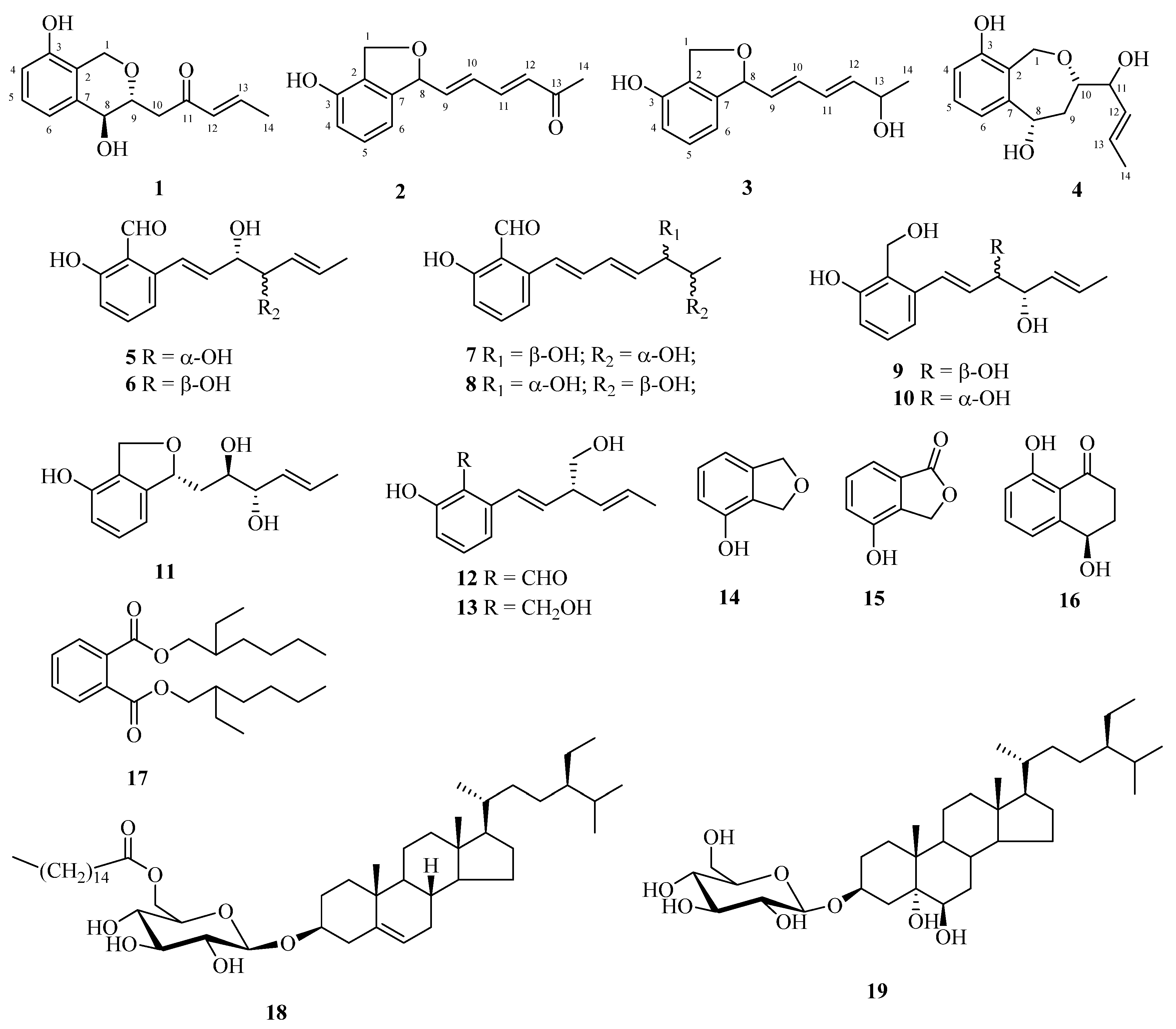
Griseaketide A (1) was isolated as a pale yellow amorphous powder. Its molecular formula C14H16O4 was determined by HR-ESI-MS and 13C NMR coupled with DEPT spectra, which indicated 7 degrees of unsaturation (Table 1). The 1H NMR spectrum exhibited signals for one methyl resonance at δH 1.94 (3H, dd, J = 6.8, 1.4 Hz), three aromatic protons at δH 6.65 (1H, d, J = 8.7 Hz), 7.11 (1H, t, J = 7.5 Hz) and 7.12 (1H, d, J = 7.5 Hz), two olefinic protons at δH 6.24 (1H, dd, J = 15.8, 1.4 Hz) and 6.99 (1H, dq, J = 15.8, 6.8 Hz) and four monooxygenated protons at δH 3.93 (1H, m), 4.49 (1H, d, J = 7.5 Hz), 4.85 (1H, d, J = 15.8 Hz), 4.70 (1H, d, J = 15.8 Hz). Sixteen carbon resonances observed in the 13C NMR spectrum coupled with the DEPT interpretations of 1 were attributable to one ketone (δC 199.2), one pair of disubstituted double bonds at δC 132.2 (d) and δC 144.6 (d), one monooxygenated 1,2,3-trisubstituted aromatic ring [δC 150.8 (s), 138.3 (s), 121.9 (s), 113.7 (d), 127.7 (d), 118.4 (d)], one primary alcohol at δC 64.3 (t), two oxymethine groups at δC 69.2 (d) and 75.4 (d), and one methyl group at δC 18.4 (q). On the basis of spectroscopic data analysis, compound 1 was deduced as an aromatic polyketide (Figure 1), similar to 3-(1’,3’-pentadienyl)-3,4-dihydro-lH-2-benzopyran-4,8-diol [7]. A preliminary linear skeleton bearing two branches was deduced to be −C-8−C-9−C-10 (−branch) −C-12−C-13−C-14 (−branch) from complete interpretations of key cross-peaks in the COSY spectrum (H-8/H-9/H-10; H-12/H-13/H-14) together with key cross-peaks in the HMBC spectrum (Figure 2): from H-8 to C-9 and C-10; from H-10 to C-8, C-9 and C-11; from H-14 to C-12 and C-13. The HMBC correlation of H-1/C-9 demonstrated that the formation of an extra pyran ring was through a bridge between C-1 and C-9. Typically, values of 3J ≤ 2 Hz suggest cis configuration, and 3J ≥5 Hz suggest trans configuration [12], so the trans-configuration was assigned to H-8 and H-9 on the basis of its larger coupling constant (7.5 Hz). As a result, the relative configuration of compound 1 is shown in Figure 1 and named as griseaketide A.
Compound 2 was obtained as optically active amorphous powder ( [ α ] D 18 –16.0 c 0.16, MeOH) and its molecular formula of C14H14O3 was established by the negative HR-ESI-MS, revealing 8 degrees of unsaturation. Analysis of the 1H and 13C NMR (Table 1) data of 2 revealed the presence of a monooxygenated 1,2,3-trisubstituted aromatic ring, two pairs of double bonds, a primary alcohol, a carbonyl group, an oxymethine and one methyl (Figure 1). These signals revealed that 2 is similar to 1 and suggested to be an aromatic polyketide [13]. The lower-field shifting of carbon signal (δC-8 84.4) in 2 and the observed HMBC correlations (Figure 2) from H-1 to C-8 suggested that C-1 and C-8 are adjacent substituents on the aromatic ring and form a 1,3-dihydroisobenzofuran system. One branch was deduced to be −C-8−C-9−C-10−C-11−C-12 from complete interpretations of key cross-peaks in the COSY spectrum (H-8/H-9/H-10/H-11/H-12) together with key cross-peaks (from H-8 to C-9 and C-10; from H-10 to C-8, C-11 and C-12; from H-11 to C-9, C-10 and C-13; from H-14 to C-12 and C-13) in the HMBC spectrum to determinate the side chain moiety (Figure 2). The configuration of conjugated double bonds was elucidated as E geometries by their coupling constants (J9−10 = 15.1 Hz, J11−12 = 15.7 Hz) [14]. The configurations of C-8 cannot be determined by the present data. Thus, the structure of 2 was determined as shown in Figure 1 and named as griseaketide B.
Compound 3 was obtained as a pale yellow oil. HR-ESI-MS analysis of 3 provided a molecular formula of C14H16O3, corresponding to an unsaturation number of 6. The UV absorption maxima at 226 and 271 nm suggested the presence of a conjugated system in the molecule. Compound 3 was very similar to compound 2, but the ketone (δC 198.8) of 2 was replaced by one hydroxyl in 3 (Table 2). The 1H−1H COSY spectrum of compound 3 revealed two structural fragments to be −C-8−C-9−C-10−C-11−C-12−C-13−C-14 and −C-4−C-5−C-6− by the clear correlations of H-8/H-9/H-10/H-11/H-12/H-13/H-14 and H-4/H-5/H-6 (Figure 2). The HMBC experiment showed correlations from H-1 to C-8 to suggest that C-1 and C-8 are adjacent substituents on the aromatic ring and formed a 1,3-dihydroisobenzofuran system (Figure 2). The E geometries of the two double bonds was inferred by coupling constants (J9−10 = 15.1 Hz, J11−12 = 15.2 Hz) [14]. The configurations of C-8 and 13-OH cannot be determined by the present data. Thus, the relative configuration of 3 is shown as Figure 1 and named as griseaketide C.
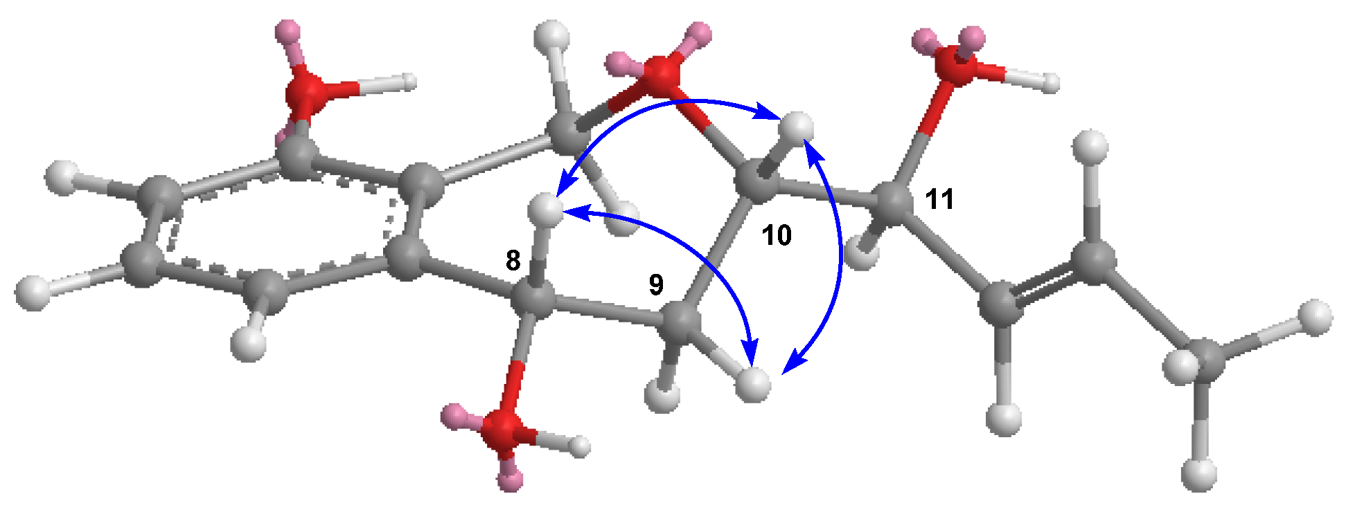
The molecular formula of griseaketide D (4) was found to be C14H18O4 by HR-ESI-MS and 13C NMR analysis. Its 1H and 13C NMR (Table 2) signals were similar to that of 1, except for the carbonyl group in 1 that was replaced by a hydroxyl group at C-11. On the basis of the consecutive COSY correlations from H-8 to H-14 (Figure 2) and the coupling constants (J12−13 = 14.6 Hz), the side chain was elucidated as 1’,3’,4’-trihydroxy-5’E-heptaenyl. The observed HMBC (Figure 2) correlations of H-1/C-10 suggested a seven membered ether ring was fused to the trisubstituted benzene ring through an oxygen bridge between C-1 and C-10. The relative configuration of 4 was deduced from ROESY correlations (Figure 3) and comparisons with data reported in the literature [15]. The ROESY cross-peaks of H-8/H-9β and H-10 indicated that they are all cofacial and assigned as β-oriented which were consistent with xylarinol B [15]. The configuration of 11-OH cannot be determined by the present data. Accordingly, the structure and relative configuration of 4 was established as shown.
The known compounds were identified as (1’E,3’S,4’S,5’E)-2-(3’,4’-dihydroxy-1’,5’-heptadienyl)-6-hydroxybenzaldehyde (5) [16], (1’E,3’S,4’R,5’E)-2-(3’,4’-dihydroxy-1’,5’-heptadienyl)-6-hydroxy benzaldehyde (6) [17], (5’R,6’S)-pyriculariol (7) [18], (5’S,6’R)-pyriculariol (8) [18], dihydropyriculol (9) [7], epi-dihydropyriculol (10) [7], pyriculin A (11) [19], (R)-pyricuol (12) [20], (1’E,3’R,4’E)-2- hydroxymethyl-3-(3’-hydroxymethylhexa-1’,4’-dienyl)phenol (13) [21], 1,3-dihydro-4-isobenzo- furanol (14) [22], 4-hydroxyphthalide (15) [23], (−)-isosclerone (16) [24], bis (2-ethylhexyl) phthalate (17) [25], daucosterol palmitate (18) [26] and 5α,6β-dihydroxydaucosterol (19) [27] by comparison of their experimental and reported spectroscopic data.
Nematicidal activityof compounds. The pure compounds 1 and 512 were tested for their nematicidal activity. Compounds 58 showed weak nematicidal activities against Caenorhabditis elegans at 400 ppm over 48 h, but compound 12 showed prominent activity that killed 94.5% of C. elegans at 400 ppm and 66.9% at 200 ppm over 24 h.

3. Discussion

Rice blast, caused by infection of the rice blast fungus, Magnaporthe grisea, is the most destructive pathogen of rice worldwide. Many metabolites from rice blast fungus have been identified and they show different activities. Pyriculol caused a necrotic lesion in a rice wounding assay and showed inhibition in a spore germination bioassay [28]. Compound pyricuol inhibited shoot growth showing a stronger effect than pyriculol and dihydropyriculol [28,29]. In our experiment, part salicylaldehyde-type products showed nematicidal activity, and among them aldehyde group-containing compounds showed nematicidal activity, which is consistent with the literature [30], while pyricuol (12) showed a prominent nematicidal activity, which was distinguished by a different substitute on the side chain, and this further provides us with an indication of the nematicidal active compounds.

4. Materials and Methods

4.1. General Experimental Procedures

The optical rotations were measured with a Horiba SEPA-300 polarimeter (Kyoto, Japan). UV spectra were recorded on a Shimadzu UV-2401PC spectrophotometer (Kyoto, Japan). The NMR spectra were obtained with an Avance III 600 spectrometer (Bruker Biospin, Rheinstetten, Germany). The ESI and HR-ESI-MS were recorded on a Finnigan LCQ-Advantage (Thermo Finnigan, San Jose, CA, USA) and VG Auto-Spec-3000 mass spectrometer (VG, Manchester, England), respectively. Column chromatography (CC) was performed on Sephadex LH-20 (Amersham Biosciences, Piscataway, NJ, USA), silica gel (200–300 mesh, Qingdao Marine Chemical Inc., Qingdao, China), and RP-18 gel (LiChroprep, 40–63 μm; Merck, Darmstadt, Germany). Semipreparative HPLC was performed on an LC3000 (Beijing Chuangxintongheng Science & Technology Co., Ltd., Beijing, China). Fractions were monitored by TLC and visualized by heating plates sprayed with 5% H2SO4 in EtOH.

4.2. Microbial Material

The fungal strain of Magnaporthe grisea M639 used in this study was isolated from the leaf spot lesions of rice collected from Yunnan province, China, in August 2012. The strain has been preserved in the State Key Laboratory for Conservation and Utilization of Bio-Resources in Yunnan, Yunnan University.

4.3. Cultivation, Extraction and Isolation

The strain M. grisea M639 was cultured on PDA solid medium at 26 °C for 7 days, and then it was inoculated into 1 L Erlenmeyer flasks each containing 200 mL of sticky rice-glucose liquid medium, which were cultivated at 26 °C for 14 days. The obtained culture filtrates were extracted by EtOAc three times to give a crude extract (1.32 g). The residual H2O portion was extracted with n-butyl alcohol to yield a residue (14.11 g). The EtOAc fraction was separated by CC on RP-18 (MeOH–H2O, 1:4 to 1:0) to produce six fractions (Fr.1–Fr.6). Fr.1 was purified by Sephadex LH-20 (CHCl3–MeOH, 1:1) and then chromatographed over a silica gel CC (CHCl3–MeOH, 100:1 to 2:1) to yield 14 (4.2 mg) and 15 (14.4 mg). Fr.2 was repeatedly subjected to silica gel CC eluting with CHCl3–MeOH (50:1 to 50:3) and then purified by Sephadex LH–20 CC (CHCl3–MeOH, 1:1) to afford 1 (5.7 mg), 9 (3.2 mg), 10 (7.2 mg), 11 (26.0 mg) and 16 (1.2 mg). Fr.3 was subjected to Sephadex LH-20 (MeOH) and then purified by silica gel to yield 4 (2.1 mg). Fr.4 was repeatedly purified by Sephadex LH-20 CC (CHCl3–MeOH, 1:1), semipreparative RP-18 CC (MeOH–H2O, 1:1) and silica gel CC (CHCl3–MeOH, 50:1) to obtain 3 (4.0 mg), 2 (1.8 mg), 5 (66.0 mg), 6 (6.5 mg), 7 (18.5 mg), 8 (5.7 mg) and 13 (1.1 mg). Fr.5 was subjected to Sephadex LH-20 CC (CHCl3–MeOH, 1:1), followed by semipreparative RP-18 CC (MeOH–H2O, 65:35) to yield 12 (7.0 mg). Fr.6 was chromatographed over a silica gel CC (petroleum ether–EtOAc, 25:1 to 3:2) and then purified by Sephadex LH-20 CC (CHCl3–MeOH, 1:1) to obtain 18 (6.7 mg) and 19 (1.8 mg). The n-butyl alcohol portion was chromatographed over silica gel CC (CHCl3–MeOH, 9:1 to 0:1) and then purified by Sephadex LH-20 CC (CHCl3–MeOH, 1:1) to give 17 (0.7 mg).
Griseaketide A (1): Pale yellow amorphous powder; [ α ] D 18 –29.1 (c 0.15, MeOH); UV (MeOH) λmax (log ε) 202 (4.53), 220 (4.34), 274 (3.43) nm; 1H NMR (CDCl3, 600 MHz) and 13C NMR (CDCl3, 150 MHz) data, see Table 1; positive ESI-MS m/z 271 [M + Na]+, 287 [M + K]+, 519 [2M + Na]+, 535 [2M + K]+; HR-ESI-MS m/z 247.0978 [M − H] (calcd for C14H15O4, 247.0976).
Griseaketide B (2): Colorless amorphous powder; [ α ] D 18 –16.0 (c 0.16, MeOH); UV (MeOH) λmax (log ε) 202 (4.23), 270 (3.90) nm; 1H NMR (CDCl3, 600 MHz) and 13C NMR (CDCl3, 150 MHz) data, see Table 1; negative ESI-MS m/z 229 [M − H]; HR-ESI-MS m/z 229.0873 [M − H] (calcd for C14H13O3, 229.0870).
Griseaketide C (3): Pale yellow oil; [ α ] D 18 +15.2 (c 0.31, MeOH); UV (MeOH) λmax (log ε) 201 (4.47), 226 (4.45), 271 (3.39) nm; 1H NMR (CDCl3, 600 MHz) and 13C NMR (CD3OD, 150 MHz) data, see Table 2; negative ESI-MS m/z 231 [M − H]; HR-ESI-MS m/z 231.1027 [M − H] (calcd for C14H16O3, 231.1027).
Griseaketide D (4): Colorless amorphous powder; [ α ] D 18 –10.6 (c 0.31, MeOH); UV (MeOH) λmax (log ε) 201 (3.44), 215 (3.07), 271 (2.54) nm; 1H NMR (CD3OD, 600 MHz) and 13C NMR (CD3OD, 150 MHz) data, see Table 2; positive ESI-MS m/z 273 [M + Na]+, 523 [2M + Na]+. HR-ESI-MS m/z 273.1096 [M + Na]+ (calcd for C14H18O4, 273.1097).

4.4. Nematicidal Activity

The saprophytic nematode C. elegans was cultured on oatmeal medium (20 g of oatmeal in 80 mL of H2O) at 25 °C for 7 days. Then the cultured nematodes were separated from the culture medium using the Baerman funnel technique, and an aqueous suspension of the nematode was prepared as a working stock. Compounds 1 and 512 were dissolved in methanol and then diluted to different concentrations (400 and 200 ppm) with sterile water. The nematicidal activity against C. elegans was assayed according to the method based on references [31,32].

Author Contributions

Investigation, Y.-H.Y., D.-S.Y. and H.-M.L.; Resources, C.-Y.L.; Writing—original draft preparation, Y.-H.Y. and D.-S.Y.; Writing—review and editing, G.-H.L., and P.-J.Z.; Supervision, G.-H.L. and P.-J.Z.; Project administration, P.-J.Z.; Funding acquisition, G.-H.L. and P.-J.Z. All authors have read and agreed to the published version of the manuscript.

Funding

This study was financially supported by the Applied Basic Research Foundation of Yunnan Province (2018FA006) and the National Natural Science Foundation of China (31760018, 31970060 and 31860015).

Conflicts of Interest

The authors declare no conflict of interest.

References

  1. Rateb, M.E.; Ebel, R. Secondary metabolites of fungi from marine habitats. Nat. Prod. Rep. 2011, 28, 290–344. [Google Scholar] [CrossRef] [PubMed]
  2. Zhong, J.J.; Xiao, J.H. Secondary metabolites from higher fungi: Discovery, bioactivity, and bioproduction. Adv. Biochem. Eng. Biotechnol. 2009, 113, 79–150. [Google Scholar] [CrossRef] [PubMed]
  3. Wolpert, T.J.; Dunkle, L.D.; Ciuffetti, L.M. Host-selective toxins and avirulence determinants: What’s in a name? Annu. Rev. Phytopathol. 2002, 40, 251–285. [Google Scholar] [CrossRef] [PubMed] [Green Version]
  4. Dufour, N.; Rao, R.P. Secondary metabolites and other small molecules as intercellular pathogenic signals. FEMS Microbiol. Lett. 2011, 314, 10–17. [Google Scholar] [CrossRef] [PubMed]
  5. Yang, Y.H.; Yang, D.S.; Li, G.H.; Pu, X.J.; Mo, M.H.; Zhao, P.J. Antibacterial diketopiperazines from an endophytic fungus Bionectria sp. Y1085. J. Antibiot. 2019, 72, 752–758. [Google Scholar] [CrossRef] [PubMed]
  6. Talbot, N.J. On the trail of a cereal killer: Exploring the biology of Magnaporthe grisea. Annu. Rev. Microbiol. 2003, 57, 177–202. [Google Scholar] [CrossRef] [PubMed] [Green Version]
  7. Kono, Y.; Sekido, S.; Yamaguchi, I.; Kondo, H.; Suzuki, Y.; Neto, G.C.; Sakurai, A.; Yaegashi, H. Structures of two novel pyriculol-related compounds and identification of naturally produced epipyriculol from Pyricularia oryzae. Agric. Biol. Chem. 1991, 55, 2785–2791. [Google Scholar] [CrossRef] [Green Version]
  8. Sviridov, S.I.; Ermolinskii, B.S. Secondary metabolites of Pyricularia oryzae I. o-nitrophenol derivatives. Chem. Nat. Compd. 1990, 26, 691–696. [Google Scholar] [CrossRef]
  9. Iwasaki, S.; Muro, H.; Nozoe, S.; Okuda, S.; Sato, Z. Isolation of 3,4-dihydro-3,4,8-trihydroxy-1(2H)-naphthalenone and tenuazonic acid from Pyricularia oryzae cavara. Tetrahedron Lett. 1972, 13, 13–16. [Google Scholar] [CrossRef]
  10. Scharf, D.H.; Heinekamp, T.; Brakhage, A.A. Human and plant fungal pathogens: The role of secondary metabolites. PLoS Pathog. 2014, 10, e1003859. [Google Scholar] [CrossRef] [Green Version]
  11. Mobius, N.; Hertweck, C. Fungal phytotoxins as mediators of virulence. Curr. Opin. Plant Biol. 2009, 12, 390–398. [Google Scholar] [CrossRef] [PubMed]
  12. Asha, K.N.; Chowdhury, R.; Hasan, C.M.; Rashid, M.A. Steroids and polyketides from Uvaria hamiltonii stem bark. Acta Pharm. 2004, 54, 57–63. [Google Scholar] [CrossRef] [PubMed]
  13. Myobatake, Y.; Takemoto, K.; Kamisuki, S.; Inoue, N.; Takasaki, A.; Takeuchi, T.; Mizushina, Y.; Sugawara, F. Cytotoxic alkylated hydroquinone, phenol, and cyclohexenone derivatives from Aspergillus violaceofuscus Gasperini. J. Nat. Prod. 2014, 77, 1236–1240. [Google Scholar] [CrossRef] [PubMed]
  14. Yang, D.S.; Peng, W.B.; Li, Z.L.; Wang, X.; Wei, J.G.; He, Q.X.; Yang, Y.P.; Liu, K.C.; Li, X.L. Chemical constituents from Euphorbia stracheyi and their biological activities. Fitoterapia 2014, 97, 211–218. [Google Scholar] [CrossRef] [PubMed]
  15. Lee, I.K.; Jang, Y.W.; Kim, Y.S.; Yu, S.H.; Lee, K.J.; Park, S.M.; Oh, B.T.; Chae, J.C.; Yun, B.S. Xylarinols A and B, two new 2-benzoxepin derivatives from the fruiting bodies of Xylaria polymorpha. J. Antibiot. 2009, 62, 163–165. [Google Scholar] [CrossRef] [Green Version]
  16. Leyva, A.; Blum, F.E.; Ley, S.V. A new synthesis of (–)-epipyriculol: A phytotoxic metabolite. Tetrahedron 2008, 64, 4711–4717. [Google Scholar] [CrossRef]
  17. Suzuki, M.; Sugiyama, T.; Watanabe, M.; Murayama, T.; Yamashita, K. Syntheses of all four stereoisomers of pyriculol. Agric. Biol. Chem. 1987, 51, 2161–2166. [Google Scholar] [CrossRef]
  18. Sasaki, A.; Tanaka, K.; Sato, Y.; Kuwahara, S.; Kiyota, H. First synthesis and absolute configuration of (–)-pyriculariol, a phytotoxin isolated from rice blast fungus, Magnaporthe grisea. Use of microwave irradiation to control Stille coupling reaction products. Tetrahedron Lett. 2009, 50, 4637–4638. [Google Scholar] [CrossRef]
  19. Masi, M.; Meyer, S.; Gorecki, M.; Mandoli, A.; Di Bari, L.; Pescitelli, G.; Cimmino, A.; Cristofaro, M.; Clement, S.; Evidente, A. Pyriculins A and B, two monosubstituted hex-4-ene-2,3-diols and other phytotoxic metabolites produced by Pyricularia grisea isolated from buffelgrass (Cenchrus ciliaris). Chirality 2017, 29, 726–736. [Google Scholar] [CrossRef]
  20. Nakamura, Y.; Kiyota, H.; Ueda, R.; Kuwahara, S. Synthesis to determine the absolute configuration of (–)-pyricuol, a phytotoxin isolated from rice blast disease fungus Magnaporthe grisea. Tetrahedron Lett. 2005, 46, 7107–7109. [Google Scholar] [CrossRef]
  21. Tanaka, K.; Nakamura, Y.; Sasaki, A.; Ueda, R.; Suzuki, Y.; Kuwahara, S.; Kiyota, H. Synthesis and plant growth inhibitory activity of both enantiomers of pyricuol, a phytotoxin isolated from rice blast disease fungus Magnaporthe grisea. Tetrahedron 2009, 65, 6115–6122. [Google Scholar] [CrossRef]
  22. Martín-matute, B.; Nevado, C.; Cárdenas, D.J.; Echavarren, A.M. Intramolecular reactions of alkynes with furans and electron rich arenes catalyzed by PtCl2: the role of platinum carbenes as intermediates. J. Am. Chem. Soc. 2003, 125, 5757–5766. [Google Scholar] [CrossRef] [PubMed]
  23. Shimizu, N.; Kuwahara, Y. 7-Hydroxyphthalide: A new natural salicylaldehyde analog from Oulenzia sp. (Astigmata: Winterschmitiidae). Biosci. Biotechnol. Biochem. 2001, 65, 990–992. [Google Scholar] [CrossRef] [PubMed]
  24. Gulder, T.A.M.; Hong, H.; Correa, J.; Egereva, E.; Wiese, J.; Imhoff, J.F.; Gross, H. Isolation, structure elucidation and total synthesis of lajollamide A from the marine fungus Asteromyces cruciatus. Mar. Drugs 2012, 10, 2912–2935. [Google Scholar] [CrossRef] [PubMed]
  25. Chang, R.J.; Wang, C.H.; Zeng, Q.; Guan, B.; Zhang, W.D.; Jin, H.Z. Chemical constituents of the stems of Celastrus rugosus. Arch. Pharm. Res. 2013, 36, 1291–1301. [Google Scholar] [CrossRef]
  26. Nguyen, A.T.; Malonne, H.; Duez, R.; Vanhaelen-Fastre, R.; Vanhaelen, M.; Fontaine, J. Cytotoxic constituents from Plumbago zeylanica. Fitoterapia 2004, 75, 500–504. [Google Scholar] [CrossRef] [PubMed]
  27. Li, S.H.; Zhang, H.J.; Yao, P.; Niu, X.M.; Xiang, W.; Sun, H.D. Non-taxane compounds from the bark of Taxus yunnanensis. J. Asian Nat. Prod. Res. 2002, 4, 147–154. [Google Scholar] [CrossRef]
  28. Kim, J.C.; Min, J.Y.; Kim, H.T.; Cho, K.Y.; Yu, S.H. Pyricuol, a new phytotoxin from Magnaporthe grisea. Biosci. Biotech. Bioch. 1998, 62, 173–174. [Google Scholar] [CrossRef]
  29. Jacob, S.; Grotsch, T.; Foster, A.J.; Schuffler, A.; Rieger, P.H.; Sandjo, L.P.; Liermann, J.C.; Opatz, T.; Thines, E. Unravelling the biosynthesis of pyriculol in the rice blast fungus Magnaporthe oryzae. Microbiology 2017, 163, 541–553. [Google Scholar] [CrossRef]
  30. Caboni, P.; Aissani, N.; Cabras, T.; Falqui, A.; Marotta, R.; Liori, B.; Ntalli, N.; Sarais, G.; Sasanelli, N.; Tocco, G. Potent nematicidal activity of phthalaldehyde, salicylaldehyde, and cinnamic aldehyde against Meloidogyne incognita. J. Agric. Food Chem. 2013, 61, 1794–1803. [Google Scholar] [CrossRef]
  31. Sajid, M.; Azim, M.K. Characterization of the nematicidal activity of natural honey. J. Agric. Food Chem. 2012, 60, 7428–7434. [Google Scholar] [CrossRef] [PubMed]
  32. Yang, Z.S.; Li, G.H.; Zhao, P.J.; Zheng, X.; Luo, S.L.; Li, L.; Niu, X.M.; Zhang, K.Q. Nematicidal activity of Trichoderma spp. and isolation of an active compound. World J. Microbiol. Biotechnol. 2010, 26, 2297–2302. [Google Scholar] [CrossRef]
Sample Availability: Samples of the compounds are available from the authors.
Figure 1. Structures of compounds 119.
Molecules 25 00072 g001
Figure 2. Selected HMBC ( Molecules 25 00072 i001) and 1H-1H COSY ( Molecules 25 00072 i002) correlations of compounds 14.
Molecules 25 00072 g002
Figure 3. Key ROESY correlations of compound 4.
Molecules 25 00072 g003
Table 1. 1H and 13C NMR Data of Compounds 1 and 2 (δ in ppm, J in Hz).
No.1 (in CDCl3)2 (in CDCl3)
δHδCHMBCδHδCHMBC
14.85, d (15.8)64.3, t2, 3, 7, 95.23, dd (11.9, 2.1)71.2, t2, 7, 8
4.70, d (15.8)2, 3, 7, 95.15, d (11.9)2, 7, 8
2-121.9, s--125.2, s-
3-150.8, s--150.3, s-
46.65, d (8.7)113.7, d66.71, d (8.0)114.5, d2, 3, 6
57.11, t (7.5, overlap)127.7, d3, 4, 77.18, t (7.7)129.4, d3, 7
67.12, d (7.5, overlap)118.4, d2, 4, 76.73, d (7.5)113.9, d2, 4, 8
7-138.3, s--142.4, s-
84.49, d (7.5)69.2, d7, 9, 105.73, brd (6.0)84.4, d9, 10
93.93, m75.4, d1, 116.25, dd (15.1, 6.8)141.8, d7, 8
102.98, dd (16.2, 4.7)43.2, t8, 9, 116.53, dd (15.1, 11.0)129.0, d8, 11, 12
3.03, dd (16.2, 7.1)-
11-199.2, s-7.15, dd (15.7, 11.0)142.4, d9, 10, 13
126.24, dd (15.8, 1.4)132.2, d11, 146.21, d (15.7)131.4, d10, 14
136.99, dq (15.8, 6.8)144.6, d11, 14-198.8, s-
141.94, dd (6.8, 1.4)18.4, q12, 132.28, s27.3, q12, 13
Table 2. 1H and 13C NMR Data of Compounds 3 and 4 (δ in ppm, J in Hz).
No.3 (in CD3OD)4 (in CD3OD)
δHδCHMBCδHδCHMBC
15.09, dd (12.0, 2.6)71.7, t2, 7, 84.91, d (15.6)65.8, t2, 3, 7, 10
4.98, brd (12.0)2, 7, 84.50, d (15.6)2, 3, 7
2-126.0, s--123.1, s-
3-153.0, s--154.0, s-
46.67, d (7.7)115.1, d1, 2, 3, 66.68, d (7.9)114.6, d3, 6
57.10, t (7.7)130.3, d3, 4, 77.08, t (7.9)128.4, d3, 6, 7
66.58, d (7.7)113.8, d2, 4, 86.85, d (7.5)122.0, d2, 4, 8
7-144.1, s--138.2, s-
85.57, d (7.7)86.6, d7, 94.28, brs67.5, d2, 6, 7
95.71, dd (15.1, 7.7)133.5, d8, 112.06, m; 1.80, m39.6, t10, 11, 12
106.38, dd (15.1, 10.6)132.8, d8, 11, 123.63, brt (6.8)76.9, d8, 9
116.24, dd (15.2, 10.6)129.3, d10, 134.30, t (6.8)71.2, d13
125.81, dd (15.2, 6.2)139.9, d10, 135.52, dd (14.6, 6.8)135.3, d11, 14
134.29, dq (6.2, 6.4)68.7, d11, 145.73, dq (14.6, 6.1)127.7, d11, 14
141.24, d (6.4)23.5, q12, 131.71, d (6.1)17.9, q12, 13