Review

Secondary Metabolites from Gorgonian Corals of the Genus Eunicella: Structural Characterizations, Biological Activities, and Synthetic Approaches

by
Dario Matulja
1,
Maria Kolympadi Markovic
2,
Gabriela Ambrožić
2,
Sylvain Laclef
3,
Sandra Kraljević Pavelić
1,* and
Dean Marković
1,*
1
Department of Biotechnology, University of Rijeka, Radmile Matejčić 2, 51000 Rijeka, Croatia
2
Department of Physics and Centre for Micro- and Nanosciences and Technologies, University of Rijeka, Radmile Matejčić 2, 51000 Rijeka, Croatia
3
Laboratoire de Glycochimie, des Antimicrobiens et des Agroressources (LG2A) UMR CNRS 7378—Institut de Chimie de Picardie FR 3085, Université de Picardie Jules Verne, 33 rue Saint Leu, FR-80039 Amiens CEDEX, France
*
Authors to whom correspondence should be addressed.
Molecules 2020, 25(1), 129; https://doi.org/10.3390/molecules25010129
Submission received: 9 December 2019 / Revised: 24 December 2019 / Accepted: 26 December 2019 / Published: 28 December 2019
(This article belongs to the Special Issue Translational Approach to Antitumor Drugs)

Abstract

:
Gorgonian corals, which belong to the genus Eunicella, are known as natural sources of diverse compounds with unique structural characteristics and interesting bioactivities both in vitro and in vivo. This review is focused primarily on the secondary metabolites isolated from various Eunicella species. The chemical structures of 64 compounds were divided into three main groups and comprehensively presented: a) terpenoids, b) sterols, and c) alkaloids and nucleosides. The observed biological activities of depicted metabolites with an impact on cytotoxic, anti-inflammatory, and antimicrobial activities were reviewed. The most promising biological activities of certain metabolites point to potential candidates for further development in pharmaceutical, cosmetic, and other industries, and are highlighted. Total synthesis or the synthetic approaches towards the desired skeletons or natural products are also summarized.

1. Introduction

Oceans and seas cover more than 70% of the surface of our planet and, therefore, are an extensive and rich source of biotechnological potential [1]. Terrestrial organisms, mainly plants, have been widely explored and utilized to obtain novel compounds with the purpose to develop novel drugs for improved treatment for various diseases [2]. In contrast, species living in the marine environment have traditionally been used as food, contributing to human health as valuable sources of proteins, polysaccharides, amino acids, vitamins, minerals, and other micronutrients [1,3]. More than 30,000 compounds have been isolated from marine organisms since biochemical research began in the second half of the last century [4,5]. Numerous sea bioprospecting programs have been performed with the aim to search for new compounds as target leads in the drug discovery processes, emphasizing on anticancer products with improved performance and/or less severe adverse effects [2,6]. Consequently, since 1969 and the approval of cytarabine, the first marine-derived product for the treatment of leukemia, six more compounds have been approved by the Food and Drug Administration (FDA) for clinical use, while dozens of others are currently in different stages of preclinical and clinical trials [7]. Besides anticancer activity, marine natural compounds have also demonstrated neuroprotective, anticardiovascular, antimicrobial, anti-inflammatory, antioxidant, antifouling, and other interesting biological activities [1].
Marine sponges from the phylum Porifera are recognized among the most interesting organisms in bioprospecting as sources of the largest number of so far characterized marine metabolites. Other invertebrates like cnidarians include more than 11,000 species, some of which are poorly or completely unexplored [4]. Moreover, corals are interesting sedentary organisms that may survive harsh internal and external marine environmental conditions. The main reason why they produce various secondary metabolites is to protect themselves from predatory species [4,8]. These characteristics may be exploited in search of novel bioactives with still unexplored biological mechanisms of action.
Eunicella coral species belong to the order Alcyonacea, family Gorgoniidae. Gorgonian corals, also known as sea fans and soft corals, are characterized by their branched, fan-like structure which can be found on both hard and soft sea bottoms in the Mediterranean, tropical, and subtropical marine areas [8,9]. Due to their tree-like morphological features, they provide nutrients and protection to other marine invertebrates and vertebrates [10]. For example, bacteria of the genus Endozoicomonas are key endosymbionts within colonies of Eunicella cavolini (Figure 1) and may contribute to the coral’s adaptation to the environment or to its metabolism [9,11].
Recently, Raimundo et al. have analyzed genomes of 15 bacteria from 12 genera, cultivated from Eunicella sp., and identified gene clusters responsible for the biosynthesis of terpenes, polyketides, and peptides [12]. The microorganisms might be also involved in symbiotic biosyntheses producing the different oxygenation and unsaturation patterns of the natural products originating from Eunicella species. Furthermore, those symbiotic organisms can be analyzed by a metagenomic approach, which allows the bioprospecting of genes encoding enzymes or discovering of biocatalysts for the synthesis of bioactive secondary metabolites [13,14,15].
Eunicella cavolini (yellow gorgonian, Koch, 1887) and Eunicella singularis (white gorgonian, Esper, 1791) are the most abundant gorgonian corals in the Mediterranean marine area [10]. The first is recognized by its orange-colored colonies and is widespread from the Tunisian coast to the Aegean Sea. It can be found up to 150 m where it can form larger, more arborescent colonies due to the environmental stability [10,16]. Other Eunicella members mentioned in this review are granulata (Grasshoff, 1992), verrucosa (Pallas, 1766), and labiata (Thomson, 1927) [17]. In addition to their potent biological activities, high susceptibility to climate change, anthropogenic influences, and other abiotic and biotic factors, together with their slow growth and recovery rates, make the Eunicella sp. A very interesting subject in different research areas.
Two previous reviews cover the isolation of bioactive metabolites from gorgonian species between 2009 and 2012 [8,18]. Herein, we summarize advances in isolation and characterization of all known compounds from Eunicella sp. during the last three decades with a focus on the research conducted from 2013 to present. The presented compounds were divided into three groups, i.e., (1) terpenoids, (2) sterols, and (3) alkaloids and nucleosides. Biogenetic pathways involved in the formation of terpenoids from geranylgeranyl diphosphate are also described. In connection to the diversity and complexity of isolated structures, their biological effects are discussed, highlighting cytotoxic, anti-inflammatory, and antimicrobial activities. The most interesting or promising compounds and their biological activities are summarized in Tables 1–3. Moreover, synthetic approaches and total syntheses of isolated metabolites are schematically depicted.

2. Terpenoids

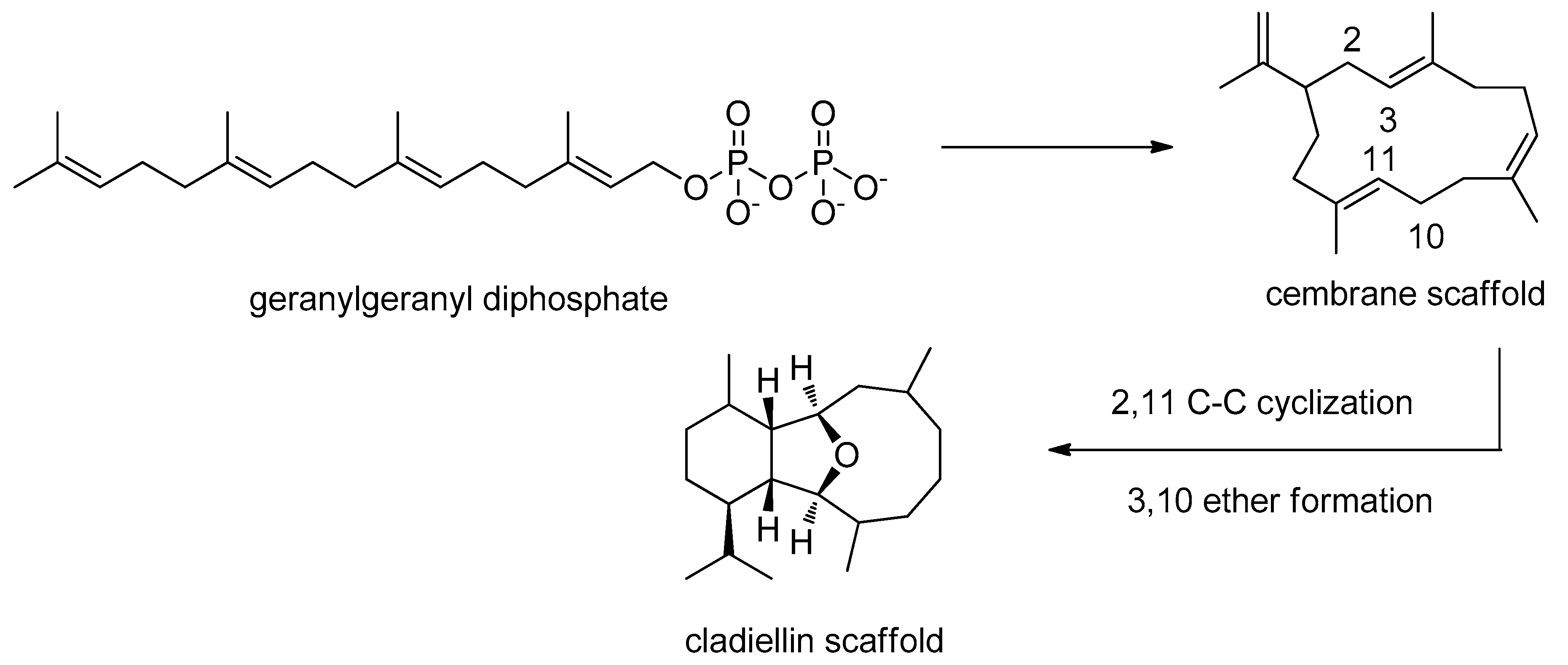
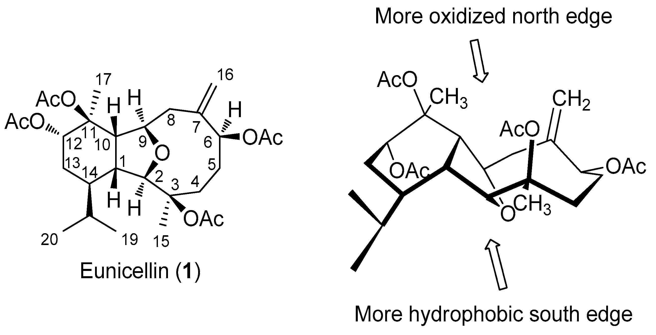
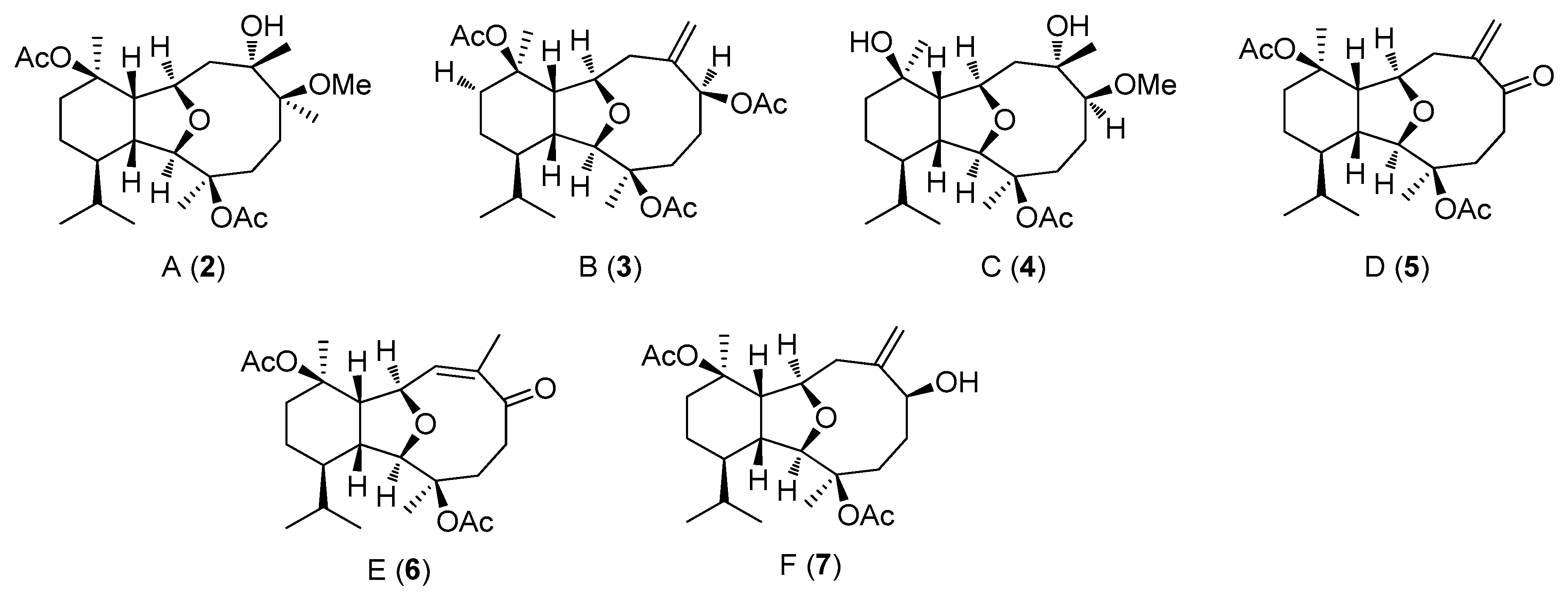
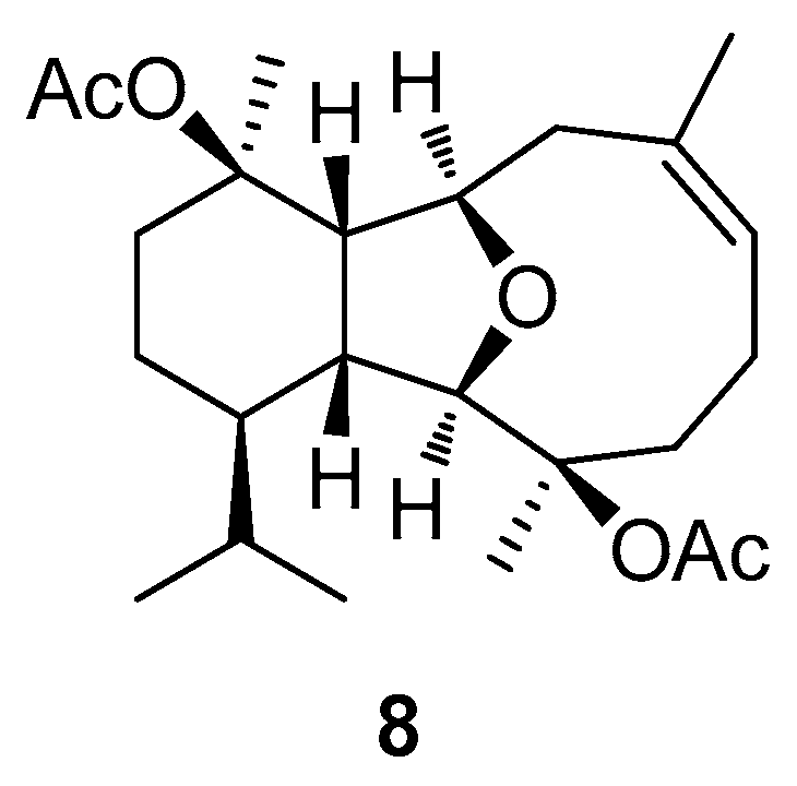
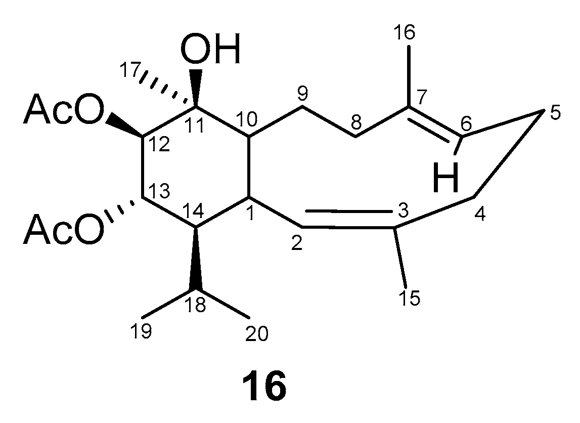
2.1. Biogenesis and Structural Analysis of Eunicellins

Among all metabolites isolated from gorgonian soft corals, those belonging to the class of terpenoids exhibited the most significant activities (IC50 values lower than 5 μg/mL) when assayed in vitro against tumor cell lines [4]. Diterpenes make up to 65% of all published metabolites from gorgonian natural resources [19]. Eunicella sp. are producers of eunicellin-type diterpenoids, i.e., eunicellin-based diterpenoids from the gorgonian Eunicella verrucosa [20] or from Eunicella cavolini [21] and Eunicella labiata [22]. These compounds have the cladiellane skeleton and contain four isoprene units linked head to tail (Figure 2). According to Welford et al., cladiellins are formed by cyclization of geranylgeranyl diphosphate, C2–C11 cyclization, and C3–C10 tetrahydrofuranyl formation from cembrane scaffold intermediate [23].
Eunicellin (1), shown in Figure 3, possess a tricyclic 6-5-9 membered structure with etheral bridge. The skeleton can be divided into the more oxidized north edge, and the more hydrophobic south edge that communicate through a tricyclic structure. Whereas the west part of the molecule is more rigid due to a bicyclic cyclohexyl-tetrahydrofuranyl system, the eastern part is more flexible as it contains an oxonane structure. A cyclohexyl ring is fused to a tetrahydrofuranyl ring through C1 and C10 pointing hydrogens syn to each other and anti to the H-atoms of the ethereal O-C2 and O-C9 bonds. Examples of deviations of this common oxygen bridge can also be found, from no ether bridge, 2,6-epoxy bridge, to compounds having two epoxy bridges. Angular methyl groups at C3 and C11 are anti-periplanar to the isopropyl group at C14, whereas at C7 is placed methylene group. Eunicellin (1) possess nine sterocenters. C1, C2, C9, and C10 form the central bridging tetrahydrofuranyl system, and still C3, C6, C11, C12, and C14 are chiral. Four acetoxy groups are placed on C3, C6, C11, and C12. Eunicellins also bear different oxygenation and unsaturation patterns with one or more common acetoxyl moieties [22,24,25,26,27,28].

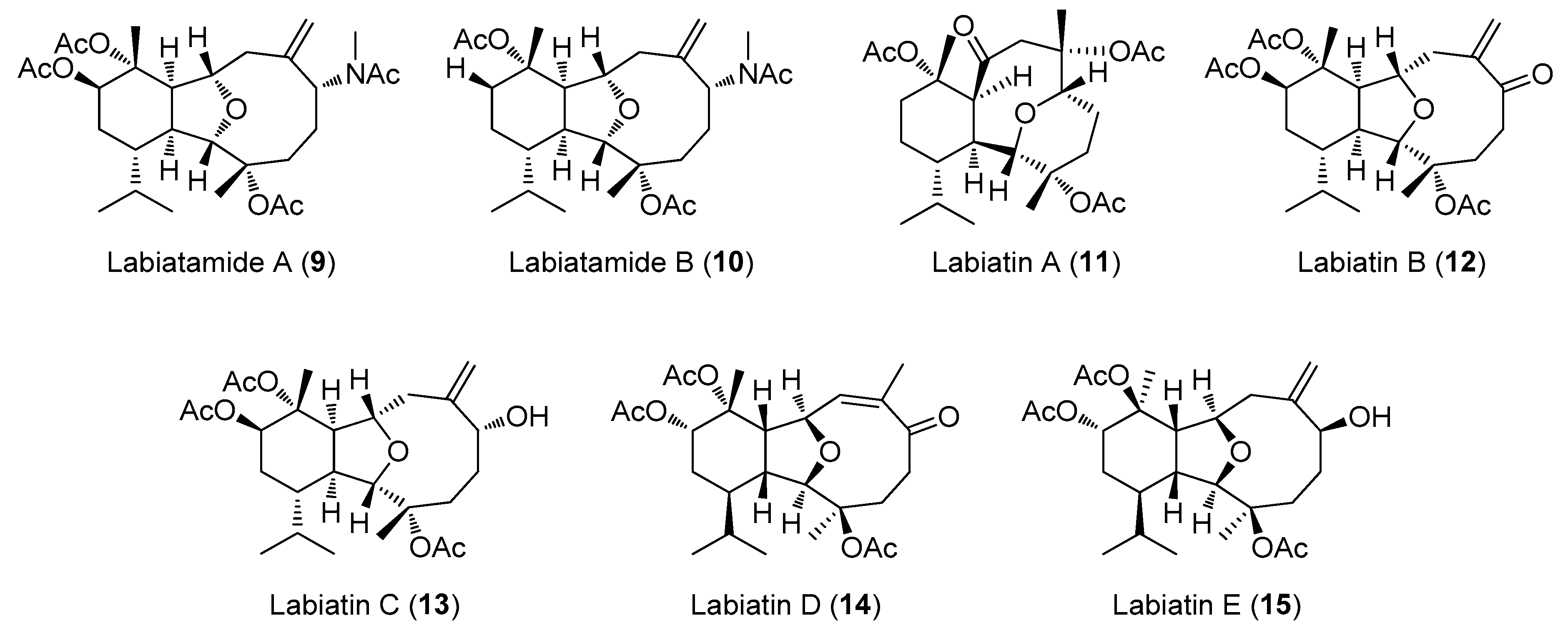
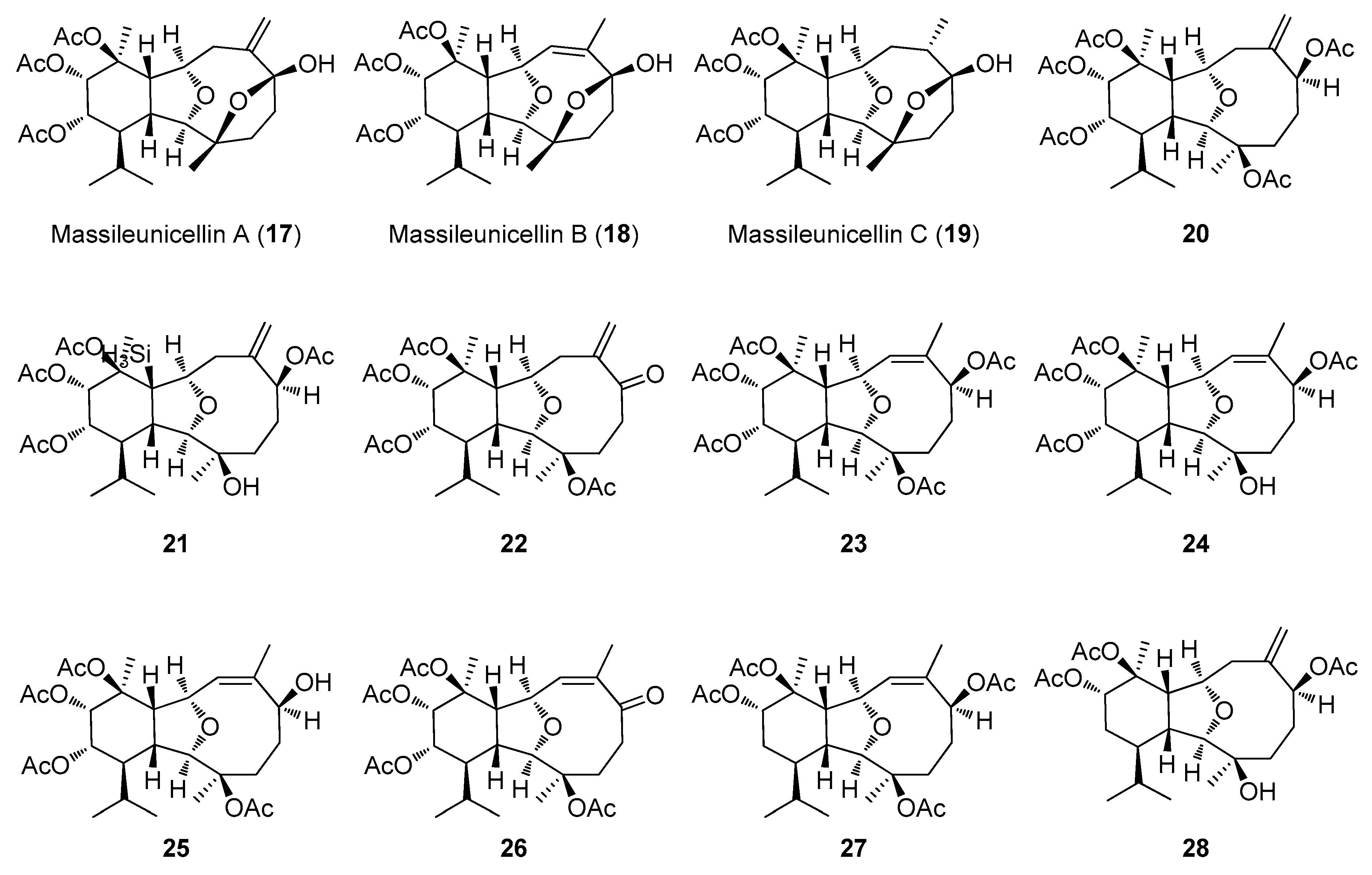
2.2. Structural Diversity of Terpenoids from Eunicella sp.
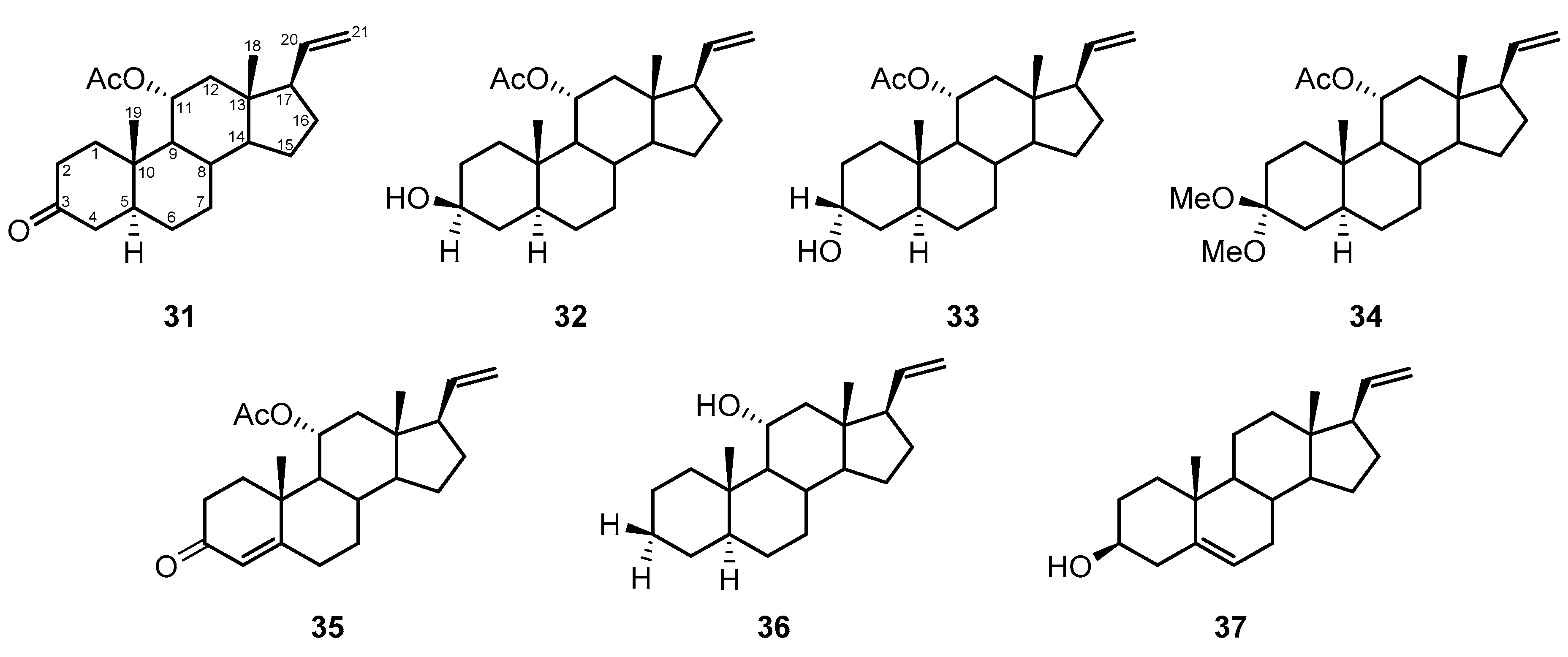
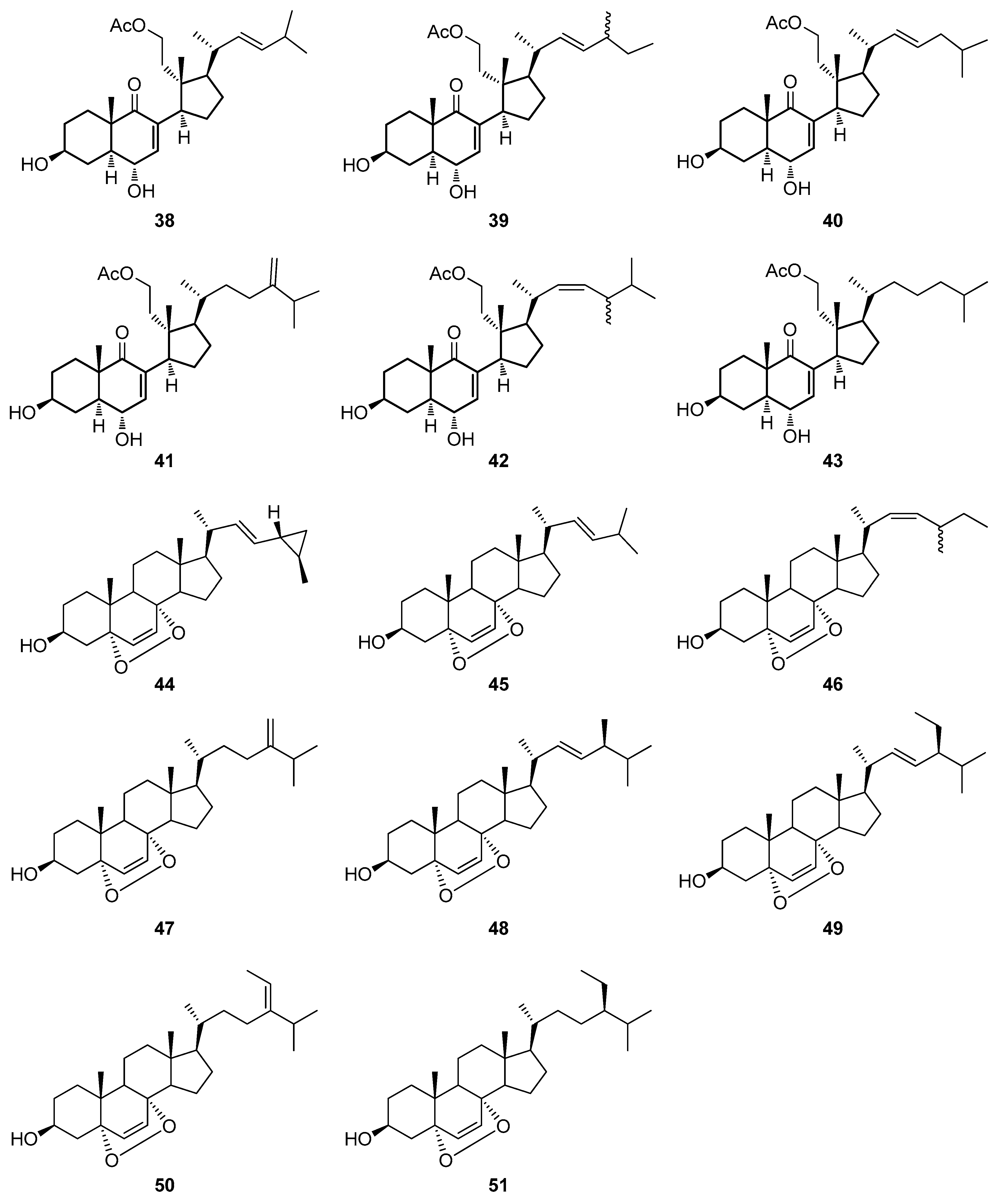
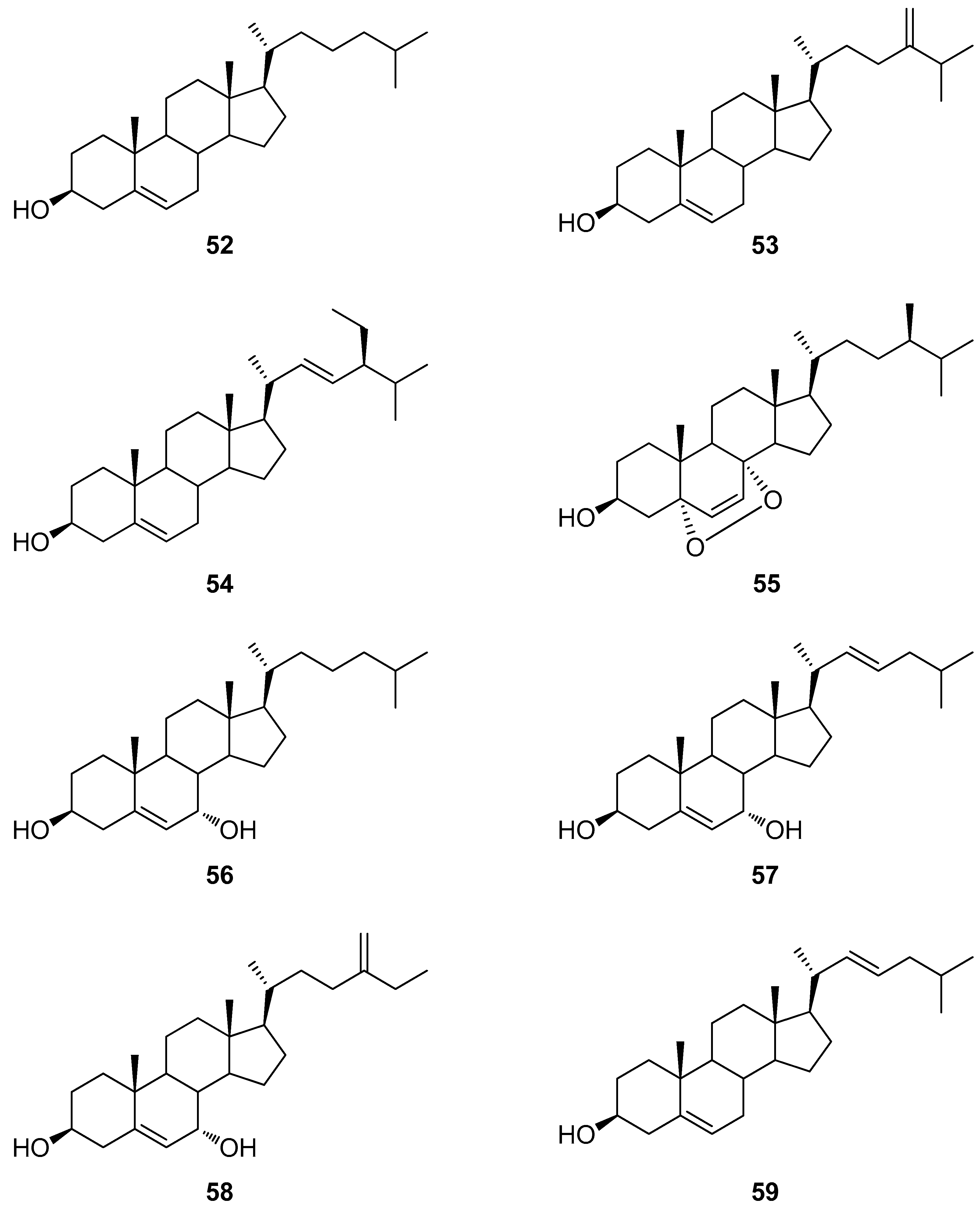
More than 40 eunicellan-type diterpenes have been isolated from gorgonians so far. However, few studies have also included evaluation of the biological activity of these compounds [8,19]. The first such metabolite was isolated in 1968 by Kennard et al., named eunicellin (1), having four acetyl groups attached to cladiellane skeleton (Figure 3) [25]. In 1993, Ortega et al. reported the isolation of five new diterpenoids 26 from the methanolic extract of Eunicella verrucosa collected near the Spanish coastal village of Palmones and named palmonines A–E (26), after the sampling site of gorgonian (Figure 4). The latter diterpenoid 6 formed white crystals, while the rest of the compounds were isolated as colorless oils. Apart from the acetate groups, they also differ in some other structural features. For example, palmonine D (5) and E (6) are two isomers bearing a conjugated keto group with an exocyclic or endocyclic double bond, respectively [20]. The same authors isolated another eunicellin-type diterpenoid from the same octocoral, palmonine F (7), which is a derivative of palmonine B (3) but displaying one less of acetoxyl NMR signals. They also performed oxidation of the secondary hydroxyl group of palmonine F (7) by Jones reagent, which led to the semisynthesis of palmonine D (5).
From 2013 to 2017, Deghrigue’s group and Ioannou’s group conducted thorough studies on E. singularis and Eunicella cavolini comprising preliminary biological evaluation of the organic extracts and fractions. It was found that the polar fractions show anti-inflammatory and analgesic potentials, as well as anti-proliferative effects on breast cancer cells MCF-7 and prostate cancer cells LNCaP, indicating the presence of novel bioactive compounds [19,29,30,31,32]. The organic extracts of the coral were prepared by maceration and were examined for the presence of several class of compounds, i.e., polyphenols, alkaloids, saponins, steroids, terpenoids, and glycosides by qualitative chemical screening tests based on visible chemical change [29,31]. Final purification of fractions resulted in isolation of 2 terpenoids, palmonines D (5) and F (7), and nine steroids of various chemical structures (Section 2.3., vide infra) [30,32]. Moreover, Rosa et al. first isolated diterpenoid 8 bearing two acetoxyl groups, from the E. cavolini acetone extract (Figure 5) [21].
Another Senegalese, deep-water gorgonian coral, E. labiate, was used to extract five diterpenoids with unique structural characteristics from the brown organic extract obtained by maceration of the natural material with dichloromethane and methanol (9:1). Roussis et al. reported the structures of labiatamides A (9) and B (10), as well as labiatins A–C (1113) (Figure 6). These compounds have an isopropyl group and a large number of acetoxyl groups, a characteristic which is also found in palmonines, mentioned previously. Labiatamides A (9) and B (10) possess a N-methyl acetamide moiety, while labiatin A (11) contains an unusual ether bridge between C2 and C6 [26]. Furthermore, Kakonikos et al. also isolated two new labiatins D (14) and E (15) from E. labiata organic extract (Figure 6). An eunicellin-type diterpenoid, labiatin D (14) is a colorless oil and showed similar spectroscopic characteristics as palmonine E (6) with one more acetyl carbonyl moiety. Labiatin E (15), a yellowish oil, also has 3 acetyl groups and spectroscopically is identical to previously isolated labiatin C (13), with the only difference being the stereochemistry of hydroxyl group at C6 position [22].
So far, all mentioned diterpenoids contain the cladiellane carbon skeleton (vide supra). However, there is an example of a metabolite having the same structure without an ethereal O-C2 and O-C9 bridge. The 12,13-Diacetoxycladiella-2,6-dien-11-ol (16) was isolated from E. labiata by Ortega et al. in 1997 (Figure 7). This secondary metabolite 16 possesses two acetoxyl groups and one hydroxyl group. The endocyclic double bond at C2 position has cis stereochemistry, whereas the one at C6 position is trans [24].
Two gorgonians, E. cavolini and E. singularis, were collected near Marseille and extracted with ethanol by Mancini et al., leading to the isolation of, in total, 20 compounds (Figure 8). Some of them, like palmonines D (5) and E (6), labiatins B–D (1214) and eunicellin (1) were already characterized by other scientific groups and discussed above (vide supra). Additionally, various oxidation numbers at the 10-membered ring and a second epoxy bridge or a differently located double bond can be observed in massileunicellins A–C (1719) [27,28]. The isolation, conformations, and reactivities of diterpenoids 2026 isolated from E. cavolini, and diterpenoids 27 and 28 from E. singularis are discussed by Mancini et al. emphasizing the various oxygenation patterns of cyclohexane ring [28].

2.3. Steroids

There have been several reports on isolation, characterization, and biological evaluation of various steroids from members of the Eunicella genus with unique structural features. These molecules differ in their degrees of oxygenation, as well as side chain patterns, which could be the reason for their miscellaneous activity, both in vitro and in vivo [19,30,31,32,33,34,35].
The acetone extract of E. cavolini was used to isolate a novel steroid, pregna-4,20-dien-11α-ol-3-one acetate (29), a pregnane derivative containing vinyl group. Besides spectroscopic elucidation, the authors also presented a chemical synthesis of 29 starting from a progesterone derivative (Figure 9) [35]. It should also be mentioned that Kashman et al. extracted verrucoside (30), a pregnane glycoside, after studying the cytotoxic activity of the DCM/MeOH extract of another gorgonian, E. verrucosa. This compound is known for having a 6-deoxy hexose derivative as a sugar unit, and Δ20 pregnane skeleton as aglycone [36].
More pregnanes were extracted from E. cavolini within a comprehensive study by Ioannou et al. to identify new natural compounds from Greek marine organisms in 2008 (Figure 10). Seven pregnanes 3137 were reported whose absolute stereochemistries were determined by Mosher’s method. Their structures differ in moieties at C3 and C11 positions of pregn-20-ene skeleton having acetoxy, methoxy, or hydroxy groups. The authors also pointed to the possibility of 34 being the product of reaction with methanol, a solvent which was used for both extraction and purification [37].
The same authors went on the isolation of steroids with more complex structures. Six 9,11-secosterols 3843 and eight 5α,8α-epidioxysterols 4451 were reported from the same gorgonian species conserving the steroidal carbon skeleton but having different aliphatic side chains (Figure 11). The first group of compounds showed C9–C11 bond cleavage in tetracyclic nucleus, while the latter one had higher oxygenation levels due to the presence of an endoperoxide bond. Those characteristic features are often found in marine invertebrates, particularly soft corals [33,34,38,39]. The 5α,8α-endoperoxides 4451 are oxidized sterols which can be formed during photooxidation reaction by addition of an oxygen molecule to a conjugated 5,7-diene system in the precursor molecule which contains the steroidal core [40,41,42,43]. This 5α,8α-endoperoxide bond is exactly responsible for the observed biological activities [40]. Sterol 48 has a structure almost identical to ergosterol peroxide, one of the most characteristic epidioxysteroidal derivatives found in both marine and terrestrial organisms. The only difference between 39 and ergosterol peroxide is the C24 stereogenic center of S- and R-configuration, respectively [42,44,45,46]. Gorgonian Verrucella umbraculum from the South China Sea is also a natural source of sterols 48, 49, and 52 [47]. Interestingly, 44 stands out from the rest of epidioxysterols found in Eunicella sp., due to the presence of a cyclopropyl moiety at the side chain [33,34].
As mentioned previously in Section 2.1. (vide supra), together with various classes of natural products, Deghrigue et al. and Lajili et al. extracted nine mono- or polyhydroxylated steroids from the Tunisian gorgonian coral E. singularis, 48 (Figure 11) and 5259 (Figure 12), of which two were epidioxysterols. Besides the number of hydroxyl groups and oxygenation of steroidal core, these products also differ in saturation patterns of side chain which was confirmed by comparison of 1D and 2D NMR data with the literature [19,30,32]. Steroid 56 was also found in other marine species, soft coral Dendronephthya gigantean and marine bryozoans, Biflustra grandicella, and Cryptosula pallasiana [48]. Furthermore, compounds 51 and 55 were also isolated from the tunicate Didemnum salary by Bensemhoun et al. [49].

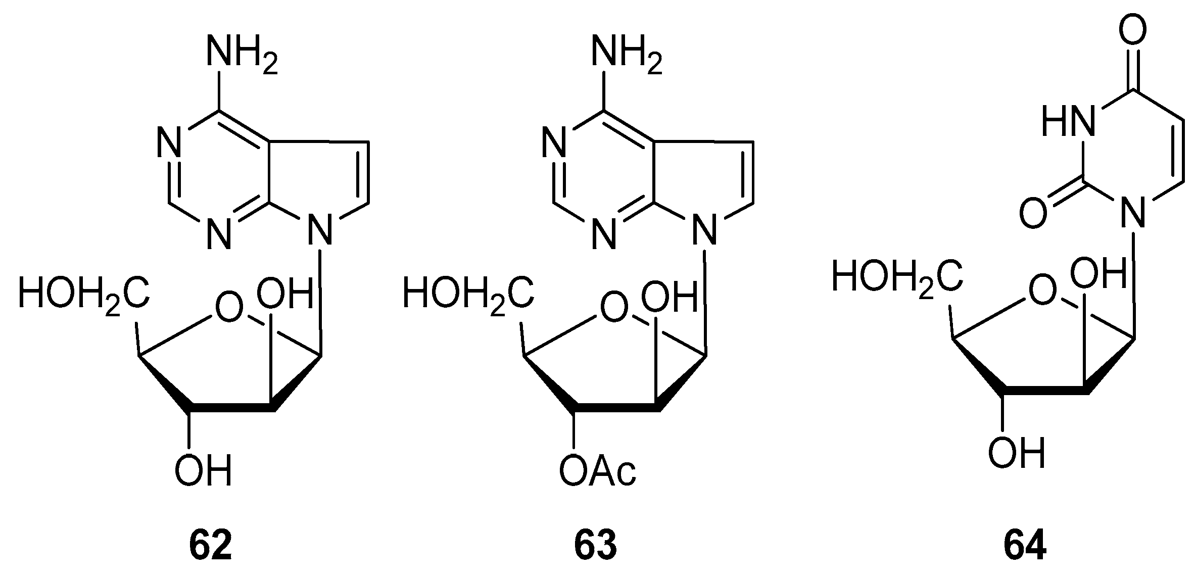
2.4. Alkaloids and Nucleosides

About 2% of metabolites isolated from all gorgonian species contain an nitrogen atom including alkaloids, nucleosides, amide, and tryptamine derivatives [8,50,51,52]. As with steroids and terpenoids, nitrogen containing compounds contribute to organisms by acting as a defense against negative external influences, increasing the biodiversity of marine habitat [50,51]. Particularly, xanthines and their methylated derivatives are often found in marine organisms included in nucleotide metabolism [50]. They have already been isolated from other corals, particularly of the genus Sinularia, exhibiting antibacterial and cardiotonic activities [53,54]. Samori et al. reported the concentration of N-heterocyclic compounds to be differently obtained following extraction of several gorgonian species with CH2Cl2/MeOH mixtures or acetonitrile during optimization of the extraction protocol for more polar metabolites. Furthermore, they observed no significant difference in the presence of nitrogen containing metabolites in three Eunicella species (verrucosa, cavolini, and singularis). They extracted hypoxanthine, guanine, and adenosine, the latter being absent in E. verrucosa and guanine being the most abundant [50].
Gorgonian E. granulata collected in Senegal was the subject of the study led by Reyes et al. to find new compounds with antitumor activity (Figure 13). The 2-propanol coral extract afforded granulatamides A (60) and B (61) after semipreparative, reverse-phased HPLC. Both novel compounds contained a tryptamine moiety connected to a fatty acid and differ in the number of double bonds in the aliphatic chain. These compounds exerted only a moderate effect in vitro against different tumor cell lines [51].
The n-butanolic extract of Eunicella cavolini was found to be a natural source of the polar nucleosides: 9-β-d-arabinofuranosyladenine (araA) (62), its 3′-O-acetyl derivative (63) and spongouridine (araU) (64) (Figure 14). The latter had already been isolated from the sponge Cryptothetia cripta. However, low solubility and susceptibility to deamination of the natural compounds led to the synthesis of several acyl derivatives that showed better properties in terms of drug administration [52].

3. Biological Activities

3.1. Cytotoxic Activity

Cnidarians, and, therefore, members of the genus Eunicella, are prominent sources of steroids and terpenoids which demonstrated antitumor activities [4]. All palmonines (27) mentioned in Section 2.1. (vide supra) were screened for their cytotoxic effect against several human or murine cancer cell lines: A549, HT29, P-388, and MEL28. The last two, mice lymphoma and human melanoma lines, respectively, were the most affected by 3, exhibiting IC50 values of 5 µg/mL [55]. Similar results were observed with excavatolides O and Q, diterpenoids with a briarane-type skeleton extracted from the gorgonian Briareum excavatum [8]. The authors hypothesized that the activity of those metabolites could arise from synergistic effects and interactions with other compounds. Since palmonine B (3) has the highest number of acetoxyl groups, that could also play a role in the observed cytotoxic activity [55]. Furthermore, significant anticancer potential of palmonines F (7) and D (5) was observed by inducing apoptosis of 92% and 93% of cells, respectively, in a breast cancer cell model (MCF-7 monolayer) at concentration of 200 µg/mL. The authors determined EC50 values from the concentration-response curves for 7 and 5, which were 13 µg/mL and 49 µg/mL, respectively. Lajili et al. also investigated the effect of two previously mentioned palmonines on MCF-7 mammospheres and reported reduction in spheroid size, which was also concentration-dependent and visible 2 days after incubation [19].
Labiatins B, C. and E (12, 13, and 15) were assayed for their cytotoxic effect against human cancer cell lines [22,26]. Labiatin B (12) was tested against colon cancer line (HCT-116) and the authors reported an IC50 value of 0.85 µg/mL [26]. The last two compounds showed a cytostatic effect against non-small cell lung cancer line (NSCLC-N6) with half inhibitory concentrations of 35 µg/mL and 7.7 µg/mL for labiatin E (15) and C (13), respectively, indicating that the stereochemistry influences the biological activity of these secondary metabolites [22].
Massileunicellins A–C (1719) were additionally assayed for antitumor and antiviral activity against KB and L1210 doxorubicin-resistant tumor cells, and Dengue virus, respectively. After observing only low cytotoxic effects of massileunicellins, Mancini et al. screened labiatin B (12) and palmonine D (5) against the same cells which also resulted in poor activity. Comparing those results with the significant cytotoxic activity reported by Ortega et al. [55] and Roussis et al. [26], they hypothesized that diterpenoids might be selective for specific tumor cells. Furthermore, they did not observed any stabilization effect of 5 and 12 on tubulin, a feature characteristic for other types of 2,11-cyclized cembranoids, i.e., sarcodictyns and eleutherobin [27]. The most promising cytotoxic diterpenoids isolated and studied from the genus Eunicella are shown in Table 1.
Steroid compounds of natural origin have recently attracted considerable attention among the scientific community due to their interference with molecular pathways of MCF-7 cells. It has been shown that steroid derivatives inhibit estrogen-dependent proliferation and survival of those cells binding to estrogen receptor, thus disrupting the interaction with transmembrane growth factor receptor [33,34,37]. Furthermore, according to Chen and his group, secosteroids and polyhydroxylated steroids could be potential inhibitors of protein-tyrosine phosphatase 1B (PTP-1B), which is involved in metabolism of glucose via hydrolyzation of insulin receptor [39,56]. However, PTP-1B was also found to have a contradictory role in cancer biology. Its deficiency has led to the growth of B lymphoma in mice while it had no role in breast tumorigenesis [57]. Nevertheless, it presents a novel target in both cancer and metabolism treatment.
Ioannou et al. assayed the growth inhibitory potential of sterols isolated from E. cavolini (3139, 41, 42, 4451) against MCF-7 cells cultured with and without a physiological concentration of 17β-estradiol (1 nM) [33,34,37]. When compared to the effect of positive control (ICI 182,780, Faslodex®, a commercially available estrogen receptor down-regulator), compounds 3135 inhibited cell growth for 40 to 49%, while the last two pregnanes (36, 37) were almost inactive. According to the authors, the presence of acetoxyl moiety at C11, observed in metabolites with significant inhibitory effect, could play a role in mediating estrogen-dependent growth of MCF-7. Another possible molecular target is the nuclear pregnane X receptor, a target of several marine-derived compounds involved in drugs and xenobiotics metabolism, oxidative stress, cancer formation, and other physiological processes which yet need to be examined [37,58,59,60]. Secosterols 38 and 39 exhibited IC50 values of 7.6 and 7.4 μM against MCF-7 cells, respectively. On the other side, growth inhibition by all compounds was significantly enhanced in MCF-7 cells cultivated in the absence of growth factors and endogenous estrogen. Since 38 was also found to be effective against proliferation of K562 and HeLa cells, it is assumed that secosterols’ antiproliferative activity is selective towards specific tumor cells, as well as the applied growth conditions. Furthermore, since tested compounds differ only in the side chain structure, the better biological activities of 38 and 39 could also be attributed to the double bond at C22 position with E geometry [34]. The gorgonian coral, Subergorgia suberosa, collected from the South China Sea, was used to extract several secosterols bearing a hydroxyl group at C11, including 38 and 43. Zhang et al. reported IC50 values of 28.1 and 23.3 μM against HeLa cells for 38 and 43, respectively, and hypothesized that the presence of a free -OH group at C11 does not contribute to the observed cytotoxic effects of the tested compounds [38]. That is also consistent with the conclusion reported by Kongkathip et al. who have synthesized and evaluated the cytotoxicity of 9,11-secosterol derivatives against KB, HeLa, and MCF-7 cells, as well as non-transformed Vero cells. According to the authors, the key to the observed biological activity is in the presence of a cholesterol-like side chain and a keto group at C9 position [61].
Epidioxysterols 4451 also displayed significant inhibition activity against growth of MCF-7 cells in the growth factor and endogenous steroids depleted medium. At a concentration of 10 µM, 44, 45, and 46 displayed almost 60% of the effect of positive control, ICI 182,780 (Faslodex®). Considering that 44 was particularly effective against MCF-7 cultured in both cell growth medium conditions, the authors assume that the cyclopropyl group at the side chain of 44 was responsible for the observed effects [33]. Luo et al. also isolated 45 from another marine organism, sponge Topsentia sp., and observed its moderate cytotoxic activity against different tumor cell lines of human origin: A549, SK-OV-3, SK-MEL-2, XF498, and HCT15 (IC50 values: 30.0, 19.4, 25.9, >30.0, 21.4 μg/mL, respectively) [62]. Another marine sponge, Monanchora sp., was used in the study conducted by Mun et al. in 2015 who isolated epidioxysterols 45 and 47. The authors evaluated the activities of these two products against renal, pancreatic, and colorectal cancer cell lines and suggested that the C24 (28) double bond, which is a feature of 47, might be responsible for its increased cytotoxic activities. At a concentration of 23 μM, 47 induced 50% inhibition of renal A-498 cells. It should be emphasized that 47 exhibited higher cytotoxicity than the positive controls, temsirolimus and 5-fluorouracil, used in the clinical treatment of the above mentioned cancers [63]. The 48 showed significant antiproliferative activity against A549, H460, and HGC27, displaying IC50 values of 10.9, 10.9, and 11.6 μg/mL, respectively, as well as moderate cytotoxicity towards human ovarian cells (A2780) with an IC50 value of 16 μg/mL [64,65]. Raslan et al. isolated epidioxysterols 49 and 51 from the marine sponge Monanchora clathrate but observed only weak cytotoxicity against MES-SA, MCF-7, and HK-2 cancer cell lines with IC50 values higher than 25 μg/mL [66].
Deghrigue et al. and Lajili et al. also investigated the cytotoxic activity of steroids isolated from E. singularis after observing promising antiproliferative, antioxidant, and other pharmacological activities of its organic extract and fractions [19,30,32]. Metabolites 52, 54, and 59 were evaluated for their anticancer activity against a monolayer and a spheroid model of MCF-7 using AnnexinV- fluorescein isothiocyanate/propidium iodide (FITC/PI) flow cytometry and spheroid size analysis, respectively. Cholesta-5,22-diene-3β-ol (59) displayed an EC50 value of 30 µg/mL against monolayer cells causing apoptosis in 91% of cancer cells at 200 µg/mL. However, steroids were more effective against the spheric model than diterpenoids (5 and 7) isolated from the same organism. The reported growth rates for 52, 54, and 59 were 1.17, 1.25, and 1.15, respectively, at a concentration of 200 µg/mL and comparable to the control, taxol at 100 µM [19]. On the contrary, according to Tian et al., 52 was inactive in vitro against HL-60, HepG2, and SGC7901 cell lines [67]. Finally, Pan et al. investigated the proapoptotic activity of the ethyl acetate extract of the hard clam Meretrix lusoria and consequently isolated a mixture of epidioxysterols containing 55. The authors observed 35% and 73% apoptotic HL-60 cells after treatment with 25 and 50 µg/mL of this mixture, respectively [68]. The antitumor activities of sterols are summarized in Table 2.
Granulatamides A (60) and B (61) were both evaluated for their cytotoxic activities against 16 different human cancer cell lines by the use of sulphorhodamine B colorimetric assay (Table 3), exhibiting low GI50 (growth inhibition concentration) values of 1.7 and 3.5 µM against two human prostate cancer cell lines, DU-145 and LN-caP, respectively [51]. Another nitrogen containing compound, nucleoside 63 was assayed for cytotoxic activity against KB cells displaying a low IC50 value of 5 µg/mL [52].

3.2. Anti-Inflammatory Activity

Discovery of novel compounds that may be used to target inflammatory processes is highly needed since the use of many existing nonsteroidal anti-inflammatory drugs have been linked with various severe adverse effects [30,32]. In this context, Deghrigue et al. have reported that the organic extract and fractions obtained by further purification with ethanol, acetone, and dichloromethane/methanol have exhibited anti-inflammatory activity in a dose-dependent manner regarding the inhibition of edema in rats induced by carrageenan. At a dose of 25 mg/kg of the ethanolic semipurified fraction, the edema was inhibited by 66% after 3 h, which is 10% higher than observed with a reference drug, acetylsalicylaze-lysine (ASL). Final purification of the ethanolic fraction led to isolation of owp palmonines, 5 and 7, and nine sterols, 48, 5259, which could contribute to the investigated biological activity [30,32]. Anti-inflammatory activity of epidioxysterols 48 and 55 was further investigated by other scientific groups [68,69,70]. Just recently, Huynh et al. studied the influence of 48 on expression of pro-inflammatory proteins, iNOS and COX-2, released from macrophages. They observed significant suppression of iNOS by 48 at 10 µM. However, dendronesterone D bearing acetoxyl moiety at C11 showed the highest suppression of inflammation [70]. Inhibition of NO production by lipopolysaccharide (LPS)-stimulated macrophages was used to assess anti-inflammatory activity of 55 in a mixture of epidioxysterols by Pan et al. NO production did not arise from the cytotoxic activity and was inhibited by 38%, 39%, 99%, and 100% at concentrations of 5, 10, 25, and 50 µg/mL, respectively [68]. Immunosuppressive activity of 47 and 48 against T and B lymphocyte cells proliferation was also examined by Yang et al. in 2019. Both epidioxysterols were extracted from a soft coral Sinularia sp. Reported IC50 values of 47 and 48 were 57.54 and 59.54 µM for T cells, and 47.57 and 19.30 µM for B lymphocytes, respectively. Thus, they showed weaker anti-inflammatory activity than cyclosporin A (IC50 = 0.04 and 0.40 µM for T and B cells, respectively), however they were less cytotoxic against murine splenocytes [69].

3.3. Antimicrobial Activity

Bioactive secondary metabolites from Eunicella sp., as well as compounds from other soft corals, showed interesting antibacterial and antiviral activities [4,52,71,72]. According to Gauvin et al., epidioxysterols are the first natural metabolites to display antiviral activity against lymphoma causer, HTLV-I retrovirus. A mixture of sterols, including 49, 51, and 55, reduced by 50% β-galactosidase activity which is correlated to virus activity at a concentration of 0.3 mg/mL. However, they were inactive against another RNA virus, HIV [71]. Polar nucleosides from E. cavolini, 9-β-d-arabinofuranosyladenine (spongoadenosine, araA) (62) and spongouridine (araU) (64), showed promising antiviral activity against DNA viruses [52]. Pharmacological properties and biosynthesis of araA (53) have been already reviewed by Huang et al. [73]. It is worth mentioning that 62 was the first drug to treat herpes encephalitis infections and diseases caused by other herpes viruses in patients with impaired immune system [73]. Synthetic O-acyl derivatives of 62 showed also promising antiviral effects but its 3′-O-acetyl derivative (63) yet needs to be examined in order to complete the structure activity relationship studies [52]. We would also like to mention that Liang et al. observed an antibacterial activity of epidioxysterols 47 with MIC50 value of 500 µM against Staphylococcus aureus Newman strain. This activity is considered significant because the MIC50 value is lower than 1 mM [72].

4. Synthetic Approaches

One of the major problems during the preclinical and clinical development of naturally derived drugs is the insufficient quantity of the isolated natural product precursors [2,4,74]. As the consequent market application by the pharmaceutical and cosmetic industries entails the desired metabolite to be available often on the ‘kilogram scale’, it is not surprising that some compounds which demonstrated promising biological activities have never reached the market. In order to obtain sufficient amounts of a natural product or its derivatives, either the extraction of the natural row material should be repeatedly performed or the natural product should be synthetized [75]. The extraction pathway may be problematic in terms of the percentage of the desired secondary metabolite in the row material and/or ecological and ethical principles. For example, in the case of Eunicella species, mass mortality events have emerged in the past few years mainly hitting the gorgonian corals in the Mediterranean Sea [76,77,78,79,80]. Furthermore, collecting the same species in various geographical areas or during different time seasons might result in a different chemical composition, thereby reducing or increasing the yield of the desired product [75,81]. Finally, several characteristics of marine natural products, such as halogenation, oxygenation substitution patterns, different degrees of carbon framework functionalization, and stereochemical diversity make chemical synthesis even more complex and challenging for organic chemists. The fulfilment of the principles of green chemistry including the application of atom-economical and high-yield reactions, the use of affordable and nontoxic starting materials and solvents, or the design of protecting group-free chemical synthesis are additional issues to also consider [2,75,82,83]. Thus, novel synthetic strategies have been developed recently, with new scientific and technological progress and insights, and are oriented toward total syntheses of natural products and their stereochemical congeners, at the same time complying with green chemical approach [2].

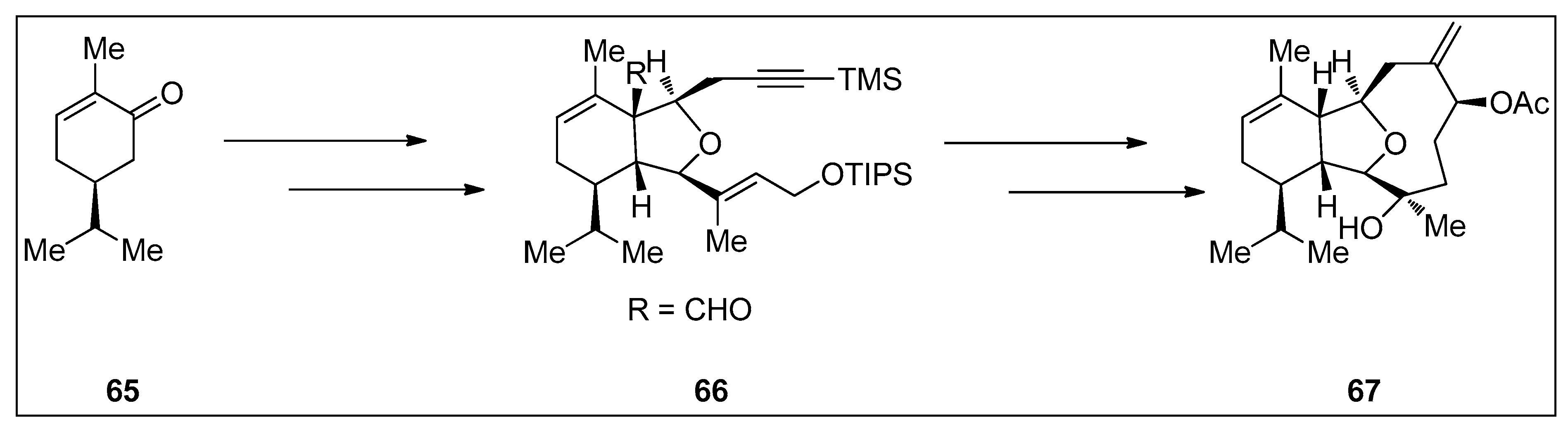
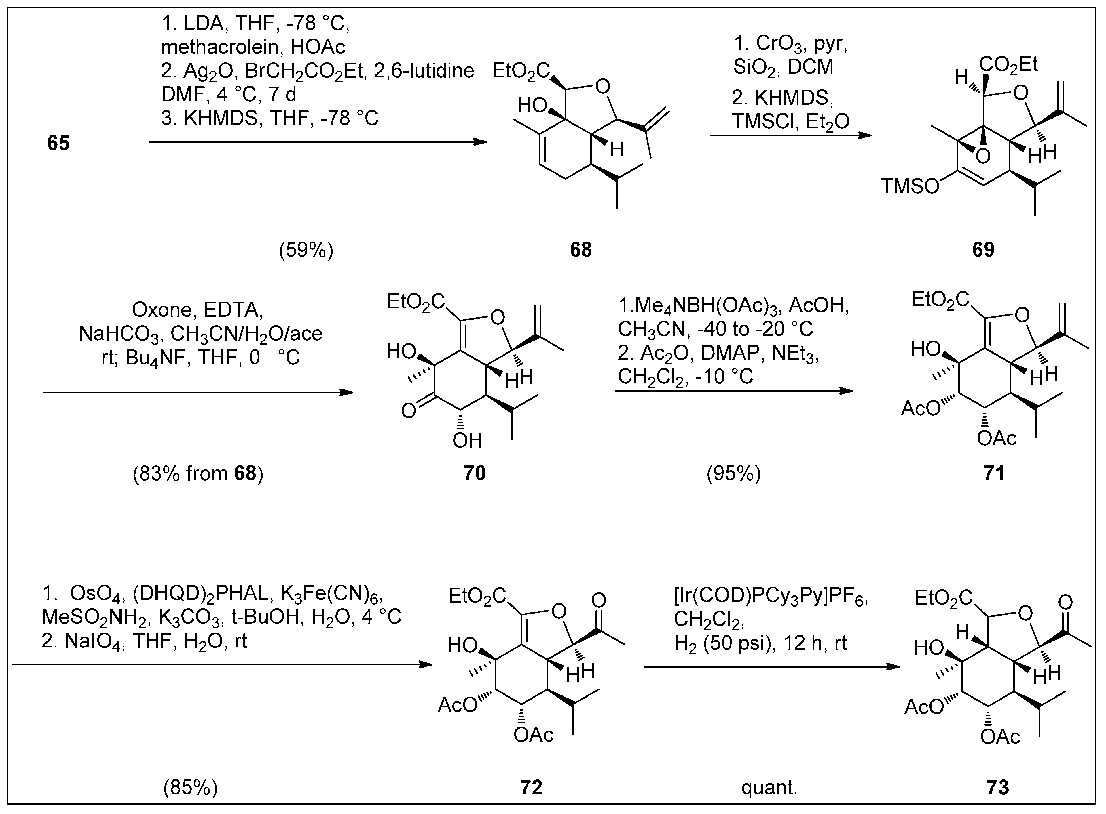
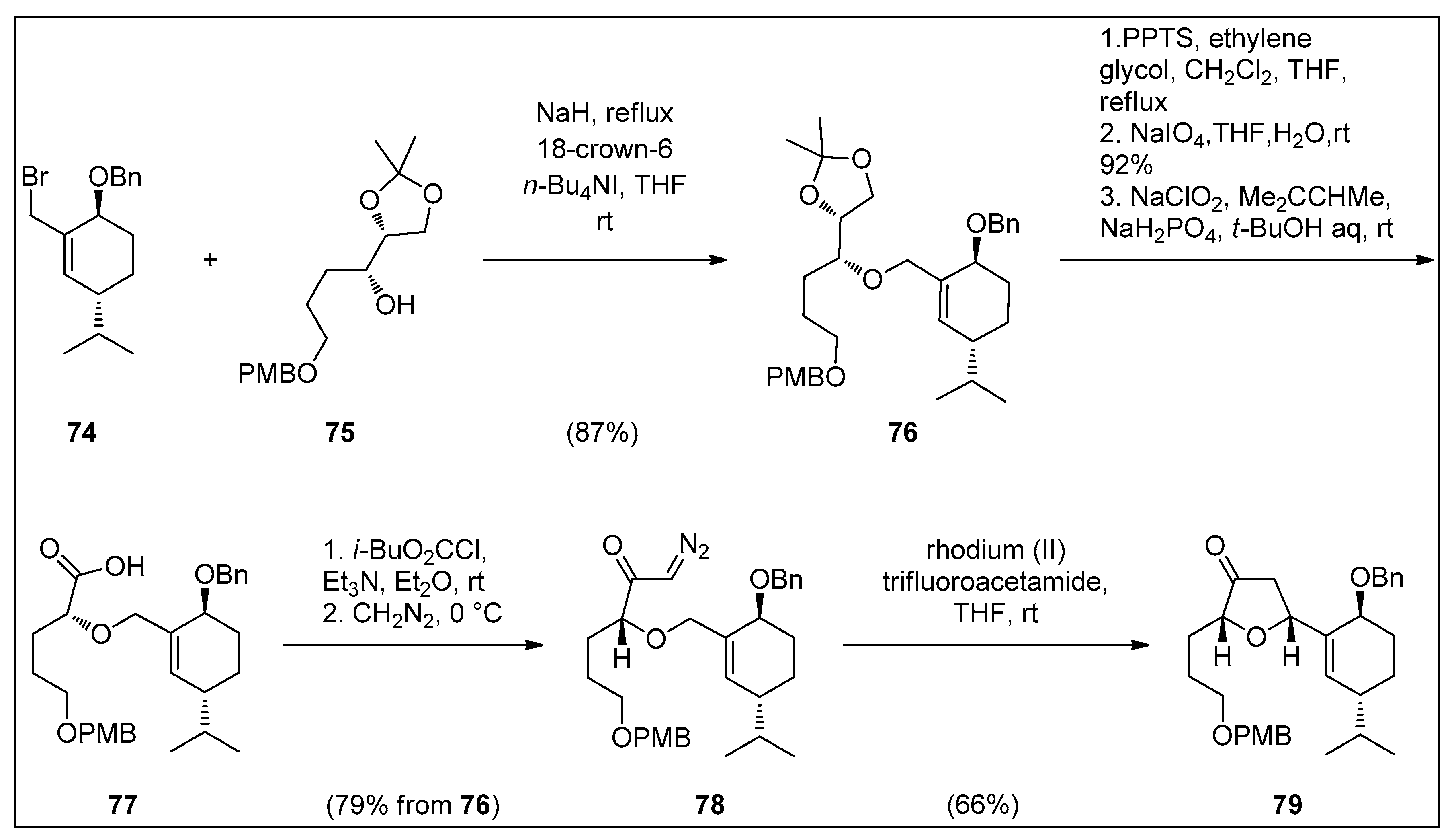
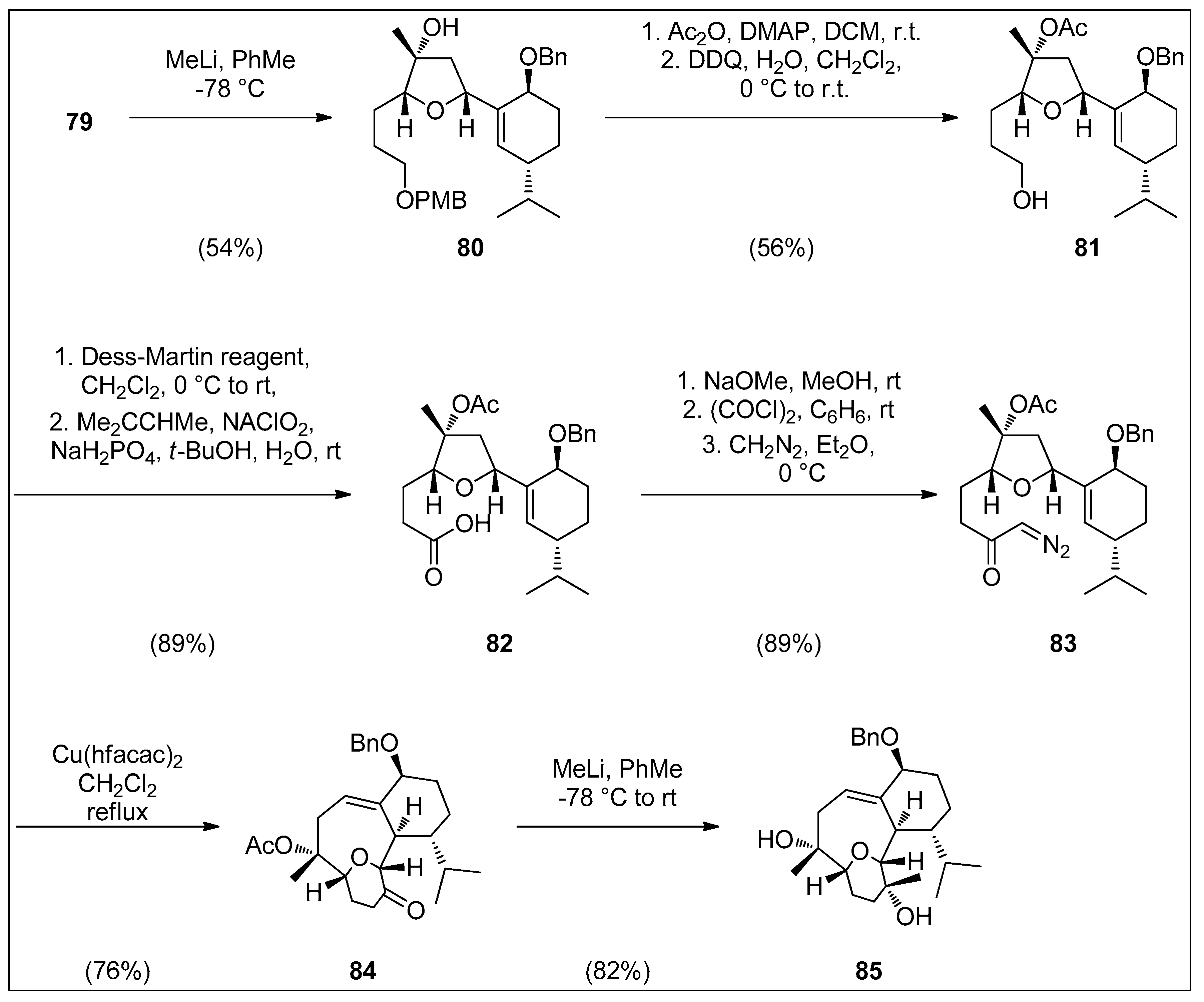
4.1. Synthetic Approaches toward the Synthesis of Terpenoids Isolated from Eunicella Species

Several studies have been conducted over the past two decades with the aim of developing a synthetic approach to obtain highly oxidized eunicellin-type diterpenes [84,85,86,87,88,89]. Two major issues had to be overcome in constructing the tricyclic skeleton of eunicellins. The first was stereoselective formation of hydroisobenzofuran core, which possesses four stereogenic centers and the second was formation of the nine-membered ring. Approaches, including a first synthesis of the isobenzofuran part followed by the cyclization of the nine-membered ring or vice versa, have been developed [88].
The first successful total synthesis of cladiellin, (−)-7-deacetoxyalcyonine (67), starting from (S)-dihydrocarvone 65, was accomplished in 1995 by MacMillian and Overman. Prins-pinacol rearrangement was employed to achieve hydroisobenzofuran 66 as a single diastereomer, followed by Nozaki–Hiyama–Kishi coupling reaction, resulting in the final tricyclic product (Scheme 1) [90]. Since that, numerous total or partial syntheses of eunicellins (cladiellins) and other types of C2–C11 cyclized cembranoids were conducted, which had been previously reviewed by Ellis et al. in 2008 [89].
Synthetic access to eunicellin-type metabolites produced by members of the genus Eunicella needs still to be developed. Up to now, profound investigations were performed by the McIntosh group to evolve the complete hydroisobenzofuran core from less oxidized compounds, thus obtaining the more complex stereochemistry of massileunicellins [86,91]. They synthesized hydroisobenzofuran of massileunicellins containing eight of a total of nine stereocenters in an overall 12 steps with a single temporary trimethylsilylation protection of hydroxyl group. As shown in Scheme 2, (S)-(+)-carvone (65) was reacted with methacrolein in aldol reaction, followed by Williamson etherification and cycloaldolization of the formed glycolate intermediate to afford allylic alcohol 68 in 85% yield [87,91]. The intermediate 68 is converted to epoxy ketone with Collins’ reagent, which in subsequent treatment with potassium bis(trimethylsilyl)amide/trimethylsilyl chloride (KHMDS/TMSCl) yielded silylated enolate 69. Rubottom oxidation by using dioxirane produced keto diol 70 in 83% yield from intermediate 68. Evans–Saksena reduction of product 70 by using borohydride reagent led to the corresponding triol, which was subsequently esterified to diacetate 71 in high yield as a single diastereomer. To obtain the desired hydroisobenzofuran product 73, 71 was subjected to dihydroxylation followed by oxidative cleavage with NaIO4, giving rise to methyl ketone 72. Finally, hydrogenation of a double bond of 72 was conducted with Crabtree’s catalyst at moderate H2 pressure to yield quantitively the hydroisobenzofuran core 73 [87].
Clark’s group previously developed synthetic approaches to synthesize the tricyclic core of labiatin A, which is an unusual eunicellin (cladiellin) possessing a C2–C6 ether bridge as discussed above (vide infra) [84,85,92,93]. The authors firstly synthesized bromide 74 in five steps and coupled it through Williams’s etherification with alcohol 75 that was easily prepared from d-mannitol (Scheme 3). The obtained ether 76 (87%) was deprotected with pyridinium p-toluensulfonate (PPTS) to cleave the acetonide moiety, and the produced syn-diol was oxidatively cleaved with NaIO4 to form a terminal aldehyde which was further oxidized to carboxylic acid 77 with NaClO2. A mixed anhydride formation of acid 77 and chloroformate followed by subsequent treatment with diazomethane gave the diazo ketone 78 in 79% yield from 76. Finally, cyclization of diazo compound 78 was performed by using rhodium (II) tifluoroacetamide as a catalyst and produced dihydrofuranone 79 in 66% yield [85].
Dihydrofuranone 79 was reacted with methyl lithium giving rise to tert-alcohol 80 (85%) (Scheme 4). Acetylation and removal of p-methoxybenzyl protecting group (PMB) with 2,3-dichloro-5,6-dicyano-1,4-benzoquinone (DDQ) of 80 produced primary alcohol 81 (56%). The latter was converted to carboxylic acid 82 (89%) after Dess–Martin and Pinick type oxidation reactions. The 82 was transformed to the corresponding acyl chloride with oxalyl chloride, and, after sequential treatment with diazomethane, resulted in the formation of diazo ketone 83 (89%) required for the final key step. Intramolecular cyclization of the electrophilic carbenoid species of intermediate 83 with copper (II) hexafluoroacetylacetonate gave tricyclic ketone 84 (76%). Finally, after 21 steps in total, tertiary alcohol 85 was formed by the reaction with MeLi leading to the carbon core of labiatin A (11) as a single isomer. Its structure was confirmed by X-ray crystallography [85].

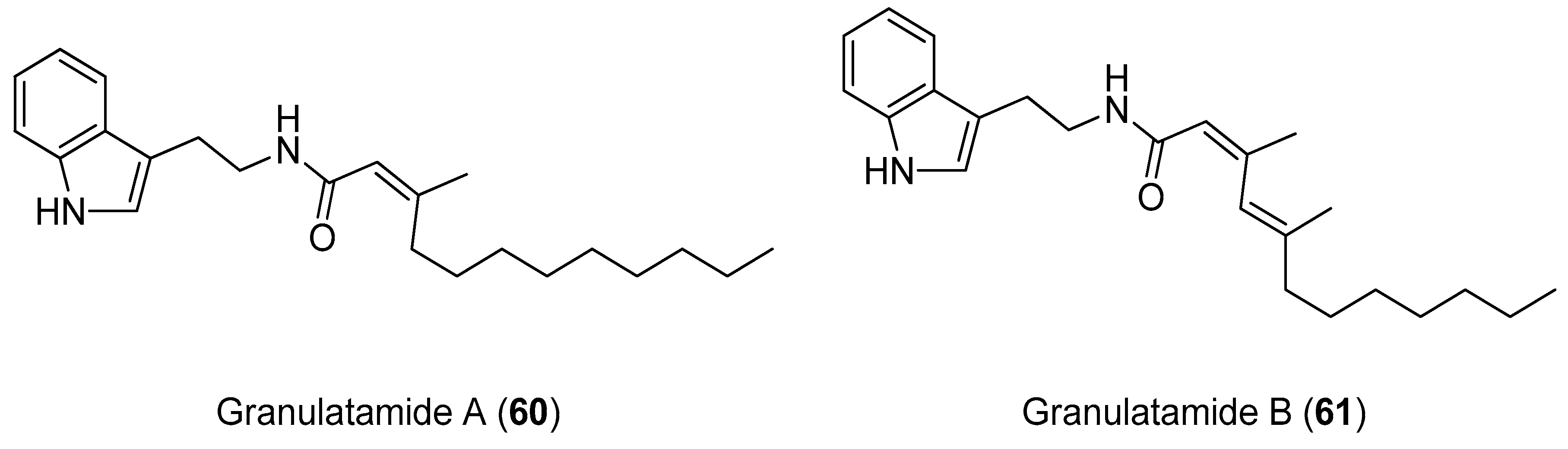
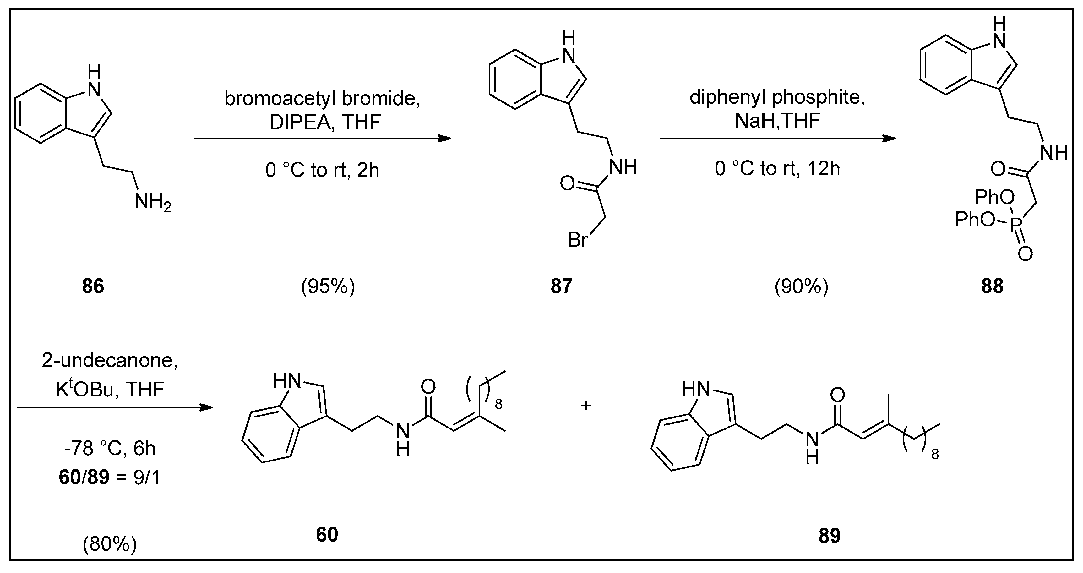
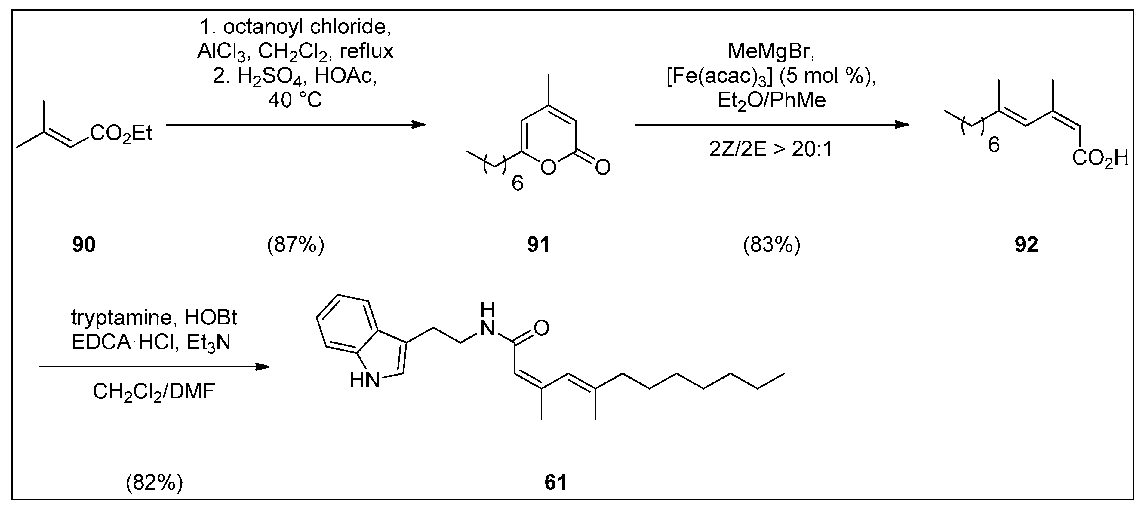
4.2. Synthesis of Granulatamide Alkaloids

In order to obtain the kinetically favored Z-isomer of cytotoxic alkaloids, Pakhare and Kusurkar reported the first total synthesis of granulatamide A (60) from tryptamine (86) via Horner–Wadsworth–Emmons olefination reaction (Scheme 5). In brief, tryptamine (86) was converted to bromoacetamide 87 which was treated with diphenyl phosphite under basic conditions to provide phosphonate 88 in 90% yield. The desired natural product granulatamide A (60) and its E-congener 89 were obtained by the reaction of phosphonate 88 with 2-undecanone in E/Z ratio = 9/1 and 80% overall yield. [94].
Sun and Fürstner developed a highly yielded three-step total synthesis of the cytotoxic tryptamine derivative granulatamide B (61) by employing their ring-opening/cross-coupling reaction of 2-pyrones methodology (Scheme 6). Pyrone 91 was obtained in multigram amounts by the reaction of the commercial crotonate 90 and octanoyl chloride. The iron catalyzed ring-opening/cross-coupling of pyrone 91 with methyl Grignard reagent smoothly afforded the acid92 as a single isomer in 83% yield. The amide bond formation was performed under standard conditions by using 1-hydroxy benzotriazole/1-ethyl-3-[3-dimethylaminopropyl] carbodiimide hydrochloride (HOBt/EDCA) coupling protocol to furnish granulatamide B (61) [95].

5. Conclusions

Natural products and their semisynthetic derivatives have been traditionally among the most important sources in the creation of novel products, especially in the pharmaceutical and cosmetic industries.
As shown in this review, the interdisciplinary studies linked to gorgonian soft corals have contributed to the discovery of new marine natural products. More than 60 metabolites were isolated from the Eunicella species in the past three decades, belonging to the class of terpenoids, sterols, alkaloids and nucleosides. These metabolites exhibited anticancer, anti-inflammatory, antiviral, analgesic, and other activities. Their full therapeutic spectra remain to be examined in detail. Already, seven compounds displayed very high cytotoxic activities against cancer cell lines in vitro, namely palmonine B (3), labiatin C (12), labiatin D (13), secosterols 38 and 39, and granualtamides A (60) and B (61) with IC50 values less than 10 μM or 10 μg/mL. Epidioxysterol 48 showed significant immunosuppressive activity, while nucleosides 62 and 64 showed relevant antiviral activity against DNA viruses.
The interesting biological activities of Eunicella’s secondary metabolites have attracted a broad attention of the scientific community with the purpose to isolate novel compounds, develop new and innovative synthetic methodologies, or conduct their partial or total synthesis. By committing studies in the direction of their synthesis, essential stereochemical and mechanistic issues were encountered and solved. The partial or total synthesis of these natural products is expected to guide advancements of new chemical approaches with wider synthetic applications. In conclusion, molecules isolated from the genus Eunicella, from their first discovery to date, present an interesting case which has stimulated research advancements in diverse scientific fields, attaining new valuable experimental and theoretical knowledge for future development in medicinal applications.

Author Contributions

D.M. (Dean Markovic) devised the concept of the manuscript. D.M. (Dario Matulja), M.K.M., G.A. and S.L. wrote parts of the manuscript. D.M. (Dario Matulja), M.K.M., G.A. and S.L. performed literature searches. S.K.P. and D.M. (Dean Markovic) participated in the manuscript writing, performed literature searches, wrote and discussed parts relevant for synthesis and biological data, finalized the paper concept, and performed the final manuscript revision. All authors have read and agreed to the published version of the manuscript.

Funding

This research is funded by European Regional Development Fund (the Competitiveness and Cohesion Operational Program, KK.01.1.1.01) through project Bioprospecting of the Adriatic Sea (KK.01.1.1.01.0002) granted to The Scientific Centre of Excellence for Marine Bioprospecting (BioProCro). Furthermore, financial support is provided by the project, Research Infrastructure for Campus-based Laboratories at University of Rijeka, co-financed by European Regional Development Fund (ERDF) and the University of Rijeka research grants, UNIRI-biomed-18-133 (1277) and UNIRI-prirod-18-102.

Acknowledgments

We would like to thank the Croatian Science Foundation project, Career Development of Young Researchers, Training of New PhDs, for funding PhD of D. Matulja. M. Kolympadi Markovic and G. Ambrožić are grateful to the Croatian Science Foundation for funding the postdoctoral position of M.K.M. through the project IP-2016-06-3568.

Conflicts of Interest

The authors declare no conflict of interest.

List of Abbreviations

A2780human ovarian carcinoma cell line
A498human renal epithelial cancer cell line
A549human lung carcinoma cell line
ASLacetylsalicylate of lysine
COX-2cyclooxygenase-2
DDQ2,3-dichloro-5,6-dicyano-1,4-benzoquinone
(DHQD)2PHALhydroquinidine 1,4-phthalazinediyl diether
DIPEAN,N-diisopropylethylamine
DMAP4-dimethylaminopyridine
DMFdimethylformamide
Du145human prostate cancer cell line
EDTAethylenediaminetetraacetic acid
H460human large cell lung cancer cell line
HCT-115human colorectal adenocarcinoma cell line
HCT-116human colon cancer cell line
HeLahuman cervical cancer cell line
HepG2human hepatocellular carcinoma cell line
Hfacachexafluoroacetylacetonat
HGC-27human gastric cancer cell line
HK-2human papillomavirus 16 (HPV-16) transformed renal cell line
HL-60human leukemia cell line
HT29human colon adenocarcinoma cell line
HTLV-1human T-lymphotropic virus 1
IC50half maximal inhibitory concentration
IGROVhuman ovarian cancer cell line
iNOSinducible nitric oxide synthase
K562human myelogenous leukemia cell line
KBkeratin-forming tumor cell line HeLa
KHMDSpotassium bis(trimethylsilyl)amide
KtOBupotassium t-butoxide
L1210murine lymphocytic leukemia cell line
LDAlithium diisopropylamide
LNcaPandrogen-sensitive human prostate adenocarcinoma cell line
LPSlipopolysaccharide
MCF-7human breast adenocarcinoma cell line
MES-SAhuman uterine carcinoma cell line
MIC50minimum inhibitory concentration required to inhibit the growth of 50% or organisms
NOnitric oxide
NSCLC-N6human non-small cell lung cancer cell line
P-388menogaril-resistant leukemia cell line
PPTSpyridinium p-toluensulfonate
SGC-7901human gastric cancer cell line
SK-BR3human breast cancer cell line
SK-MEL-2human melanoma cell line derived from metastasis on skin of thigh
SK-OV-3human ovarian carcinoma cell line
THFtetrahydrofuran
TMSCltrimethylsilyl chloride
Veroepithelial cell line derived from kidney of Cercopithecus aethiops
XF498human glioblastoma cell line

References

  1. Hamed, I.; Özogul, F.; Özogul, Y.; Regenstein, J.M. Marine Bioactive Compounds and Their Health Benefits: A Review. Compr. Rev. Food Sci. Food Saf. 2015, 14, 446–465. [Google Scholar] [CrossRef]
  2. Lefranc, F.; Koutsaviti, A.; Ioannou, E.; Kornienko, A.; Roussis, V.; Kiss, R.; Newman, D. Algae metabolites: From in vitro growth inhibitory effects to promising anticancer activity. Nat. Prod. Rep. 2019, 36, 810–841. [Google Scholar] [CrossRef]
  3. Suleria, H.A.R.; Gobe, G.; Masci, P.; Osborne, S.A. Marine bioactive compounds and health promoting perspectives; innovation pathways for drug discovery. Trends Food Sci. Technol. 2016, 50, 44–55. [Google Scholar] [CrossRef]
  4. Rocha, J.; Peixe, L.; Gomes, N.C.M.; Calado, R. Cnidarians as a source of new marine bioactive compounds—An overview of the last decade and future steps for bioprospecting. Mar. Drugs 2011, 9, 1860–1886. [Google Scholar] [CrossRef]
  5. Ercolano, G.; De Cicco, P.; Ianaro, A. New Drugs from the Sea: Pro-Apoptotic Activity of Sponges and Algae Derived Compounds. Mar. Drugs 2019, 17, 31. [Google Scholar] [CrossRef] [Green Version]
  6. Simmons, T.L.; Andrianasolo, E.; Mcphail, K.; Flatt, P.; Gerwick, W.H. Marine natural products as anticancer drugs Minireview Marine natural products as anticancer drugs. Mol. Cancer Ther. 2005, 4, 333–342. [Google Scholar] [PubMed]
  7. Dyshlovoy, S.A.; Honecker, F. Marine compounds and cancer: 2017 updates. Mar. Drugs 2018, 16, 41. [Google Scholar] [CrossRef] [PubMed] [Green Version]
  8. Qi, S.-H. Bioactive Compounds from Marine Gorgonian Corals. Stud. Nat. Prod. Chem. 2012, 38, 325–351. [Google Scholar]
  9. Sánchez, J.A.; Dueñas, L.F.; Rowley, S.J.; Gonzalez-Zapata, F.L.; Vergara, D.C.; Montaño-Salazar, S.M.; Calixto-Botía, I.; Gómez, C.E.; Abeytia, R.; Colin, P.L.; et al. Gorgonian Corals. In Mesophotic Coral Ecosystems; Loya, Y., Puglise, K.A., Bridge, T.C.L., Eds.; Springer: Cham, Switzerland, 2019; pp. 729–747. [Google Scholar]
  10. Angiolillo, M.; Canese, S. Deep Gorgonians and Corals of the Mediterranean Sea. In Corals in a Changing World; Duque Beltran, C., Tello Camacho, E., Eds.; IntechOpen: Rijeka, Croatia, 2018; pp. 29–49. [Google Scholar]
  11. Bayer, T.; Arif, C.; Ferrier-Pagès, C.; Zoccola, D.; Aranda, M.; Voolstra, C.R. Bacteria of the genus Endozoicomonas dominate the microbiome of the Mediterranean gorgonian coral Eunicella cavolini. Mar. Ecol. Prog. Ser. 2013, 479, 75–84. [Google Scholar] [CrossRef] [Green Version]
  12. Raimundo, I.; Silva, S.G.; Costa, R.; Keller-Costa, T. Bioactive Secondary Metabolites from Octocoral-Associated Microbes—New Chances for Blue Growth. Mar. Drugs 2018, 16, 485. [Google Scholar] [CrossRef] [Green Version]
  13. Kodzius, R.; Gojobori, T. Marine metagenomics as a source for bioprospecting. Mar. Genomics 2015, 24, 21–30. [Google Scholar] [CrossRef] [PubMed] [Green Version]
  14. Popovic, A.; Tchigvintsev, A.; Tran, H.; Chernikova, T.N.; Golyshina, O.V.; Yakimov, M.M.; Golyshin, P.N.; Yakunin, A.F. Metagenomics as a tool for enzyme discovery: Hydrolytic enzymes from marine-related metagenomes. In Advances in Experimental Medicine and Biology; Krogan, N.J., Babu, M., Eds.; Springer International Publishing: Cham, Switzerland, 2015; Volume 883, pp. 1–20. [Google Scholar]
  15. Ngara, T.R.; Zhang, H. Recent Advances in Function-based Metagenomic Screening. Genom. Proteom. Bioinforma. 2018, 16, 405–415. [Google Scholar] [CrossRef]
  16. Sini, M.; Kipson, S.; Linares, C.; Koutsoubas, D.; Garrabou, J. The yellow gorgonian Eunicella cavolini: Demography and disturbance levels across the Mediterranean Sea. PLoS ONE 2015, 10, e0126253. [Google Scholar] [CrossRef] [PubMed] [Green Version]
  17. WoRMS—World Register of Marine Species. Available online: http://marinespecies.org/index.php (accessed on 27 October 2019).
  18. Berrue, F.; Kerr, R.G. Diterpenes from gorgonian corals. Nat. Prod. Rep. 2009, 26, 681–710. [Google Scholar] [CrossRef] [PubMed]
  19. Lajili, S.; Deghrigue, M.; Abdelhamid, A.; Bjelogrlić, S.; Muller, C.D.; D’auria, M.V.; Bouraoui, A. Anticancer activity of diterpenes and steroids from Eunicella singularis against two- and threedimensional breast cancer cell models. J. Coast. Life Med. 2017, 5, 531–539. [Google Scholar] [CrossRef]
  20. Ortega, M.J.; Zubia, E.; He, H.-Y.; Salva, J. New Eunicellin-Based Diterpenoids from the Gorgonian Eunicella verrucosa. Tetrahedron 1993, 49, 7823–7828. [Google Scholar] [CrossRef]
  21. De Rosa, S.; Cimino, G.; De Giulio, A.; Milone, A.; Crispino, A.; Iodice, C. A New Bioactive Eunicellin-Type Diterpene from the Gorgonian Eunicella cavolini. Nat. Prod. Lett. 1995, 7, 259–265. [Google Scholar] [CrossRef]
  22. Kakonikos, C.; Vagias, C.; Roussis, V.; Roussakis, C.; Kornprobst, J.M. New eunicellin type diterpenoids from the gorgonian coral Eunicella labiata. Nat. Prod. Lett. 1999, 13, 89–95. [Google Scholar] [CrossRef]
  23. Welford, A.J.; Collins, I. The 2,11-cyclized cembranoids: Cladiellins, Asbestinins, and Briarellins (Period 1998-2010). J. Nat. Prod. 2011, 74, 2318–2328. [Google Scholar] [CrossRef]
  24. Ortega, M.J.; Zubía, E.; Salva, J. A New Cladiellane Diterpenoid from Eunicella labiata. J. Nat. Prod. 1997, 60, 485–487. [Google Scholar] [CrossRef]
  25. Kennard, O.; Watson, D.G.; di Sanseverino, L.R.; Tursch, B.; Bosmans, R.; Djerassi, C. Chemical studies of marine invertebrates. IV. Terpenoids LXII. Eunicellin, a diterpenoid of the gorgonian. X-ray diffraction analysis of Eunicellin dibromide. Tetrahedron Lett. 1968, 9, 2879–2884. [Google Scholar] [CrossRef]
  26. Roussis, V.; Fenical, W.; Kornprobst, J.M.; Vagias, C.; Miralles, J. Labiatamides A, B, and other eunicellan diterpenoids from the Senegalese gorgonian Eunicella labiata. Tetrahedron 1996, 52, 2735–2742. [Google Scholar] [CrossRef]
  27. Mancini, I.; Guella, G.; Zibrowius, H.; Laurent, D.; Pietra, F. A novel type of a second epoxy bridge in eunicellane diterpenes: Isolation and characterization of massileunicellins A - C from the gorgonian Eunicella cavolinii. Helv. Chim. Acta 1999, 82, 1681–1689. [Google Scholar] [CrossRef]
  28. Mancini, I.; Guella, G.; Zibrowius, H.; Pietra, F. Configuration, conformation, and reactivity of highly functionalized eunicellane diterpenes isolated from the gorgonians Eunicella cavolinii and Eunicella singularis from Marseille. Helv. Chim. Acta 2000, 83, 1561–1575. [Google Scholar] [CrossRef]
  29. Deghrigue, M.; Dellai, A.; Bouraoui, A. In vitro antiproliferative and antioxidant activities of the organic extract and its semi-purified fractions from the Mediterranean gorgonian Eunicella singularis. Int. J. Pharm. Pharm. Sci. 2013, 5, 432–439. [Google Scholar]
  30. Deghrigue, M.; Festa, C.; Ghribi, L.; D’Auria, M.V.; De Marino, S.; Ben Jannet, H.; Bouraoui, A. Anti-inflammatory and analgesic activities with gastroprotective effect of semi-purified fractions and isolation of pure compounds from Mediterranean gorgonian Eunicella singularis. Asian Pac. J. Trop. Med. 2015, 8, 606–611. [Google Scholar] [CrossRef] [Green Version]
  31. Deghrigue, M.; Dellai, A.; Akremi, N.; Le Morvan, V.; Robert, J.; Bouraoui, A. Evaluation of antiproliferative and antioxidant activities of the organic extract and its polar fractions from the Mediterranean gorgonian Eunicella singularis. Environ. Toxicol. Pharmacol. 2013, 36, 339–346. [Google Scholar] [CrossRef]
  32. Deghrigue, M.; Festa, C.; Ghribi, L.; D’auria, M.V.; de Marino, S.; Ben Jannet, H.; Ben Said, R.; Bouraoui, A. Pharmacological evaluation of the semi-purified fractions from the soft coral Eunicella singularis and isolation of pure compounds. Daru 2014, 22, 64. [Google Scholar] [CrossRef] [Green Version]
  33. Ioannou, E.; Abdel-Razik, A.F.; Zervou, M.; Christofidis, D.; Alexi, X.; Vagias, C.; Alexis, M.N.; Roussis, V. 5alpha,8alpha-Epidioxysterols from the gorgonian Eunicella cavolini and the ascidian Trididemnum inarmatum: isolation and evaluation of their antiproliferative activity. Steroids 2009, 74, 73–80. [Google Scholar] [CrossRef]
  34. Ioannou, E.; Abdel-Razik, A.F.; Alexi, X.; Vagias, C.; Alexis, M.N.; Roussis, V. 9,11-Secosterols with antiproliferative activity from the gorgonian Eunicella cavolini. Bioorg. Med. Chem. 2009, 17, 4537–4541. [Google Scholar] [CrossRef]
  35. Cimino, G.; Desiderio, B.; De Stefano, S.; Sodano, G. Chemistry of mediterranean gorgonians. II. Pregna 4,20-dien-11α-ol-3-one acetate, a novel steroid from the gorgonian Eunicella cavolini. Experientia 1979, 35, 298–299. [Google Scholar] [CrossRef]
  36. Kashman, Y.; Green, D.; Garcia, C.; Garcia Arevalos, D. Verrucoside, A new cytotoxic pregnane glycoside from a gorgonian eunicella verrucosa. J. Nat. Prod. 1991, 54, 1651–1655. [Google Scholar] [CrossRef] [PubMed]
  37. Ioannou, E.; Abdel-Razik, A.F.; Alexi, X.; Vagias, C.; Alexis, M.N.; Roussis, V. Pregnanes with antiproliferative activity from the gorgonian Eunicella cavolini. Tetrahedron 2008, 64, 11797–11801. [Google Scholar] [CrossRef]
  38. Zhang, J.; Liu, L.L.; Zhong, B.L.; Liao, X.J.; Xu, S.H. 9,11-Secosteroids with cytotoxic activity from the South China Sea gorgonian coral Subergorgia suberosa. Steroids 2015, 98, 100–106. [Google Scholar] [CrossRef]
  39. Chen, W.T.; Liu, H.L.; Yao, L.G.; Guo, Y.W. 9,11-Secosteroids and polyhydroxylated steroids from two South China Sea soft corals Sarcophyton trocheliophorum and Sinularia flexibilis. Steroids 2014, 92, 56–61. [Google Scholar] [CrossRef]
  40. Bu, M.; Yang, B.; Hu, L. Natural Bioactive Sterol 5α,8α-endoperoxides as Drug Lead Compounds. Med. Chem. 2014, 4, 709–716. [Google Scholar] [CrossRef] [Green Version]
  41. Bu, M.; Yang, B.; Hu, L. Natural Endoperoxides as Drug Lead Compounds. Curr. Med. Chem. 2016, 23, 383–405. [Google Scholar] [CrossRef]
  42. Pereira, R.B.; Pereira, D.M.; Jiménez, C.; Rodríguez, J.; Nieto, R.M.; Videira, R.A.; Silva, O.; Andrade, P.B.; Valentaõ, P. Anti-inflammatory effects of 5α,8α-epidioxycholest-6-en-3β-ol, a steroidal endoperoxide isolated from aplysia depilans, based on bioguided fractionation and NMR analysis. Mar. Drugs 2019, 17, 330. [Google Scholar] [CrossRef] [Green Version]
  43. Zhou, X.; Sun, J.; Ma, W.; Fang, W.; Chen, Z.; Yang, B.; Liu, Y. Bioactivities of six sterols isolated from marine invertebrates. Pharm. Biol. 2014, 52, 187–190. [Google Scholar] [CrossRef] [Green Version]
  44. Zhong, C.; Jiang, C.; Xia, X.; Mu, T.; Wei, L.; Lou, Y.; Zhang, X.; Zhao, Y.; Bi, X. Antihepatic Fibrosis Effect of Active Components Isolated from Green Asparagus (Asparagus officinalis L.) Involves the Inactivation of Hepatic Stellate Cells. J. Agric. Food Chem. 2015, 63, 6027–6034. [Google Scholar] [CrossRef]
  45. Hua, L.; Li, Y.; Wang, F.; Lu, D.F.; Gao, K. Biologically active steroids from the aerial parts of Vernonia anthelmintica Willd. Fitoterapia 2012, 83, 1036–1041. [Google Scholar] [CrossRef] [PubMed]
  46. Bao, F.; Yang, K.; Wu, C.; Gao, S.; Wang, P.; Chen, L.; Li, H. New natural inhibitors of hexokinase 2 (HK2): Steroids from Ganoderma sinense. Fitoterapia 2018, 125, 123–129. [Google Scholar] [CrossRef] [PubMed]
  47. Sun, L.L.; Shao, C.L.; Mei, W.L.; Huang, H.; Wang, C.Y.; Dai, H.F. Chemical constituents of gorgonian Verrucella umbraculum from the South China Sea. Biochem. Syst. Ecol. 2010, 38, 1085–1087. [Google Scholar] [CrossRef]
  48. Tian, X.R.; Tang, H.F.; Li, Y.S.; Lin, H.W.; Chen, X.L.; Ma, N.; Yao, M.N.; Zhang, P.H. New cytotoxic oxygenated sterols from the marine bryozoan Cryptosula pallasiana. Mar. Drugs 2011, 9, 162–183. [Google Scholar] [CrossRef] [Green Version]
  49. Bensemhoun, J.; Bombarda, I.; Aknin, M.; Faure, R.; Gaydou, E.M. 5α,8α-Epidioxysterols from the tunicate Didemnum salary. Biochem. Syst. Ecol. 2008, 36, 942–944. [Google Scholar] [CrossRef]
  50. Samorì, C.; Costantini, F.; Galletti, P.; Tagliavini, E.; Abbiati, M. Inter- and Intraspecific Variability of Nitrogenated Compounds in Gorgonian Corals via Application of a Fast One-Step Analytical Protocol. Chem. Biodivers. 2018, 15, e1700449. [Google Scholar] [CrossRef]
  51. Reyes, F.; Martín, R.; Fernández, R. Granulatamides A and B, Cytotoxic Tryptamine Derivatives from the Soft Coral Eunicella granulata. J. Nat. Prod. 2006, 69, 668–670. [Google Scholar] [CrossRef]
  52. Cimino, G.; De Rosa, S.; De Stefano, S. Antiviral agents from a gorgonian, Eunicella cavolini. Experientia 1984, 40, 339–340. [Google Scholar] [CrossRef]
  53. Kazlauskas, R.; Marwood, J.F.; Wells, R.J. 2-phenyiethylamides of a novel lipid acid; Atrial stimulants from the soft coral sinularia flexibilis. Aust. J. Chem. 1980, 33, 1799–1803. [Google Scholar] [CrossRef]
  54. Kazlauskas, R.; Murphy, P.T.; Ravi, B.N.; Lance Sanders, R.; Wells, J.R. Spermidine derivatives and 9,11-secosteroids from a soft coral (Sinularia sp.). Aust. J. Chem. 1982, 35, 69–75. [Google Scholar] [CrossRef]
  55. Ortega, M.J.; Zubía, E.; Salva, J. Structure and absolute configuration of palmonine F, a new eunicellin-based diterpene from the gorgonian Eunicella verrucosa. J. Nat. Prod. 1994, 57, 1584–1586. [Google Scholar] [CrossRef] [PubMed]
  56. Byon, J.C.H.; Kusari, A.B.; Kusari, J. Protein-tyrosine phosphatase-1B acts as a negative regulator of insulin signal transduction. Mol. Cell. Biochem. 1998, 182, 101–108. [Google Scholar] [CrossRef] [PubMed]
  57. Bentires-Alj, M.; Neel, B.G. Protein-tyrosine phosphatase 1B is required for HER2/Neu-induced breast cancer. Cancer Res. 2007, 67, 2420–2424. [Google Scholar] [CrossRef] [PubMed] [Green Version]
  58. Sepe, V.; Di Leva, F.S.; D’Amore, C.; Festa, C.; De Marino, S.; Renga, B.; D’Auria, M.V.; Novellino, E.; Limongelli, V.; D’Souza, L.; et al. Marine and semi-synthetic hydroxysteroids as new scaffolds for pregnane X receptor modulation. Mar. Drugs 2014, 12, 3091–3115. [Google Scholar] [CrossRef] [Green Version]
  59. Carazo, A.; Mladěnka, P.; Pávek, P. Marine Ligands of the Pregnane X Receptor (PXR): An Overview. Mar. Drugs 2019, 17, 554. [Google Scholar] [CrossRef] [Green Version]
  60. Fiorucci, S.; Distrutti, E.; Bifulco, G.; D’Auria, M.V.; Zampella, A. Marine sponge steroids as nuclear receptor ligands. Trends Pharmacol. Sci. 2012, 33, 591–601. [Google Scholar] [CrossRef]
  61. Kongkathip, B.; Hasakunpaisarn, A.; Boonananwong, S.; Kongkathip, N. Synthesis of cytotoxic novel 9,11-secosterol analogs: Structure/activity studies. Steroids 2010, 75, 834–847. [Google Scholar] [CrossRef]
  62. Luo, X.; Li, F.; Shinde, P.B.; Hong, J.; Lee, C.O.; Im, K.S.; Jung, J.H. 26,27-cyclosterols and other polyoxygenated sterols from a marine sponge Topsentia sp. J. Nat. Prod. 2006, 69, 1760–1768. [Google Scholar] [CrossRef]
  63. Mun, B.; Wang, W.; Kim, H.; Hahn, D.; Yang, I.; Won, D.H.; Kim, E.H.; Lee, J.; Han, C.; Kim, H.; et al. Cytotoxic 5α,8α-epidioxy sterols from the marine sponge Monanchora sp. Arch. Pharm. Res. 2015, 38, 18–25. [Google Scholar] [CrossRef]
  64. Wu, S.B.; Bao, Q.Y.; Wang, W.X.; Zhao, Y.; Xia, G.; Zhao, Z.; Zeng, H.; Hu, J.F. Cytotoxic triterpenoids and steroids from the bark of Melia azedarach. Planta Med. 2011, 77, 922–928. [Google Scholar] [CrossRef]
  65. Cao, S.; Brodie, P.; Miller, J.S.; Birkinshaw, C.; Rakotondrafara, A.; Andriantsiferana, R.; Rasamison, V.E.; Kingston, D.G.I. Antiproliferative compounds of Helmiopsis sphaerocarpa from the Madagascar rainforest. Nat. Prod. Res. 2009, 23, 638–643. [Google Scholar] [CrossRef] [PubMed]
  66. Raslan, A.E.; Radwan, M.M.; Ahmed, S.A.; Nafady, A.M.; Zaki, M.A.; Wanas, A.S.; Abou-Karam, M.; Shier, T.W.; Hassanean, H.A.; ElSohly, M.A. Monanchoramides A–D, ceramides from the marine sponge Monanchora clathrata with cytotoxic activity. Phytochem. Lett. 2018, 23, 83–89. [Google Scholar] [CrossRef]
  67. Tian, X.R.; Gao, Y.Q.; Tian, X.L.; Li, J.; Tang, H.F.; Li, Y.S.; Lin, H.W.; Ma, Z.Q. New cytotoxic secondary metabolites from marine bryozoan Cryptosula pallasiana. Mar. Drugs 2017, 15, 120. [Google Scholar] [CrossRef] [PubMed] [Green Version]
  68. Pan, M.H.; Huang, Y.T.; Chang, C.I.; Ho, C.T.; Pan, B.S. Apoptotic-inducing epidioxysterols identified in hard clam (Meretrix lusoria). Food Chem. 2007, 102, 788–795. [Google Scholar] [CrossRef]
  69. Yang, M.; Liang, L.F.; Li, H.; Tang, W.; Guo, Y.W. A new 5α,8α-epidioxysterol with immunosuppressive activity from the South China Sea soft coral Sinularia sp. Nat. Prod. Res. 2019, 1–6. [Google Scholar] [CrossRef]
  70. Huynh, T.-H.; Chen, P.-C.; Yang, S.-N.; Lin, F.-Y.; Su, T.-P.; Chen, L.-Y.; Peng, B.-R.; Hu, C.-C.; Chen, Y.-Y.; Wen, Z.-H.; et al. New 1,4-Dienonesteroids from the Octocoral Dendronephthya sp. Mar. Drugs 2019, 17, 530. [Google Scholar] [CrossRef] [Green Version]
  71. Gauvin, A.; Smadja, J.; Aknin, M.; Faure, R.; Gaydou, E.M. Isolation of bioactive 5α,8α-epidioxy sterols from the marine sponge Luffariella cf. variabilis. Can. J. Chem. 2000, 78, 986–992. [Google Scholar] [CrossRef]
  72. Liang, L.F.; Wang, X.J.; Zhang, H.Y.; Liu, H.L.; Li, J.; Lan, L.F.; Zhang, W.; Guo, Y.W. Bioactive polyhydroxylated steroids from the Hainan soft coral Sinularia depressa Tixier-Durivault. Bioorganic Med. Chem. Lett. 2013, 23, 1334–1337. [Google Scholar] [CrossRef]
  73. Huang, R.M.; Chen, Y.N.; Zeng, Z.; Gao, C.H.; Su, X.; Peng, Y. Marine nucleosides: Structure, bioactivity, synthesis and biosynthesis. Mar. Drugs 2014, 12, 5817–5838. [Google Scholar] [CrossRef] [Green Version]
  74. Calado, R.; Leal, M.C.; Gaspar, H.; Santos, S.; Marques, A.; Nunes, M.L.; Vieira, H. How to Succeed in Marketing Marine Natural Products for Nutraceutical, Pharmaceutical and Cosmeceutical Markets. In Grand Challenges in Marine Biotechnology; Springer: Berlin, Germany, 2018; pp. 317–403. [Google Scholar]
  75. Martins, A.; Vieira, H.; Gaspar, H.; Santos, S. Marketed marine natural products in the pharmaceutical and cosmeceutical industries: Tips for success. Mar. Drugs 2014, 12, 1066–1101. [Google Scholar] [CrossRef] [Green Version]
  76. Turicchia, E.; Abbiati, M.; Sweet, M.; Ponti, M. Mass mortality hits gorgonian forests at Montecristo Island. Dis. Aquat. Organ. 2018, 131, 79–85. [Google Scholar] [CrossRef] [PubMed]
  77. de la Linde-Rubio, A.; Tierno de Figueroa, J.M.; López-Rodríguez, M.J.; Sánchez-Tocino, L. Mass mortality of Eunicella singularis (Anthozoa: Octocorallia) in the Chafarinas Islands (north Africa, western Mediterranean Sea). Rev. Biol. Mar. Oceanogr. 2018, 53, 285–290. [Google Scholar] [CrossRef]
  78. Ezzat, L.; Merle, P.L.; Furla, P.; Buttler, A.; Ferrier-Pagès, C. The Response of the Mediterranean Gorgonian Eunicella singularis to Thermal Stress Is Independent of Its Nutritional Regime. PLoS ONE 2013, 8, e64370. [Google Scholar] [CrossRef] [PubMed] [Green Version]
  79. Carella, F.; Aceto, S.; Saggiomo, M.; Mangoni, O.; De Vico, G. Gorgonian disease outbreak in the Gulf of Naples: Pathology reveals cyanobacterial infection linked to elevated sea temperatures. Dis. Aquat. Organ. 2014, 111, 69–80. [Google Scholar] [CrossRef] [PubMed] [Green Version]
  80. Linares, C.; Cebrian, E.; Kipson, S.; Garrabou, J. Does thermal history influence the tolerance of temperate gorgonians to future warming? Mar. Environ. Res. 2013, 89, 45–52. [Google Scholar] [CrossRef] [PubMed]
  81. Leal, M.C.; Calado, R.; Sheridan, C.; Alimonti, A.; Osinga, R. Coral aquaculture to support drug discovery. Trends Biotechnol. 2013, 31, 555–561. [Google Scholar] [CrossRef]
  82. Jansen, D.J.; Shenvi, R.A. Synthesis of medicinally relevant terpenes: reducing the cost and time of drug discovery. Futur. Med Chem 2014, 6, 1127–1148. [Google Scholar] [CrossRef] [Green Version]
  83. Mandrekar, V.K.; Gawas, U.B.; Majik, M.S. Brominated Molecules From Marine Algae and Their Pharmacological Importance. In Studies in Natural Products Chemistry; Rahman, A., Ed.; Elsevier B.V.: Amsterdam, The Netherlands, 2018; Volume 61, pp. 461–490. [Google Scholar]
  84. Clark, J.S.; Baxter, C.A.; Castro, J.L. Synthesis of the tricyclic core of the marine natural product labiatin A. Synthesis (Stuttg.) 2005, 19, 3398–3404. [Google Scholar] [CrossRef]
  85. Clark, J.S.; Vignard, D.; Parkin, A. Synthesis of the tricyclic core of labiatin A and Australin A. Org. Lett. 2011, 13, 3980–3983. [Google Scholar] [CrossRef]
  86. Chai, Y.; Vicic, D.A.; McIntosh, M.C. Cycloaldol approach to the isobenzofuran core of eunicellin diterpenes. Org. Lett. 2003, 5, 1039–1042. [Google Scholar] [CrossRef]
  87. Chai, Y.; Mou, Z.; McIntosh, M.C. Studies directed toward the synthesis of the massileunicellins. Part 2. Tetrahedron Lett. 2010, 51, 2393–2395. [Google Scholar] [CrossRef] [PubMed]
  88. Hooper, J.F.; White, J.M.; Holmes, A.B. Development of the claisen rearrangement/organocatalytic diels-alder approach for the synthesis of eunicellins. Aust. J. Chem. 2014, 67, 1189–1194. [Google Scholar] [CrossRef]
  89. Ellis, J.M.; Crimmins, M.T. Strategies for the total synthesis of C2-C11 cyclized cembranoids. Chem. Rev. 2008, 108, 5278–5298. [Google Scholar] [CrossRef] [PubMed]
  90. MacMillan, D.W.; Overman, L.E. Enantioselective Total Synthesis of (−)-7-Deacetoxyalcyonin Acetate. First Synthesis of a Eunicellin Diterpene. J. Am. Chem. Soc. 1995, 117, 10391–10392. [Google Scholar] [CrossRef]
  91. Chai, Y.; McIntosh, M.C. Studies directed toward the synthesis of the massileunicellins. Tetrahedron Lett. 2004, 45, 3269–3272. [Google Scholar] [CrossRef] [Green Version]
  92. Clark, J.S.; Hayes, S.T.; Wilson, C.; Gobbi, L. A concise total synthesis of (±)-vigulariol. Angew. Chemie Int. Ed. 2007, 46, 437–440. [Google Scholar] [CrossRef]
  93. Clark, J.S.; Berger, R.; Hayes, S.T.; Thomas, L.H.; Morrison, A.J.; Gobbi, L. Enantioselective total syntheses of three cladiellins (Eunicellins): A general approach to the entire family of natural products. Angew. Chemie Int. Ed. 2010, 49, 9867–9870. [Google Scholar] [CrossRef]
  94. Pakhare, D.; Kusurkar, R. Application of Horner-Wadsworth-Emmons olefination for the synthesis of granulatamide A, its: E isomer and other amides of tryptamine. New J. Chem. 2016, 40, 5428–5431. [Google Scholar] [CrossRef]
  95. Legros, J.; Figadère, B. Iron-promoted C-C bond formation in the total synthesis of natural products and drugs. Nat. Prod. Rep. 2015, 32, 1541–1555. [Google Scholar] [CrossRef] [Green Version]
Sample Availability: Samples of the compounds are not available from the authors.
Figure 1. Eunicella cavolini from the Adriatic Sea (Rtina, Paška vrata, Croatia), geographical coordinates: Geographical longitude 44°19′14′’, geographical latitude 15°55′42′’.
Molecules 25 00129 g001
Figure 2. Biosynthetic pathway to cembranoid and cladiellin cores [23].
Molecules 25 00129 g002
Figure 3. Eunicellin (1).
Molecules 25 00129 g003
Figure 4. Palmonines A–F (27) isolated by Ortega’s group.
Molecules 25 00129 g004
Figure 5. Diterpenoid (8) isolated by Rosa et al. from E. cavolini.
Molecules 25 00129 g005
Figure 6. Labiatamides A (9) and B (10) and labiatins A–E (1115) isolated from E. labiata.
Molecules 25 00129 g006
Figure 7. The 12,13-diacetoxycladiella-2,6-dien-11-ol (16) isolated from E. labiata.
Molecules 25 00129 g007
Figure 8. Massileunicellins A–C (1719) and diterpenoids (2026) isolated from E. cavolini, as well as (27, 28) isolated from E. singularis.
Molecules 25 00129 g008
Figure 9. Pregna-4,20-dien-11α-ol-3-one acetate (29) and verrucoside (30).
Molecules 25 00129 g009
Figure 10. Structures of pregnanes (3137).
Molecules 25 00129 g010
Figure 11. Structures of nine 11-secosterols (3843) and 5α,8α-epidioxysterols (4451).
Molecules 25 00129 g011
Figure 12. Structures of steroids (5259) isolated from the Tunisian gorgonian coral E. singularis.
Molecules 25 00129 g012
Figure 13. Structures of granulatamides A (60) and B (61) isolated from E. granulata collected in Senegal.
Molecules 25 00129 g013
Figure 14. Structures of nucleosides 9-β-d-arabinofuranosyladenine (araA) (62), its 3′-O-acetyl derivative (63), and spongouridine (araU) (64) isolated from Eunicella cavolini.
Molecules 25 00129 g014
Scheme 1. MacMillian’s and Overman’s total synthesis of cladiellin (67) [90].
Molecules 25 00129 sch001
Scheme 2. McIntosh’s synthetic approach toward the hydroisobenzofuran core (73) of the massileunicellins [85,86].
Molecules 25 00129 sch002
Scheme 3. Clark’s synthesis of dihydrofuranone (79) via intramolecular dihydrofuranone formation through diazo ketone (78) [85].
Molecules 25 00129 sch003
Scheme 4. Synthesis of the tricyclic core (85) of labiatin A (11).
Molecules 25 00129 sch004
Scheme 5. Pakhare’s and Kusurkar’s synthesis of granulatamide A (60) via Z-favored Horner–Wadsworth–Emmons olefination [94].
Molecules 25 00129 sch005
Scheme 6. Sun’s and Fürstner’s Fe-catalyzed synthesis of granulatamide B (61) via formal ring-opening/cross-coupling reaction of 2-pyrones [95].
Molecules 25 00129 sch006
Table 1. The most promising diterpenoids with remarkable cytotoxic activity isolated and studied from corals of the genus Eunicella.
CompoundNameSourceTumor Cell LineIC50/(µg/mL)Reference
3Palmonine BE. verrucosaP-388
MEL28
5
5
[55]
5Palmonine DE. verrucosa
E. singularis
MCF-749[19]
7Palmonine FE. verrucosa
E. singularis
MCF-713[19]
12Labiatin BE. labiataHCT-1160.85[26]
13Labiatin CE. labiataNSCLC-N67.7[22]
15Labiatin EE. labiataNSCLC-N635[22]
Table 2. The most promising sterols with remarkable cytotoxic activity isolated and studied from the genus Eunicella.
CompoundClassSourceTumor Cell LineIC50 *Reference
38SecosterolE. cavoliniMCF-7
HeLa
7.6 a
28.1 a
[34]
[38]
39SecosterolE. cavoliniMCF-77.4 a[34]
43SecosterolE. cavolini
S. suberosa
HeLa23.3 a[38]
45EpidioxysterolE. cavolini
Topsentia sp.
A549
SK-OV-3
SK-MEL-2
HCT15
30.0 b
19.4 b
25.9 b
21.4 b
[62]
47EpidioxysterolE. cavolini
Monanchora sp.
A-49823 a[63]
48EpidioxysterolE. cavolini
E. singularis
M. azedarach
H. sphaerocarpa
A549
H460
HGC27
A2780
10.9 b
10.9 b
11.6 b
16 b
[64,65]
59SterolE. singularisMCF-730 b[19]
* Values for IC50 are expressed as either μM (a) or μg/mL (b).
Table 3. Growth inhibitory activity of granulatamides A (60) and B (61) against various tumor cell lines [51].
CompoundIC50/µM
Du-145LN-caPSK-BR3HT29IGROVA549K-562
601.74.72.72.26.76.76.8
617.73.56.0>108.28.94.3

