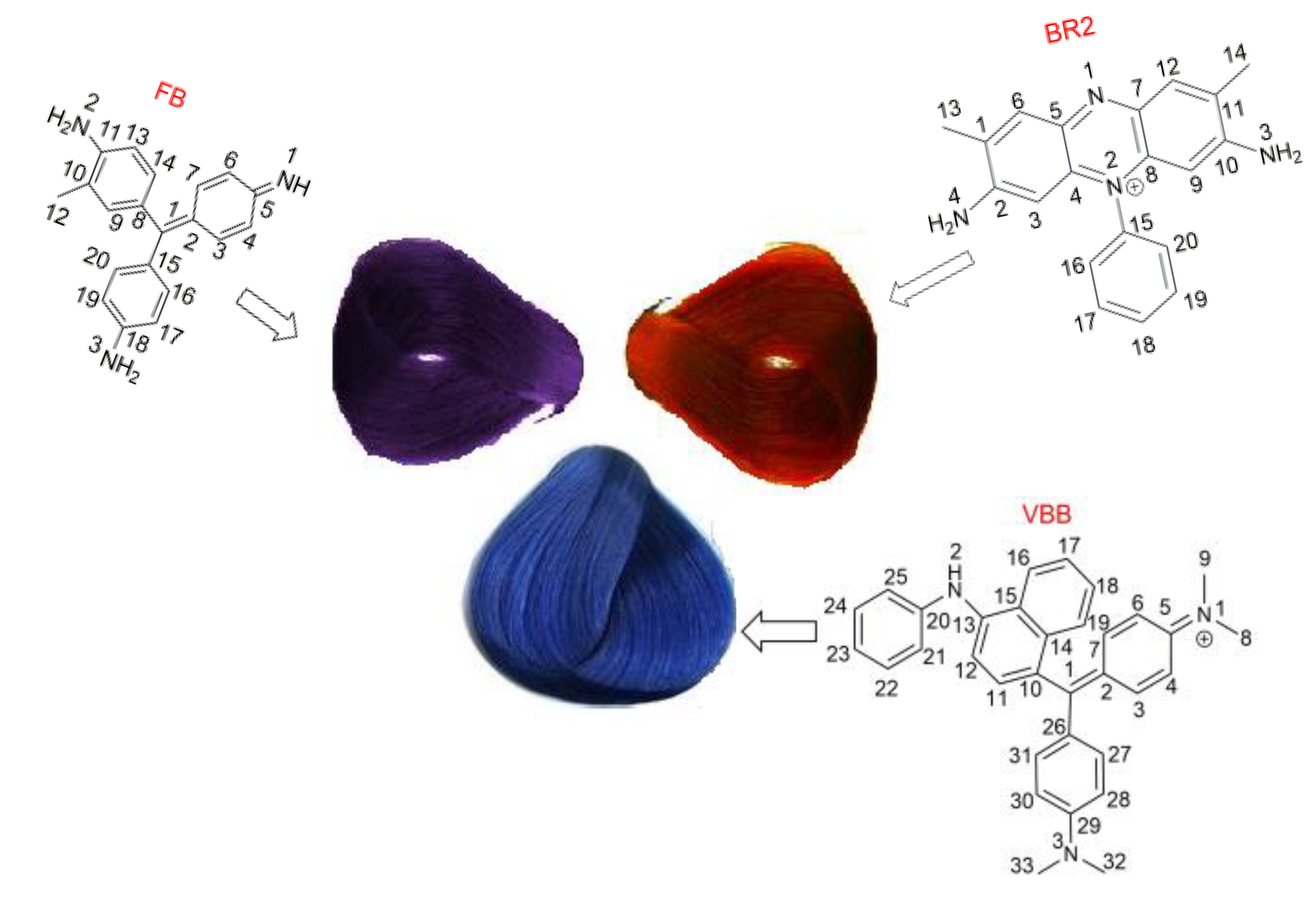
The Bio-Safety Concerns of Three Domestic Temporary Hair Dye Molecules: Fuchsin Basic, Victoria Blue B and Basic Red 2

Abstract
1. Introduction
2. Results
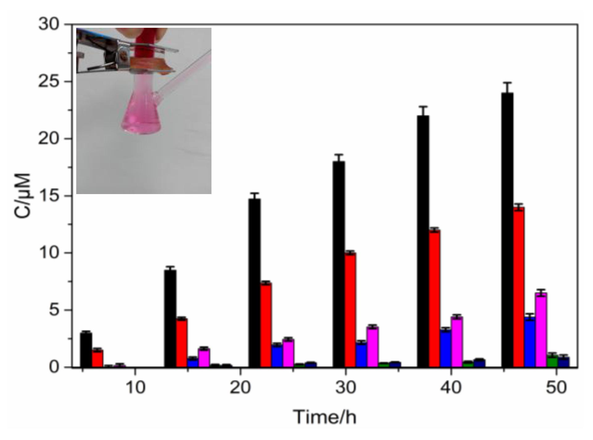
2.1. Percutaneous Absorption
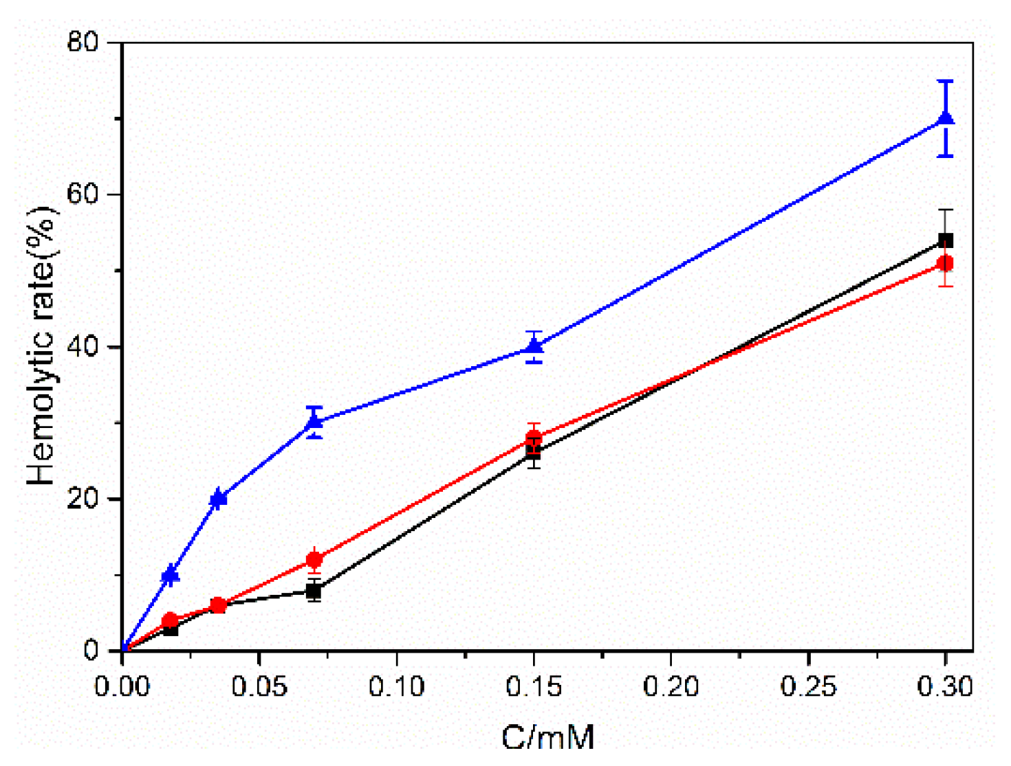
2.2. Hemolytic and Bacteria Cytotoxic Effects of Hair Dyes
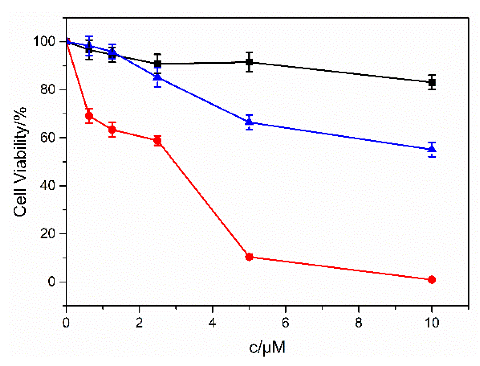
2.3. Cell Cytotoxicity of Dyes
2.4. BSA Interactions of Dyes
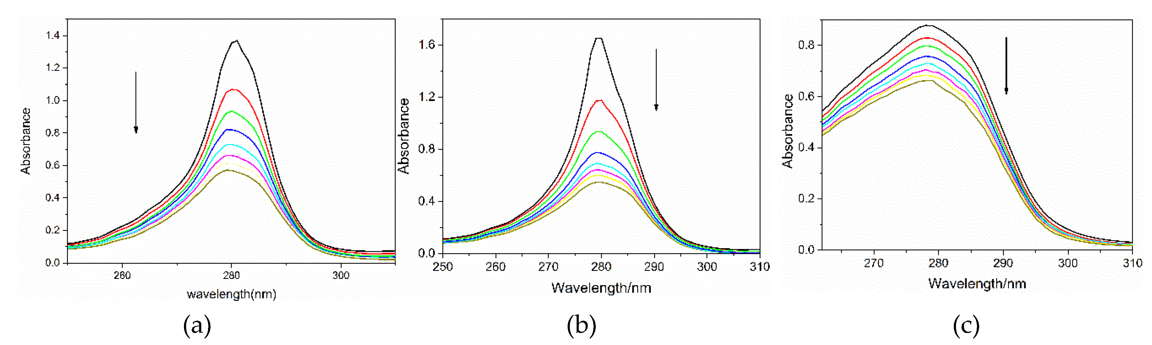
2.4.1. BSA Absorption Change with Dyes
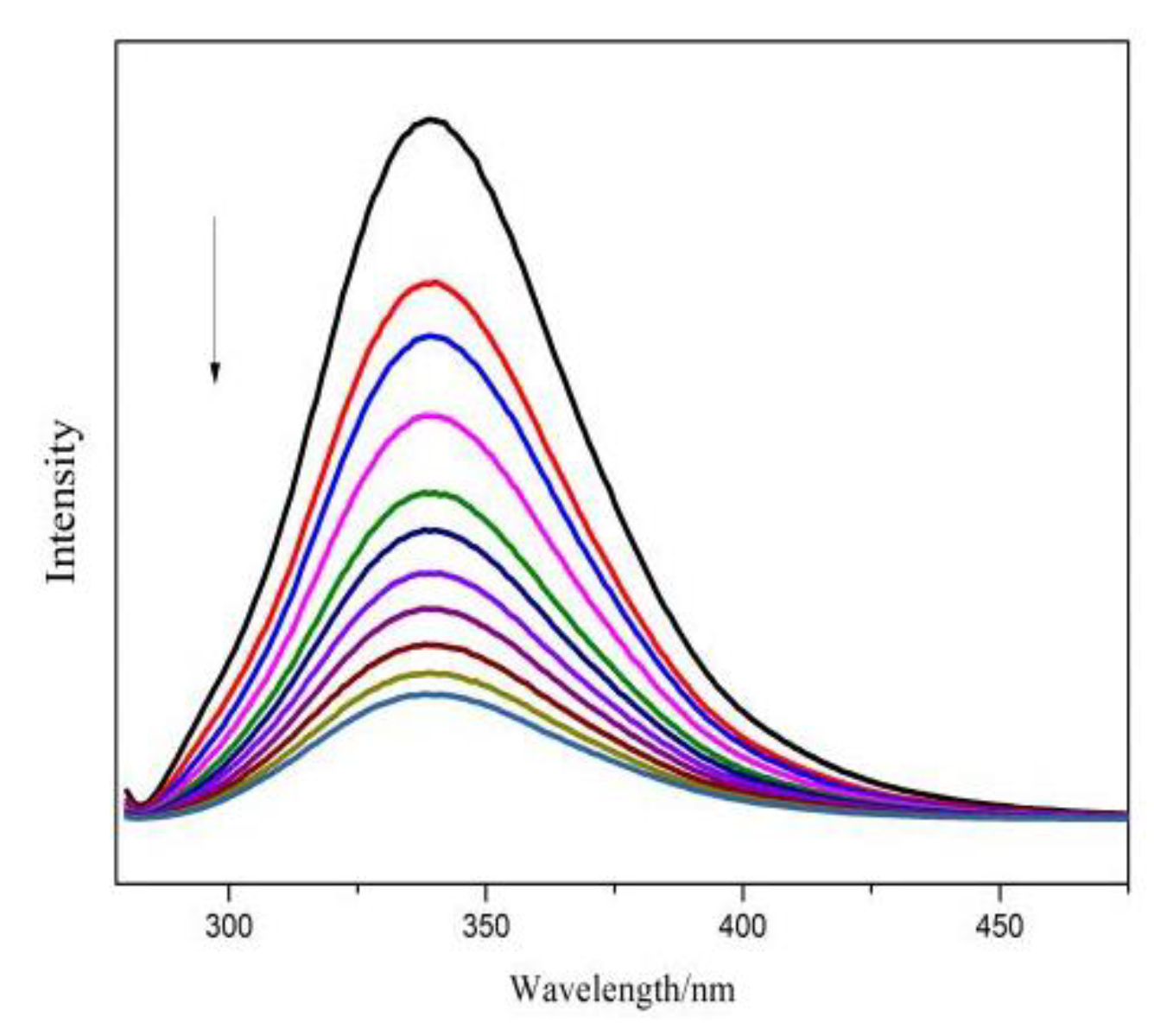
2.4.2. Dyes’ Fluorescence Emission Change with BSA
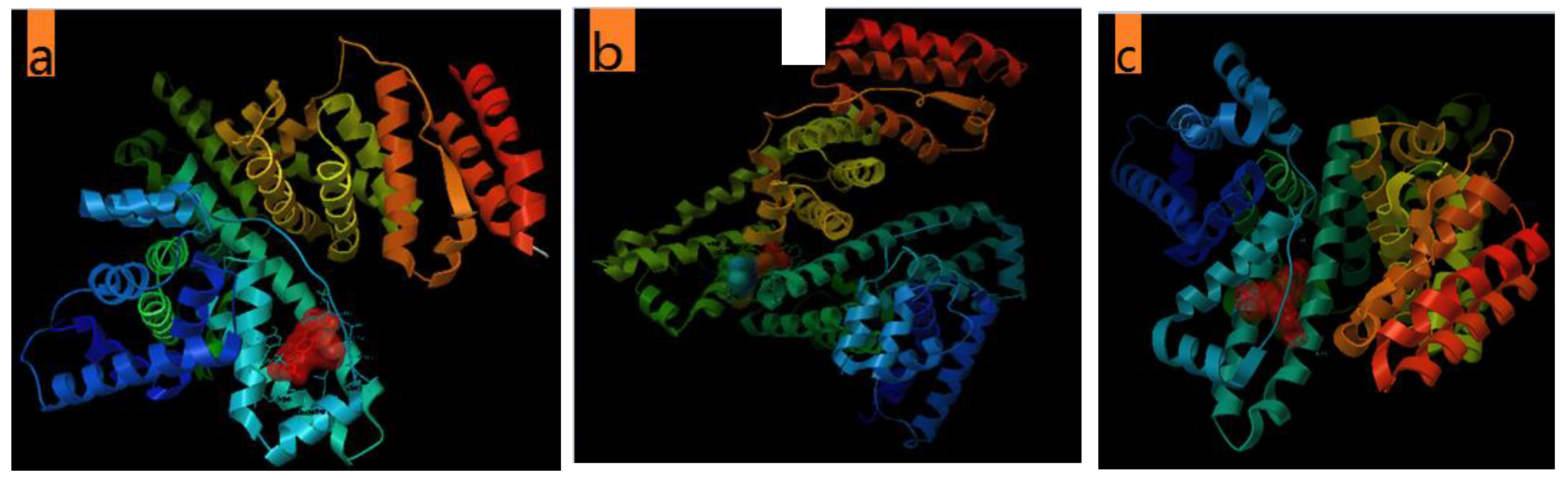
2.4.3. The Molecular Docking of the Dyes with BSA
2.5. DNA Interactions of Dyes
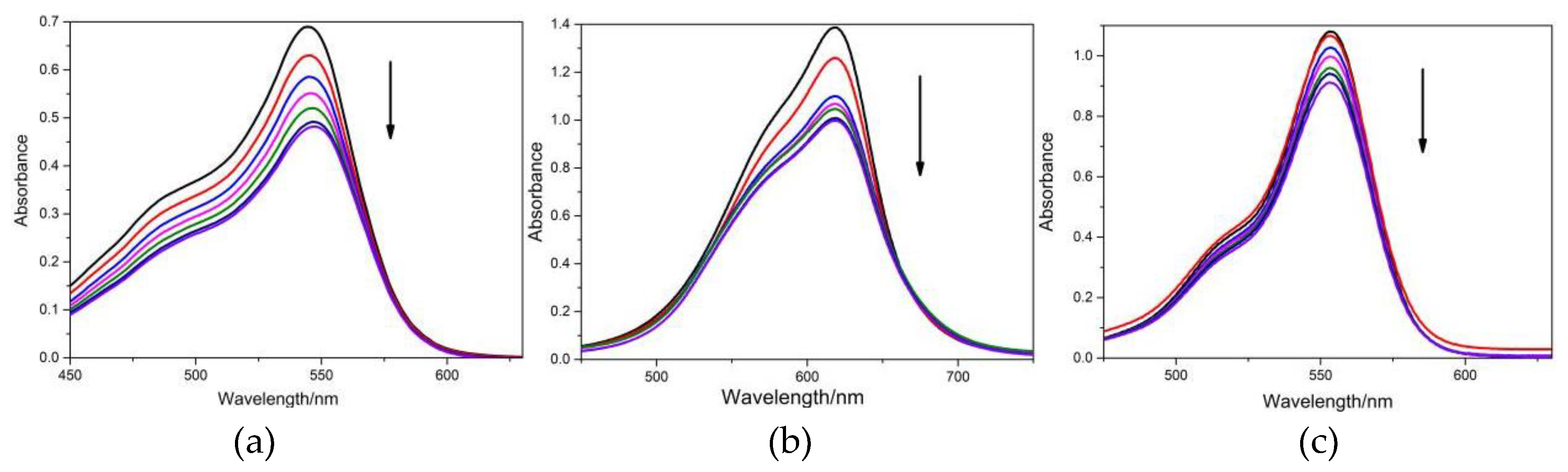
2.5.1. DNA Absorption Change with Dyes
2.5.2. Dyes’ Fluorescence Emission Change with CT-DNA
2.5.3. The Molecular Docking of the Dyes with CT-DNA
3. Discussion
3.1. Percutaneous Absorption
3.2. Effects to Human
3.3. BSA and DNA Interactions of Dyes
4. Materials and Methods
4.1. Materials and Instruments
4.2. Methods
4.2.1. Percutaneous Absorption
4.2.2. Hemolytic Effect of Hair Dye
4.2.3. Measurement of Cell Viability
4.2.4. Spectral Experiments
4.2.5. Molecular Model
5. Conclusions
Supplementary Materials
Author Contributions
Funding
Conflicts of Interest
References
- Swati, M.; Sudarson, S.S.; Mukesh, S. Microbial decolorization and detoxification of emerging environmental pollutant: Cosmetic hair dyes. J. Hazard Mater. 2017, 338, 356–363. [Google Scholar]
- Tai, S.Y.; Hsieh, H.M.; Huang, S.P.; Wu, M.T. Hair dye use, regular exercise, and the risk and prognosis of prostate cancer: Multicenter case-control and case-only studies. BMC Cancer. 2016, 16, 242–249. [Google Scholar] [CrossRef] [PubMed]
- Zanoni, T.B.; Hudari, F.; Munnia, A.; Peluso, M.; Godschalk, R.W.; Zanoni, M.V.; den Hartog, G.J.; Bast, A.; Barros, S.B.; Maria-Engler, S.S.; et al. The oxidation of p-phenylenediamine, an ingredient used for permanent hair dyeing purposes leads to the formation of hydroxyl radicals: Oxidative stress and DNA damage in human immortalized keratinocytes. Toxicol. Lett. 2015, 239, 194–204. [Google Scholar] [CrossRef]
- Guo, H.; Bassig, B.A.; Lan, Q.; Zhu, Y.; Zhang, Y.; Holford, T.R.; Leaderer, B.; Boyle, P.; Qin, Q.; Zhu, C.; et al. Polymorphisms in DNA repair genes, hair dye use, and the risk of non-Hodgkin lymphoma. Cancer Cause Control. 2014, 25, 1261–1270. [Google Scholar] [CrossRef]
- Wu, S.; Li, W.Q.; Cho, E.; Harris, J.E.; Speizer, F.; Qureshi, A.A. Use of permanent hair dyes and risk of vitiligo in women. Pigment Cell Melanoma Res. 2015, 28, 744–746. [Google Scholar] [CrossRef]
- He, J.; Jaka, S.; Miao, J.; Sun, H.n.; Dai, J.; Zhang, C.; Zhou, W.; Shao, Z.P. A highly sensitive perovskite oxide sensor for detection of p-phenylenediamine in hair dyes. J. Hazard Mater. 2019, 369, 699–706. [Google Scholar] [CrossRef] [PubMed]
- Qin, M.R.; Chen, R.H.; Huang, Z.Y.; Wang, J.P.; Xu, S.W.; Jiang, R.Q.; Li, J.P.; Xian, J.W.; Wang, X.W.; Lu, Y.; Ning, L.X.; et al. Application of the in vivo Pig-a gene mutation assay to test the potential genotoxicity of p-phenylenediamine. Food Chem. Toxicol. 2019, 123, 424–430. [Google Scholar] [CrossRef] [PubMed]
- IARC Monographs. Occupational Exposures of Hairdressers and Barbersand Personal Use of Hair Colourants. IARC Monogr. Eval. Carcinog. Risks Hum. 1993, 57, 43–118. Available online: https://www.ncbi.nlm.nih.gov/books/NBK385481/ (accessed on 5 April 2019).
- De Groot, A.C. Side-effects of henna and semi-permanent ‘black henna’ tattoos: A full review. Contact Dermat. 2013, 69, 1–25. [Google Scholar] [CrossRef]
- Piérard, G.E.; Goffin, V.; Piérard-Franchimont, C. Corneosurfametry: A predictive assessment of the interaction of personal-care cleansing products with human stratum corneum. Dermatology 1994, 189, 152–156. [Google Scholar] [CrossRef]
- Diamante, C.; Bergfeld, W.F.; Belsito, D.V.; Klaassen, C.D.; Marks, J.G.Jr.; Shank, R.C.; Slaga, T.J.; Snyder, P.W.; Alan, A.F. Final report on the safety assessment of Basic Violet 1, Basic Violet 3, and Basic Violet 4. Int. J. Toxicol. 2009, 28, 193S–204S. [Google Scholar] [CrossRef] [PubMed]
- Preston, R.J.; Skare, J.A.; Aardema, M.J. A review of biomonitoring studies measuring genotoxicity in humans exposed to hair dyes. Mutagenesis 2010, 25, 17–23. [Google Scholar] [CrossRef]
- Lizier, T.M.; Zanoni, T.B.; de Oliveria, D.P.; Zanoni, M.V.B. Electrochemicalreduction as a powerful tool to highlight the possible formation of by-productsmore toxic than Sudan III dye. Int. J. Electrochem. Sci. 2012, 7, 7784–7796. [Google Scholar]
- Liu, H.; Chen, J.; Jiang, J.; Giesy, J.; Yu, J.P.; Wang, H. Cytotoxicity of HC Orangeno. 1 to L929 fibroblast cells. Environ. Toxicol. Pharmacol. 2008, 26, 309–314. [Google Scholar] [CrossRef]
- Zanoni, T.B.; Tiago, M.; Faião-Flores, F.; de Moraes Barros, SB.; Bast, A.; Hageman, G.; de Oliveira, DP.; Maria-Engler, S.S. Basic Red 51, a permitted semi-permanent hair dye, is cytotoxic to human skin cells: Studies in monolayer and 3D skin model using human keratinocytes (HaCaT). Toxicol. Lett. 2014, 227, 139–149. [Google Scholar] [CrossRef]
- Hansen, H.S.; Johansen, J.D.; Thyssen, J.P.; Linneberg, A.; Søsted, H. Personal use of hair dyes and temporary black tattoos in Copenhagen hairdressers. Ann. Occup. Hyg. 2010, 54, 453–458. [Google Scholar] [PubMed]
- Sahinturk, V.; Kacar, S.; Vejselova, D.; Kutlu, H.M. Acrylamide exerts its cytotoxicity in NIH/3T3 fibroblast cells by apoptosis. Toxicol. Ind. Health 2018, 34, 481–489. [Google Scholar] [CrossRef] [PubMed]
- Desposito, D.; Chollet, C.; Taveau, C.; Descamps, V.; Alhenc-Gelas, F.; Roussel, R.; Bouby, N.; Waeckel, L. Improvement of skin wound healing in diabetic mice by kinin B2 receptor blockade. Clin. Sci. 2016, 130, 45–56. [Google Scholar] [CrossRef] [PubMed]
- Ye, Y.; Chen, X.L.; Guo, Y. Research on the interaction between pheophorbide and bovine serum albumin. Adv. Mater. Res. 2013, 749, 471–476. [Google Scholar] [CrossRef]
- Sayo, O.F.; Austria, T.; Ashley, M. Determination of mercury (II) ion concentrations in human serum albumin using fluorescence spectroscopy and multivariate regression analysis. Appl. Spectrosc. 2012, 66, 999–1004. [Google Scholar]
- Nohynek, G.J.; Antignac, E.; Re, T.; Toutain, H. Safety assessment of personal care products/cosmentics and their ingredients. Toxicol. Appl. Pharmacol. 2010, 243, 239–259. [Google Scholar] [CrossRef]
- Liu, Y.H.; Ji, F.Y.; Liu, R.T. The interaction of bovine serum albumin with doxorubicin-loaded superparamagnetic iron oxide nanoparticles: Spectroscope and molecular modelling identification. Nanotoxicology 2013, 7, 97–104. [Google Scholar] [CrossRef]
- Zhao, P.; Huang, J.W.; Ji, L.N. Cationic pyridinium porphyrins appending different peripheral substituents: Spectroscopic studies on their interactions with bovine serum albumin. Spectrochim. Acta 2012, 88, 130–136. [Google Scholar] [CrossRef]
- Eftink, M.R.; Ghiron, C.A. Fluorescence quenching studies with proteins. Anal. Biochem. 1981, 114, 199–227. [Google Scholar] [CrossRef]
- Trommel, J.S.; Marzilli, L.G. Synthesis and DNA binding of novel water-Soluble cationic methylcobalt porphyrins. Inorg. Chem. 2001, 40, 4374–4379. [Google Scholar] [CrossRef]
- Lakowicz, J.R.; Weber, G. Quenching of fluorescence by oxygen. Probe for structural fluctuations in macromolecules. Biochemistry 1973, 12, 4161–4170. [Google Scholar] [CrossRef]
- Yeggoni, D.P.; Rachamallu, A.; Subramanyam, R. Protein stability, conformational change and binding mechanism of human serum albumin upon binding of embelin and its role in disease control. J. Photochem. Photobiol. B 2016, 160, 248–259. [Google Scholar] [CrossRef] [PubMed]
- Saito, K.O.; Ikeda, R.; Endo, K.; Tsujino, Y.; Takagi, M.; Tamiya, E. Isolation of a novel alkaline-induced laccase from Flammulina velutipes and its application for hair coloring. J. Biosci. Bioeng. 2012, 113, 575–579. [Google Scholar] [CrossRef] [PubMed]
- Gondal, M.A.; Maganda, Y.W.; Dastageer, M.A.; Al Adel, F.F.; Naqvi, A.A.; Qahtan, T.F. Detection of carcinogenic chromium in synthetic hair dyes using laser induced breakdown spectroscopy. Appl. Opt. 2014, 53, 1636–1643. [Google Scholar] [CrossRef]
- Platzek, T. Risk from exposure to arylamines from consumer products and hair dyes. Front. Biosci. 2010, 2, 1169–1183. [Google Scholar] [CrossRef]
- Wang, H.C.; Jiang, X.H.; Zhou, L.M. Interaction of NAEn-s-n gemini surfactants with bovine serum albumin: A structure-activity probe. J. Lumin. 2013, 134, 138–147. [Google Scholar] [CrossRef]
- Bronaugh, R.L.; Maibach, H.I. Percutaneous Absorption of Nitroaromatic Compounds: In vivo and studies in the human and monkey. J. Investig. Dermatol. 1985, 84, 180–183. [Google Scholar] [CrossRef]
- Jardes, D.J.; Ross, L.A.; Markovich, J.E. Hemolytic anemia after ingestion of the natural hair dye Lawsonia inermis (henna) in a dog. J. Vet. Emerg. Crit. Care 2013, 23, 648–651. [Google Scholar] [CrossRef]
- Maiti, S.; Sasmal, K.; Sinha, S.S.; Singh, M. Analysis of cytotoxicity and genotoxicity on E. coli.; human blood cells and Allium cepa suggests a greater toxic potential of hair dye. Ecotoxicol. Environ. Saf. 2016, 124, 248–254. [Google Scholar] [CrossRef] [PubMed]
- Ma, X.; Yan, J.; Xu, K.; Guo, L.; Li, H. Binding mechanism of trans-N-caffeoyltyramine and human serum albumin: Investigation by multi-spectroscopy and docking simulation. Bioorg. Chem. 2016, 66, 102–110. [Google Scholar] [CrossRef]
- Zhao, P.; Li, J.; Yang, L.J.; Lu, J.Z.; Guo, H.M.; Ma, L.N.; Ou, B.H. DNA binding ad photocleavage properties of cationic porphyrin-polypyridyl ruthenium(II) hybrids. J. Coord. Chem. 2013, 66, 4220–4236. [Google Scholar] [CrossRef]
- Ji, L.N.; Zou, X.H.; Liu, J.G. Shape- and enantioselective interaction of Ru(II)/Co(III) polypyridyl complexes with DNA. Coord. Chem. Rev. 2001, 216–217, 513–516. [Google Scholar] [CrossRef]
Sample Availability: Samples of the compounds are not available from the authors. |


| a LD50 | BSA Binding | Calf Thymus (CT)-DNA Binding | ||||
|---|---|---|---|---|---|---|
| RBC (mM) | NIH/3T3 (μM) | Kq × 1011 | Kb × 103 | n | K’b × 102 | |
| FB | 0.23 ± 0.01 | 10.66 ± 0.31 | 23.4 ± 0.22 | 118 ± 1.82 | 1.21 ± 0.02 | 1.2 ± 0.04 |
| BR2 | 0.29 ± 0.02 | 22.77 ± 0.43 | 1.1 ± 0.01 | 4.37 ± 0.01 | 0.88 ± 0.01 | 530 ± 3.35 |
| VBB | 0.16 ± 0.01 | 2.318 ± 0.03 | 21.9 ± 0.31 | 30.1 ± 0.54 | 1.03 ± 0.03 | 140 ± 2.48 |
| Dye | Atom | BSA | Distance (Å) | CT-DNA | Distance (Å) |
|---|---|---|---|---|---|
| FB | N1 | His145 NE2 | 2.88 | dg10 N2 | 3.21 |
| FB | N2 | dc9 O4 | 2.96 | ||
| FB | N2 | dt8 O2 | 3.25 | ||
| BR2 | N2 | Lys499 N2 | 2.89 | ||
| BR2 | N4 | Glu125 OE2 | 2.91 | dt19 O3 | 3.06 |
| BR2 | N3 | Leu115 O | 2.77 | ||
| BR2 | N4 | Gln416 OE | 2.55 | ||
| VBB | N2 | da6 O4 | 2.17 |
© 2019 by the authors. Licensee MDPI, Basel, Switzerland. This article is an open access article distributed under the terms and conditions of the Creative Commons Attribution (CC BY) license (http://creativecommons.org/licenses/by/4.0/).
Share and Cite
Liu, B.; Jin, S.-F.; Li, H.-C.; Sun, X.-Y.; Yan, S.-Q.; Deng, S.-J.; Zhao, P. The Bio-Safety Concerns of Three Domestic Temporary Hair Dye Molecules: Fuchsin Basic, Victoria Blue B and Basic Red 2. Molecules 2019, 24, 1744. https://doi.org/10.3390/molecules24091744
Liu B, Jin S-F, Li H-C, Sun X-Y, Yan S-Q, Deng S-J, Zhao P. The Bio-Safety Concerns of Three Domestic Temporary Hair Dye Molecules: Fuchsin Basic, Victoria Blue B and Basic Red 2. Molecules. 2019; 24(9):1744. https://doi.org/10.3390/molecules24091744
Chicago/Turabian StyleLiu, Bing, Shu-Fang Jin, Hua-Chao Li, Xiang-Yu Sun, Si-Qi Yan, Shu-Jun Deng, and Ping Zhao. 2019. "The Bio-Safety Concerns of Three Domestic Temporary Hair Dye Molecules: Fuchsin Basic, Victoria Blue B and Basic Red 2" Molecules 24, no. 9: 1744. https://doi.org/10.3390/molecules24091744
APA StyleLiu, B., Jin, S.-F., Li, H.-C., Sun, X.-Y., Yan, S.-Q., Deng, S.-J., & Zhao, P. (2019). The Bio-Safety Concerns of Three Domestic Temporary Hair Dye Molecules: Fuchsin Basic, Victoria Blue B and Basic Red 2. Molecules, 24(9), 1744. https://doi.org/10.3390/molecules24091744