Article

Design, Synthesis, and Evaluation of Monoamine Oxidase A Inhibitors–Indocyanine Dyes Conjugates as Targeted Antitumor Agents

1
Key Laboratory of Structure-Based Drug Design & Discovery of Ministry of Education, Shenyang Pharmaceutical University, Shenyang 110016, China
2
School of life sciences and biological pharmacy, Shenyang Pharmaceutical University, Shenyang 110016, China
3
Wuya College of innovation, Shenyang Pharmaceutical University, Shenyang 110016, China
*
Authors to whom correspondence should be addressed.
These authors contributed equally.
Molecules 2019, 24(7), 1400; https://doi.org/10.3390/molecules24071400
Submission received: 18 March 2019 / Revised: 5 April 2019 / Accepted: 6 April 2019 / Published: 10 April 2019
(This article belongs to the Special Issue Drug Design)

Abstract

:
Monoamine oxidase A (MAOA) is an important mitochondria-bound enzyme that catalyzes the oxidative deamination of monoamine neurotransmitters. Accumulating evidence suggests a significant association of increased MAOA expression and advanced high-grade prostate cancer (PCa) progression and metastasis. Herein, a series of novel conjugates combining the MAOA inhibitor isoniazid (INH) and tumor-targeting near-infrared (NIR) heptamethine cyanine dyes were designed and synthesized. The synthesized compounds G1G13 were evaluated in vitro for their cytotoxicity against PC-3 cells using the MTT assay, and molecular docking studies were performed. Results showed that most tested compounds exhibited improved antitumor efficacy compared with INH. Moreover, conjugates G10 and G11 showed potent anticancer activity with IC50 values (0.85 and 0.4 μM respectively) comparable to that of doxorubicin (DOX). This may be attributable to the preferential accumulation of these conjugates in tumor cells. G10, G11, and G12 also demonstrated moderate MAOA inhibitory activities. This result and the results of molecular docking studies were consistent with their cytotoxicity activities. Taken together, these data suggest that a combination of the MAOA inhibitor INH with tumor-targeting heptamethine cyanine dyes may prove to be a highly promising tool for the treatment of advanced prostate cancer.

Graphical Abstract

1. Introduction

Global prostate cancer (PCa) incidence and mortality have substantially increased, compounded by an increase in the proportion of the elderly population and in the frequency of diagnosis. Although the majority of cases with metastatic PCa respond to the available therapeutic modalities, hormone-refractory PCa and advanced metastatic PCa remain inevitable [1,2,3,4]. Therefore, novel and highly effective treatments with high tumor-targeting specificity, fewer side effects, and improved efficacy are desirable for hormone-refractory PCa and metastatic PCa.
Monoamine oxidase A (MAOA) is an important mitochondria-bound enzyme that catalyzes the oxidative deamination of monoamine neurotransmitters including serotonin (5-hydroxytryptamine), catecholamines, and biogenic amines [5,6,7,8]. This process subsequently generates their catalytic by-product hydrogen peroxide (H2O2), a predominant source of reactive oxygen species (ROS), which may predispose cancer cells to DNA damage, leading to tumor initiation and progression. Thus, although MAOA was initially recognized as a crucial neurotransmitter regulator, emerging evidence revealed its significant roles in oncogenesis [9,10,11,12]. Moreover, recent studies have also reported that the overexpression of MAOA could promote the invasion and metastasis of PCa cells by mediating epithelial-mesenchymal transition (EMT) [13,14]. Conversely, the inhibition of MAOA by the inhibitor clorgyline attenuated the proliferation of PCa cells and inhibited tumor growth and metastasis in mouse xenograft models [15,16,17,18,19]. Therefore, MAOA inhibitors can serve as a potentially promising agent for the effective therapy of advanced human PCa.
Near-infrared (NIR) heptamethine carbocyanine dyes are a novel class of heterocyclic polymethine cyanine analogues with cancer imaging ability due to their high extinction coefficients and large Stokes shifts, exhibiting characteristic fluorescence emission in the range of 700–1000 nm [20,21,22]. Recent in vitro and in vivo studies revealed that hypoxia—one of the elements of the tumor endogenous microenvironment—induced the preferential uptake of NIR cyanine dyes by human cancer cells and tissues through the hypoxia inducible factor-1α/organic anion-transporting polypeptides (HIF-1α/OATPs) signaling axis [23,24,25,26,27,28,29,30,31,32]. Second, tumor cells usually possess higher negative mitochondrial transmembrane potentials than normal cells. Therefore, some lipophilic cations preferentially accumulate in the mitochondria of tumor cells [33,34]. The higher mitochondrial membrane potential in tumor cells may play a critical role in the preferential accumulation of heptamethine cyanine dyes in tumors owing to the lipophilic and electropositive characteristics of these dyes [35]. Wang et al. confirmed that NIR heptamethine cyanine dyes IR-780 and MHI-148 could be retained in the mitochondria of tumor cells, increase ROS production, decrease mitochondrial membrane potential, and induce tumor cell apoptosis [36]. Taken together, these findings suggest that heptamethine carbocyanine dyes hold great promise for tumor targeting and to some extent possess antitumor efficacy.
Isoniazide (INH) is a small molecule inhibitor of MAOA and is now clinically used as the first-line anti-tuberculosis medicine. Studies have shown that INH hydrazone and N-acetylated derivatives are more efficient and less hepatotoxic than INH because of the blockage of the terminal amino group of hydrazine [37,38,39]. Herein, we designed and synthesized a series of novel conjugates (Figure 1) through the conjugation of structurally diverse heptamethine carbocyanine dyes with INH via hydrazine bond and investigated their cytotoxicity against PC-3 cells. Further, molecular docking studies were performed to elucidate the binding mode of these conjugates with MAOA. MAOA inhibition assay was also carried out.

2. Results and Discussion
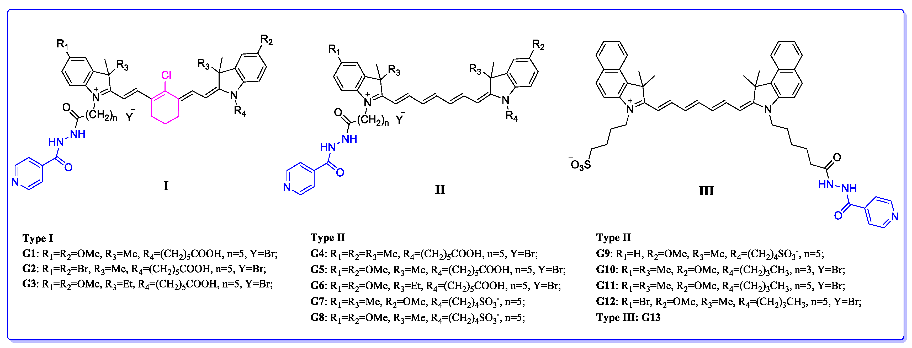
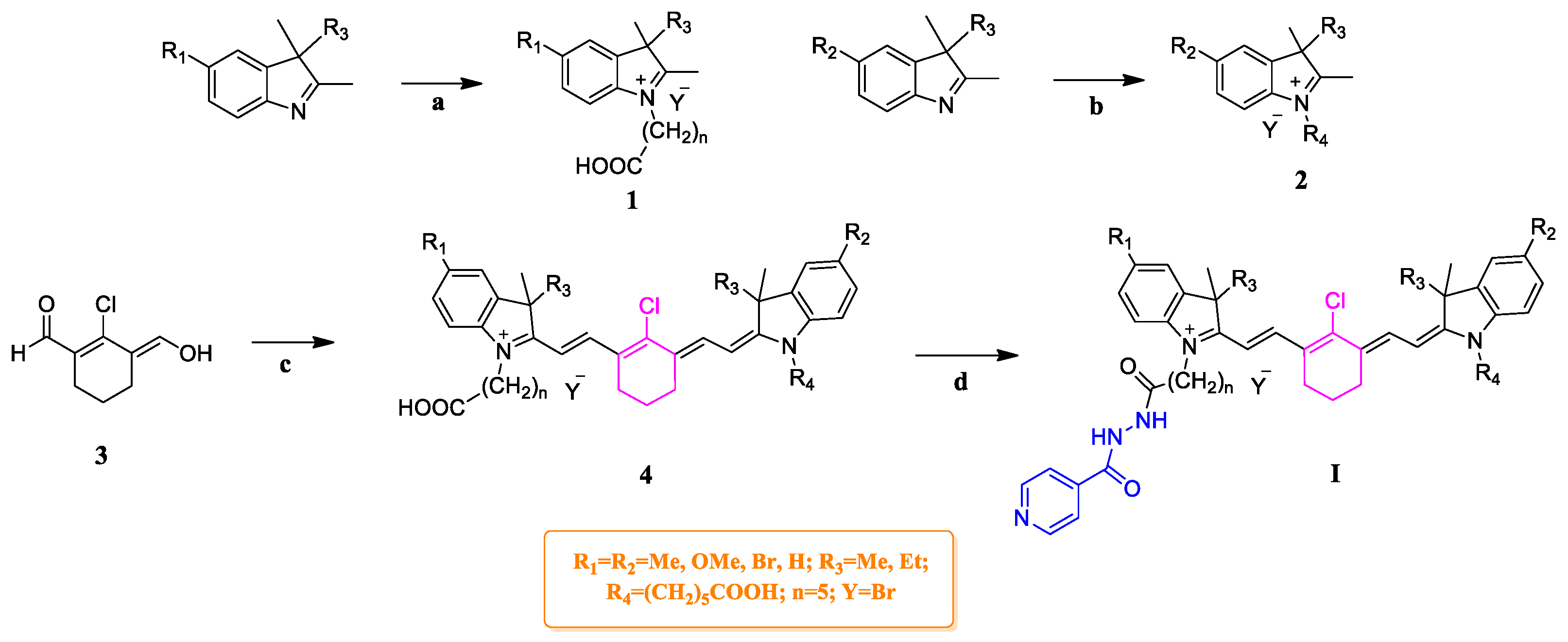
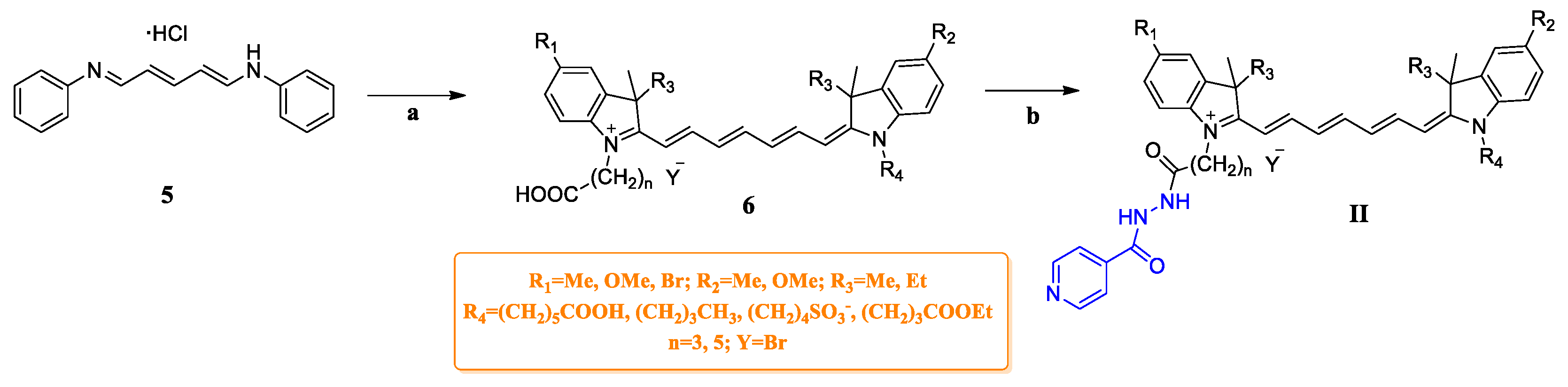
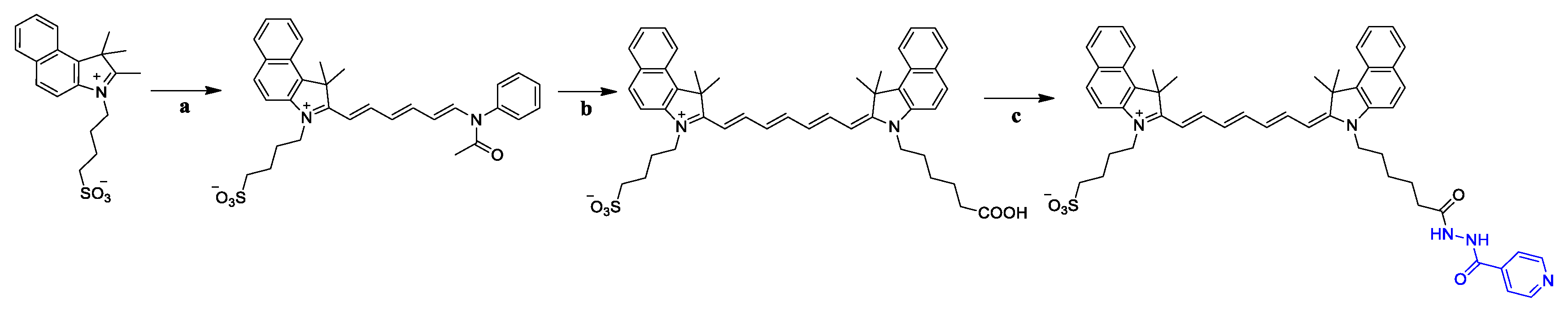
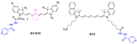
2.1. Chemistry

The synthetic route for the type I compounds is outlined in Scheme 1. Intermediates 1 and 2 were both synthesized by electrophilic substitution of 5-substitutional 3H-indole. The synthesis of key intermediate 4 was achieved according to procedures described in the literature [26]. Briefly, both intermediates 1 and 2 condensed with 3 via base-catalyzed (AcONa) aldol condensation reaction to give intermediate 4. The target compounds G1G3 were generated through the coupling reaction of intermediate 4 with INH in the presence of dicyclohexylcarbodiimide (DCC) and dimethylaminopyridine (DMAP).
As illustrated in Scheme 2, symmetric heptamethine cyanine dye intermediate 6 was synthesized by one-step base-catalyzed (Et3N) aldol reaction of intermediates 1 and 2. However, asymmetric heptamethine cyanine dye intermediate 6 was afforded by the two-step aldol condensation reaction of intermediates 1, 2, and 5 in Ac2O with pyridine as catalyst. Finally, the desired compounds G4G12 were generated by the coupling of 6 with INH in anhydrous DCM with DCC and DMAP acting as coupling agents. Meanwhile, the type III compound G13 was obtained in the sequence of steps outlined in Scheme 3.

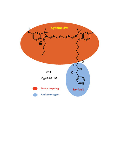
2.2. Cytotoxicity against PC-3

The antitumor activities of G1G13 were evaluated by MTT (3-(4,5-dimethylthiazol-2-yl)-2,5-diphenyltetrazolium bromide) assay using a human PC-3 cell line. The IC50 values of G1G13 against PC-3 cells are summarized in Table 1.
MTT assay results revealed that nearly all tested compounds showed improved antitumor activity when compared with their parent compound, the MAOA inhibitor INH. The moderate cytotoxicity of these conjugates may be due to their preferential uptake by PC-3 cells mediated by OATPs and subsequent accumulation in the mitochondria of PC-3 cells, disrupting mitochondrial activities. The fact that INH exhibited negligible cytotoxicity may be attributed to its poor passive diffusion into PC-3 cells. G10, G11 (IC50 values of 0.85 and 0.40 μM, respectively) possessed potent cytotoxicity comparable to that of doxorubicin (DOX) (IC50 = 0.21 μM). G10 and G11 have very similar chemical structures except for the different chain length of carboxyalkyl connected to the N atom in indole moieties. Their structural similarity may explain their similar antitumor efficacy. These results indicated that the conjugation with INH was conducive to the enhancement of anti-tumor activity against PC-3 in vitro.

2.3. MAOA Inhibitory Activity of G10, G11, and G12

Because the prostate cancer cell line LNCaP expressed a relatively high level of MAOA [17,40], we tested the inhibitory effect of compounds G10, G11, and G12 on MAOA using LNCaP cells (Table 2). Our data showed that three synthesized novel compounds displayed an enhanced inhibitory action on MAOA levels as compared with the parent compound INH, while clorgyline—another MAOA inhibitor—exhibited the most potent inhibitory effect. The fact that heptamethine dye–INH conjugates possessed higher MAOA inhibition efficacy than INH may be attributed to the modification of the terminal hydrazine group in INH. This assay result is consistent with that of the cell viability assay, suggesting that the tested compounds inhibited cell growth by suppressing MAOA activity.

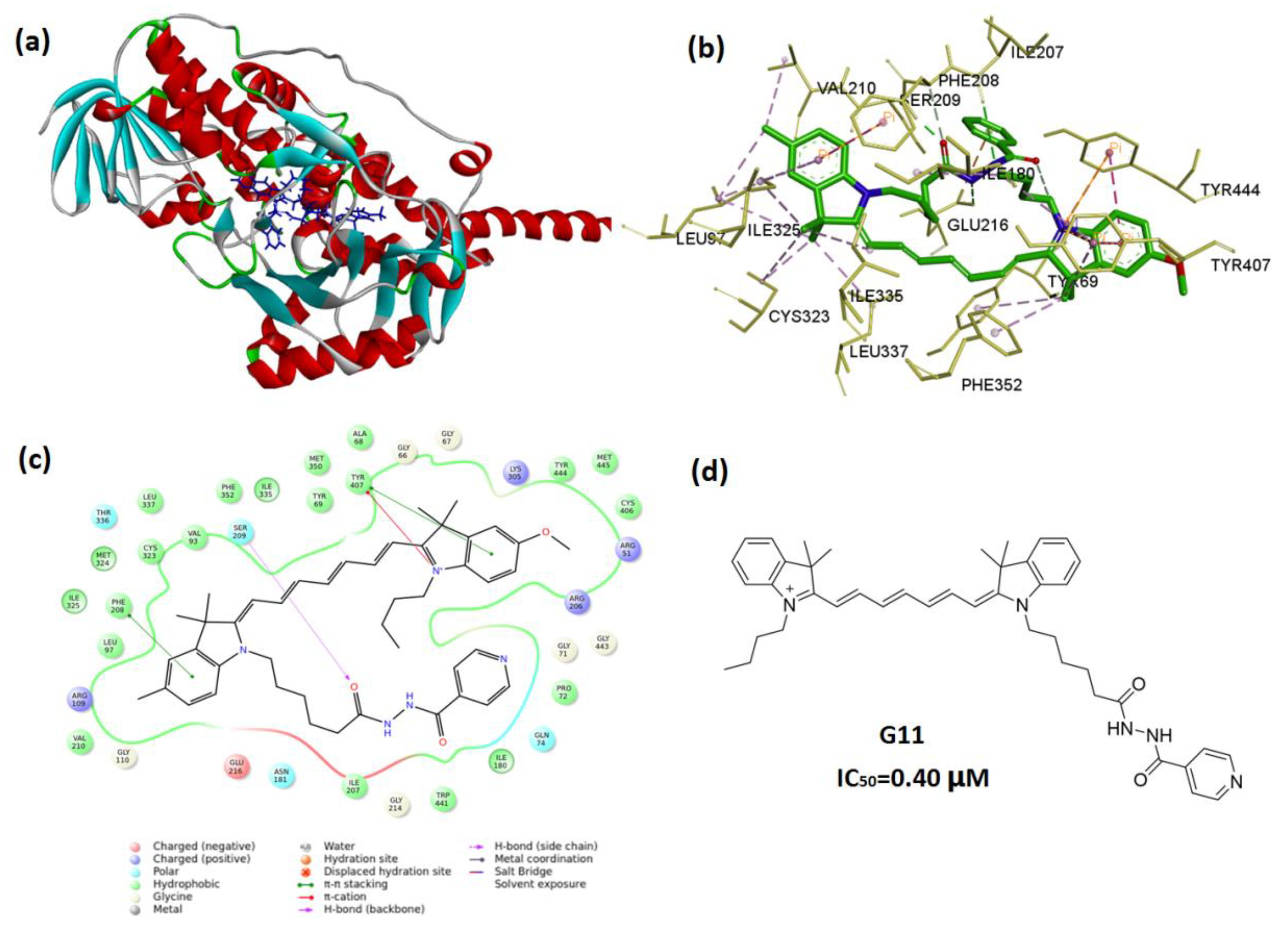
2.4. In Silico Molecular Docking

To clarify the underlying mechanism of action of these compounds at the molecular level, molecular docking of 2BXR and G11 was performed. 2BXR is a human monoamine oxidase A (hMAOA). De Colibus et al. revealed that the active binding site of hMAOA was formed primarily by a single cavity consisting of residues 210–216 (all active site residues: Tyr69, Gln74, Val91, Val93, Leu97, Ile207, Phe208, Ser209, Val210, Glu216, Cys323, Ile325, Ile335, Leu337, Met350, Phe352, Tyr407, Tyr444) [41]. Since G11 showed the best in vitro antitumor activity, it was selected as the ligand for this docking analysis. The molecular docking analysis showed the heptamethine chain and indole moieties of G11 could be integrated closely with the hydrophobic active site cavity (Figure 2) with the low binding energy of (ΔG) −12.27 kcal/mol. The docked pose of G11 (in 2BXR) was enclosed by a partially hydrophobic cavity with amino acid residues including Tyr69, Val93, Leu97, Ile180, Ile207, Phe208, Phe208, Val210, Cys323, Ile325, Ile335, Leu337, Met350, Phe352, Tyr407, and Tyr444. A hydrogen bond interaction between O44 and Ser209 was also observed.

3. Materials and Methods

3.1. Materials

All reagents and solvents were obtained from commercial sources and were used as received unless otherwise stated. Phenylhydrazine hydrochloride, 4-bromophenylhydrazine hydrochloride, 4-methylphenylhydrazine hydrochloride, 4-methoxyphenylhydrazine hydrochloride, 6-bromohexanoic acid, ethyl 4-bromobutyrate, and 1-bromobutane were purchased from Meryer (Shanghai, China). 3-Methyl-2-butanone, 3-methyl-2-pentanone, 1,4-butane sultone, and isoniazid were purchased from Aladdin (Shanghai, China). Clorgyline was obtained from Bide Pharmatech Ltd. (Shanghai, China). All other reagents were synthesized in our laboratory. 1640 DMEM and 0.25% trypsin EDTA were obtained from Gibco (Sigma-Aldrich, St. Louis, MO, USA). (3-(4,5-Dimethylthiazol-2-yl)-2,5-diphenyltetrazolium bromide). MTT reagent was acquired from Sigma-Aldrich (St. Louis, MO, USA). PC-3 and LNCaP cells were obtained from the Cell Bank of the Chinese Academy of Sciences (Shanghai, China).

3.2. Molecular Docking

The crystal structure of MAOA with a resolution of 3 Å was retrieved from the Protein Data Bank (PDBID: 2BXR). AutoDock 4.2 suite, an automated docking tool that uses a Lamarckian genetic algorithm (LGA), was used to perform the molecular docking simulations [42]. Briefly, all the bound water molecules, ligands, and co-factors were eliminated from the protein, and the polar hydrogen was added to the proteins. Gasteiger charges were computed in AutoDockTools 4.2 [43]. 2D structures of G11 were constructed using ChemBioDraw ultra12.0 and were transformed to 3D. Subsequently, 3D structures of the aforementioned compounds were also deduced and then the structure was energetically minimized by using an MMFF94 force field.
In the docking process the grid point maps were calculated using AutoGrid 4.2. The program AutoGrid was used to generate the grid maps. Each grid was centered at the structure of the corresponding receptor. The grid dimensions for the grid maps were fixed at a spacing of 0.375 Å with the grid box of size 90 × 70 × 60 (number of points in x-, y-, and z- axes for both proteins). For each docking experiment, default parameters for docking run included number of final conformations, 100; population size, 150; maximum number of energy evaluations, 2.5 × 10⁶, and maximum number of generations, 27,000. Some low-binding-energy docked poses were investigated in detail for their binding interactions with the binding cavity of the target protein, using Discovery Studio Visualizer (DSV). The docked compound complexes were built using the low-free-energy binding positions.

3.3. Cytotoxicity Evaluation

The in vitro cytotoxicity activity of G1G13 were measured by the colorimetric MTT assay. Briefly, PC-3 cells were incubated in 96-well plates at a density of 3 × 10³ cells/well for 24 h. Then, the cells were exposed to various concentrations of tested compounds, INH and DOX, respectively for 96 h. After incubation, 10 μL of MTT (5 mg/mL) solution was added and the plates were incubated for 4 h at 37 °C. Next, the medium was replaced with 200 μL of DMSO to dissolve the formed formazan crystals. The absorbance was measured at 570 nm on a microplate reader (Synergy-HT, BioTek Instruments, Winooski, VT, USA).

3.4. MAOA Inhibition Activity Assay

In a 10-mm dish, 6 × 10⁵ LNCaP cells were plated in medium supplemented with 10% FBS. After 24 h, cells were treated with various concentrations (10 pM, 1 nM, 100 nM, 10 μM) of the tested compounds G10, G11, G12, isoniazid, and clorgyline for 48 h. The reaction products were extracted and MAOA activities were measured as described in the introduction for the Cell MAOA Assay Kit (Shanghai Chengong Biotechnology, CHN, Lot No: 1-201838-10). The MAOA activity was assessed based on the substrate p-tyramine level after treatment with MAOB inhibitor pargyline.

3.5. General Procedure for the Synthesis of Compounds G1G3

(E)-2-Chloro-3-(hydroxymethylene)-cyclohex-1-enecarbaldehyde (3) (500 mg, 2.9 mmol), 5-methoxyl-1-(5-carboxypentyl)-2,3,3-trimethyl-3H-indol-1-ium bromide (1a) (2.23 g, 5.8 mmol) and sodium acetate (401 mg, 2.9 mmol) were suspended in acetic anhydride, the mixture was stirred at 60 °C for 1 h. Next the resulting solution was poured into water. Precipitation was allowed to proceed for 6 h and then mixture was filtered to obtain intermediate 4a as a gold solid. DCC (104 mg, 0.506 mmol) and DMAP (6 mg, 0.051 mmol) were added to a solution of 4a (400 mg, 0.506 mmol) in DCM in an ice-salt bath. INH (58 mg, 0.422 mmol) was added dropwise, the mixture was stirred for 24 h, and was then filtered. Filtrate was evaporated to dryness under reduced pressure and the residue was purified by silica gel column to yield G1 (50 mg, 13%). G2 and G3 were prepared by the same procedure as G1 (Supplementary Materials).
2-(2-(3-(2-(5-Methoxyl-1-(5-carboxypentyl)-3,3-dimethyl-indol-2-ylidene)ethylidene)-2-chloro-1-cyclohexen-1-yl)vinyl)-5-methoxyl-1-(6-(2-isonicotinoylhydrazinyl)-6-oxohexyl)-3,3-dimethyl-3H-indol-1-ium bromide (G1). MS (ESI) m/z: 862.4 [M + H]+. 1H NMR (400 MHz, CDCl3) δ 8.63–8.56 (br, 2H), 8.14–8.12 (m, 2H), 7.89–7.83 (br, 2H), 7.11 (d, 1H), 6.89 (s, 2H), 6.87 (s, 2H), 6.84 (s, 2H), 6.14 (d, 1H), 6.00 (d, 1H), 5.92 (d, 1H), 4.09–4.01 (br, 2H), 3.94–3.87 (br, 2H), 3.82 (s, 3H), 3.80 (s, 3H), 2.58–2.54 (br, 2H), 2.50–2.45 (br, 2H), 2.43–2.34 (br, 2H), 2.23 (d, 4H), 1.83–1.78 (br, 2H), 1.77–1.70 (br, 6H), 1.65 (s, 6H), 1.63 (s, 6H), 1.56 (d, 4H). 13C NMR (151 MHz, CD3OD) δ 180.10, 174.51, 172.99, 172.86, 166.31, 159.81, 159.74, 150.86, 149.56, 144.20, 144.16, 144.02, 143.94, 141.91, 141.89, 136.92, 136.90, 126.93, 126.90, 124.92, 124.73, 122.96, 118.49, 114.75, 112.89, 112.82, 109.80, 101.80, 101.67, 66.69, 56.26, 50.57, 50.54, 48.94, 45.15, 45.08, 38.22, 34.22, 28.15, 28.11, 27.89, 27.65, 27.15, 27.12, 27.06, 26.94, 25.88, 23.76, 21.97, 15.26.
2-(2-(3-(2-(5-Bromo-1-(5-carboxypentyl)-3,3-dimethyl-indol-2-ylidene)ethylidene)-2-chloro-1-cyclohexen-1-yl)vinyl)-5-bromo-1-(6-(2-isonicotinoylhydrazinyl)-6-oxohexyl)-3,3-dimethyl-3H-indol-1-ium bromide (G2). MS (ESI) m/z: 960 [M + H]+. 1H NMR (400 MHz, CD3OD) δ 8.71 (d, 2H), 8.42 (d, 1H), 8.39 (d, 1H), 7.81 (dd, 2H), 7.71 (d, 2H), 7.69–7.65 (m, 1H), 7.58–7.57 (m, 1H), 7.56 (dd, 1H), 7.55 (d, 1H), 7.28 (d, 1H), 7.27 (d, 1H), 6.30 (d, 1H), 6.28 (d, 1H), 2.75–2.71 (br, 2H), 2.69 (t, 2H), 2.36 (t, 2H), 2.20 (t, 2H), 1.92–1.89 (m, 4H), 1.79 (dt, 2H), 1.73 (d, 12H), 1.71–1.65 (m, 4H), 1.58 (dt, 7.7 Hz, 2H), 1.50–1.44 (m, 4H). 13C NMR (151 MHz, DMSO) δ 175.40, 171.45, 163.96, 156.11, 150.48, 147.97, 143.87, 141.01, 140.49, 139.78, 132.07, 130.69, 130.45, 129.95, 129.63, 128.38, 124.89, 124.77, 124.56, 123.74, 121.51, 110.78, 108.44, 108.17, 108.15, 107.96, 99.69, 99.62, 93.24, 48.85, 45.44, 40.14, 33.45, 33.24, 28.71, 28.12, 27.72, 27.43, 27.22, 26.61, 26.51, 26.15, 26.00, 25.47, 25.22, 24.97, 21.27, 19.66.
2-(2-(3-(2-(5-Methoxyl-1-(5-carboxypentyl)-5-ethyl-3-methyl-indol-2-ylidene)ethylidene)-2-chloro-1-cyclohexen-1-yl)vinyl)-5-methoxyl-1-(6-(2-isonicotinoylhydrazinyl)-6-oxohexyl)-5-ethyl-3-methyl-3H-indol-1-ium bromide (G3). MS (ESI) m/z: 891.5 [M + H]+. 1H NMR (400 MHz, CDCl3) δ 8.59 (s, 1H), 8.21 (d, 1H), 8.14 (d, 1H), 7.77–7.70 (br, 2H), 7.68–7.62 (br, 1H), 7.57–7.50 (br, 1H), 7.18–7.10 (br, 1H), 7.07 (d, 1H), 6.97 (d, 2H), 6.91–6.87 (m, 1H), 6.85 (d, 1H), 6.16 (d, 2H), 5.98 (d, 1H), 4.06–3.97 (br, 2H), 3.95–3.88 (br, 2H), 3.83 (d, 6H), 2.56–2.50 (br, 2H), 2.50–2.44 (br, 2H), 2.38–2.32 (br, 2H), 2.30–2.21 (br, 4H), 2.13–2.07 (m, 2H), 2.00–1.94 (br, 4H), 1.73–1.69 (m, 4H), 1.64 (d, 6H), 1.53–1.48 (br, 2H), 1.44–1.39 (br, 2H), 1.26 (d, 2H), 1.21 (d, 6H). 13C NMR (151 MHz, CD3OD) δ 174.66, 171.35, 171.17, 166.41, 159.80, 159.75, 150.87, 143.53, 143.29, 142.20, 142.13, 141.71, 137.99, 137.96, 128.75, 127.04, 126.93, 125.23, 125.14, 122.93, 118.44, 114.84, 114.79, 112.68, 112.55, 112.19, 109.74, 102.33, 102.11, 56.26, 55.73, 55.70, 55.66, 55.64, 45.16, 45.05, 35.77, 35.75, 35.39, 34.18, 28.11, 28.04, 27.92, 27.36, 27.10, 25.84, 21.92, 21.47, 15.23, 15.01, 14.92, 9.03.

3.6. General Procedure for the Synthesis of Compounds G4G6

1-Phenylamino-5-phenylimino-1,3-pentadiene hydrochloride (5) (500 mg, 1.76 mmol), 1-(5-carboxypentyl)-2,3,3,5-tetramethyl-3H-indol-1-ium bromide (1a) (1.30 g, 3.52 mmol), and triethylamine (1 mL) were dissolved in acetic anhydride, and the mixture was stirred at 60 °C for 1 h. Afterwards, the resulting solution was added to water and allowed to stand for 1 h followed by extraction with ethyl acetate. The organic layer was dried over Mg2SO4 and concentrated. The residue was purified by chromatography on silica gel to get 6a. Next, DCC (86 mg, 0.419 mmol) and DMAP (5 mg, 0.04 mmol) were added to a solution of 6a (300 mg, 0.419 mmol) in DCM in an ice-salt bath. After cooling to room temperature, INH (52 mg, 0.377 mmol) was added, and the mixture was stirred for 24 h. After filtration, the filtrate was evaporated in vacuo and the residue was purified by silica gel column to yield G4 (60 mg, 17%). G5 and G6 were prepared by the same procedure as G4.
2-(7-(1-(5-carboxypentyl)-1,3-dihydro-3,3,5-trimethyl-2H-indol-2-ylidene)-1,3,5-heptatrien-1-yl)-1-(6-(2-isonicotinoylhydrazinyl)-6-oxohexyl)-3,3,5-trimethyl-3H-indol-1-ium bromide (G4). MS (ESI) m/z: 756.4 [M + H]+. 1H NMR (400 MHz, CD3OD) δ 8.73 (d, 2H), 7.87 (dd, 2H), 7.83 (d, 2H), 7.57–7.49 (br, 1H), 7.28 (s, 2H), 7.20 (d, 1H), 7.18 (d, 1H), 7.14 (t, 2H), 6.51 (t, 1H), 6.43 (t, 1H), 6.25 (d, 1H), 6.19 (d, 1H), 4.05 (dt, 4H), 2.39 (d, 6H), 2.36 (t, 2H), 2.25–2.18 (br, 2H), 1.88–1.82 (m, 2H), 1.81–1.76 (m, 4H), 1.68 (s, 2H), 1.66 (s, 6H), 1.65 (s, 6H), 1.57 (dt, 2H), 1.51–1.43 (m, 2H). 13C NMR (151 MHz, MeOD) δ 177.39, 174.89, 170.49, 166.76, 166.66, 159.54, 156.55, 151.24, 151.10, 142.14, 142.12, 141.94, 138.33, 130.83, 128.94, 126.46, 126.38, 125.30, 125.14, 123.16, 118.65, 114.71, 114.69, 112.48, 112.25, 112.23, 109.97, 104.82, 104.73, 66.87, 56.40, 55.53, 45.00, 44.97, 36.22, 35.46, 34.38, 28.38, 28.24, 27.79, 27.59, 27.24, 26.31, 26.06, 22.12, 15.44, 9.23.
2-(7-(5-methoxyl-1-(5-carboxypentyl)-1,3-dihydro-3,3-dimethyl-2H-indol-2-ylidene)-1,3,5-heptatrien-1-yl)-5-methoxyl-1-(6-(2-isonicotinoylhydrazinyl)-6-oxohexyl)-3,3-dimethyl-3H-indol-1-ium bromide (G5). MS (ESI) m/z: 788 [M + H]+. 1H NMR (400 MHz, CDCl3) δ 8.71 (d, 2H), 8.63–8.57 (br, 2H), 7.89–7.80 (br, 2H), 7.72 (d, 1H), 7.66 (d, 2H), 7.52–7.39 (m, 1H), 7.15–7.10 (m, 1H), 7.03–7.10 (br, 1H), 6.98 (d, 1H), 6.90 (d, 1H), 6.86 (s, 2H), 6.53–6.42 (br, 1H), 6.25 (d, 1H), 5.93 (d, 1H), 3.96–3.86 (br, 2H), 3.82 (s, 3H), 3.82 (s, 3H), 3.48 (q, 2H), 2.31 (d, 2H), 1.79–1.61 (br, 6H), 1.58 (s, 3H), 1.57 (s, 3H), 1.56–1.53 (m, 2H), 1.52–1.48 (br, 2H), 1.44–1.39 (br, 2H), 1.21 (t, 2H). 13C NMR (151 MHz, MeOD) δ 178.22, 174.91, 173.45, 172.13, 166.65, 159.65, 159.52, 156.50, 151.68, 151.21, 151.11, 144.33, 144.19, 141.94, 137.22, 130.81, 126.38, 123.15, 114.66, 114.65, 113.28, 112.65, 112.47, 110.96, 110.53, 109.98, 104.35, 104.32, 56.40, 50.42, 49.57, 44.96, 44.94, 36.93, 36.89, 34.38, 28.23, 28.08, 28.01, 27.58, 27.15, 26.56, 26.07, 24.62, 22.67.
2-(7-(5-methoxyl-1-(5-carboxypentyl)-1,3-dihydro-5-ethyl-3-methyl-2H-indol-2-ylidene)-1,3,5-heptatrien-1-yl)-5-methoxyl-1-(6-(2-isonicotinoylhydrazinyl)-6-oxohexyl)-5-ethyl-3-methyl-3H-indol-1-ium bromide (G6). MS (ESI) m/z: 816.6 [M + H]+. 1H NMR (400 MHz, CD3OD) δ 8.73 (s, 2H), 7.84 (d, 2H), 7.80 (d, 2H), 7.51–7.43 (br, 1H), 7.19 (t, 2H), 7.03 (t, 2H), 6.97–6.95 (m, 1H), 6.95–6.94 (m, 1H), 6.49 (t, 1H), 6.42 (t, 1H), 6.27 (d, 1H), 6.21 (d, 1H), 4.05 (dd, 4H), 3.84 (s, 3H), 3.83 (s, 3H), 2.36 (t, 2H), 2.32 (dd, 2H), 2.26–2.13 (br, 4H), 1.85–1.81 (m, 2H), 1.81–1.77 (m, 4H), 1.70–1.67 (br, 2H), 1.66 (s, 3H), 1.65 (s, 3H), 1.61–1.56 (br, 2H), 1.52–1.44 (br, 2H). 13C NMR (151 MHz, MeOD) δ 177.36, 174.89, 170.49, 166.76, 166.66, 159.54, 156.55, 151.24, 151.10, 151.05, 142.14, 142.12, 141.94, 138.33, 130.83, 128.94, 126.38, 125.30, 125.14, 123.16, 118.65, 114.71, 114.69, 112.48, 112.25, 112.23, 109.97, 104.82, 104.73, 66.87, 56.40, 55.53, 49.57, 45.00, 44.97, 36.22, 35.46, 34.38, 28.38, 28.24, 27.79, 27.59, 27.24, 26.31, 26.06, 22.12, 20.54, 15.44, 9.23.

3.7. General Procedure for the Synthesis of Compounds G7G13

1-Phenylamino-5-phenylimino-1,3-pentadiene hydrochloride (5) (500 mg, 1.76 mmol) and 4-(5-methoxy-2,3,3-trimethyl-3H-indol-1-ium-1-yl)butane-1-sulfonate (2a) were added to acetic anhydride with continuous stirring at 50 °C for 1 h. The mixture was poured into diethyl ether after cooling to room temperature. Then, precipitate (A) was filtered and used in further reaction without any purification.
1-(5-Carboxypentyl)-2,3,3,5-tetramethyl-3H-indol-1-ium bromide (1a) (352 mg, 0.956 mmol) was added to a solution of A (500 mg, 0.956 mmol) in 25 mL pyridine. The resulting solution was stirred at 60 °C for 30 min. Diethyl ether was added in excess to the mixture to obtain the precipitate after cooling to room temperature. The resulting mixture was acidified with hydrochloric acid to get 6d. Subsequently, DCC (92 mg, 0.445 mmol) and DMAP (5 mg, 0.04 mmol) were added to a solution of 6d (300 mg, 0.445 mmol) in DCM in an ice-salt bath. After the mixture was cooled to room temperature, INH (76 mg, 0.557 mmol) was added slowly. The mixture was stirred for 24 h and then filtered. Filtrate was evaporated to dryness in vacuo and the residue was purified by silica gel to yield G7 (80 mg, 23%). G8G13 were prepared by the same procedure as G7.
3H-Indolium,2-(7-(5-methoxy-1-(4-sulfonatobutyl)-3,3-dimethyl-2H-indolin-2-ylidene)-1,3,5-heptatrien-1-yl)-1-(6-(2-isonicotinoylhydrazinyl)-6-oxohexyl)-1,3-dihydro-3,3,5-trimethyl-, inner salt (G7). MS (ESI) m/z: 816.3 [M + Na]+. 1H NMR (400 MHz, CDCl3) δ 8.63 (s, 1H), 8.58–8.55 (br, 2H), 7.79–7.76 (br, 2H), 7.69 (s, 1H), 7.46–7.34 (m, 2H), 7.11–7.06 (br, 2H), 7.02 (d, 1H), 6.99 (s, 1H), 6.86 (d, 1H), 6.74 (s, 2H), 6.39–6.32 (br, 2H), 6.24–6.17 (br, 1H), 6.03–5.93 (br, 1H), 4.04–3.97 (br, 2H), 3.85–3.77 (br, 2H), 3.46 (s, 3H), 3.04–2.99 (m, 2H), 2.41–2.35 (br, 2H), 2.28 (s, 3H), 2.07–2.02 (br, 2H), 1.88–1.83 (br, 2H), 1.71–1.65 (m, 4H), 1.45 (s, 12H), 1.33 (d, 2H). 13C NMR (151 MHz, CD3OD) δ 174.66, 173.09, 171.11, 166.60, 166.40, 159.70, 150.91, 150.75, 144.27, 142.15, 141.74, 141.54, 136.74, 135.32, 129.82, 126.32, 124.88, 124.70, 123.75, 122.94, 122.60, 118.47, 114.63, 112.88, 112.30, 110.87, 109.73, 103.44, 56.23, 55.55, 51.58, 50.49, 49.61, 44.92, 44.44, 43.57, 34.24, 27.95, 27.77, 27.71, 27.22, 27.00, 25.92, 23.31, 21.06.
3H-Indolium,2-(7-(5-methoxy-1-(4-sulfonatobutyl)-3,3-dimethyl-2H-indolin-2-ylidene)-1,3,5-heptatrien-1-yl)-5-methoxy-1-(6-(2-isonicotinoylhydrazinyl)-6-oxohexyl)-1,3-dihydro-3,3-dimethyl-, inner salt (G8). MS (ESI) m/z: 832.3 [M + Na]+. 1H NMR (400 MHz, CDCl3) δ 8.46–8.35 (br, 2H), 7.72–7.62 (br, 2H), 7.47 (d, 2H), 6.99–6.90 (br, 2H), 6.87–6.83 (br, 1H), 6.82–6.76 (br, 2H), 6.75–6.70 (br, 2H), 6.64–6.60 (br, 1H), 6.60–6.55 (br, 1H), 6.32–6.22 (br, 2H), 6.05–5.98 (br, 1H), 5.97–5.89 (br, 1H), 3.83–3.75 (br, 2H), 3.71 (s, 3H), 3.52 (s, 3H), 2.92–2.82 (br, 2H), 2.33–2.19 (br, 2H), 1.93–1.82 (br, 2H), 1.57–1.43 (m, 6H), 1.39 (s, 6H), 1.34 (s, 6H), 1.29–1.25 (m, 4H). 13C NMR (151 MHz, CD3OD) δ 174.84, 172.15, 171.92, 166.84, 166.59, 159.52, 159.43, 151.12, 150.96, 144.19, 144.08, 141.91, 137.19, 137.16, 128.85, 126.48, 125.17, 124.99, 123.16, 122.84, 118.69, 114.67, 114.63, 112.55, 112.48, 109.95, 104.52, 104.32, 66.85, 56.41, 55.61, 51.83, 50.42, 50.34, 45.01, 44.82, 43.64, 34.41, 28.10, 28.03, 27.33, 27.15, 26.08, 23.51, 23.42.
3H-Indolium,2-(7-(5-methoxy-1-(4-sulfonatobutyl)-3,3-dimethyl-2H-indolin-2-ylidene)-1,3,5-heptatrien-1-yl)-1-(6-(2-isonicotinoylhydrazinyl)-6-oxohexyl)-1,3-dihydro-3,3-dimethyl-, inner salt (G9). MS (ESI) m/z: 780.3 [M + H]+. 1H NMR (400 MHz, CD3OD) δ 8.68 (d, 2H), 7.95–7.89 (m, 1H), 7.86 (d, 2H), 7.79 (t, 1H), 7.70–7.64 (m, 1H), 7.57–7.46 (m, 1H), 7.39 (d, 1H), 7.36–7.31 (m, 2H), 7.26–7.23 (m, 1H), 7.17–7.14 (m, 1H), 6.99 (dd, 1H), 6.54–6.45 (m, 2H), 6.40 (d, 1H), 6.12 (d, 1H), 4.21–4.15 (m, 2H), 4.02–3.98 (m, 2H), 3.85 (s, 3H), 2.89 (t, 2H), 2.35 (t, 2H), 2.01–1.94 (m, 2H), 1.96–1.93 (m, 2H), 1.83–1.77 (m, 4H), 1.69 (s, 6H), 1.66 (s, 6H), 1.58–1.55 (br, 2H). 13C NMR (151 MHz, MeOD) δ 174.90, 174.49, 170.13, 166.66, 160.34, 151.12, 150.96, 150.25, 144.81, 144.11, 141.86, 136.69, 129.54, 128.91, 126.61, 125.21, 124.98, 124.78, 123.16, 122.82, 118.64, 115.00, 113.64, 112.52, 110.86, 109.90, 106.59, 102.93, 66.87, 56.44, 54.82, 51.71, 51.10, 45.40, 44.34, 34.42, 28.25, 27.83, 27.78, 27.51, 27.22, 26.13, 23.48, 15.44.
2-(7-(5-Methoxy-1-butyl-3,3,5-trimethyl-2H-indolin-2-ylidene)-1,3,5-heptatrien-1-yl)-1-(6-(2-isonicotinoylhydrazinyl)-4-oxobutyl)-1,3-dihydro-3,3-dimethyl-3H-indolium bromide (G10). MS (ESI) m/z: 686.4 [M − Br]+. 1H NMR (600 MHz, CDCl3) δ 8.61 (d, 2H), 7.84 (d, 2H), 7.67–7.61 (m, 2H), 7.60–7.53 (m, 1H), 7.34–7.28 (br, 1H), 7.13 (s, 2H), 7.10 (d, 1H), 7.06 (s, 1H), 6.91 (d, 1H), 6.88 (s, 1H), 6.83 (d, 1H), 6.62–6.54 (br, 1H), 6.35 (dd, 2H), 5.89 (d, 1H), 4.19–4.14 (br, 2H), 3.91–3.84 (m, 2H), 3.82 (s, 3H), 3.14 (td, 2H), 2.70–2.64 (br, 2H), 2.34 (s, 3H), 2.10–2.04 (m, 2H), 1.75–1.69 (m, 2H), 1.60 (s, 6H), 1.57 (s, 6H), 0.97 (t, 3H). 13C NMR (151 MHz, MeOD) δ 173.68, 171.84, 170.86, 165.09, 160.04, 156.87, 152.97, 151.03, 150.54, 149.88, 144.64, 144.46, 142.21, 141.67, 136.89, 135.35, 130.01, 126.79, 126.65, 123.94, 123.23, 122.95, 114.87, 113.15, 110.87, 109.91, 105.64, 103.38, 56.42, 50.86, 49.71, 47.36, 45.23, 43.87, 31.70, 30.80, 28.18, 27.84, 27.33, 23.97, 21.25, 21.12, 14.23.
2-(7-(5-Methoxy-1-butyl-3,3,5-trimethyl-2H-indolin-2-ylidene)-1,3,5-heptatrien-1-yl)-1-(6-(2-isonicotinoylhydrazinyl)-4-oxohexyl)-1,3-dihydro-3,3-dimethyl-3H-indolium bromide (G11). MS (ESI) m/z: 714.5 [M − Br]+. 1H NMR (400 MHz, CDCl3) δ 8.62 (s, 2H), 7.67 (s, 4H), 7.43–7.36 (br, 1H), 7.11 (d, 1H), 7.08 (s, 1H), 6.95 (t, 2H), 6.89 (d, 1H), 6.86 (dd, 1H), 6.55–6.48 (m, 1H), 6.40 (t, 1H), 6.06 (d, 1H), 5.98 (d, 1H), 3.90 (s, 4H), 3.83 (s, 3H), 2.34 (s, 3H), 1.63 (s, 6H), 1.60 (s, 6H), 1.54–1.50 (m, 2H), 1.43 (dq, 2H), 1.32 (d, 2H), 0.96 (t, 3H). 13C NMR (151 MHz, CD3OD) δ 173.32, 172.08, 171.28, 164.43, 159.87, 156.96, 152.77, 151.27, 150.28, 149.73, 145.36, 144.53, 142.33, 141.70, 136.93, 135.45, 130.00, 126.65, 126.43, 123.92, 123.21, 122.90, 114.80, 112.98, 111.00, 109.89, 105.29, 103.47, 66.86, 56.42, 50.74, 49.78, 45.12, 44.55, 35.21, 30.74, 28.15, 28.02, 27.89, 27.30, 26.52, 21.26, 21.10, 15.43, 14.24.
2-(7-(1-Butyl-3,3,5-trimethyl-2H-indolin-2-ylidene)-1,3,5-heptatrien-1-yl)-5-bromo-1-(6-(2-isonicotinoylhydrazinyl)-4-oxohexyl)-1,3-dihydro-3,3-dimethyl-3H-indolium bromide (G12). MS (ESI) m/z: 778.3 [M − Br]+. 1H NMR (600 MHz, CDCl3) δ 8.60 (s, 2H), 7.75 (t, 1H), 7.70–7.65 (br, 2H), 7.57 (t, 1H), 7.41 (t, 1H), 7.36 (d, 1H), 7.29 (s, 1H), 7.07 (d, 1H), 6.92 (s, 1H), 6.90 (d, 1H), 6.85 (d, 1H), 6.46 (dd, 2H), 6.17 (d, 1H), 5.90 (d, 1H), 4.03–3.98 (br, 2H), 3.84 (s, 3H), 3.80–3.76 (br, 2H), 2.36–2.29 (br, 2H), 1.78–1.75 (m, 2H), 1.72–1.67 (br, 4H), 1.64 (s, 6H), 1.57 (s, 6H), 1.50–1.46 (br, 2H),1.45– 1.42 (m, 2H), 0.96 (t, 3H). 13C NMR (151 MHz, CD3OD) δ 175.73, 174.00, 167.39, 165.87, 160.66, 156.27, 154.33, 150.69, 150.18, 148.38, 145.15, 143.60, 143.53, 142.60, 136.21, 132.08, 127.55, 126.77, 126.22, 122.96, 122.74, 116.16, 115.09, 114.04, 111.64, 109.59, 107.76, 102.00, 66.68, 56.29, 51.37, 47.13, 45.62, 34.43, 30.84, 28.05, 27.42, 27.39, 27.16, 27.11, 27.03, 26.05, 20.87, 13.99.
1H-Benz[e]indolium,2-(7-(3,3-dimethyl-1-(4-sulfonatobutyl)benz(e)indolin-2-ylidene)hepta-1,3,5-trien-1-yl)-1-(6-(2-isonicotinoylhydrazinyl)-6-oxohexyl)-1,3-dihydro-3,3-dimethyl-, inner salt (G13). MS (ESI) m/z: 872.5 [M + H]+. 1H NMR (400 MHz, CDCl3) δ 8.65 (d, 1H), 8.55 (s, 4H), 7.99–7.91 (m, 2H), 7.84–7.79 (br, 4H), 7.74 (s, 2H), 7.68 (d, 1H), 7.60–7.56 (br, 1H), 7.53–7.46 (m, 2H), 7.42–7.37 (br, 1H), 7.35 (d, 2H), 7.31–7.29 (m, 1H), 7.22–7.10 (br, 1H), 6.61–6.52 (br, 1H), 6.50–6.43 (br, 1H), 6.37–6.28 (br, 1H), 6.26–6.17 (br, 1H), 4.17–4.07 (br, 2H), 4.07–3.95 (br, 2H), 3.08–3.00 (br, 2H), 2.47–2.31 (br, 2H), 2.07 (s, 4H), 1.98–1.89 (br, 2H), 1.78 (s, 6H), 1.75 (s, 6H), 1.58–1.50 (m, 2H). 13C NMR (151 MHz, MeOD) δ 174.85, 174.48, 174.24, 166.89, 166.59, 151.07, 151.00, 150.94, 144.53, 142.01, 141.11, 134.80, 134.64, 133.26, 133.23, 131.69, 131.05, 130.82, 129.44, 128.85, 128.66, 128.62, 127.17, 125.92, 125.84, 125.14, 124.94, 123.31, 123.27, 123.15, 122.82, 118.67, 112.50, 112.08, 112.05, 104.47, 66.88, 55.63, 54.82, 52.13, 51.82, 45.00, 43.65, 34.41, 28.36, 27.61, 27.12, 26.10, 23.79, 23.52, 15.43.

4. Conclusions

In this study, we developed a series of novel near-infrared heptamethine dye–INH conjugates. Evaluation of IC50 values indicated that nearly all conjugates showed moderate antitumor efficacy. Among them, conjugates G10 and G11 exhibited strong cytotoxicity comparable to doxorubicin in inhibiting PC-3 cell growth while INH showed negligible in vitro antitumor activity. Furthermore, MAOA inhibitory activities of G10, G11, and G12 and molecular docking analysis were consistent with the cell viability results, suggesting that the enhanced antitumor activity of these conjugates may be attributed to the preferential accumulation in cancer cells mediated by OATPs and the increased MAOA inhibitory activity derived from INH N-acylation. All these above results suggested that a combination of the MAOA inhibitor INH and tumor-specific targeting heptamethine cyanine dyes may provide a promising tool for the treatment of advanced prostate cancer.

Supplementary Materials

The following are available online at https://www.mdpi.com/1420-3049/24/7/1400/s1.

Author Contributions

Conceptualization, D.W. and C.G.; methodology, X.-G.Y., Y.-H.M. and Y.-J.W.; software, J.W. and M.D.; formal analysis, X.-G.Y. and R.-H.K.; investigation, X.-G.Y., Y.-H.M., Y.-Y.L.; data curation, X.-G.Y.; writing—original draft preparation, X.-G.Y.; writing—review and editing, D.W. and X.-G.Y.; supervision, D.W. and C.G.; project administration, D.W. and C.G.

Funding

This research received no external funding.

Acknowledgments

The authors would like to thank Prof. Lihui Wang for his technical support in MAOA Inhibition Activity Assay.

Conflicts of Interest

The authors declare no conflict of interest.

References

  1. Aggarwal, R.; Ryan, C.J. Castration-resistant prostate cancer: Targeted therapies and individualized treatment. Oncologist 2011, 16, 264–275. [Google Scholar] [CrossRef]
  2. Heidegger, I.; Massoner, P.; Eder, I.E.; Pircher, A.; Pichler, R.; Aigner, F.; Bektic, J.; Horninger, W.; Klocker, H. Novel therapeutic approaches for the treatment of castration-resistant prostate cancer. J. Steroid Biochem. Mol. Biol. 2013, 138, 248–256. [Google Scholar] [CrossRef]
  3. Kgatle, M.M.; Kalla, A.A.; Islam, M.M.; Sathekge, M.; Moorad, R. Prostate cancer: Epigenetic alterations, risk factors, and therapy. Prostate Cancer. 2016, 2016, 1–11. [Google Scholar] [CrossRef] [PubMed]
  4. Stavridi, F.; Karapanagiotou, E.M.; Syrigos, K.N. Targeted therapeutic approaches for hormone-refractory prostate cancer. Cancer Treat. Rev. 2010, 36, 122–130. [Google Scholar] [CrossRef] [PubMed]
  5. Mou, Q.; Ma, Y.; Zhu, X.; Yan, D. Inhibition of excessive monoamine oxidase A/B activity protects against stress-induced neuronal death in huntington disease. J. Control. Release 2016, 230, 34–44. [Google Scholar]
  6. Shih, J.C.; Chen, K.; Ridd, M.J. Monoamine oxidase: From genes to behavior. Annu Rev. Neurosci. 1999, 22, 197–217. [Google Scholar] [CrossRef]
  7. Wu, H.F.; Chen, K.J.; Shih, C. Site-directed mutagenesis of monoamine oxidase A and B: Role of cysteines. Mol. Pharmacol. 1993, 43, 888–893. [Google Scholar] [PubMed]
  8. Sandi, C.; Haller, J. Stress and the social brain: Behavioural effects and neurobiological mechanisms. Nat. Rev. Neurosci. 2015, 16, 290–304. [Google Scholar] [CrossRef]
  9. Sun, W.; Choi, J.; Cha, Y.; Koo, J. Evaluation of the Expression of Amine Oxidase Proteins in Breast Cancer. Int. J. Mol. Sci. 2017, 18, 2775. [Google Scholar] [CrossRef]
  10. Liu, F.; Hu, L.; Ma, Y.; Huang, B.; Xiu, Z.; Zhang, P.; Zhou, K.; Tang, X. Increased expression of monoamine oxidase A is associated with epithelial to mesenchymal transition and clinicopathological features in non-small cell lung cancer. Oncol. Lett. 2018, 15, 3245–3251. [Google Scholar] [CrossRef]
  11. Li, P.C.; Siddiqi, I.N.; Mottok, A.; Loo, E.Y.; Wu, C.H.; Cozen, W.; Steidl, C.; Shih, J.C. Monoamine oxidase A is highly expressed in classical Hodgkin lymphoma. J. Pathol. 2017, 243, 220–229. [Google Scholar] [CrossRef]
  12. Schwartz, T.L. A neuroscientific update on monoamine oxidase and its inhibitor. CNS Spectrums 2013, 18, 25–33. [Google Scholar] [CrossRef]
  13. True, L.; Coleman, I.; Hawley, S.; Huang, C.Y.; Gifford, D.; Coleman, R.; Beer, T.M.; Gelmann, E.; Datta, M.; Mostaghel, E.; et al. A molecular correlate to the Gleason grading system for prostate adenocarcinoma. Proc. Natl. Acad. Sci. USA 2006, 103, 10991–10996. [Google Scholar] [CrossRef] [Green Version]
  14. Peehl, D.M.; Coram, M.; Khine, H.; Reese, S.; Nolley, R.; Zhao, H. The significance of monoamine oxidase-A expression in high grade prostate cancer. J. Urol. 2008, 180, 2206–2211. [Google Scholar] [CrossRef]
  15. Liao, C.P.; Lin, T.P.; Li, P.C.; Geary, L.A.; Chen, K.; Vaikari, V.P.; Wu, J.B.; Lin, C.H.; Gross, M.E.; Shih, J.C. Loss of MAOA in epithelia inhibits adenocarcinoma development, cell proliferation and cancer stem cells in prostate. Oncogene 2018, 37, 5175–5190. [Google Scholar] [CrossRef]
  16. Zhao, H.; Flamand, V.; Peehl, D.M. Anti-oncogenic and pro-differentiation effects of clorgyline, a monoamine oxidase A inhibitor, on high grade prostate cancer cells. BMC Med. Genom. 2009, 2, 55. [Google Scholar] [CrossRef]
  17. Xu, S.; Adisetiyo, H.; Tamura, S.; Grande, F.; Garofalo, A.; Roy-Burman, P.; Neamati, N. Dual inhibition of survivin and MAOA synergistically impairs growth of PTEN-negative prostate cancer. Br. J. Cancer 2015, 113, 242–251. [Google Scholar] [CrossRef] [Green Version]
  18. Wu, J.B.; Lin, T.P.; Gallagher, J.D.; Kushal, S.; Chung, L.W.; Zhau, H.E.; Olenyuk, B.Z.; Shih, J.C. Monoamine oxidase A inhibitor-near-infrared dye conjugate reduces prostate tumor growth. J. Am. Chem. Soc. 2015, 137, 2366–2374. [Google Scholar] [CrossRef]
  19. Flamand, V.; Zhao, H.; Peehl, D.M. Targeting monoamine oxidase A in advanced prostate cancer. J. Cancer Res. Clin. 2010, 136, 1761–1771. [Google Scholar] [CrossRef] [Green Version]
  20. Alan, S.; Waggoner, P.P. Cyanine Dye as Labeling Reagents for Detection of Biological and othEr Materials by Luminescence Methods. U.S. Patent 5627027, 6 May 1997. [Google Scholar]
  21. Chen, X.; Peng, X.; Cui, A.; Wang, B.; Wang, L.; Zhang, R. Photostabilities of novel heptamethine 3H-indolenine cyanine dyes with different N-substituents. J. Photochem. Photobiol. A 2006, 181, 79–85. [Google Scholar] [CrossRef]
  22. Song, F.; Peng, X.; Lu, E.; Zhang, R.; Chen, X.; Song, B. Syntheses, spectral properties and photostabilities of novel water-soluble near-infrared cyanine dyes. J. Photochem. Photobiol. A 2004, 168, 53–57. [Google Scholar] [CrossRef]
  23. Khemthongcharoen, N.; Jolivot, R.; Rattanavarin, S.; Piyawattanametha, W. Advances in imaging probes and optical microendoscopic imaging techniques for early in vivo cancer assessment. Adv. Drug Deliv. Rev. 2014, 74, 53–74. [Google Scholar] [CrossRef] [PubMed]
  24. Shi, C.; Wu, J.B.; Chu, G.; Li, Q.; Wang, R.; Zhang, C.; Zhang, Y.; Kim, H.L.; Wang, J.; Zhau, H.E.; et al. Heptamethine carbocyanine dye-mediated near-infrared imaging of canine and human cancers through the HIF-1α/OATPs signaling axis. Oncotarget 2014, 5, 10114–10126. [Google Scholar] [CrossRef] [PubMed] [Green Version]
  25. Luo, S.; Tan, X.; Qi, Q.; Guo, Q.; Ran, X.; Zhang, L.; Zhang, E.; Liang, Y.; Weng, L.; Zheng, H.; et al. A multifunctional heptamethine near-infrared dye for cancer theranosis. Biomaterials 2013, 34, 2244–2251. [Google Scholar] [CrossRef]
  26. Henary, M.; Pannu, V.; Owens, E.A.; Aneja, R. Near infrared active heptacyanine dyes with unique cancer-imaging and cytotoxic properties. Bioorg. Med. Chem. Lett. 2012, 22, 1242–1246. [Google Scholar] [CrossRef]
  27. Yang, X.; Shi, C.; Tong, R.; Qian, W.; Zhau, H.E.; Wang, R.; Zhu, G.; Cheng, J.; Yang, V.W.; Cheng, T.; et al. Near IR Heptamethine Cyanine Dye-Mediated Cancer Imaging. Clin Cancer Res. 2010, 16, 2833–2844. [Google Scholar] [CrossRef] [PubMed]
  28. Wu, J.B.; Shao, C.; Li, X.; Shi, C.; Li, Q.; Hu, P.; Chen, Y.T.; Dou, X.; Sahu, D.; Li, W.; et al. Near-infrared fluorescence imaging of cancer mediated by tumor hypoxia and HIF1alpha/OATPs signaling axis. Biomaterials 2014, 35, 8175–8185. [Google Scholar] [CrossRef]
  29. Hong, G.; Antaris, A.L.; Dai, H. Near-infrared fluorophores for biomedical imaging. Nat. Biomed. Eng. 2017, 1, 0010. [Google Scholar] [CrossRef]
  30. Tan, X.; Luo, S.; Wang, D.; Su, Y.; Cheng, T.; Shi, C. A NIR heptamethine dye with intrinsic cancer targeting, imaging and photosensitizing properties. Biomaterials 2012, 33, 2230–2239. [Google Scholar] [CrossRef]
  31. Luo, S.; Zhang, E.; Su, Y.; Cheng, T.; Shi, C. A review of NIR dyes in cancer targeting and imaging. Biomaterials 2011, 32, 7127–7138. [Google Scholar] [CrossRef]
  32. Sevick-Muraca, E.M. Translation of near-infrared fluorescence imaging technologies: Emerging clinical applications. Annu. Rev. Med. 2012, 63, 217–231. [Google Scholar] [CrossRef]
  33. Lv, Q.; Wang, D.; Yang, Z.; Yang, J.; Zhang, R.; Yang, X.; Wang, M.; Wang, Y. Repurposing antitubercular agent isoniazid for treatment of prostate cancer. Biomater. Sci. 2018, 7, 296–306. [Google Scholar] [CrossRef]
  34. Lv, Q.; Yang, X.; Wang, M.; Yang, J.; Qin, Z.; Kan, Q.; Zhang, H.; Wang, Y.; Wang, D.; He, Z. Mitochondria-targeted prostate cancer therapy using a near-infrared fluorescence dye-monoamine oxidase A inhibitor conjugate. J. Control. Release 2018, 279, 234–242. [Google Scholar] [CrossRef]
  35. Wang, G.L.; Jiang, B.H.; Rue, E.A.; Semenza, G.L. Hypoxia-inducible factor 1 is a basic-helix-loop-helix-PAS heterodimer regulated by cellular O2 tension. Proc. Natl. Acad. Sci. USA 1995, 92, 5510–5514. [Google Scholar] [CrossRef]
  36. Wang, Y.; Liu, T.; Zhang, E.; Luo, S.; Tan, X.; Shi, C. Preferential accumulation of the near infrared heptamethine dye IR-780 in the mitochondria of drug-resistant lung cancer cells. Biomaterials 2014, 35, 4116–4124. [Google Scholar] [CrossRef]
  37. Oliveira, P.F.M.; Guidetti, B.; Chamayou, A.; Andre-Barres, C.; Madacki, J.; Kordulakova, J.; Mori, G.; Orena, B.S.; Chiarelli, L.R.; Pasca, M.R.; et al. Mechanochemical synthesis and biological evaluation of novel isoniazid derivatives with potent antibumercular activity. Molecules 2017, 22, 1457. [Google Scholar] [CrossRef]
  38. Pahontu, E.; Ilies, D.C.; Shova, S.; Oprean, C.; Paunescu, V.; Olaru, O.T.; Radulescu, F.S.; Gulea, A.; Rosu, T.; Draganescu, D. Synthesis, characterization, antimicrobial and antiproliferative activity evaluation of Cu(II), Co(II), Zn(II), Ni(II) and Pt(II) complexes with isoniazid-derived compound. Molecules 2017, 22, 650. [Google Scholar] [CrossRef]
  39. Vila-Vicosa, D.; Victor, B.I.; Ramos, J.; Machado, D.; Viveiros, M.; Switala, J.; Loewen, P.C.; Leitao, R.; Martins, F.; Machuqueiro, M. Insights on the mechanism of action of INH-C10 as an antitubercular prodrug. Mol. Pharm. 2017, 14, 4597–4605. [Google Scholar] [CrossRef]
  40. Wu, J.B.; Shao, C.; Li, X.; Li, Q.; Hu, P.; Shi, C.; Li, Y.; Chen, Y.T.; Yin, F.; Liao, C.P.; et al. Monoamine oxidase A mediates prostate tumorigenesis and cancer metastasis. J. Clin. Invest. 2014, 124, 2891–2908. [Google Scholar] [CrossRef] [Green Version]
  41. Colibus, L.D.; Li, M.; Binda, C.; Lustig, A.; Edmondson, D.E.; Mattevi, A. Three-dimensional structure of human monoamine oxidase A (MAOA): Relation to the structures of rat MAOA and human MAOB. Proc. Natl. Acad. Sci. USA 2005, 102, 12684–12689. [Google Scholar] [CrossRef]
  42. Morris, G.M.; Goodsell, D.S.; Halliday, R.S.; Huey, R.; Hart, W.E.; Belew, R.K.; Olson, A.J. Automated Docking Using a Lamarckian Genetic Algorithm and an Empirical Binding Free Energy Function. J. Comput. Chem. 1998, 19, 1639–1662. [Google Scholar] [CrossRef]
  43. Morris, G.M.; Huey, R.; Lindstrom, W.; Sanner, M.F.; Belew, R.K.; Goodsell, D.S.; Olson, A.J. AutoDock4 and AutoDockTools4: Automated docking with selective receptor flexibility. J. Comput. Chem. 2009, 30, 2785–2791. [Google Scholar] [CrossRef]
Sample Availability: Samples of the compounds G1G13 are available from the authors.
Figure 1. Structures of the designed compounds.
Molecules 24 01400 g001
Scheme 1. Synthesis of target compounds G1G3. Reagents and conditions: (a) Y-(CH2)nCOOH, toluene, 100 °C, 20–25 h; (b) Y-R4 or 1,4-butane sultone, MeCN, 60 °C, 30–40 h; (c) Intermediates 1 and 2, AcONa, Ac2O, 60 °C, 1 h; (d) INH, dicyclohexylcarbodiimide (DCC), dimethylaminopyridine (DMAP), dichloromethane (DCM), 24 h, r.t.
Molecules 24 01400 sch001
Scheme 2. Synthesis of target compounds G4G12. Reagents and conditions: (a) i. Intermediate 2, Et3N, Ac2O, 60 °C, 30 min; ii. Intermediate 1, Py, 70 °C, 1 h; (b) INH, DCC, DMAP, DCM, r.t., 24 h.
Molecules 24 01400 sch002
Scheme 3. Synthesis of compound G13. Reagents and conditions: (a) Intermediate 5, Et3N, Ac2O, 60 °C, 30 min; (b) 3-(5-Carboxypentyl)-1,1,2-trimethyl-1H-benzo[e]indol-3-ium bromide, Py, 70 °C, 1 h; (c) INH, DCC, DMAP, DCM, r.t., 24 h.
Molecules 24 01400 sch003
Figure 2. Molecular docking model of compound G11 interacting with the active site of 2BXR. (a) The docking site of G11 (blue) in 2BXR. (b) 3D ligand interactions diagram. (c) 2D ligand interactions diagram. (d) The structure of G11.
Molecules 24 01400 g002
Table 1. Anti-proliferative activities of compounds against PC-3 cells (IC50, μM). DOX: doxorubicin.
Molecules 24 01400 i001
CompoundR1R2R3R4nYIC50 (μM)
G1OMeOMeMe(CH2)5COOH5Br18.41
G2BrBrMe(CH2)5COOH5Br75.00
G3OMeOMeEt(CH2)5COOH5Br13.29
G4MeMeMe(CH2)5COOH5Br46.66
G5OMeOMeMe(CH2)5COOH5Br5.06
G6OMeOMeEt(CH2)5COOH5Br37.07
G7MeOMeMe(CH2)4SO35-28.59
G8OMeOMeMe(CH2)4SO35-101.80
G9HOMeMe(CH2)4SO35-90.53
G10MeOMeMe(CH2)3CH33Br0.85
G11MeOMeMe(CH2)3CH35Br0.40
G12BrOMeMe(CH2)3CH35Br1.55
G13------10.54
INH------>200
DOX------0.21
Table 2. Monoamine oxidase A (MAOA) inhibitory activity of G10G12 in LNCaP cells (IC50, nM).
CompoundG10G11G12IsoniazidClorgyline
IC50 (nM)221.1469.5660.6>10001.8

