Article

Effect of the Incorporation of Functionalized Cyclodextrins in the Liposomal Bilayer

1
Dipartimento di Farmacia, Università “G. d’Annunzio”, Via dei Vestini snc, I-66100 Chieti, Italy
2
Normandie Univ, INSA Rouen, UNIROUEN, CNRS, COBRA (UMR 6014 & FR3038), 76000 Rouen, France
3
Dipartimento di Scienze Fisiche e Chimiche, Università di L’Aquila, Via Vetoio snc, I-67100 L’Aquila, Italy
*
Authors to whom correspondence should be addressed.
Molecules 2019, 24(7), 1387; https://doi.org/10.3390/molecules24071387
Submission received: 9 March 2019 / Revised: 5 April 2019 / Accepted: 6 April 2019 / Published: 9 April 2019
(This article belongs to the Special Issue Cyclodextrin Chemistry 2018)

Abstract

:
Liposomes loaded with drug–cyclodextrin complexes are widely used as drug delivery systems, especially for species with low aqueous solubility and stability. Investigation of the intimate interactions of macrocycles with liposomes are essential for formulation of efficient and stable drug-in-cyclodextrin-in-liposome carriers. In this work, we reported the preparation of unilamellar vesicles of 1-palmitoyl-2-oleoyl-sn-glycero-3-phosphocholine (POPC) embedded with native β-cyclodextrin and two synthetic derivatives: heptakis(2,3,6-tri-O-methyl)-β-cyclodextrin (TMCD) and heptakis(2,3-di-O-acetyl)-β-cyclodextrin (DACD). We then studied the effect of these macrocycles on the liposomal size, membrane viscosity, and liposomal stability at different temperatures and concentrations. We observed that TMCD and DACD affected vesicle size and the change of size was related to CD concentration. Irrespective of its nature, the macrocycle established interactions with the phospholipidic head groups, preventing cyclodextrins to diffuse into the lipid bilayer, as confirmed by molecular dynamics simulations. Such supramolecular structuring improves liposome stability making these colloid systems promising carriers for biologically active compounds.

Graphical Abstract

1. Introduction

Nanocarriers are able to change the physicochemical properties of the incorporated molecules, affect the pharmacokinetic profiles of embedded drugs, as well as allow the incorporated molecules to overcome biological barriers. For these reasons nanovehicles are used to enhance the effectiveness of the drug, decrease severe side effects, protect the drug from chemical degradation that may compromise their efficiency [1], or lead to the formation of secondary products, which are sometimes toxic.
Liposomes are spherical lipid vesicles formed in aqueous solution by phospholipid molecules organized in bilayers enclosing an aqueous compartment. These aggregates are useful in biological, biomedical, and biotechnological applications as drug delivery systems due to their extraordinary capacity to encapsulate hydrophilic drugs in the aqueous core and to trap lipophilic compounds within the membrane [2].
Nevertheless, the incorporation of highly hydrophobic molecules into liposomes can destabilize the lipid membrane, leading to a rapid release of the drug from the bilayer. In an attempt to overcome this problem, in 1994, McCormack and Gregoriadis engineered, for the first time, the possibility of forming a combined system of cyclodextrins (CDs) and liposomes called drug-in-cyclodextrin-in-liposomes (DCLs) [3]. These systems are able to combine the capability of CDs to form CD/drug inclusion complexes with the use of liposomes as shuttle systems. Recently, different DCLs have been designed to overcome drawbacks such as the low solubility and low stability of many type of active compounds comprising aromatics, essential oils, and hydrophobic drugs [4,5,6,7,8,9,10,11,12,13].
CDs are water soluble oligosaccharides that contain hydrophobic cavities in which they can accommodate via host–guest interactions hydrophobic drugs while their outer surface permits aqueous miscibility. The embedded drug molecule (guest molecule) is situated in the cavity of CD (host molecule) without significantly affecting the host framework structure. The native and most common CDs are composed of six (α-CD), seven (β-CD), and eight (γ-CD) glycosyl units [14]. In order to increase their aqueous solubility, different chemical modifications have been designed to improve water solubility of natural occurring CDs [15,16,17,18,19,20].
DCLs combine the advantages of both carriers and thereby provide a controlled drug release [13,21,22,23]. The DCL system was applied to encapsulate a variety of lipophilic drugs [4,24,25].
Despite the increasing interest on DCLs, often including randomly substituted CD, the effects of differently functionalized single isomer CDs on the stability and fluidity of lipid bilayers are still poorly understood. The presence of CDs in the aqueous compartments of liposomes demonstrated to affect liposome characteristics and membrane integrity depending on CD functionalization and liposome composition and structure [26,27,28,29,30].
In order to deepen the mechanisms and capacity of functionalized CDs to interact with phospholipidic membranes, we investigated the effect of some entrapped modified CDs (Figure 1), at different phospholipidic ratio, in the inner aqueous phase of pure 100-nm 1-palmitoyl-2-oleoyl-sn-glycero-3-phosphocholine (POPC) unilamellar liposomes on membrane chemicophysical characteristics and correlated it with CDs affinity with the phospholipidic bilayer. In particular we selected three CDs having different properties, especially a distinct solubility in water and in an organic medium such as chloroform, the native (nonfunctionalized) β-CD was therefore chosen as a reference molecule. The heptakis(2,3,6-tri-O-methyl)-β-cyclodextrin (TMCD) [31,32] was used because it was demonstrated to interact with the lipid bilayer [33]. The last derivative—heptakis(2,3-di-O-acetyl)-β-cyclodextrin (DACD) [34,35]—to the best of our knowledge, had never been used for studying liposome interactions. Experimental evidence were also confirmed by molecular dynamics (MD) simulations, in particular, Umbrella Sampling (US) calculations, aimed at quantifying the energetics of cyclodextrin–membrane (mimicking the liposome) interaction. The comprehensive experimental and theoretical results allowed us to clarify the behavior of DLCs and tune and optimize the DCLs formulation.

2. Results and Discussion

2.1. Liposome Size

The liposome dispersions are quite homogenous (Table 1), with the polydispersity of each dispersion always lower than 0.3. On addition of CDs the size of liposomes slightly increases. This increase is then approximately correlated to the concentration of added CD. Since the ability of CD to break and solubilize liposomes has been demonstrated for concentrations from 100 to 1000 higher than those investigated in this study [27], the increase in dimensions is probably related to the capacity of CDs to interact with the liposomal surface without disrupting completely the vesicles. The effect is particularly evident for functionalized CDs. In the case of POPC/TMCD the increase could be partly related to the capacity of TMCD, as a consequence of the presence of three additional methyl groups per glycosidic unit, to interact more deeply with the bilayer partly contacting the methylene groups of the alkyl chains close to the head moieties, in agreement with previous measurements [33,36]. Due to its high water solubility, no evidence of TMCD embedding in the bilayer was obtained from viscosity and stability measurements (see below). It is likely that TMCD entails additional hydrophobic interactions mostly at the level of the POPC head groups and these interactions affect the radius of the liposomes. It is worth mentioning that the specific interactions observed [36] for TMCD and 1,2-dipalmitoyl-sn-glycero-3-phosphocholine (DPPC), supposed to penetrate the hydrophobic region of the bilayer, could not hold for POPC. Indeed it has been demonstrated that different phospholipids interact differently with CDs, despite vesicles composed of saturated phospholipid were found to be mainly more stable than vesicles obtained with nonsaturated phospholipids [27].
In the case of DACD, the presence of two additional acetyl groups per glycosidic unit with respect to β-CD may affect the interaction with the liposomes. The effect may be connected to the capacity of ester groups to be a good hydrogen bond acceptor [37]. Recent studies have highlighted that surfactants containing specific moieties such as amide bonds are able to stabilize POPC liposomes thanks to the capability of these surfactants to form strong hydrogen bonds (HB) with the neighboring phospholipids of the membrane [38,39]. Likely DACD is able to organize, through additional hydrogen bonds, water molecules on the phospholipidic surface thus accounting for the increase of liposomal dimensions (i.e., DLS monitors the hydrodynamic diameter and therefore takes into account the solvated liposomes).

2.2. Viscosity of the Membrane

Measurement of membrane viscosity allows to get information on the organization and packing of POPC in the bilayer following the interaction with proper guests. The microviscosity is defined as the homogenous solution viscosity, which results in the same behavior as that in the microenvironment of the phospholipidic bilayer [40]. Therefore, we measured the microviscosity of the lipid bilayer of liposomal membranes (Table 2) by using the capacity of a fluorescent probe, i.e., pyrene, to form excimer when solubilized in a proper lipophilic environment [41]. The excimer forms from the encounter of an electronically excited pyrene molecule and another pyrene molecule in its ground state [42]. The ratio of the fluorescence intensities associated to the excimer and that of the monomer (IE/IM) provides details on the translational diffusivity of the probe pyrene in the bilayer. The obtained data refer to the microviscosity of the system [41,43] since a low value of IE/IM, associated to a low mobility of the pyrene within the bilayer, implies a high viscosity of the system whereas the C′h value, i.e., the reciprocal of the slope of the straight line obtained by plotting IE/IM vs. pyrene concentration (see Experimental Section 4.4), is proportional to the viscosity of the microenvironment.
It is interesting to note that a similar behavior can be highlighted at both the two temperatures investigated, although the effect is higher at 37 °C. As expected, the viscosity of all the systems reduces on increasing the temperature, in agreement with previous measurements [26]. The addition of CDs increases the viscosity of the bilayer, but, on increasing the CD concentration, the viscosity reduces. In particular at 25 °C the viscosity at the highest investigated CD concentrations becomes lower than that of the pure bilayer, whereas at 37 °C even at the highest concentrations of CD investigated, the viscosity reaches a value higher than that of the pure bilayer.
These pieces of evidence suggest interactions of the CDs with the liposomes. In particular, strongly hydrogen bonding CDs, i.e., mainly β-CD and, to a lesser extent, DACD, may favor superficial interactions of the CDs with the phospholipidic head groups that may yield a corresponding reorganization of the phospholipidic hydrocarbon chains that resembles a sort of gel-like phase. These data are in agreement with previously published DSC data evidencing depression of the pretransition peak (Lβ→Pβ) of DPPC vesicles and higher Tm values of the main transition peak (Pβ→Lα) of DPPC vesicles in the presence of different CDs, with respect to that for pure DPPC dispersions. Both these effects were imputed to hydrogen bonding between CDs and phospholipidic bilayer′s head groups [36]. On the other hand the decreased viscosity observed at the highest investigated CDs concentrations, particularly evident in the case of highly OH-rich β-CDs at 25° C, may be imputed to the fact that once the number of hydrogen bond interactions reaches a certain threshold, the structuring of CDs involves an out-and-out stabilizing effect with reduction of the spontaneous pore formation and a promotion of the encounter of pyrene molecules in the bilayer. Indeed, the same effect could be imputed to specific interactions of functionalized CDs with the distal region of the hydrophobic chains of the bilayer; nevertheless, this hypothesis should be discarded as the same effect was observed for nonfunctionalized CDs.

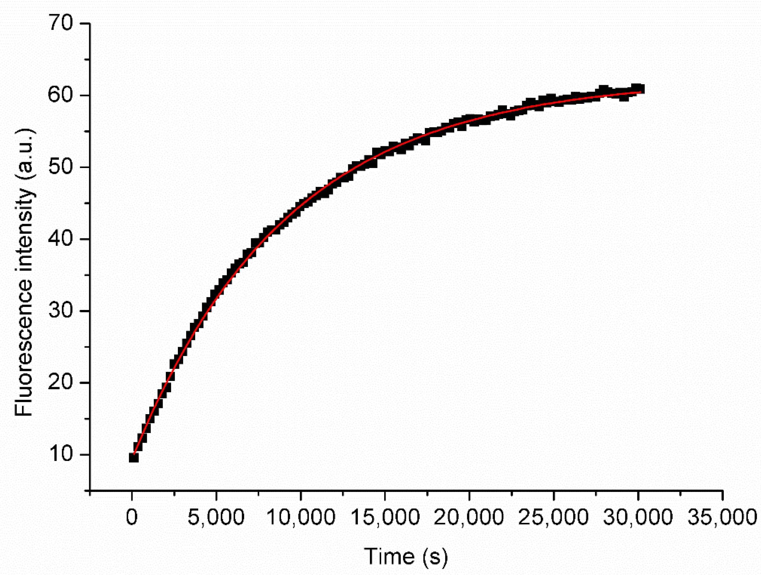
2.3. Liposome Stability

The kinetic stability of the POPC liposomes was evaluated at two different temperatures (25.0 ± 0.1 °C and 37.0 ± 0.1 °C) and three different POPC/CD molar ratios (12, 5, and 2.5) by investigating the time-dependent leakage of the 5(6)-carboxyfluorescein (CF3−) [44] anionic dye from the liposomal aqueous core. The high self-quenching concentration of CF3− used for the hydration of the phospholipidic thin film ensures very low initial fluorescence intensity. When liposomes start to release the dye, the dilution-dependent dequenching of CF3− causes an increase of the fluorescence intensity that is consistent with first-order kinetics (Figure 2, and Supplementary Materials). Therefore, for each breakdown experiment, in the absence or in the presence of CDs, an apparent first-order rate constant kobs can be determined according to the following Equation (1).
d[CF3−]/dt = kobs[CF3−]
The rate constants, kobs, for the investigated liposomal systems are reported in Table 3. The most accredited mechanism of leakage of CF3− from POPC liposomes, initially proposed by Kashchiev and Exerova [45], is that the leakage is due to the spontaneous formation in the liposomal bilayer of pores or defects.
A simple diffusion (dissolution) of the probe into the bilayer is very unlikely because CF3− is nearly insoluble in the hydrophobic bilayer. It has been demonstrated that changing the composition of the bilayer affects its chemicophysical properties and, subsequently, the rate of the release of CF3− [26,38,46,47,48,49,50,51].
The data on CF3- leakage are in perfect agreement with viscosity and dimensional evidence. The addition of functionalized CDs determined mainly a small stabilizing effect for the highest concentrations of CD investigated. This is in agreement with the above mentioned stabilizing effect due to the reduction of spontaneous pore formation associated to the strong interactions between the CDs and the head groups of the phospholipids. The small destabilizing effect observed on addition of β-CD and low concentrations of functionalized CDs may be imputed to the rearrangement consequent to the partial restructuring of CD’s on the liposomal surface. No evidence of penetration of CDs in the liposomal bilayer or extraction of phospholipid to form POPC–CD complexes has been observed. The obtained data are in agreement with a previous study [27] in which POPC SUV liposomes demonstrated to be relatively stable upon the addition of methyl and hydroxypropyl-β-CD (HP-CD) as compared to MLV. Only at very high concentrations of methylated CDs (i.e., 50 times the concentration of phospholipids) SUVs stability was affected, likely due to the extraction of phospholipids (PL) and formation of the relevant complexes. The authors speculated that the curvature of the lipid membrane of substantially smaller SUVs (compared to MLV) did not allow functionalized CD molecules to establish the contact angle needed for lipid extraction from the lipid bilayer. A similar conclusion can be drawn from stability data of MLV in the presence of β-CD [26] and the present data. As a matter of fact, the addition of β-CD to POPC MLVs renders the liposomes ca. 2 times less stable. On the other hand, in the present case the addition of β-CD to POPC SUVs at the highest concentration of β-CD investigated (i.e., the POPC/CD ratio is 0.65 [26]) does not alter the leakage rate of SUVs at 37 °C.

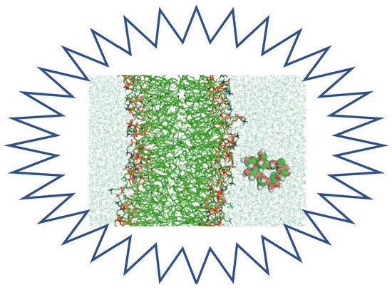
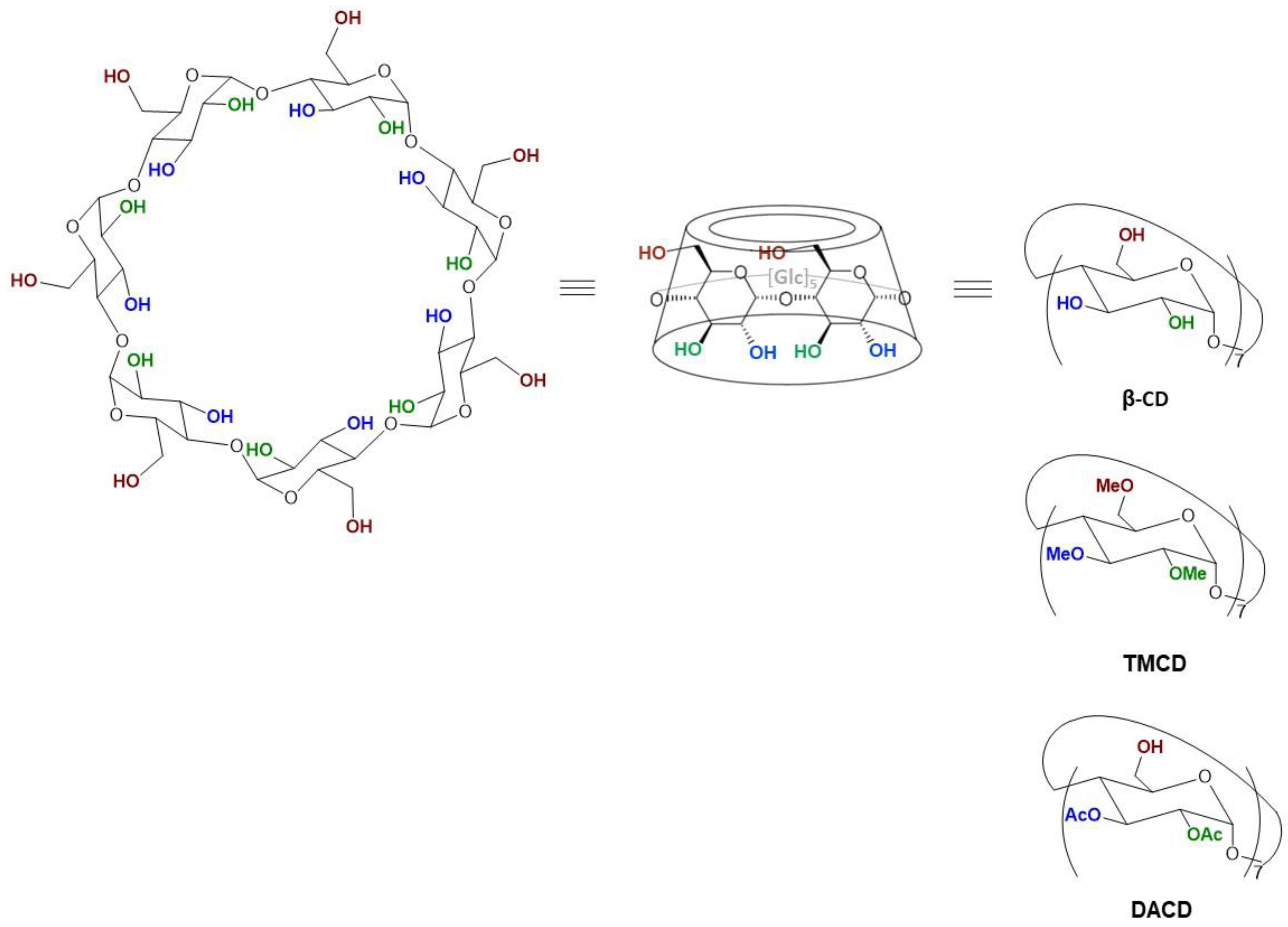
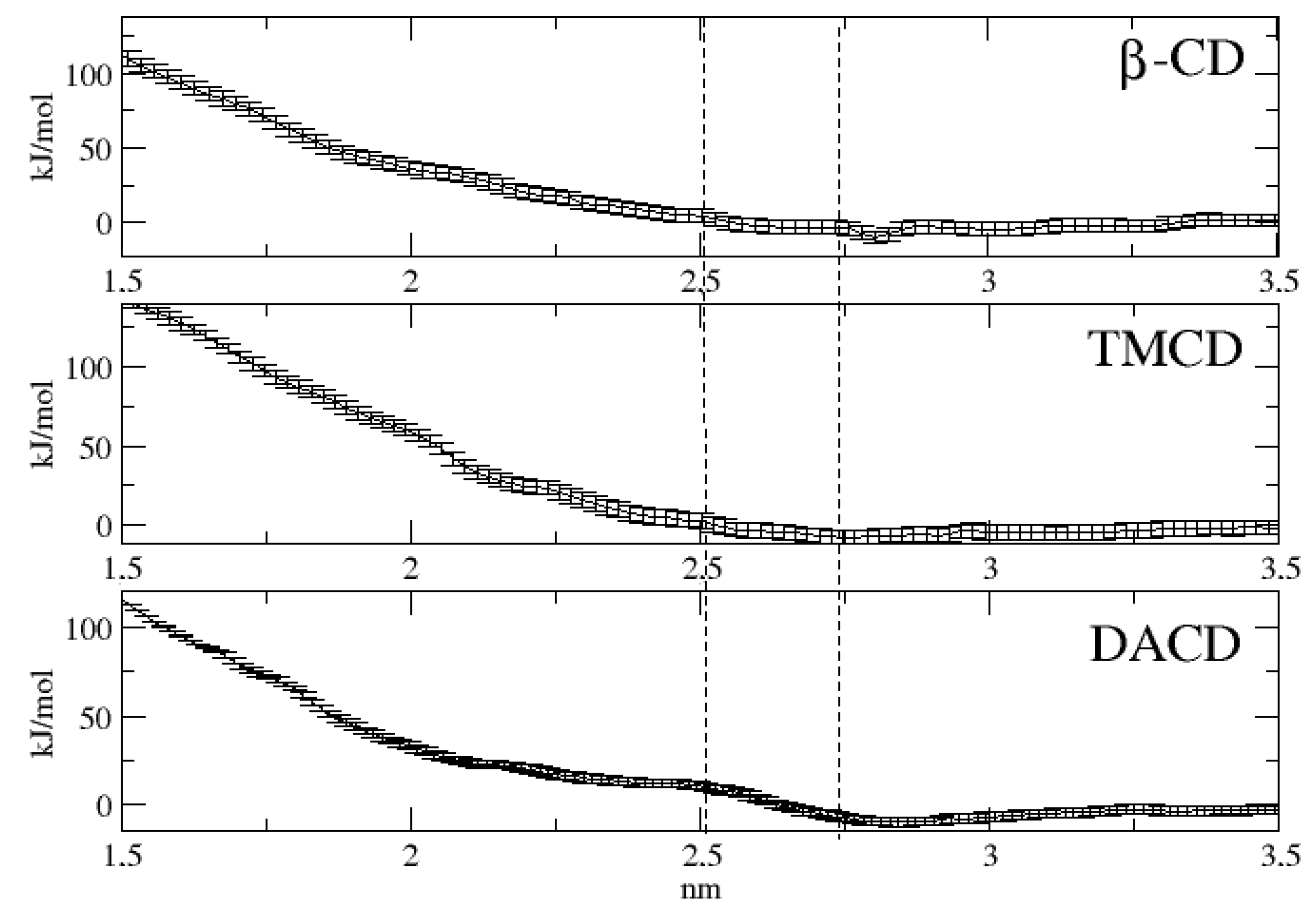
2.4. Molecular Dynamics Simulation

As already reported in the introduction, US calculations were performed with the only aim of estimating the energetics associated to the insertion of each of the three investigated cyclodextrins into the lipid bilayer mimicking the liposome surface. In Figure 3, we report the free energy at 298 K as a function of the direction normal to lipid bilayer whose center of the mass is set to zero. For the sake of clarity we have also reported, in the same figure with vertical dotted lines, the range spanned by the membrane surface along the simulations and, moreover, we have also represented, in Figure 4, three different cyclodextrin–membrane distances taken as snapshot along the simulations.
The obtained free energy profiles turn out to be quite similar in all the cases. In fact, the three cyclodextrins appear to approach the membrane through a diffusion-like motion up to approximately 2.4 nm, i.e., in correspondence of the membrane surface. Subsequently, the free energy profiles appear to undergo a sudden increase, reaching values much larger than 180 kJ/mol at the center of the bilayer. This latter result clearly indicates that, according to our data and in line with previous computational studies [52], penetration into the membrane represents a process practically impossible both by thermodynamic and kinetic point of view. In other words all the three investigated cyclodextrins, because of the favorable interactions with the POPC heads are expected to mainly reside on the liposome surface.

3. Conclusions

Functionalized CDs were embedded into POPC liposomes at different concentrations and the obtained colloidal systems were characterized from various points of view. Dimensions slightly increase on addition of CDs and the increase is proportional to the concentration of TMCD and DACD, whereas no size change was recorded in the presence of different concentration of native β-CD. The viscosity and stability data demonstrate that at the investigated concentrations of CDs, independently of the functionalization, a supramolecular structuring of the CDs around the phospholipidic head groups via hydrogen bonding and van der Waals interactions ensures a stabilization of the liposomes, with reduction of the spontaneous pore formation and a consequent promotion of excimer formation. Molecular dynamic simulations confirm that the CDs have no tendency to enter the lipid bilayer.

4. Experimental Section

4.1. Materials

1-Palmitoyl-2-oleoyl-sn-glycero-3-phosphocholine (POPC) was purchased from Avanti Polar Lipids (Alabaster, AL, USA). CF3− (95% purity) and Sephadex G-25 were purchased from Merck KGaA (Darmstadt, Germany). Native β-cyclodextrin (β-CD) was purchased as CAVAMAX® W7 PHARMA BETADEX from Wacker Chimie S.A.S. (Lyon, France). Heptakis(2,3,6-tri-O-methyl)-β-cyclodextrin (TMCD) was synthesized according to a previously described method [32]. Heptakis(2,3-di-O-acetyl)-β-cyclodextrin (DACD) was prepared following previously described methods [34,35]. β-CD, TMCD, and DACD were dried under vacuum at 80 °C for 48 h prior to use.

4.2. Instruments

The extrusion was performed on a nitrogen-driven Lipex Biomembranes (Vancouver, BC, Canada) apparatus operating at room temperature. UV/Vis absorption measurements were performed on Jasco V-550UV/Vis (Cremella, Italy). Luminescence intensity measurements were performed with Jasco FP-6200 and Jasco FP-6500 spectrofluorimeters. Measurements of vesicle size were performed by using a 90Plus/BI-MAS ZetaPlus multiangle particle size analyzer (Brookhaven Instruments Corp., Holtsville, NY, USA) on dilute samples. The osmolarity of the solutions was checked using a micro-osmometer (Advanced Instruments Model 3300, Norwood, MA, USA), while the pH of the solutions was checked on a pHM93 pH meter from Radiometer (Brønshøj, Denmark).

4.3. POPC Liposome Preparation

An appropriate amount of a stock solution of POPC in chloroform (25 mg mL−1) was evaporated at 40 °C under reduced pressure by rotary evaporation to form a thin phospholipid film on the inside wall of a flask. The thin lipid film formed was vacuum dried and kept at 4 °C overnight before rehydration with an aqueous buffer solution containing cyclodextrins in order to obtain phospholipid/cyclodextrin molar ratios of 12:1; 5:1, and 2.5:1.
The resulting multilamellar vesicle dispersions were extruded five times through polycarbonate filters with 100-nm pores [26,38,48,49,50,51].
For quenching/dequenching measurements, the rehydration of the lipidic film was performed by using 50 mM CF3− buffered aqueous solution. To get rid of unentrapped dye the liposomes were passed through a Sephadex G-25 column. Extrusion and gel permeation column chromatography were performed at room temperature.
Prior to use, the liposomal solutions were diluted with the appropriate buffer (isosmotic to the one used during the rehydration step) to give a final concentration of POPC equal to 1.32 × 10⁻⁵ M for stability measurements and 2.64 × 10⁻⁴ M for viscosity measurements, assuming 100% elution of the lipid during the gel filtration process.
The buffer (pH 7.4) used for the stability measurements was made of 121.5 mM NaCl, 25.2 mM Na2HPO4, 4.8 mM KH2PO4, and 50 mM CF3−. The buffer (pH 7.4) used for viscosity measurements was made of 121.5 mM NaCl, 25.2 mM Na2HPO4, and 4.8 mM KH2PO4.

4.4. Fluorimetric Measurements

The viscosity and the polarity of the liposomal membrane have been fluorimetrically determined at two different temperatures (25.0 ± 0.1 °C and 37.0 ± 0.1 °C) using pyrene as the fluorescent probe. The fluorescence intensity ratio, IE/IM, was used [41] for membrane microviscosity estimation (were IE stays for fluorescence intensity of the excimer at λ = 480 nm and IM for fluorescence intensity of the pyrene monomer at λ = 395 nm). Indeed, the relative intensities of excimer and monomer fluorescence of pyrene have been shown to be a simple linear function of the viscosity of a homologous series of solvents [53,54]. This relationship was demonstrated to be valid as well for micellar [53] and liposomal systems [54] and may be represented by Equation (2).
C′h = (IM/IE) × C = 1/K × η
where C is the concentration of pyrene, K is a constant that depends on the temperature, and η is the viscosity. C′h can therefore be obtained as the reciprocal of the slope of the plot of IE/IM versus the concentration of pyrene, C. Fluorescence emission spectra were taken at the excitation wavelength λex = 335 nm. The final concentrations of POPC, pyrene, and CDs in the final liposomal suspension were 2.64 × 10⁻⁴ M, 1 × 10⁻⁵ M, and 2.2 × 10⁻⁵ M, 5.28 × 10⁻⁵ M and 1.06 × 10⁻⁴ M for POPC/CD ratio 12, 5, and 2.5, respectively.
Stability measurements were made monitoring CF3− leakage from liposome at 25.0 ± 0.1 and 37.0 ± 0.1 °C. The fluorescence intensity was measured at an emission wavelength of 516 nm using λex = 490 nm as the excitation wavelength.

4.5. Molecular Dynamics Simulations

Cyclodextrins have been extensively studied through MD simulations. For this reason we have based our computational setup on the available literature [55,56,57,58,59]. All simulations were carried out using the Gromacs package [60,61] version 5.0.2 (Science for Life Laboratory, Stockholm, Sweden). The three investigated cyclodextrins, i.e., β-CD, TMCD, and DACD, whose structure was constructed with the program Molden [62] and minimized with the same level of calculations utilized for calculating the atomic charges, were modeled by adapting the Gromos force field (gromos53a6) [63] with the atomic charges calculated ex novo using standard fitting procedures with the program Gaussian 09 [64] in the framework of Density Functional Theory with the CAM-B3LYP functional [65] in conjunction with the 6-31G* basis set. Details of the cyclodextrins topologies are reported in the Supplementary Materials.
For the lipid bilayer (POPC) and water representation we adopted the force field taken from the literature [66] and the Simple Point Charge (spc) model [67], respectively. The simulations were then performed in the NVT ensemble with a time step of 2.0 fs adopting a rather standard protocol: after an initial energy minimization, the system was gradually heated from 50 to 250 K using short (20 ps) MD simulations. Finally a further preequilibration of the system, arrived at 298K, was carried out by running 1.0 ns of MD simulation in all the systems. The temperature was kept constant using the velocity rescaling procedure [68]. The LINCS algorithm was employed to constraint all bond lengths [69]. Long-range electrostatic interactions were computed by the Particle Mesh Ewald method with 34 wave vectors in each dimension and a fourth-order cubic interpolation, and a cut-off of 1.1 nm was used [70].
Determination of the free energy of insertion of the cyclodextrins into the lipid bilayer was carried out using the following procedure.
(i) Generation of initial configurations: at this purpose, we utilized constant velocity-steered MD simulations. The center of mass of the substrate was pulled in the z-dimension (normal to the bilayer) forcing the substrate to move from the solvent (water) toward the middle of the membrane. A harmonic potential with a force constant of 1000 kJ mol−1 nm−2 was adopted in conjunction with a center of mass constant velocity of 0.001 nm ps−1.
(ii) Umbrella sampling: The umbrella sampling (US) procedure [71] with the weighted histogram analysis method (WHAM) [72] was performed with a harmonic potential of 3000 kJ mol−1 nm−2 in the z direction between the center of mass of the substrate and the center of mass of the POPC bilayer. Twenty windows separated by 0.15 nm were produced and independently simulated for 10 ns. The error was evaluated through standard bootstrap analysis [73].

Supplementary Materials

The following are available online, Figures S1–S20: Representative plots of IE/IM vs. pyrene concentration for pure POPC, POPC/β-CD, POPC/TMCD and POPC/DACD liposomes at 25 °C and 37 °C, Figures S21–S40: Representative kinetic profiles of the release of CF3− from pure POPC, POPC/β-CD, POPC/TMCD and POPC/DACD liposomes at 25 °C and 37 °C, Cartesian coordinates of β-CD, TMCD and DACD, atom-types and charges for topology of β-CD, TMCD and DACD.

Author Contributions

Conceptualization, A.F.; methodology, B.C. and R.Z.; software, M.A.; validation, M.A. and R.Z.; formal analysis, R.Z., S.P. and M.A.; investigation, B.C. and R.Z.; resources, A.F., F.E. and G.S.; data curation, G.S.; writing—original draft preparation, R.Z.; writing—review and editing, A.F., B.C. and F.E.; supervision, A.F.; project administration, F.E.; funding acquisition, F.E.

Funding

This work was carried out with the support from the University ‘G. d’Annunzio’ of Chieti-Pescara and MIUR. BC and FE thank the Labex SYNORG (ANR-11-LABX-0029), the Région Normandie (CRUNCh network) and ERDF for support.

Conflicts of Interest

The authors declare no conflict of interest.

References

  1. Loftsson, T.; Brewster, M.E. Pharmaceutical applications of cyclodextrins. 1. Drug solubilization and stabilization. J. Pharm. Sci. 1996, 85, 1017–1025. [Google Scholar] [CrossRef] [PubMed]
  2. Rodríguez, J.; Martín, M.J.; Ruiz, M.A.; Clares, B. Current encapsulation strategies for bioactive oils: From alimentary to pharmaceutical perspectives. Food Res. Int. 2016, 83, 41–59. [Google Scholar] [CrossRef]
  3. McCormack, B.; Gregoriadis, G. Drugs-in-cyclodextrins-in liposomes: A novel concept in drug delivery. Int. J. Pharm. 1994, 112, 249–258. [Google Scholar] [CrossRef]
  4. Aloisio, C.; Antimisiaris, S.G.; Longhi, M.R. Liposomes containing cyclodextrins or meglumine to solubilize and improve the bioavailability of poorly soluble drugs. J. Mol. Liq. 2017, 229, 106–113. [Google Scholar] [CrossRef]
  5. Gharib, R.; Auezova, L.; Charcosset, C.; Greige-Gerges, H. Drug-in-cyclodextrin-in-liposomes as a carrier system for volatile essential oil components: Application to anethole. Food Chem. 2017, 218, 365–371. [Google Scholar] [CrossRef] [PubMed]
  6. Kfoury, M.; Lounès-Hadj Sahraoui, A.; Bourdon, N.; Laruelle, F.; Fontaine, J.; Auezova, L.; Fourmentin, S. Solubility, photostability and antifungal activity of phenylpropanoids encapsulated in cyclodextrins. Food Chem. 2016, 196, 518–525. [Google Scholar] [CrossRef]
  7. Szente, L.; Szejtli, J. Cyclodextrins as food ingredients. Trends Food Sci. Technol. 2004, 15, 137–142. [Google Scholar] [CrossRef]
  8. Azzi, J.; Jraij, A.; Auezova, L.; Fourmentin, S.; Greige-Gerges, H. Novel findings for quercetin encapsulation and preservation with cyclodextrins, liposomes, and drug-in-cyclodextrin-in-liposomes. Food Hydrocolloids 2018, 81, 328–340. [Google Scholar] [CrossRef]
  9. Gharib, R.; Greige-Gergesa, H.; Fourmentin, S.; Charcosset, C. Hydroxypropyl-β-cyclodextrin as a membrane protectant during freeze drying of hydrogenated and non-hydrogenated liposomes and molecule-in-cyclodextrin-in- liposomes: Application to trans-anethole. Food Chem. 2018, 267, 67–74. [Google Scholar] [CrossRef]
  10. Azzi, J.; Auezova, L.; Danjou, P.-E.; Fourmentin, S.; Greige-Gergesa, H. First evaluation of drug-in-cyclodextrin-in-liposomes as an encapsulating system for nerolidol. Food Chem. 2018, 255, 399–404. [Google Scholar] [CrossRef] [PubMed]
  11. Matloob, A.H.; Mourtas, S.; Klepetsanis, P.; Antimisiaris, S.G. Increasing the stability of curcumin in serum with liposomes or hybrid drug-in-cyclodextrin-in-liposome systems: A comparative study. Int. J. Pharm. 2014, 476, 108–115. [Google Scholar] [CrossRef]
  12. Bhatt, P.; Lalani, R.; Vhora, I.; Patil, S.; Amrutiya, J.; Misra, A.; Mashru, R. Liposomes encapsulating native and cyclodextrin enclosed paclitaxel: Enhanced loading efficiency and its pharmacokinetic evaluation. Int. J. Pharm. 2018, 536, 95–107. [Google Scholar] [CrossRef]
  13. Yakavets, I.; Lassalle, H.-P.; Scheglmann, D.; Wiehe, A.; Zorin, V.; Bezdetnaya, L. Temoporfin-in-cyclodextrin-in-liposome. A new approach for anticancer drug delivery: The optimization of composition. Nanomaterials 2018, 8, 847. [Google Scholar] [CrossRef]
  14. Szejtli, J. Introduction and general overview of cyclodextrin chemistry. Chem. Rev. 1998, 98, 1743–1754. [Google Scholar] [CrossRef]
  15. Crini, G. Review: A history of cyclodextrins. Chem. Rev. 2014, 114, 10940–10975. [Google Scholar] [CrossRef]
  16. de Miranda, J.C.; Martins, T.E.A.; Veiga, F.; Ferraz, H.G. Cyclodextrins and ternary complexes: Technology to improve solubility of poorly soluble drugs. Braz. J. Pharm. Sci. 2011, 47, 665–681. [Google Scholar] [CrossRef]
  17. Liu, Y.; Han, B.H.; Li, B.; Zhang, Y.M.; Zhao, P.; Chen, Y.T.; Wada, T.; Inoue, Y. Molecular recognition study on supramolecular systems. Synthesis of modified cyclodextrin and their inclusion complexation thermodynamics with L-tryptophan and some naphthalene derivatives. J. Org. Chem. 1998, 63, 1444–1454. [Google Scholar] [CrossRef]
  18. Qu, Q.; Tucker, E.; Christian, S.D. Sulfoalkyl ether β-cyclodextrin derivatives: Synthesis and characterizations. J. Incl. Phenom. Macrocycl. Chem. 2002, 43, 213–222. [Google Scholar] [CrossRef]
  19. Grachev, M.K.; Kurochkina, G.I.; Mishina, V.Y.; Mustafin, I.G.; Nifant’ev, E.E. Phosphorylated derivatives on the basis of per-6-O-(tert-butyldimethylsilyl)cyclodextrins. Russ. J. Gen. Chem. 1999, 69, 1702–1707. [Google Scholar]
  20. Brandt, L.; Felcht, U.H. Water Soluble Mixed Ether of Beta-cyclodextrin, and Process for Its Preparation. Patent EP0146841(A2), 3 July 1985. [Google Scholar]
  21. Chen, J.; Lu, W.L.; Gu, W.; Lu, S.S.; Chen, Z.-P.; Cai, B.C.; Yang, X.-X. Drug-in-cyclodextrin-in-liposomes: A promising delivery system for hydrophobic drugs. Expert Opin. Drug Deliv. 2014, 11, 565–577. [Google Scholar] [CrossRef]
  22. Gharib, R.; Greige-Gerges, H.; Fourmentin, S.; Charcosset, C.; Auezova, L. Liposomes incorporating cyclodextrin-drug inclusion complexes: Current state of knowledge. Carbohydr. Polym. 2015, 129, 175–186. [Google Scholar] [CrossRef]
  23. Palazzo, C.; Laloy, J.; Delvigne, A.-S.; Nys, G.; Fillet, M.; Dogne, J.-M.; Pequeux, C.; Foidart, J.-M.; Evrard, B.; Piel, G. Development of injectable liposomes and drug-in-cyclodextrin-in-liposome formulations encapsulating estetrol to prevent cerebral ischemia of premature babies. Eur. J. Pharm. Sci. 2019, 127, 52–59. [Google Scholar] [CrossRef]
  24. Bragagni, M.; Maestrelli, F.; Mennini, N.; Ghelardini, C.; Mura, P. Liposomal formulations of prilocaine: Effect of complexation with hydroxypropyl-β-cyclodextrin on drug anesthetic efficacy. J. Liposome Res. 2010, 20, 315–322. [Google Scholar] [CrossRef]
  25. Sebaaly, C.; Charcosset, C.; Stainmesse, S.; Fessi, H.; Greige-Gerges, H. Clove essential oil-in-cyclodextrin-in-liposomes in the aqueous and lyophilized states: From laboratory to large scale using a membrane contactor. Carbohydr. Polym. 2016, 138, 75–85. [Google Scholar] [CrossRef]
  26. Gasbarri, C.; Guernelli, S.; Boncompagni, S.; Angelini, G.; Siani, G.; De Maria, P.; Fontana, A. Fine-tuning of POPC liposomal leakage by the use of β-cyclodextrin and several hydrophobic guests. J. Liposome Res. 2010, 20, 202–210. [Google Scholar] [CrossRef]
  27. Hatzi, P.; Mourtas, S.; Klepetsanis, P.G.; Antimisiaris, S.G. Integrity of liposomes in presence of cyclodextrins: Effect of liposome type and lipid composition. Int. J. Pharm. 2007, 333, 167–176. [Google Scholar] [CrossRef]
  28. Maestrelli, F.; González-Rodríguez, M.L.; Rabasco, A.M.; Ghelardini, C.; Mura, P. New “drug-in cyclodextrin-in deformable liposomes” formulations to improve the therapeutic efficacy of local anaesthetics. Int. J. Pharm. 2010, 395, 222–231. [Google Scholar] [CrossRef]
  29. Angelini, G.; Campestre, C.; Boncompagni, S.; Gasbarri, C. Liposomes entrapping β-cyclodextrin/ibuprofen inclusion complex: Role of the host and the guest on the bilayer integrity and microviscosity. Chem. Phys. Lipids 2017, 209, 61–65. [Google Scholar] [CrossRef]
  30. Ghariba, R.; Fourmentin, S.; Charcosset, C.; Greige-Gergesa, H. Effect of hydroxypropyl-β-cyclodextrin on lipid membrane fluidity, stability and freeze-drying of liposomes. J. Drug Deliv. Sci. Technol. 2018, 44, 101–107. [Google Scholar] [CrossRef]
  31. Wenz, G. Influence of intramolecular hydrogen bonds on the binding potential of methylated β-cyclodextrin derivatives. Belstein J. Org. Chem. 2012, 8, 1890–1895. [Google Scholar] [CrossRef]
  32. Szejtli, J.; Lipták, A.; Jodál, I.; Fügedi, P.; Nánási, P.; Neszmélyi, A. Synthesis and 13C-NMR spectroscopy of methylated beta-cyclodextrins. Starch Staerke 1980, 32, 165–169. [Google Scholar] [CrossRef]
  33. Nishijo, J.; Mizuno, H. Interactions of cyclodextrins with DPPC liposomes. Differential scanning calorimetry study. Chem. Pharm. Bull. 1998, 46, 120–124. [Google Scholar] [CrossRef]
  34. Wang, X.; Fan, H.; Zhang, F.; Qi, Y.; Qiu, W.; Yang, F.; Tang, J.; He, P. Synthesis of a β-cyclodextrin derivate and its molecular recognition behavior on modified glassy carbon electrode by diazotization. Tetrahedron 2010, 66, 7815–7820. [Google Scholar] [CrossRef]
  35. Takeo, K.; Mitoh, H.; Uemura, K. Selective chemical modification of cyclomaltooligosaccha-rides via tert-butyldimethylsilylation. Carbohydr. Res. 1989, 187, 203–221. [Google Scholar] [CrossRef]
  36. Puglisi, G.; Fresta, M.; Ventura, C.A. Interaction of natural and modified β-cyclodextrins with a biological membrane model of dipalmitoylphosphatidylcholine. J. Colloid Interface Sci. 1996, 180, 542–547. [Google Scholar] [CrossRef]
  37. Besseau, F. Hydrogen-bond basicity of esters, lactones and carbonates. J. Chem. Soc. Perkin Trans. 1994, 2, 485–489. [Google Scholar] [CrossRef]
  38. De Maria, P.; Fontana, A.; Gasbarri, C.; Velluto, D. Effects of fullerene guests on the stability of 1-palmitoyl-2-oleoylphosphatidylcholine liposomes. Soft Matter 2006, 2, 595–602. [Google Scholar] [CrossRef]
  39. Silvander, M.; Bergstrand, N.; Edwards, K. Linkage identity is a major factor in determining the effect of PEG-ylated surfactants on permeability of phosphatidylcholine liposomes. Chem. Phys. Lipids 2003, 126, 77–83. [Google Scholar] [CrossRef]
  40. Bahri, M.A.; Heyne, B.L.; Hans, P.; Mouithys Mickalad, A.A.; Hoebeke, M.D. Quantification of lipid bilayer effective microviscosity and fluidity effect induced by propofol. Biophys. Chem. 2005, 114, 53–61. [Google Scholar] [CrossRef] [Green Version]
  41. Pownall, H.J.; Smith, L.C. Viscosity of the hydrocarbon region of micelles. Measurement by excimer fluorescence. J. Am. Chem. Soc. 1973, 95, 3136–3140. [Google Scholar] [CrossRef]
  42. Birks, J.B. Excimers. Rep. Prog. Phys. 1975, 38, 903–974. [Google Scholar] [CrossRef]
  43. Turro, N.J.; Lei, X.-G.; Ananthapadmanabhan, K.P.; Aronson, M. Spectroscopic probe analysis of protein-surfactant interactions: The BSA/SDS system. Langmuir 1995, 11, 2525–2533. [Google Scholar] [CrossRef]
  44. Aschi, M.; D’Archivio, A.A.; Fontana, A.; Formiglio, A. Physicochemical properties of fluorescent probes: Experimental and computational determination of the overlapping pKa values of carboxyfluorescein. J. Org. Chem. 2008, 73, 3411–3417. [Google Scholar] [CrossRef]
  45. Kashchiev, D.; Exerova, D. Bilayer lipid membrane permeation and rupture due to hole formation. Biochim. Biophys. Acta Biomembr. 1983, 732, 133–145. [Google Scholar] [CrossRef]
  46. Iwata, T.; Lee, S.; Oishi, O.; Aoyagi, H.; Ohno, M.; Anzai, K.; Kirino, Y.; Sugihara, G. Design and synthesis of amphipathic 3(10)-helical peptides and their interactions with phospholipid bilayers and ion channel formation. J. Biol. Chem. 1994, 269, 4928–4933. [Google Scholar]
  47. Scrimin, P.; Tecilla, P.; Tonellato, U.; Veronese, A.; Crisma, M.; Formaggio, F.; Toniolo, C. Zinc(II) as an allosteric regulator of liposomal membrane permeability induced by synthetic template-assembled tripodal polypeptides. Chem. Eur. J. 2002, 8, 2753–2763. [Google Scholar] [CrossRef]
  48. De Maria, P.; Filippone, P.; Fontana, A.; Gasbarri, C.; Siani, G.; Velluto, D. Cardanol as a replacement for cholesterol into the lipid bilayer of POPC liposomes. Colloid Surf. B Biointerfaces 2005, 40, 11–18. [Google Scholar] [CrossRef]
  49. Angelini, G.; Boncompagni, S.; De Maria, P.; Fontana, A.; Gasbarri, C.; Siani, G. Kinetic evaluation of the effect of layer by layer deposition of polyeletrolytes on the stability of POPC liposomes. Colloid Surf. A Physicochem. Eng. Asp. 2008, 322, 234–238. [Google Scholar] [CrossRef]
  50. Zappacosta, R.; Semeraro, M.; Baroncini, M.; Silvi, S.; Aschi, M.; Credi, A.; Fontana, A. Liposome destabilization by a 2,7-diazapyrenium derivative through formation of transient pores in the lipid bilayer. Small 2010, 6, 952–959. [Google Scholar] [CrossRef]
  51. Velluto, D.; Gasbarri, C.; Angelini, G.; Fontana, A. Use of simple kinetic and reaction order measurements for the evaluation of the mechanism of surfactant-liposome interactions. J. Phys. Chem. B 2011, 115, 8130–8137. [Google Scholar] [CrossRef]
  52. Khuntawee, W.; Wolschann, P.; Rungrotmongkol, T.; Wong-Ekkabut, J.; Hannongbua, S. Molecular dynamics simulations of the interaction of beta-cyclodextrin with a lipid bilayer. J. Chem. Inf. Model. 2015, 55, 1894–1902. [Google Scholar] [CrossRef]
  53. Kalyanasundaram, K.; Thomas, J.K. Environmental effects on vibronic band intensities in pyrene monomer fluorescence and their application in studies of micellar systems. J. Am. Chem. Soc. 1977, 99, 2039–2044. [Google Scholar] [CrossRef]
  54. Han, S.K.; Kim, J.-S.; Lee, Y.-S.; Kim, M. Effect of drug substances on the microviscosity of lipid bilayer of liposomal membrane. Arch. Pharm. Res. 1990, 13, 192–197. [Google Scholar] [CrossRef]
  55. Lopez, C.A.; de Vries, A.H.; Marrink, S.J. Computational microscopy of cyclodextrin mediated cholesterol extraction from lipid model membranes. Sci. Rep. 2013, 3, 2071. [Google Scholar] [CrossRef]
  56. Ivanov, P.M.; Jaime, C. Insights into the structure of large-ring cyclodextrins through molecular dynamics simulations in solution. J. Phys. Chem. B 2004, 108, 6261–6274. [Google Scholar] [CrossRef]
  57. Nutho, B.; Khuntawee, W.; Rungnim, C.; Pongsawasdi, P.; Wolschann, P.; Karpfen, A.; Kungwan, N.; Rungrotmongkol, T. Binding mode and free energy prediction of fisetin/β-cyclodextrin inclusion complexes. Beilstein J. Org. Chem. 2014, 10, 2789–2799. [Google Scholar] [CrossRef] [Green Version]
  58. Zhang, H.; Tan, T.; Feng, W.; van der Spoel, D. Molecular recognition in different environments: β-cyclodextrin dimer formation in organic solvents. J. Phys. Chem. B 2012, 116, 12684–12693. [Google Scholar] [CrossRef]
  59. Zhang, H.; Feng, W.; Li, C.; Tan, T. Investigation of the inclusions of puerarin and daidzin with β-cyclodextrin by molecular dynamics simulation. J. Phys. Chem. B 2010, 114, 4876–4883. [Google Scholar] [CrossRef]
  60. Berendsen, H.J.C.; van der Spoel, D.; van Drunen, R. GROMACS: A message-passing parallel molecular dynamics implementation. Comput. Phys. Commun. 1995, 91, 43–56. [Google Scholar] [CrossRef] [Green Version]
  61. Van der Spoel, D.; Lindahl, E.; Hess, B.; Groenhof, G.; Mark, A.E.; Berendsen, H.J.C. GROMACS: Fast, flexible and free. J. Comput. Chem. 2005, 26, 1701–1718. [Google Scholar] [CrossRef]
  62. Schaftenaar, G.; Noordik, J.H. Molden: A pre- and post-processing program for molecular and electronic structures. J. Comput. Aided Mol. Des. 2000, 14, 123–134. [Google Scholar] [CrossRef]
  63. Van Gunsteren, W.F.; Billeter, S.R.; Eising, A.A.; Hunenberger, P.H.; Kruger, P.; Mark, A.E.; Scott, W.R.P.; Tironi, I.G. Biomolecular Simulation: The GROMOS96 Manual and User Guide; Hochschulverlag AG an der ETH Zurich: Zurich, Switzerland, 1996. [Google Scholar]
  64. Frisch, M.J.; Trucks, G.W.; Schlegel, H.B.; Scuseria, G.E.; Robb, M.A.; Cheeseman, J.R.; Scalmani, G.; Barone, V.; Mennucci, B.; Petersson, G.A.; et al. Gaussian 09, Revision E.01; Gaussian, Inc.: Wallingford, CT, USA, 2009. [Google Scholar]
  65. Yanai, T.; Tew, D.; Handy, N. A new hybrid exchange-correlation functional using the Coulomb-attenuating method (CAM-B3LYP). Chem. Phys. Lett. 2014, 393, 51–57. [Google Scholar] [CrossRef]
  66. Kukol, A. Lipid models for united-atom molecular dynamics simulations of proteins. J. Chem. Theory Comput. 2009, 5, 615–626. [Google Scholar] [CrossRef]
  67. Berendsen, J.C.; Postma, J.P.M.; van Gunsteren, W.F.; Hermans, J. Interaction models for water in relation to protein hydration. In Intermolecular Forces; Pullman, B., Ed.; Reidel: Dordrecht, The Netherlands, 1981; pp. 331–342. [Google Scholar]
  68. Bussi, G.; Donadio, D.; Parrinello, M. Canonical sampling through velocity rescaling. J. Chem. Phys. 2007, 126, 014101. [Google Scholar] [CrossRef] [Green Version]
  69. Hess, B.; Bekker, H.; Berendsen, H.J.C.; Frajie, J.C.E.M. LINCS: A linear constraint solver for molecular simulations. J. Comput. Chem. 1997, 18, 1463–1472. [Google Scholar] [CrossRef] [Green Version]
  70. Darden, T.A.; York, D.M.; Pedersen, L.G. Particle mesh Ewald: An N·log (N) method for Ewald sums in large systems. J. Chem. Phys. 1993, 98, 10089–10092. [Google Scholar] [CrossRef]
  71. Roux, B. The calculation of the potential of mean force using computer simulations. Comput. Phys. Commun. 1995, 91, 275–282. [Google Scholar] [CrossRef]
  72. Kumar, S.; Rosenberg, J.M.; Bouzida, D.; Swendsen, R.H.; Kollman, P.A. The weighted histogram analysis method for free energy calculations in biomolecules. I. The method. J. Comput. Chem. 1992, 13, 1011–1021. [Google Scholar] [CrossRef]
  73. Hub, J.S.; de Groot, B.L.; van der Spoel, D. g_wham: A free weighted histogram analysis implementation including robust error and autocorrelation estimates. J. Chem. Theory Comput. 2010, 6, 3713–3720. [Google Scholar] [CrossRef]
Sample Availability: Samples of the compounds TMCD and DACD are available from the authors.
Figure 1. β-Cyclodextrins and functionalized cyclodextrins (CDs).
Molecules 24 01387 g001
Figure 2. Representative kinetic profile of the release of CF3− from POPC/β-CD liposomes at POPC/β-CD = 5 and 25° C. The red curve is the fitting curve for a first order kinetics.
Molecules 24 01387 g002
Figure 3. Free energy profile at 298 K as a function of the distance between cyclodextrin and bilayer centers of mass.
Molecules 24 01387 g003
Figure 4. Snapshots taken from US calculations representing three different distances between β-CD and bilayer.
Molecules 24 01387 g004
Table 1. Size of pure and CD-doped 1-palmitoyl-2-oleoyl-sn-glycero-3-phosphocholine (POPC) liposomes measured at 25.0 ± 0.1 °C.
EntryLiposome CompositionPOPC/CD [a]Diameter (nm)Polydispersity
1POPC 118.0 ± 0.50.02 ± 0.01
2POPC/β-CD12131.6 ± 1.50.05 ± 0.01
3POPC/β-CD5130.5 ± 2.30.05 ± 0.02
4POPC/β-CD2.5132.2 ± 0.70.08 ± 0.01
5POPC/TMCD12128.6 ± 4.70.18 ± 0.01
6POPC/TMCD5136.6 ± 1.00.13 ± 0.01
7POPC/TMCD2.5144.1 ± 3.00.11 ± 0.01
8POPC/DACD12144.6 ± 2.30.11 ± 0.02
9POPC/DACD5160.7 ± 4.20.19 ± 0.03
10POPC/DACD2.5185.8 ± 0.60.29 ± 0.01
[a] Molar ratio.
Table 2. Bilayer viscosity (C′h) [a] in pure and CD-doped POPC liposomes.
EntryLiposome CompositionPOPC/CD [b] C′h × 105 (M) at 25.0 ± 0.1 °CC′h × 105 (M) at 37.0 ± 0.1 °C
1POPC 13.80 ± 0.104.52 ± 0.01
2POPC/β-CD1211.70 ± 0.408.31 ± 0.58
3POPC/β-CD57.44 ± 0.115.37 ± 0.15
4POPC/β-CD2.57.16 ± 0.086.14 ± 0.15
5POPC/TMCD1214.40 ± 0.4011.70 ± 0.10
6POPC/TMCD513.90 ± 0.059.37 ± 0.20
7POPC/TMCD2.56.75 ± 0.545.20 ± 0.18
8POPC/DACD1215.10 ± 0.6011.00 ± 0.90
9POPC/DACD515.80 ± 1.5011.00 ± 0.09
10POPC/DACD2.58.07 ± 0.205.76 ± 0.11
[a] C′h is the reciprocal of the slope of the plot of IE/IM vs. pyrene concentration; [b] Molar ratio.
Table 3. First-order rate constants for the release of carboxyfluorescein (CF3−) from pure and CD-doped POPC liposomes.
EntryLiposome CompositionPOPC/CD [a]10−4 kobs (s−1) at 25.0 ± 0.1 °C10−4 kobs (s−1) at 37.0 ± 0.1 °C
1POPC 1.31 ± 0.050.90 ± 0.04
2POPC/β-CD121.18 ± 0.051.15 ± 0.05
3POPC/β-CD51.18 ± 0.131.40 ± 0.19
4POPC/β-CD2.50.28 ± 0.030.93 ± 0.02
5POPC/TMCD121.48 ± 0.051.24 ± 0.06
6POPC/TMCD50.89 ± 0.030.60 ± 0.03
7POPC/TMCD2.50.32 ± 0.070.33 ± 0.08
8POPC/DACD121.40 ± 0.151.54 ± 0.23
9POPC/DACD51.62 ± 0.151.74 ± 0.16
10POPC/DACD2.50.30 ± 0.050.58 ± 0.08
[a] Molar ratio.

