Article

The Meta-Position of Phe4 in Leu-Enkephalin Regulates Potency, Selectivity, Functional Activity, and Signaling Bias at the Delta and Mu Opioid Receptors

1
Department of Medicinal Chemistry and Molecular Pharmacology, College of Pharmacy, Purdue University, West Lafayette, IN 47907, USA
2
Department of Medicinal Chemistry, The University of Kansas, Lawrence, KS 66045, USA
3
Department of Chemistry, Purdue University, West Lafayette, IN 47907, USA
4
College of Wuya, Shenyang Pharmaceutical University, Shenyang 110016, China
5
Purdue Institute for Drug Discovery, Purdue University, West Lafayette, IN 47907, USA
6
Purdue Institute for Integrative Neuroscience, Purdue University, West Lafayette, IN 47907, USA
*
Authors to whom correspondence should be addressed.
These authors contributed equally to this work.
Molecules 2019, 24(24), 4542; https://doi.org/10.3390/molecules24244542
Submission received: 20 November 2019 / Revised: 8 December 2019 / Accepted: 10 December 2019 / Published: 12 December 2019

Abstract

:
As tool compounds to study cardiac ischemia, the endogenous δ-opioid receptors (δOR) agonist Leu5-enkephalin and the more metabolically stable synthetic peptide (d-Ala2, d-Leu5)-enkephalin are frequently employed. However, both peptides have similar pharmacological profiles that restrict detailed investigation of the cellular mechanism of the δOR’s protective role during ischemic events. Thus, a need remains for δOR peptides with improved selectivity and unique signaling properties for investigating the specific roles for δOR signaling in cardiac ischemia. To this end, we explored substitution at the Phe4 position of Leu5-enkephalin for its ability to modulate receptor function and selectivity. Peptides were assessed for their affinity to bind to δORs and µ-opioid receptors (µORs) and potency to inhibit cAMP signaling and to recruit β-arrestin 2. Additionally, peptide stability was measured in rat plasma. Substitution of the meta-position of Phe4 of Leu5-enkephalin provided high-affinity ligands with varying levels of selectivity and bias at both the δOR and µOR and improved peptide stability, while substitution with picoline derivatives produced lower-affinity ligands with G protein biases at both receptors. Overall, these favorable substitutions at the meta-position of Phe4 may be combined with other modifications to Leu5-enkephalin to deliver improved agonists with finely tuned potency, selectivity, bias and drug-like properties.

Graphical Abstract

1. Introduction

Leu5-enkephalin (Tyr1-Gly2-Gly3-Phe4-Leu5, Figure 1) is an endogenous opioid peptide produced in vertebrate species including rodents, primates and humans [1,2,3,4] that results from the metabolism of proenkephalin or dynorphin [5]. Pharmacologically, Leu5-enkephalin agonizes the δ opioid receptor (δOR) with moderate selectivity over the µ opioid receptor (µOR), but does not significantly interact with the κ opioid receptor [6]. As a neurotransmitter in pain circuits, Leu5-enkephalin possesses antinociceptive properties [7], whereas peripherally, the peptide demonstrates cardiovascular effects [8]. Over the last two decades, improved pharmacological characterization of opioid pathways has revealed that activation of an opioid receptor can trigger two distinct pathways, β-arrestin-dependent or β-arrestin-independent (i.e., G protein-mediated) and that these pathways differentially modulate antinociception and side effect profiles [9]. Despite the increasing number of studies that implicate δOR mediated β-arrestin recruitment with various (patho)physiological effects, such as tolerance [10], alcohol intake [11,12] and δOR agonist-induced seizures [10], the role of β-arrestin recruitment towards δOR-induced cardioprotection remains unclear.
From a translational perspective, peptide-based probes provide an ideal tool for studying the cardioprotective effects of the δOR, given their low brain penetration. While numerous enkephalin-like peptides have been synthesized that interact with excellent potency and selectivity for δORs and µORs [13], a majority of studies [14] investigating δOR involvement in ischemia have utilized the synthetic peptide d-Ala2, d-Leu5-enkephalin (DADLE, Figure 1) [15,16], because DADLE possesses improved proteolytic stability and improved selectivity for δORs over µORs relative to Leu5-enkephalin [6,17]. However, DADLE’s discovery in 1977 [18], predated identification of β-arrestin as a modulator of opioid signaling [14,19,20,21], With the use of contemporary cellular assays it is now apparent that DADLE pharmacologically signals similarly to Leu5-enkephalin, though it recruits β-arrestin 2 (arrestin 3) slightly more efficaciously. Given the similarities between DADLE and Leu5-enkephalin, it is unclear to what degree β-arrestin recruitment contributes to or detracts from these peptides’ in vivo cardioprotective efficacy, and new analogs with distinct pharmacoloogical profiles are necessary to probe these contributions.
To better investigate the role of δOR-mediated β-arrestin signaling in ischemic protection, the development of δOR selective agonists that have either low, intermediate or high β-arrestin is desired; however, reports of δOR selective peptide-based biased ligands remain limited. Recently, the naturally occurring peptides rubiscolin-5 and -6 (Figure 1) were classified as G protein-biased (β-arrestin 2 efficacy = 15% and 20%, respectively; δOR bias factor = 2.0) δOR selective peptides, albeit with only micromolar potencies [22]. While a number of synthetic peptides display nanomolar potencies at δORs, such as aza-β-homoleucine-enkephalin (β-arrestin 2 efficacy = 64%; δOR bias factor = 5.2) [23] and Dmt-Tic analogs [24], these peptides all recruit β-arrestin 2 far more efficaciously than the rubiscolins (UFP-512: cAMP potency 0.4 nM, β-arrestin 2 potency = 20 nM, efficacy = 60%) [23,24,25]. Additionally, these latter biased compounds, including aza-β-homoleucine-enkephalin and Dmt-Tic-Gly-NH-Ph, have at best 10-fold selectivity for δORs over µORs and actually super-recruit β-arrestin 2 at µOR [23,24], which is likely to cause adverse, undesired in vivo effects [26,27]. As such, our goal was to identify novel and potent δOR peptide agonists with varying degrees of β-arrestin recruitment efficacy, and that importantly have improved δOR selectivity over µOR while limiting β-arrestin recruitment at µOR.
A collection of previously published structure activity relationship studies on Leu5-enkephalin directed us to Phe4 as a position that can modulate δOR and µOR potency and selectivity (Figure 2A) [28]. Specifically, ortho and para substitutions on Phe4 can modulate binding affinity and selectivity between opioid receptors [28], and halogenation of the para-position of Leu5-enkephalin and endomorphin 1 in particular appears to enhance δOR affinity while reducing µOR affinity [28]. Corroborating research has further shown that halogenation of the para position of Phe4 of [D-Pen2,D-Pen5]enkephalin (DPDPE) enhanced δOR selectivity, potency, peptide stability, central nervous system (CNS) distribution, and antinociceptive potency compared to unsubstituted DPDPE [29,30,31]. Thus, halogenation at Phe4 may provide Leu5-enkephalin derivatives that can be used to study not only cardiac but also cerebral ischemia [32,33]. We herein report structure-activity-relationship trends at the meta-position of Phe4 of Leu5-enkephalin (Figure 2B), demonstrating that substitution at this position variously regulates δOR and µOR affinity and G-protein activity, enables the fine-tuning of β-arrestin 2 recruitment to both δOR and µOR, and further increases the plasma stability of the derived peptides. Combined, these features provide a clear direction for designing the next-generation of Leu5-enkephalin analogs with well-defined biases and improved stabilities.

2. Results and Discussion

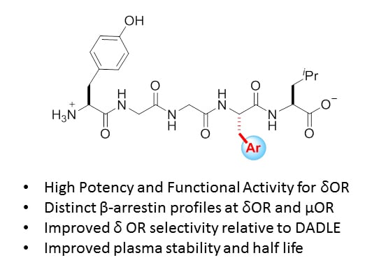
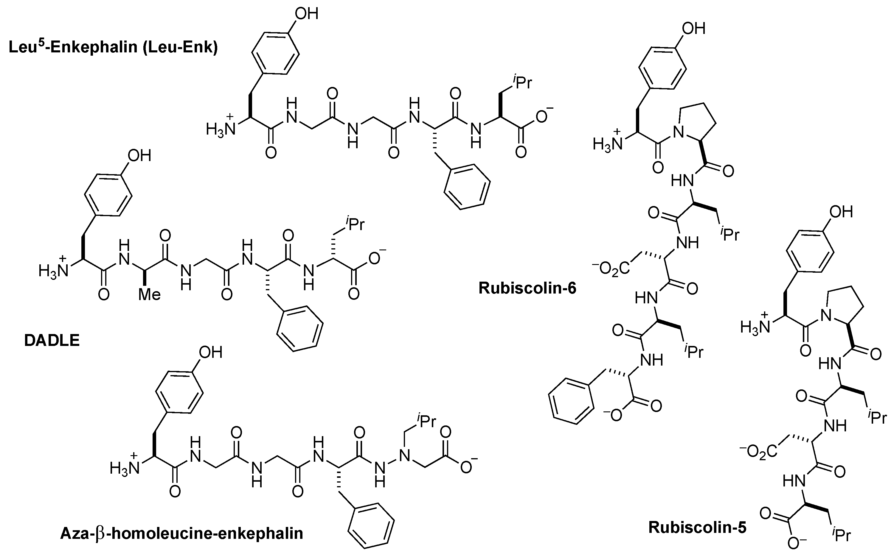
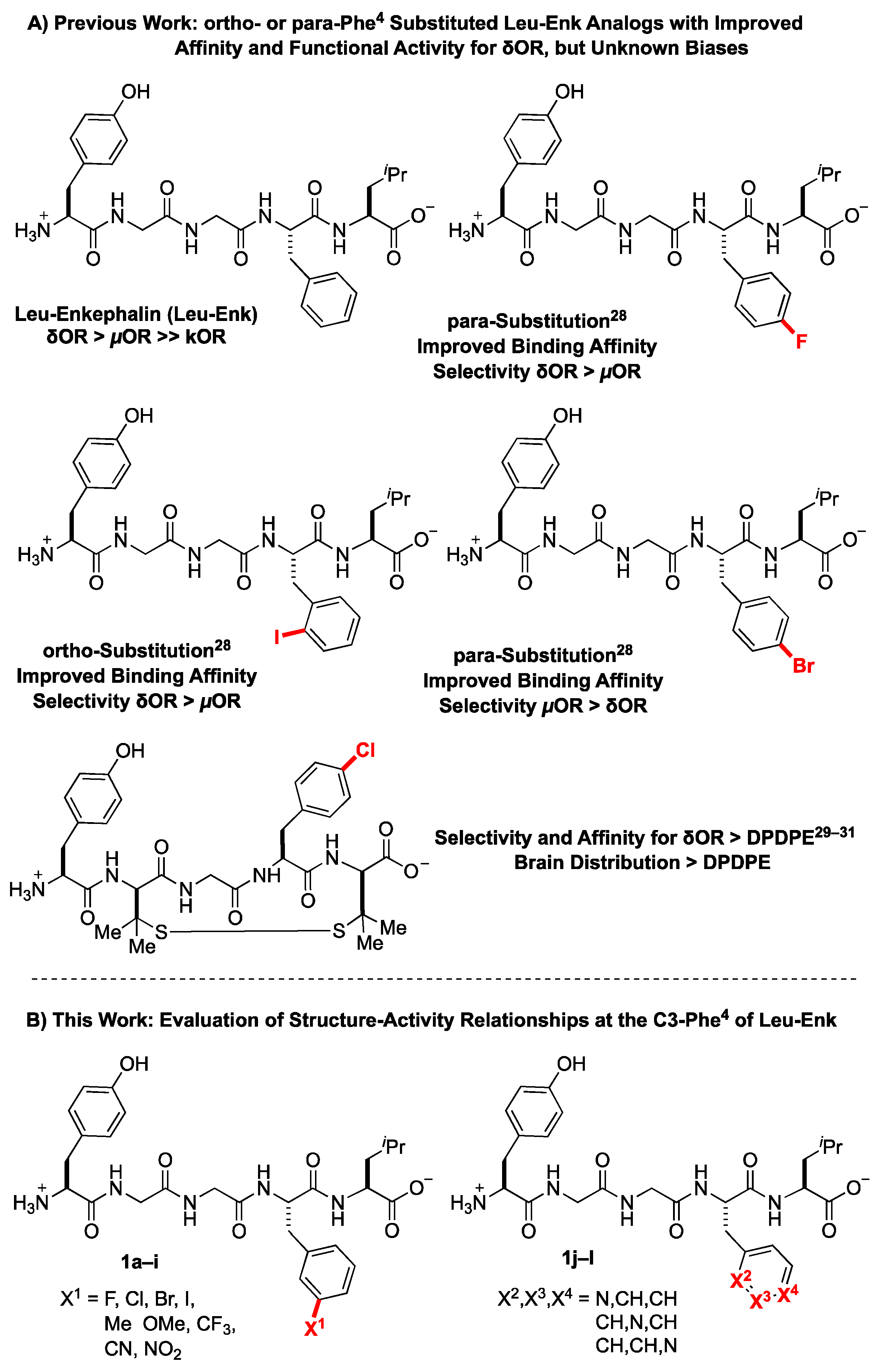
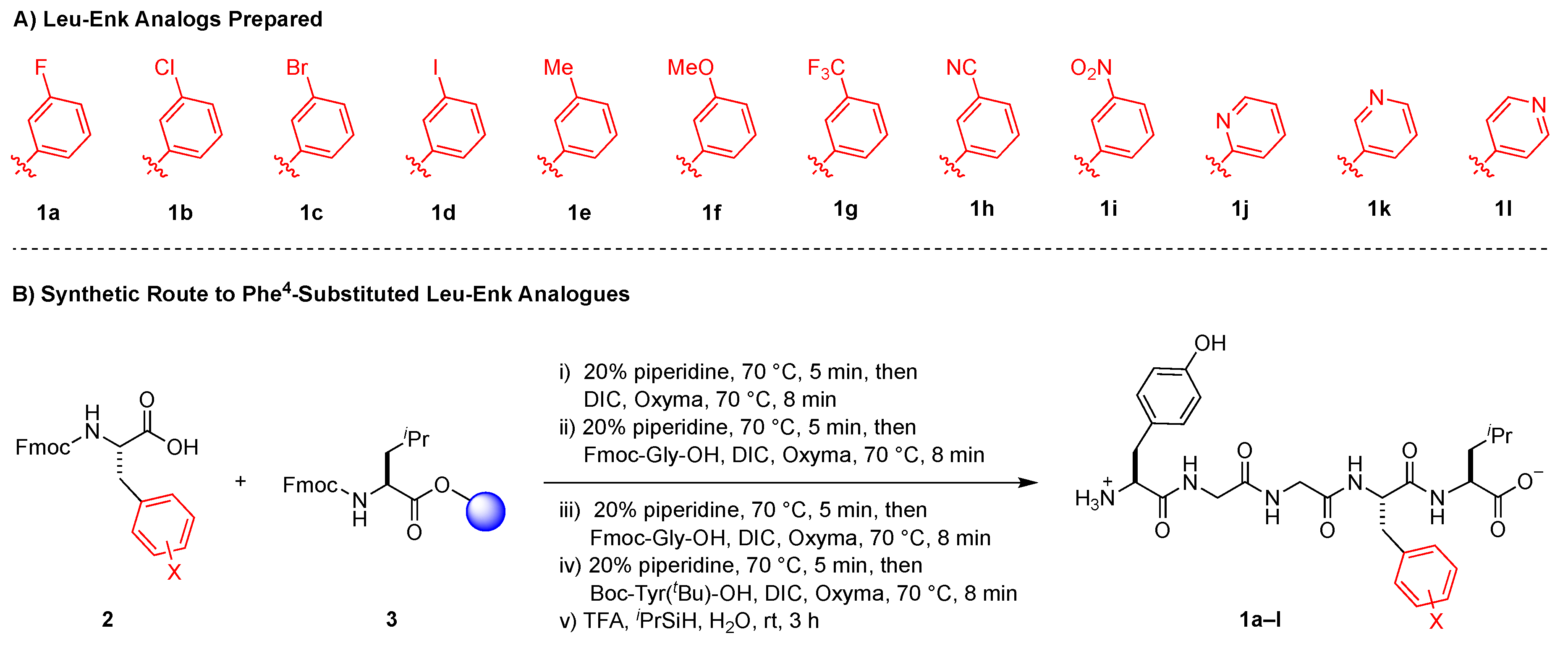
2.1. Design Considerations

To probe the meta position of Phe4, we initially considered known structure-activity-relationship trends at the ortho and para positions of this residue. Considering that halogenated substituents at these positions perturbed binding affinity, δOR selectivity, and stability properties of the Leu5-enkephalin [28], we initially hypothesized that meta-halogenated analogs might similarly perturb the parent scaffold (Figure 3A, 1a1d). An additional set of analogs bearing electron-donating (1e1f) and -withdrawing groups (1g1i) would further probe interactions at this site, including the electronic character of the Phe4 ring. Finally, pyridine analogs (1j1l) would present H-bond accepting contacts about the ring, as well as provide analogs that present dipoles at similar vectors as to previously successful halogenated substituents.

2.2. Solid Phase Synthesis of Peptides

All peptides were synthesized using a rapid solid phase peptide synthesis protocol on an automated peptide synthesizer using an Fmoc protection strategy [34,35] and N,N′-diisopropylcarbodiimide and oxyma as the coupling reagents (Figure 3B). Fmoc-Leu-Wang resin was utilized as a starting template for this synthetic protocol. All coupling steps and Fmoc-deprotection steps were carried out at 70 °C under an atmosphere of N2. Cleavage from the resin was performed using TFA/triisopropylsilane/H2O. Purification of the synthesized peptides was performed by reverse-phase high performance liquid chromatography (RP-HPLC), and analysis of purity was performed using ultra-performance liquid chromatography (UPLC). All desired peptides were obtained in ≥95% purity before submitting for pharmacological evaluation.

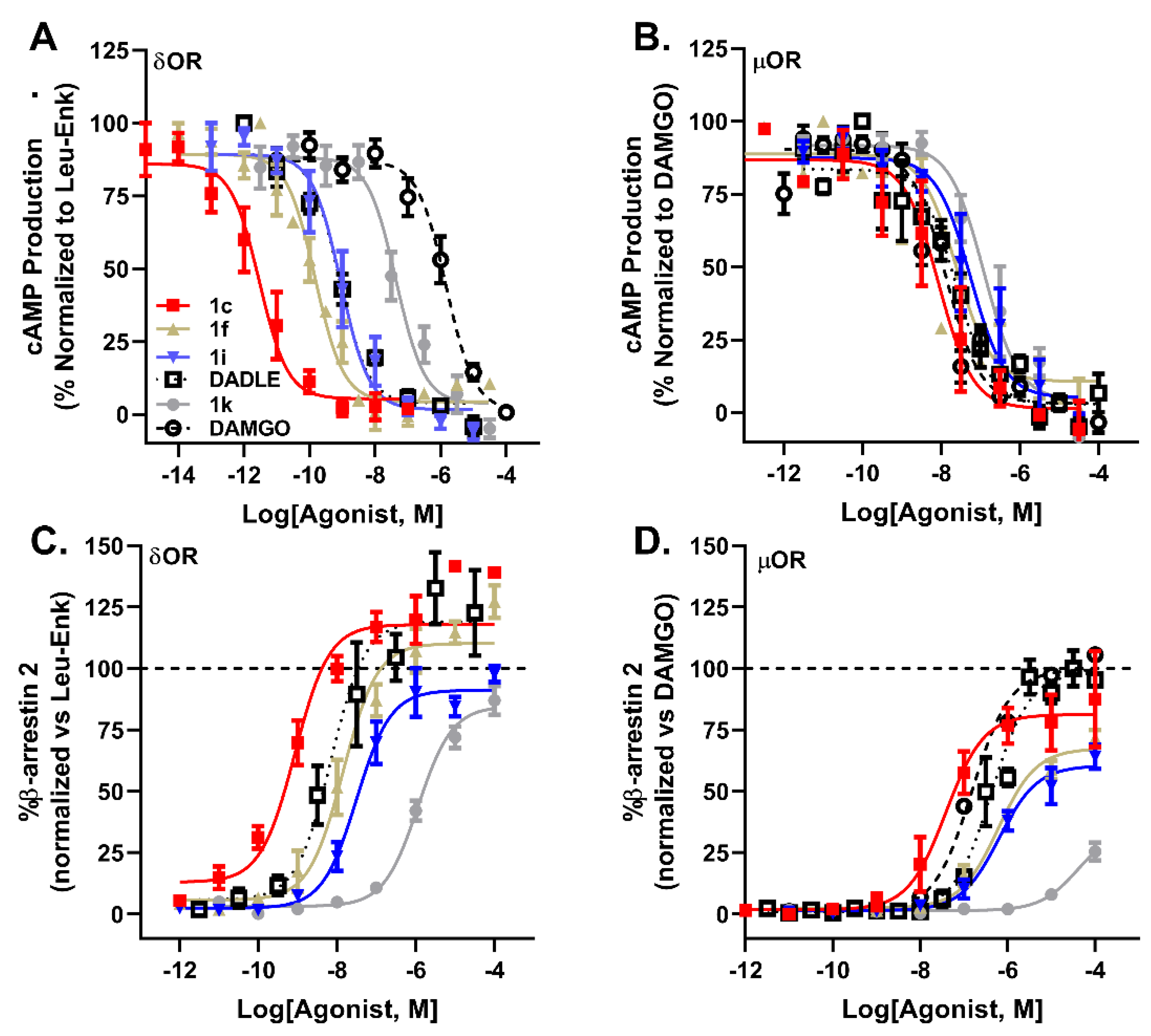
2.3. Pharmacological Characterization

To characterize our substituted analogs, we assessed binding affinity by competition radioligand binding, G protein potency and efficacy using a cAMP GloSensor assay and β-arrestin 2 recruitment via PathHunter assays at both the δOR and µOR. Using Leu5-enkephalin as well as DAMGO (for µOR), as reference compounds, substitution of the meta position of Phe4 (1a1i) generally increased binding affinity for the δOR (Table 1, Ki = 0.023–0.93 nM; Leu5-enkephalin = 1.26 nM) and µOR (Ki = 0.059–0.98 nM; Leu5-enkephalin = 1.7 nM). The improved binding affinity correlated with improved functional activation of the G protein pathway at both the δOR and µOR, while providing near-full agonist activity at the δOR (Table S1, 92–100% efficacy) relative to Leu5-enkephalin, and mostly near-full agonist activities at the µOR (Table S1, 85–105% efficacy; Leu5-enkephalin = 100% efficacy vs. DAMGO).
At the δOR, meta-substituted analogs recruited β-arrestin 2 with a range of potencies (Table 2, EC50: 0.56–49 nM; Leu5-enkephalin = 8.9 nM) with near-full efficacies (Table 2, 91–130%; Leu-enkephalin = 100%), and likewise at the µOR, analogs recruited β-arrestin 2 with a broad range of potencies (Table 2, EC50: 36–589 nM; Leu-enkephalin = 977 nM) and efficacies (Table 2, 60–96% relative to DAMGO; Leu-enkephalin = 60%; Figure 4). Interestingly, DADLE recruited β-arrestin 2 more efficaciously than Leu5-enkephalin at both δOR (126%) and µOR (99%). Picoline analogs 1j1l, generally showed reduced affinity at both receptors (Table 1, δOR Ki = 6.2–33 nM; µOR Ki = 9–158 nM), which further correlated with decreased G protein activation of the receptors (Table 2, δOR IC50 = 4.6–48 nM; µOR IC50 = 41–302 nM). At the δOR, the low affinity of pyridyl-substituted analogs correlated with low β-arrestin 2 recruitment (Table 2, EC50 = 100–1122 nM; 84–98% efficacy), though these substitutions drastically affected β-arrestin 2 recruitment though the µOR (Table 2, EC50 = 1.3–41.6 µM; 36–70% efficacy).
Within these overall trends, specific analogs show unique profiles. Meta− chlorination, −bromination, or −iodination (1b1d) produced high-affinity analogs of Leu5-enkephalin that super-recruited β-arrestin 2 and that drastically increased functional selectivity (Table 3). These increases in affinity at δOR linearly correlate (R2 = 0.998) with the atomic van der Waals radii (H, 1.20 Å; Cl, 1.77 Å; Br, 1.92 Å; I, 2.06 Å) [36], and might suggest a halogen bonding interaction similar to those previously explored at the ortho position of Phe4 [28]. However, the distinct vectors at which a sigma-hole would present from the ortho vs. meta positions suggest that the specific residues engaged through meta-substitution are likely distinct from those previously identified through ortho-substitution [28], and future computational collaborative work might enable a better understanding of the unique binding interactions between the δOR and meta-halogenated analogs of Leu5-enkephalin. Notably, the meta-chlorinated and −brominated analogs (1b, and 1c) are 500–900-fold more potent in cAMP assays at δOR than at µOR, despite exhibiting little differences in binding affinity at δOR relative to µOR (Table 1). Interestingly, the meta-Cl analog (1b) had stronger bias towards G protein-signaling at δOR (Table 3, bias factor 1.6), but towards β-arrestin 2 recruitment at µOR relative to Leu5-enkephalin (Table 3, bias factor 0.004) as well as DAMGO (Table 3, bias factor 0.3), which provides a unique pharmacological profile for future uses. In contrast, meta-F and −CN and −NO2-substitutions (1a,h,i) improved δOR functional selectivity (Table 3), but lost β-arrestin 2 potency at δOR relative to Leu5-enkephalin. Additionally, meta-OMe and −NO2 substitution (1f, 1i) provided both potent and biased analogs, with G protein coupling activities comparable to Leu5-enkephalin (Table 2, IC50 = 0.14–0.47 nM vs. 1.02 nM), but with improved bias factors relative to Leu5-enkephalin at both δOR (Table 3, bias factor 6.9–7.5) and µOR (Table 3, bias factor 8.4–11.1). Of these two analogs, the -NO2-substituted analog 1i exhibited higher δOR functional G protein selectivity (Table 3, 104-fold δOR selectivity) relative to the −OMe analog 1f (Table 3, 58-fold δOR selectivity). Though pyridyl-substituted analogs (1j1l) showed poor potency and efficacy for the δOR and µOR relative to Leu5-enkephalin (Table 2), the 3- and 4-pyridyl analogs (1k1l) showed strong bias at µOR (Table 3, bias factor 17–331), when compared to the full agonist DAMGO. However, if instead of DAMGO, Leu5-enkephalin was used as the reference compound, most analogs, with the exception of 1k and 1l lost G protein bias (Table 3, bias factor < 1), because the analogs generally were more potent and efficacious than Leu5-enkephalin in recruiting β-arrestin 2 at µOR (Table 2), with the exception of 1k (Table 2, 36% recruitment efficacy). Given that exogenous Leu5-enkephalin analogs in vivo would compete with endogenous Leu5-enkephalin and not DAMGO, our results highlight the limitations of interpreting bias factors (particularly using an unnatural compound, such as DAMGO, as a reference) and the associated risk of using bias factor as a major driver of lead optimization.
Compared to DADLE, the Leu5-enkephalin analogs 1b and 1c displayed a similar signaling bias profile, but were more potent and selective for δOR, and thus may provide a better tool compound to target δORs for the treatment of cardiac ischemia. However, compounds 1b and 1c are also more potent than DADLE at recruiting β-arrestin 2 to µOR, resulting in a lower bias factor (Figure 4). In contrast, analog 1i has a favorable pharmacology relative to DADLE, with similar selectivity as 1c, but improved G-protein bias at δOR and µOR relative to DADLE. Given the potential adverse effects associated with strong µOR-mediated β-arrestin 2 recruitment, analog 1k is potentially useful, as it exhibited weak β-arrestin 2 recruitment (36% efficacy) and concomitant strong bias factor for G-protein signaling. However, cAMP potency for 1k at δOR was more than one log unit weaker than DADLE, and thus it may be necessary further optimize the ligand by increasing δOR potency in the cAMP assay, while retaining the low µOR β-arrestin 2 recruitment efficacy (Figure 4).

2.4. Stability

The stability of all compounds to Sprague Dawley rat plasma was assessed to study the influence of the meta− and picoline/pyridine-substitutions at Phe4 relative to Leu5-enkephalin (Table 4). For this parent compound, the predominant known routes of metabolism and clearance occur through cleavage of Tyr1-Gly2 by aminopeptidase N [37,38], and of Gly3-Phe4 by angiotensin converting enzyme [39], and combined, the plasma metabolism occurs with a half-life (t1/2) of < 10 min. In general, meta substituted Phe4 analogs exhibited improved plasma stability compared with Leu5-enkephalin with half-lives typically >20 min. The 3-fluoro derivative (1a) was the most stable analog with a half-life of 82.3 min. From UPLC-mass spectrometry analysis of degradation fragments, meta-substitution did not greatly impede the proteolysis at the Tyr1-Gly2 site, but instead slowed digestion at the Gly3-Phe4 site (Table 4). Thus, the improved stability of our Phe4-substituted analogs presumably derived from perturbation/deceleration of angiotensin-converting enzyme activity. Picoline peptides also displayed improved stability though interestingly, UPLC-mass spectrometry analysis indicated that degradation of all pyridyl-substituted analogs (1j1l) predominantly occurred through Tyr1-Gly2 as opposed to meta-substituted analogs 1a1i that degraded through cleavage of Gly3-Phe4.

3. Methods

3.1. Synthetic Chemistry-General Considerations

Unless specified, all chemicals were purchased from commercial sources and used without further purification. All solvents used for synthesis were of analytical grade and used without further purification. Proton nuclear magnetic resonance (1H-NMR) spectra, and carbon nuclear magnetic resonance (13C-NMR) spectra were recorded on a Bruker AVIII 500 AVANCE spectrometer with a CPDUL cryoprobe (500 and 126 MHz, respectively) or a Bruker DRX 500 spectrometer (500 and 126 MHz, respectively). Fluorine nuclear magnetic resonance (19F-NMR) spectra were recorded on a Bruker AVIII 500 AVANCE spectrometer with a CPDUL BBFO cryoprobe (376 MHz) or a Bruker DRX 500 spectrometer (376 MHz). Chemical shifts (δ) for protons are reported in parts per million (ppm) downfield from tetramethylsilane, and are referenced to proton resonance of solvent residual peak in the NMR solvent (MeOD-d4: δ = 4.87 ppm or DMSO-d6: δ = 2.50 ppm). Chemical shifts (δ) for carbon are reported in ppm downfield from tetramethylsilane, and are referenced to the carbon resonances of the solvent residual peak (MeOD-d4: δ = 49.1 ppm or DMSO-d6: δ = 39.52 ppm). 19F-NMR chemical shifts (δ) are reported in ppm with respect to added internal standard: PhCF3 (δ = −63.72 ppm). NMR data are represented as follows: chemical shift (ppm), multiplicity (s = singlet, d = doublet, dd = doublet of doublets, t = triplet, q = quartet, sept = septet, m = multiplet), coupling constant in Hertz (Hz), and integration. Exact mass determinations were obtained by using electrospray ionization using a time of flight mass analyzer (Waters LCT Premiere).
Peptides were synthesized using an Aapptec Focus XC automated peptide synthesizer coupled with a heating system using a solid phase peptide synthesis protocol using Fmoc chemistry. Preparative RP-HPLC was performed using an appropriate column and solvent system (described below) and final purity of peptides was determined by UV area % from UPLC analysis. Peptides were purified by Teledyne ISCO EZ Prep system on RediSep® C18 Prep column (30 × 250 mm, 100 Å). Purity analysis of final peptides was carried out using a Waters UPLC Aquity system equipped with a PDA eλ detector (200 to 400 nm) and a HSS T3 C18 column, (1.8 μM, 2.1 × 50 mm column), using one of two methods. Protocol A: gradient elution of 2% MeCN with 0.1% formic acid in H2O to 98% MeCN over 2.5 min, then holding at 98% MeCN for 3 min at a flow rate of 0.6 mL/min. Protocol B: gradient elution of 2% MeCN with 0.1% formic acid in H2O to 98% MeCN over 2.5 min, then holding at 98% MeCN for 1 min at a flow rate of 0.7 mL/min at 40 °C. Plasma stability was also assessed using the above UPLC column and solvent gradient program using a Waters H class Plus Acquity UPLC system coupled with a QDa detector using protocol B.

3.2. Synthesis of Peptides

Peptides were synthesized using a solid phase peptide synthesis protocol using an Aapptec Focus XC automated peptide synthesizer coupled with a heating system using the Fmoc chemistry and Wang resin as solid support [34]. To prepare the resin for synthesis, a reaction vessel equipped with a sintered glass bottom was charged with Fmoc-Leu-Wang resin (0.2 mM), and swelled in a mixture of dichloromethane and DMF (1:1) for 15 min. The resin was then transferred to a peptide synthesizer reaction vessel. The resin was deprotected twice using 20% piperidine in DMF for 5 min at 70 °C. Subsequently, an Fmoc-protected amino acid was double coupled with the Leucine-wang resin by treating with N,N′-diisopropylcarbodiimide (3.0 equiv., 0.2 M in DMF) and Oxyma (3.0 equiv., 0.2 M in DMF) at 70 °C for 8 min. Completion of coupling reactions was monitored by a Kaiser’s test for the initial peptide [40]. Each coupling was followed by removal of the Fmoc group using 20% piperidine in DMF at 70 °C for 5 min and repeated once. The cycle of the Fmoc removal and coupling was repeated with subsequent Fmoc-protected amino acids to generate the desired resin-bound peptide. Cleavage of the peptide from resin and concomitant deprotection of the side chain protecting groups was carried out by shaking in TFA/triisopropylsilane/H20 (95/2.5/2.5; 5 mL), at ambient temperature for 3 h. Subsequent filtration afforded the peptide in the filtrate and the volume was reduced to 0.2 mL. Then, the crude peptides were precipitated by adding cold diethyl ether, and the crude peptides were then purified by RP-HPLC. Synthesized peptides were characterized by NMR, and the high-resolution mass spectroscopy.

3.3. Materials Used in the Cellular Signaling Assays

Leu5-enkephalin and forskolin, were purchased from Sigma-Aldrich (St. Louis, MO USA). (d-Ala2, N-MePhe4, Gly-ol) enkephalin (DAMGO) was purchased from Tocris Bioscience (Minneapolis, MN, USA). Radiolabels were from Perkin Elmer (Waltham, MA, USA).

3.4. Cell Culture and Biased Signaling Assays

cAMP inhibition and β-arrestin 2 recruitment assays were performed as previously described [12]. In brief, for cAMP inhibition assays HEK 293 (Life Technologies, Grand Island, NY, USA) cells were transiently transfected in a 1:3 ratio with FLAG-mouse δOR, or HA-mouse µOR and pGloSensor22F-cAMP plasmids (Promega, Madison, WI, USA) using Xtremegene9 (Sigma). Two days post-transfection cells (20,000 cells/well, 7.5 µL) were seeded in low volume Greiner 384-well plates (#82051-458, VWR, Batavia, IL, USA) and were incubated with Glosensor reagent (Promega, 7.5 µL, 2% final concentration) for 90 min at room temperature. Cells were stimulated with 5 µL drug solution for 20 min at room temperature prior to stimulation with 5 µL forskolin (final concentration 30 µM) for an additional 15 min at room temperature. For β-arrestin recruitment assays, CHO-human µOR PathHunter β-arrestin 2 cells or CHO-human δOR PathHunter β-arrestin 2 cells (DiscoverX, Fremont, CA, USA) were plated (2,500 cells/well, 10 µL) one day prior to stimulation with 2.5 µL drug solution for 90 min at 37 °C/5% CO2, after which cells were incubated with 6 µL cell PathHunter assay buffer (DiscoverX) for 60 min at room temperature as per the manufacturer’s protocol. Luminescence for each of these assays was measured using a FlexStation3 plate reader (Molecular Devices, Sunnyvale, CA, USA).

3.5. Radioligand Binding Assay

For the binding assay 50 µL of a dilution series of peptide was added to 50 µL of 3.3 nM [3H]DPDPE (Kd = 3.87 nM) or 2.35 nM of [3H]DAMGO (Kd = 1.07 nM) in a clear 96 well plate. Next, 100 µL of membrane suspension containing 7 µg protein was added to the agonist wells and incubated for 90 min at room temperature. The reaction mixture was then filtered over a GF-B filter plate (Perkin Elmer) followed by four quick washes with ice-cold 50 mM Tris HCl. The plate was dried overnight, after which 50 µL scintillation fluid (Ultimagold uLLT) was added and radioactivity was counted on a Packard TopCount NXT scintillation counter. All working solutions were prepared in a radioligand assay buffer containing 50 mM Tris HCl, 10 mM MgCl2, and 1 mM ethylenediaminetetraacetic acid at pH 7.4.

3.6. Calculation of Bias Factor

Bias factors were calculated using the operational model equation in Prism 8 to calculate Log R (τ/KA) (Table S2) as previously described [12]. Subsequently, bias factors were calculated using Leu5-enkephalin as reference compound for δOR and using either DAMGO or Leu5-enkephalin as reference compound for µOR, respectively. Leu5-enkephalin and DAMGO were more potent in the cAMP (G protein) assay than in the β-Arrestin 2 recruitment assay, and thus were not unbiased, but rather G protein-biased to begin with. A bias factor > 1 meant that the agonist was more G protein-biased than the reference compound; A bias factor < 1 meant that the agonist was less G protein-biased than the reference compound.

3.7. Data and Statistical Analysis

All data are presented as means ± standard error of the mean, and analysis was performed using GraphPad Prism 8 software (GraphPad Software, La Jolla, CA). For in vitro assays, nonlinear regression was conducted to determine pIC50 (cAMP) or pEC50 (β-arrestin 2 recruitment). Technical replicates were used to ensure the reliability of single values, specifically each data point for binding and β-arrestin recruitment was run in duplicate, and for the cAMP assay in triplicate. The averages of each independent run were counted as a single experiment and combined to provide a composite curve in favor of providing a ‘representative’ curve. In each experimental run, a positive control/reference compound was utilized to allow the data to be normalized and to calculate the log bias value. A minimum of three independent values were obtained for each compound in each of the cellular assays.

3.8. Assessment of Plasma Stability

Sprague Dawley rat plasma containing K2-ethylenediaminetetraacetic acid (Innovative Research, MI, USA) was transferred into 300 µL aliquots and stored at −20 °C until use. The plasma stabilities of Leu5-enkephalin and its synthesized analogs were determined in plasma diluted to 50% with saline (isotonic sodium chloride solution; 0.9% w/v) [41]. An aliquot of the resulting solution (25 μL) was incubated at 37 °C for 15 min before the addition of a solution of a Leu5-enkephalin analog (25 μL of a 100 μM isotonic NaCl solution; 0.9% w/v). After adding analog, an aliquot of the mixture was removed at each indicated time point (0, 5, 10, 15, 20, 30, 60, 120, 240 min) and immediately quenched with 100 μL of a methanol solution containing 20 μM Fmoc-Leu-OH as an internal standard. The resulting peptide solutions were centrifuged at 13,000 rpm for 15 min at 4 °C on tubes equipped with 2000 MW filtrate tubes (Sartorius, USA). Then, the filtrate was transferred into vials and a 5 μL sample of the resulting solution was analyzed on an UPLC system coupled with a QDa detector. For quantitative determination, area-under-the-curve for the peaks corresponding to the UV chromatogram was measured for both the Leu5-enkephalin analog and the internal standard. Determination of half-life (t1/2) was carried out by using the GraphPad Prism one-phase decay method.

4. Conclusions

Meta-substitutions of Phe4 of Leu5-enkephalin were generally well tolerated and certain substitutions improved affinity, potency, δOR selectivity and stability of this endogenous opioid. The generated pharmacological data herein may aid computational modeling efforts to reveal ligand-receptor interactions at δOR and µOR that will guide the development of novel peptides with tuned selectivity and signaling profiles. The novel Leu5-enkephalin analogs have superior δOR selectivity over µOR relative to DADLE, the gold-standard peptide for studying the role of δOR in cardiac ischemia. Additionally, the analogs generally have lower β-arrestin recruitment efficacy at µOR, which could further reduce potential adverse in vivo effects. Finally, relative to DADLE, the meta-substituents tune the bias profile at δOR (either more or less biased towards G-protein signaling), and the resulting tool compounds should be useful for investigating the importance of δOR mediated β-arrestin signaling in the peptides cardioprotective effects.

Supplementary Materials

The following are available online, Specifically, for compounds 1a–l: Additional pharmacological characterization, stability data, NMR spectra and characterization data for peptides, UPLC traces for determining purity. Table S1: Efficacy of cAMP inhibition of Meta-substituted Phe4 Analogs of Leu5-enkephalin at δOR and µOR. Table S2: LogR values for Meta-substituted Phe4 Analogs of Leu-enkephalin at δOR and µOR in the cAMP and β-arrestin 2 (β-arr2) assays. 95% confidence intervals are presented between parentheses.

Author Contributions

R.J.C. data acquisition and analysis, supervision and original draft preparation. K.K.S. design, synthesis, and analysis of analogs, original draft preparation. H.S. data acquisition and analysis. B.R.C. data acquisition and analysis. H.C. data acquisition and analysis. K.L.M. data acquisition and analysis. A.T.B. data acquisition and analysis. R.A.A. design and conceptualization, data analysis, original draft preparation, funding acquisition and supervision. R.M.v.R.: design and conceptualization, data analysis, original draft preparation, funding acquisition and supervision. All authors proofread and approved the final draft.

Funding

This research was funded by the National Institute on Alcohol Abuse and Alcoholism grants AA025368, AA026949, AA026675 (R.M.v.R.) and the National Institute on Drug Abuse grants DA045897 (R.M.v.R.) and DA036730 (R.A.A.) of the National Institutes of Health, and funds provided by the Purdue Institute for Drug Discovery and the Purdue University Department of Medicinal Chemistry and Molecular Pharmacology. NMR Instrumentation was provided by NIH Shared Instrumentation Grants (S10OD016360 and S10RR024664), NSF Major Research Instrumentation Grants (9977422 and 0320648), and an NIH Center Grant (P20GM103418). The APC was funded by DA036730 (R.A.A.).

Conflicts of Interest

All authors declare no biomedical financial interests or potential conflicts of interests.

References

  1. Hughes, J.; Smith, T.W.; Kosterlitz, H.W.; Fothergill, L.A.; Morgan, B.A.; Morris, H.R. Identification of two related pentapeptides from the brain with potent opiate agonist activity. Nature 1975, 258, 577–580. [Google Scholar] [CrossRef]
  2. Comb, M.; Seeburg, P.H.; Adelman, J.; Eiden, L.; Herbert, E. Primary structure of the human Met- and Leu-enkephalin precursor and its mRNA. Nature 1982, 295, 663–666. [Google Scholar] [CrossRef]
  3. Ikeda, Y.; Nakao, K.; Yoshimasa, T.; Yanaihara, N.; Numa, S.; Imura, H. Existence of Met-enkephalin-Arg6-Gly7-Leu8 with Met-enkephalin, Leu-enkephalin and Met-enkephalin-Arg6-Phe7 in the brain of guinea pig, rat and golden hamster. Biochem. Biophys. Res. Commun. 1982, 107, 656–662. [Google Scholar] [CrossRef]
  4. Simantov, R.; Kuhar, M.J.; Pasternak, G.W.; Snyder, S.H. The regional distribution of a morphine-like factors enkephalin in monkey brain. Brain Res. 1976, 106, 189–197. [Google Scholar] [CrossRef]
  5. Akil, H.; Owens, C.; Gutstein, H.; Taylor, L.; Curran, E.; Watson, S. Endogenous opioids: Overview and current issues. Drug Alcohol Depend. 1998, 51, 127–140. [Google Scholar] [CrossRef]
  6. Raynor, K.; Kong, H.; Chen, Y.; Yasuda, K.; Yu, L.; Bell, G.I.; Reisine, T. Pharmacological characterization of the cloned kappa-, delta-, and mu-opioid receptors. Mol. Pharmacol. 1994, 45, 330–334. [Google Scholar]
  7. Belluzzi, J.D.; Grant, N.; Garsky, V.; Sarantakis, D.; Wise, C.D.; Stein, L. Analgesia induced in vivo by central administration of enkephalin in rat. Nature 1976, 260, 625–626. [Google Scholar] [CrossRef]
  8. Schaz, K.; Stock, G.; Simon, W.; Schlor, K.H.; Unger, T.; Rockhold, R.; Ganten, D. Enkephalin effects on blood pressure, heart rate, and baroreceptor reflex. Hypertens. (Dallas, Tex. 1979) 1980, 2, 395–407. [Google Scholar] [CrossRef] [Green Version]
  9. Violin, J.D.; Crombie, A.L.; Soergel, D.G.; Lark, M.W. Biased ligands at G-protein-coupled receptors: Promise and progress. Trends Pharmacol. Sci. 2014, 35, 308–316. [Google Scholar] [CrossRef]
  10. Vicente-Sanchez, A.; Dripps, I.J.; Tipton, A.F.; Akbari, H.; Akbari, A.; Jutkiewicz, E.M.; Pradhan, A.A. Tolerance to high-internalizing delta opioid receptor agonist is critically mediated by arrestin 2. Br. J. Pharmacol. 2018, 175, 3050–3059. [Google Scholar] [CrossRef]
  11. Robins, M.T.; Chiang, T.; Mores, K.L.; Alongkronrusmee, D.; van Rijn, R.M. Critical Role for Gi/o-Protein Activity in the Dorsal Striatum in the Reduction of Voluntary Alcohol Intake in C57Bl/6 Mice. Front. Psychiatry 2018, 9, 112. [Google Scholar] [CrossRef] [Green Version]
  12. Chiang, T.; Sansuk, K.; Van Rijn, R.M. β-Arrestin 2 dependence of δ opioid receptor agonists is correlated with alcohol intake. Br. J. Pharmacol. 2016, 173, 332–343. [Google Scholar] [CrossRef] [Green Version]
  13. Remesic, M.; Lee, Y.S.; Hruby, V.J. Cyclic Opioid Peptides. Curr. Med. Chem. 2016, 23, 1288–1303. [Google Scholar] [CrossRef] [Green Version]
  14. Zhang, J.; Ferguson, S.S.G.; Barak, L.S.; Bodduluri, S.R.; Laporte, S.A.; Law, P.-Y.; Caron, M.G. Role for G protein-coupled receptor kinase in agonist-specific regulation of -opioid receptor responsiveness. Proc. Natl. Acad. Sci. 1998, 95, 7157–7162. [Google Scholar] [CrossRef] [Green Version]
  15. Borlongan, C.V. Delta opioid peptide (D-ALA 2, D-LEU 5) enkephalin: Linking hibernation and neuroprotection. Front. Biosci. 2004, 9, 3392. [Google Scholar] [CrossRef] [Green Version]
  16. Wolf, A.; Lusczek, E.R.; Beilman, G.J. Hibernation-Based Approaches in the Treatment of Hemorrhagic Shock. SHOCK 2018, 50, 14–23. [Google Scholar] [CrossRef]
  17. Uchiyama, T.; Kotani, A.; Kishida, T.; Tatsumi, H.; Okamoto, A.; Fujita, T.; Murakami, M.; Muranishi, S.; Yamamoto, A. Effects of various protease inhibitors on the stability and permeability of [D-Ala2,D-Leu5]enkephalin in the rat intestine: Comparison with leucine enkephalin. J. Pharm. Sci. 1998, 87, 448–452. [Google Scholar] [CrossRef]
  18. Beddell, C.R.; Clark, R.B.; Lowe, L.A.; Wilkinson, S.; Chang, K.J.; Cuatrecasas, P.; Miller, R. A conformational analysis for leucine-enkephalin using activity and binding data of synthetic analogues. Br. J. Pharmacol. 1977, 61, 351–356. [Google Scholar] [CrossRef] [Green Version]
  19. Kovoor, A.; Nappey, V.; Kieffer, B.L.; Chavkin, C. μ and δ Opioid Receptors Are Differentially Desensitized by the Coexpression of β-Adrenergic Receptor Kinase 2 and β-Arrestin 2 in Xenopus Oocytes. J. Biol. Chem. 1997, 272, 27605–27611. [Google Scholar] [CrossRef] [Green Version]
  20. Bohn, L.M.; Lefkowitz, R.J.; Gainetdinov, R.R.; Peppel, K.; Caron, M.G.; Lin, F.T. Enhanced morphine analgesia in mice lacking beta-arrestin 2. Science 1999, 286, 2495–2498. [Google Scholar] [CrossRef]
  21. Whistler, J.L.; Chuang, H.; Chu, P.; Jan, L.Y.; von Zastrow, M. Functional Dissociation of μ Opioid Receptor Signaling and Endocytosis. Neuron 1999, 23, 737–746. [Google Scholar] [CrossRef] [Green Version]
  22. Cassell, R.J.; Mores, K.L.; Zerfas, B.L.; Mahmoud, A.H.; Lill, M.A.; Trader, D.J.; van Rijn, R.M. Rubiscolins are naturally occurring G protein-biased delta opioid receptor peptides. Eur. Neuropsychopharmacol. 2019, 29, 450–456. [Google Scholar] [CrossRef]
  23. Bella Ndong, D.; Blais, V.; Holleran, B.J.; Proteau-Gagné, A.; Cantin-Savoie, I.; Robert, W.; Nadon, J.-F.; Beauchemin, S.; Leduc, R.; Piñeyro, G.; et al. Exploration of the fifth position of leu-enkephalin and its role in binding and activating delta (DOP) and mu (MOP) opioid receptors. Pept. Sci. 2018, 111, e24070. [Google Scholar] [CrossRef]
  24. Vezzi, V.; Onaran, H.O.; Molinari, P.; Guerrini, R.; Balboni, G.; Calo, G.; Costa, T.; Calò, G.; Costa, T. Ligands raise the constraint that limits constitutive activation in G protein-coupled opioid receptors. J. Biol. Chem. 2013, 288, 23964–23978. [Google Scholar] [CrossRef] [Green Version]
  25. Aguila, B.; Coulbault, L.; Boulouard, M.; Leveille, F.; Davis, A.; Toth, G.; Borsodi, A.; Balboni, G.; Salvadori, S.; Jauzac, P.; et al. In vitro and in vivo pharmacological profile of UFP-512, a novel selective delta-opioid receptor agonist; correlations between desensitization and tolerance. Br. J. Pharmacol. 2007, 152, 1312–1324. [Google Scholar] [CrossRef] [Green Version]
  26. Raehal, K.M.; Walker, J.K.L.; Bohn, L.M. Morphine Side Effects in β-Arrestin 2 Knockout Mice. J. Pharmacol. Exp. Ther. 2005, 314, 1195–1201. [Google Scholar] [CrossRef] [Green Version]
  27. Soergel, D.G.; Subach, R.A.; Burnham, N.; Lark, M.W.; James, I.E.; Sadler, B.M.; Skobieranda, F.; Violin, J.D.; Webster, L.R. Biased agonism of the μ-opioid receptor by TRV130 increases analgesia and reduces on-target adverse effects versus morphine: A randomized, double-blind, placebo-controlled, crossover study in healthy volunteers. Pain 2014, 155, 1829–1835. [Google Scholar] [CrossRef]
  28. Rosa, M.; Caltabiano, G.; Barreto-Valer, K.; Gonzalez-Nunez, V.; Gómez-Tamayo, J.C.; Ardá, A.; Jiménez-Barbero, J.; Pardo, L.; Rodríguez, R.E.; Arsequell, G.; et al. Modulation of the Interaction between a Peptide Ligand and a G Protein-Coupled Receptor by Halogen Atoms. ACS Med. Chem. Lett. 2015, 6, 872–876. [Google Scholar] [CrossRef] [Green Version]
  29. Toth, G.; Kramer, T.H.; Knapp, R.; Lui, G.; Davis, P.; Burks, T.F.; Yamamura, H.I.; Hruby, V.J. [D-Pen2,D-Pen5]enkephalin analogues with increased affinity and selectivity for delta opioid receptors. J. Med. Chem. 1990, 33, 249–253. [Google Scholar] [CrossRef]
  30. Weber, S.J.; Greene, D.L.; Sharma, S.D.; Yamamura, H.I.; Kramer, T.H.; Burks, T.F.; Hruby, V.J.; Hersh, L.B.; Davis, T.P. Distribution and analgesia of [3H][D-Pen2, D-Pen5]enkephalin and two halogenated analogs after intravenous administration. J. Pharmacol. Exp. Ther. 1991, 259, 1109–1117. [Google Scholar]
  31. Weber, S.J.; Greene, D.L.; Hruby, V.J.; Yamamura, H.I.; Porreca, F.; Davis, T.P. Whole body and brain distribution of [3H]cyclic [D-Pen2,D-Pen5] enkephalin after intraperitoneal, intravenous, oral and subcutaneous administration. J. Pharmacol. Exp. Ther. 1992, 263, 1308–1316. [Google Scholar] [PubMed]
  32. Grant Liska, M.; Crowley, M.G.; Lippert, T.; Corey, S.; Borlongan, C.V. Delta Opioid Receptor and Peptide: A Dynamic Therapy for Stroke and Other Neurological Disorders. In Handbook of Experimental Pharmacology, 1st ed.; Emily, J., Ed.; Springer: Cham, Switzerland, 2017; pp. 277–299. [Google Scholar]
  33. He, X.; Sandhu, H.K.; Yang, Y.; Hua, F.; Belser, N.; Kim, D.H.; Xia, Y. Neuroprotection against hypoxia/ischemia: δ-opioid receptor-mediated cellular/molecular events. Cell. Mol. Life Sci. 2013, 70, 2291–2303. [Google Scholar] [CrossRef]
  34. Shenmar, K.; Sharma, K.K.; Wangoo, N.; Maurya, I.K.; Kumar, V.; Khan, S.I.; Jacob, M.R.; Tikoo, K.; Jain, R. Synthesis, stability and mechanistic studies of potent anticryptococcal hexapeptides. Eur. J. Med. Chem. 2017, 132, 192–203. [Google Scholar] [CrossRef] [Green Version]
  35. Mahindra, A.; Sharma, K.K.; Jain, R. Rapid microwave-assisted solution-phase peptide synthesis. Tetrahedron Lett. 2012, 53, 6931–6935. [Google Scholar] [CrossRef]
  36. Bondi, A. van der Waals Volumes and Radii. J. Phys. Chem. 1964, 68, 441–451. [Google Scholar] [CrossRef]
  37. Weinberger, S.B.; Martinez, J.L.J. Characterization of Hydrolysis of [ Leu ] enkephalin in Rat Plasma. J. Pharmacol. Exp. Ther. 1988, 247, 129–135. [Google Scholar]
  38. Roques, B.; Noble, F. Dual inhibitors of enkephalin-degrading enzymes (neutral endopeptidase 24.11 and aminopeptidase N) as potential new medications in the management of pain and opioid addiction. NIDA Res. Monogr. 1995, 147, 104–145. [Google Scholar]
  39. Suzanne, E.T.; Kenneth, L.A. Leucine-Enkephalin Metabolism in Brain Microvessel Endothelial Cells. Peptides 1994, 15, 109–116. [Google Scholar]
  40. Sarin, V.K.; Kent, S.B.; Tam, J.P.; Merrifield, R.B. Quantitative monitoring of solid-phase peptide synthesis by the ninhydrin reaction. Anal. Biochem. 1981, 117, 147–157. [Google Scholar] [CrossRef]
  41. Beaudeau, J.L.; Blais, V.; Holleran, B.J.; Bergeron, A.; Pineyro, G.; Guérin, B.; Gendron, L.; Dory, Y.L. N-Guanidyl and C-Tetrazole Leu-Enkephalin Derivatives: Efficient Mu and Delta Opioid Receptor Agonists with Improved Pharmacological Properties. ACS Chem. Neurosci. 2019, 10, 1615–1626. [Google Scholar] [CrossRef]
Sample Availability: Upon reasonable request and based on material in hand, compounds 1a1l can be distributed in a timely fashion by Dr. Altman. However, as supplies are finite, we cannot guarantee every request can be accommodated.
Figure 1. Overview of unbiased (Leu5-enkephalin and DADLE) and biased (Aza-β-Homoleucine-Enkephalin, Rubiscolin-5 and -6) δOR peptides.
Molecules 24 04542 g001
Figure 2. Substituents of Phe4 of Leu5-enkephalin Affect Pharmacodynamic, Stability, and Distribution Properties.
Molecules 24 04542 g002
Figure 3. Peptide Synthesis of Phe4-Substituted Analogs of Leu5-enkephalin.
Molecules 24 04542 g003
Figure 4. Modifications of Phe4 of Leu5-enkephalin Produces Analogs with Divergent and Distinct Signaling Profiles. Inhibition of cAMP production by 1c (), 1f (), 1i (), 1k (), Leu5-enkephalin (□) and DAMGO (◯) in HEK cells expressing δOR (A) and µOR (B). Recruitment of β-arrestin 2 by 1c, 1f, 1i, 1k, Leu-Enk and DAMGO in CHO cells expressing δOR (C) and µOR (D).
Molecules 24 04542 g004
Table 1. Meta Substituted Phe4 Analogs of Leu5-enkephalin Increase Affinity at δOR and µOR.
pKi ± SEM (δOR)Ki (nM)pKi ± SEM (µOR)Ki (nM)Binding Selectivity (δOR vs µOR)
1a (F)9.48 ± 0.10.339.12 ± 0.40.762.3
1b (Cl)9.87 ± 0.10.1310.14 ± 0.20.0720.55
1c (Br)10.35 ± 0.20.0459.90 ± 0.30.132.9
1d (I)10.64 ± 0.20.0239.86 ± 0.50.146.1
1e (Me)9.86 ± 0.10.149.86 ± 0.10.141.0
1f (OMe)9.31 ± 0.10.499.07 ± 0.10.851.7
1g (CF3)9.93 ± 0.10.1210.23 ± 0.30.0590.49
1h (CN)9.17 ± 0.30.689.52 ± 0.10.300.44
1i (NO2)9.03 ± 0.20.939.01 ± 0.10.981.05
1j (2-pyr)8.21 ± 0.16.178.04 ± 0.29.121.5
1k (3-pyr)7.48 ± 0.133.16.80 ± 0.11584.8
1l (4-pyr)7.69 ± 0.120.47.41 ± 0.138.91.9
DADLE9.01 ± 0.10.988.80 ± 0.11.581.6
Leu5-enkephalin8.90 ± 0.1 1.268.77 ± 0.11.701.3
DAMGO--9.01 ± 0.10.98-
All compounds were tested in three independent trials.
Table 2. Meta-substituted Phe4 Analogs of Leu5-enkephalin Display Enhanced δOR and µOR Potency for cAMP Inhibition and β-arrestin 2 Recruitment, But Vary in β-arrestin Recruitment Efficacy.
cAMPβ-Arrestin 2
CompoundpIC50 ± SEM (δOR)IC50 (nM)pIC50 ± SEM (µOR)IC50 (nM)pEC50 ± SEM (δOR)EC50 (nM)δOR Efficacy (% ± SEM)pEC50 ± SEM (µOR)EC50 (nM)µOR Efficacy (% + SEM)
1a (F)9.47 ± 0.20.398.10 ± 0.27.948.01 ± 0.29.77107 ± 96.24 ± 0.157575 ± 13
1b (Cl)10.66 ± 0.30.0227.52 ± 0.130.28.77 ± 0.21.70130 ± 107.17 ± 0.267.696 ± 8
1c (Br)10.52 ± 0.30.0308.00 ± 0.4109.25 ± 0.20.56116 ± 77.45 ± 0.235.579 ± 6
1d (I)10.61 ± 0.20.0258.37 ± 0.34.278.95 ± 0.21.12111 ± 77.33 ± 0.246.870 ± 3
1e (Me)10.41 ± 0.30.0398.43 ± 0.33.728.46 ± 0.23.46105 ± 56.73 ± 0.218671 ± 3
1f (OMe)9.84 ± 0.30.148.09 ± 0.38.127.93 ± 0.211.7106 ± 56.23 ± 0.158967 ± 3
1g (CF3)9.97 ± 0.40.118.61 ± 0.42.458.39 ± 0.24.07108 ± 97.25 ± 0.256.271 ± 3
1h (CN)9.46 ± 0.30.357.92 ± 0.312.07.77 ± 0.217.091 ± 56.8 ± 0.215868 ± 4
1i (NO2)9.33 ± 0.30.477.31 ± 0.449.07.31 ± 0.249.096 ± 66.27 ± 0.153760 ± 6
1j (2-pyr)8.34 ± 0.24.577.39 ± 0.440.77.00 ± 0.210092 ± 135.87 ± 0.1134970 ± 4
1k (3-pyr)7.32 ± 0.247.96.63 ± 0.42345.95 ± 0.1112285 ± 64.38 ± 0.14168736 ± 7
1l (4-pyr)7.52 ± 0.230.26.52 ± 0.33026.40 ± 0.239898 ± 64.43 ± 0.13715364 ± 11
DADLE9.09 ± 0.20.817.45 ± 0.135.57.92 ± 0.412.0126 ± 106.33 ± 0.146899 ± 3
Leu5-enkephalin8.99 ± 0.11.027.34 ± 0.245.78.05 ± 0.18.911006.01 ± 0.197761 ± 3
DAMGO5.91 ± 0.212307.82 ± 0.115.1<5 -6.80 ± 0.1158100
Table 3. Meta-substituted Phe4 Analogs of Leu5-enkephalin Display a Range of Selectivity and Bias Profiles.
CompoundG Protein Selectivity (δOR vs µOR)Bias Factor
δORµOR (DG = Ref)µOR (LE = Ref)
1a (F)201.822.20.36
1b (Cl)13723.20.30.004
1c (Br)3331.31.20.02
1d (I)1713.53.20.05
1e (Me)954.311.50.19
1f (OMe)587.58.40.14
1g (CF3)222.04.30.07
1h (CN)346.85.40.09
1i (NO2)1046.911.10.18
1j (2-pyr)8.94.017.20.27
1k (3-pyr)4.96.7331.15.89
1l (4-pyr)102.263.71.08
DADLE440.52.30.035
Leu5-enkephalin4514.91
DAMGO0.012-10.003
Aza-β-homoleucine 9.9 [23]5.2 -1.2
Rubiscolin-5-2.0 [22]--
from reference 26 using BRET (β-arrestin 2) and EPAC (cAMP) assays. DG = DAMGO, LE = Leu5-enkephalin.
Table 4. Rat Plasma Stability of Leu5-enkephalin and Its Analogs.
CompoundHalf-Life (min)95% CIDegradation Products (First Appearance)
Leu5-enkephalin9.46.3−4.5Gly-Gly-Phe-Leu (5 min)
Phe-Leu (10 min)
1a (F)82.368.0–102.6Gly-Gly-(meta-F)Phe-Leu (5 min)
(meta-F)Phe-Leu (60 min)
1b (Cl)37.826.5–56.6Gly-Gly-(meta-Cl)Phe-Leu (5 min)
(meta-Cl)Phe-Leu (30 min)
1c (Br)21.512.7–38.7Gly-Gly-(meta-Br)Phe-Leu (5 min)
(meta-Br)Phe-Leu (15 min)
1d (I)13.211.4–15.2Gly-Gly-(meta-I)Phe-Leu (5 min)
(meta-I)Phe-Leu (15 min)
1e (Me)39.530.0–53.5Gly-Gly-(meta-Me)Phe-Leu (5 min)
(meta-Me)Phe-Leu (20 min)
1f (OMe)46.130.4–75.6Gly-Gly-(meta-OMe)Phe-Leu (5 min)
(meta-OMe)Phe-Leu (20 min)
1g (CF3)44.535.0–59.1Gly-Gly-(meta-CF3)Phe-Leu (5 min)
(meta- CF3)Phe-Leu (30 min)
1h (CN)33.024.0–47.0Gly-Gly-(meta-CN)Phe-Leu (5 min)
(meta-CN)Phe-Leu (20 min)
1i (NO2)28.816.8–55.8Gly-Gly-(meta-NO2)Phe-Leu (5 min)
(meta- NO2)Phe-Leu (20 min)
1j (2-pyr)26.815.4–53.7(2-pyridyl)Ala-Leu (5 min)
Gly-Gly-(2-pyridyl)Ala-Leu (30 min)
1k (3-pyr)54.029.0–127.0(3-pyridyl)Ala-Leu (10 min)
Gly-Gly-(3-pyridyl)Ala-Leu (20 min)
1l (4-pyr)78.147.5–165.4(4-pyridyl)Ala-Leu (10 min)
Gly-Gly-(4-pyridyl)Ala-Leu (30 min)

