Article

First Attempts of the Use of 195Pt NMR of Phenylbenzothiazole Complexes as Spectroscopic Technique for the Cancer Diagnosis

by
Bruna T. L. Pereira
1,
Mateus A. Gonçalves
1,
Daiana T. Mancini
1,
Kamil Kuca
2,* and
Teodorico C. Ramalho
1,2,*
1
Department of Chemistry, Federal University of Lavras, P.O. Box 3037, Lavras 37200-00, MG, Brazil
2
Department of Chemistry, Faculty of Science, University of Hradec Kralove, 500 02 Hradec Kralove, Czech Republic
*
Authors to whom correspondence should be addressed.
Molecules 2019, 24(21), 3970; https://doi.org/10.3390/molecules24213970
Submission received: 30 September 2019 / Revised: 30 October 2019 / Accepted: 31 October 2019 / Published: 2 November 2019
(This article belongs to the Section Computational and Theoretical Chemistry)

Abstract

:
Platinum complexes have been studied for cancer treatment for several decades. Furthermore, another important platinum characteristic is related to its chemical shifts, in which some studies have shown that the 195Pt chemical shifts are very sensitive to the environment, coordination sphere, and oxidation state. Based on this relevant feature, Pt complexes can be proposed as potential probes for NMR spectroscopy, as the chemical shifts values will be different in different tissues (healthy and damaged) Therefore, in this paper, the main goal was to investigate the behavior of Pt chemical shifts in the different environments. Calculations were carried out in vacuum, implicit solvent, and inside the active site of P13K enzyme, which is related with breast cancer, using the density functional theory (DFT) method. Moreover, the investigation of platinum complexes with a selective moiety can contribute to early cancer diagnosis. Accordingly, the Pt complexes selected for this study presented a selective moiety, the 2-(4′aminophenyl)benzothiazole derivative. More specifically, two Pt complexes were used herein: One containing chlorine ligands and one containing water in place of chlorine. Some studies have shown that platinum complexes coordinated to chlorine atoms may suffer hydrolyses inside the cell due to the low chloride ion concentration. Thus, the same calculations were performed for both complexes. The results showed that both complexes presented different chemical shift values in the different proposed environments. Therefore, this paper shows that platinum complexes can be a potential probe in biological systems, and they should be studied not only for cancer treatment, but also for diagnosis.

1. Introduction

Cancer is currently one of the most discussed diseases, as it presents incredibly high mortality rates [1,2]. More specifically, cancer is a term employed for a group of diseases characterized by the acute growth of abnormal cells. Breast cancer is the class with the highest incidence in women, and it is associated with the high mortality rates in women with cancer [3,4,5].
Early-stage diagnosis is very important for cancer treatment, as it can reduce mortality rates and increase treatment possibilities [6,7,8]. Some imaging techniques are used as main approaches in cancer diagnosis, including mammography, magnetic resonance imaging (MRI), positron-emission tomography (PET), and computed tomography (CT). Furthermore, the use of these techniques with the help of biochemical biomarkers, such as proteins, could increase the chances of early diagnosis [3,9,10].
The use of imaging techniques associated with biomarkers as proteins, RNAs, microRNAs, and enzymes could also increase the chances of early diagnosis [3,11,12]. Furthermore, the use of compounds that have already been employed in medicine could potentially probe diseases [3,13,14]. In this context, Mavroid and colleagues synthesized the cis-dichloro(2′pyridinyl)methylamineplatinum(II) bonded to 2-(4′aminophenyl)benzothiazole derivative in order to study its properties as an antitumor agent [13].
This complex has two important moieties in cancer treatment: The platinum and the benzothiazole group. In 1965, the carcinogenic properties of the platinum group were discovered with the emergence of cisplatin, an important drug in chemotherapy [15,16,17]. Since then, different drugs using platinum has been developed to improve the treatment of different types of cancer [16,18,19]. Regarding the benzothiazole moiety, some studies have revealed that these types of molecules show potent and selective antitumor activity in vitro and in vivo against breast cancer cells, among other types of cancer [14,20,21].
Accordingly, the purpose of this work was to use the cis-dichloro(2߰pyridinyl)methylamineplatinum(II) bonded to 2-(4′aminophenyl)benzothiazole derivative (Figure 1) as a potential probe in breast cancer diagnosis. It is also important to mention that this coordinated complex has already been investigated as a potential anticancer agent [13]. Furthermore, the strategy chosen for this work was to perform chemical shift analysis of 195Pt in different chemical environments (vacuum, implicit solvent, and inside active enzyme P13K), which was done through nuclear magnetic resonance (NMR) calculations. These calculations were performed for the monoaquated complex (Figure 2), as it is well-known that platinum complexes are hydrolyzed inside the cell [18,19,22]. Researcher groups have studied different platinum complexes with different ligands, as well as different oxidation numbers. Synthesis, characterization, and NMR analysis were also performed, and it was possible to see the sensitivity of the 195Pt chemical shift [23,24,25,26].

2. Results and Discussion

This discussion is divided into three main parts following the order established in the methodology section. Initially, it is worth mentioning that the validation step of the theoretical methodology was based on another work about NMR platinum complexes developed by Paschoal and collaborators (2017) [27]. In this research, the authors developed a basis set with relativistic correction for Pt(II) chemical shift calculations [27]. Moreover, for the validation step, we used two different methodologies for the platinum atom. In this step, optimization calculations were carried out at B3LYP/lanl2dz, B3LYP/aug-cc-pVTZ-pp, and Zora/B3LYP/Def2-TZVP levels, which is a relativistic method. The results are described in the supplementary material. In the second part, the 195Pt NMR chemical shift for complex 1 (Figure 1) was calculated in the gas phase, in solution, using implicit solvent (PCM) model, and in the P13K enzyme active site. In order to evaluate the thermal effects, the dynamic simulations were also carried out. In the last part, the same calculations were performed for the monoaquated complex (Figure 2).
For NMR chemical shift calculations, the notation was as follows: Level of shielding computation (PBEPBE)//level of geometry optimization (B3LYP) or MD simulation. For example, (PBEPBE//B3LYP) means shielding computation in vacuum//geometry optimization in vacuum, while (PBEPBE/PCM(H2O)//B3LYP/PCM(H2O)) means shielding computation with implicit solvent (PCM)//geometry optimization with implicit solvent (PCM). The same notation was employed when including the dynamic effect (ADMP). For example, (PBEPBE//ADMP) means shielding computation in vacuum//dynamic simulations in vacuum, while (PBEPBEPCM(H2O)//ADMP/PCM(H2O)) means shielding computation with implicit solvent (PCM)//dynamic simulations with implicit solvent (PCM).

2.1. Validation of 195Pt NMR Chemical Shifts Theoretical Methodology

In the study developed by Paschoal and collaborators, a basis set (labeled as NMR-DKH) was developed for 195Pt NMR, which allowed the recovery of relativistic effects. The authors performed several NMR chemical shift calculations for many Pt(II) complexes using different theoretical methodologies. Then, a comparison was made among the three different methodologies [27].
The studied complexes were initially optimized using two different basis sets. After this step, NMR calculations were performed using NMR-DKH basis set, as follows: (PBEPBE/NMR-DKH/IEFPCM(UFF)//B3LYP/LANL2DZ/Def2-TZVPP/IEFPCM(UFF)) (Model 2) and (PBEPBE/NMR-DKH/IEFPCM(UFF)//B3LYP/LANL2DZ/Def2-SVP/IEFPCM(UFF)) (Model 3). Then, a comparison was performed using a Hamiltonian relativistic operator (ZORA), as follows: COSMO-PBE-SO-ZORA/TZ2P (Model 1) [27]. A statistical study using absolute deviation (MAD) and the relative deviation (MRD) were employed for a methodology comparison. The statistical results showed that the MAD and the MRD were 200 ppm and 6% (Model 1), 182 ppm and 6% (Model 2), and 168 ppm and 5% (Model 3), respectively [27].
As a result, the methodology chosen by Paschoal and collaborators showed an excellent agreement between the different models used. In the same way, the theoretical values obtained by relativistic calculations using NMR-DKH basis set agreed with the experimental data [27].

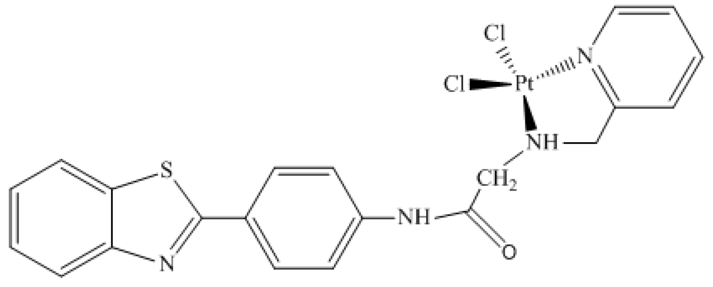
2.2. The 195Pt Nuclear Magnetic Resonance Chemical Shift

The importance of investigating how temperature, coordination, and chemical environment effects influence the chemical shifts is well-known. This investigation is even more important when the goal is to use this NMR parameter to propose a compound as a probe in biological systems. Keeping that in mind, the importance of geometry, chemical environment, solvent, and thermal effects for the 195Pt NMR chemical shift in Complex 1 (Figure 1) were analyzed. The corresponding δ(195Pt) values were collected and are reported in Table 1.
Analyzing the geometry effect on the 195Pt chemical shifts (Table 1), we observed that the δ values revealed a variation of 335.64 ppm when comparing δe(PBEPBE//B3LYP) and δe(PBEPBE//B3LYP/PCM(H2O)). Regarding the chemical shift effects for 195Pt, when comparing the NMR calculations with and without implicit solvent δe(PBEPBE//B3LYP/PCM(H2O)) and δ(PBEPBE/PCM(H2O) //B3LYP/PCM(H2O)), a variation of 512.33 ppm was observed (Table 1). It is worth mentioning that this large difference in chemical shift values for different methodologies (vacuum and implicit solvent) was expected. According to the literature, the platinum chemical shift is very sensitive to geometry, electronic parameters, and chemical environments [28].
In this line, thermal and dynamic effects are other important contributions to be analyzed when studying chemical shifts. When the dynamic effect was included in the gas phase comparing δe(PBEPBE//B3LYP) and δ310K(PBEPBE//ADMP)), the chemical shift value decreased by 295.51 ppm (Table 1). Moreover, when the implicit solvent effect was considered along with the dynamic effect (comparing δe(PBEPBE/PCM[H2O]//B3LYP/PCM[H2O]) and δ310K (PBEPBE/PCM[H2O]//ADMP/PCM[H2O])), the chemical shift value decreased by 2285.76 ppm.
Indeed, the methodology including thermal and dynamic effects with the implicit solvent was the closest approximation to real systems used in this work. In this sense, this methodology was compared to the Pt chemical shift inside the enzyme in the next topic.

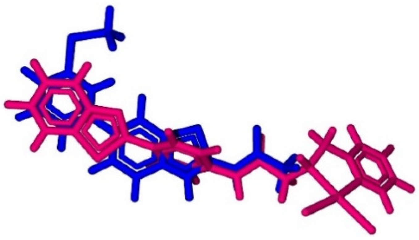
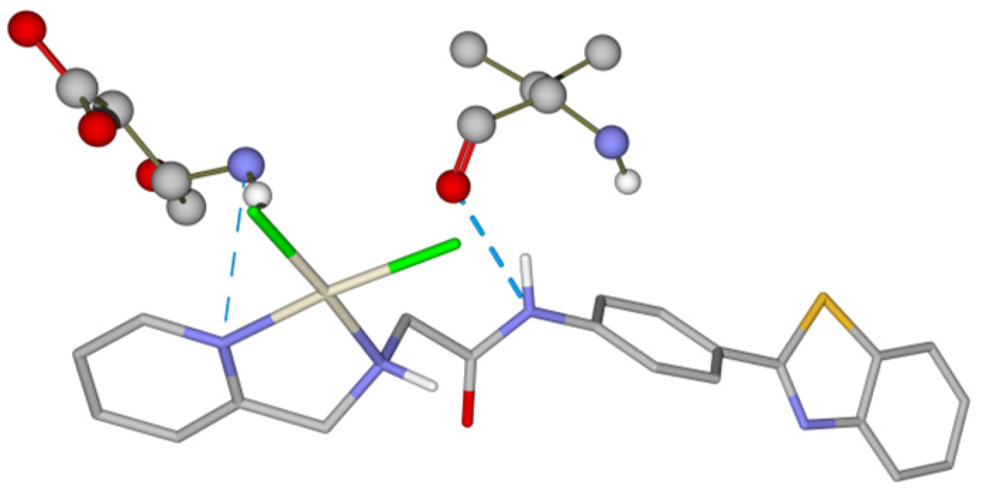
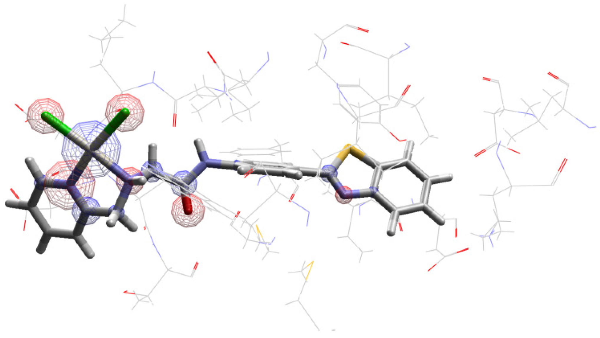
The 195Pt Chemical Shift in PI3K Enzyme Active Site

Docking calculations are a fundamental tool for studies involving ligand and receptor systems [29]. In this work, a docking study was carried out to analyze the 195Pt chemical shift inside the enzyme PI3K, which is related to breast cancer. Then, the overlap between the complex conformations, generated during docking analysis, and the active ligand (PDB ligand) (benzothiazole derived) was performed inside the active site of the enzyme. The best superposition between the two structures is shown in Figure 3.
Furthermore, the complex behavior inside of the receptor active site was also evaluated to check the stability of the complex in this chemical environment. The complex established hydrogen bonds with Val882 and Asp 884 amino acids (Figure 4).
Furthermore, other interactions happened to give stability to the system receptor–ligand, for example, as electrostatic and hydrophobic interactions. In this sense, the electrostatic interactions were also shown in Figure 5.
Accordingly, it can be suggested that the platinum complex is stable inside the active site of the enzyme. The platinum complex formed interactions inside the active site of enzyme and presented an intermolecular interaction energy around −150 kcal mol−1. To validate this methodology, a redocking procedure was performed. However, the docking study is a theoretical research and the preliminary results do not take into account pharmacokinetic proprieties. Therefore, an additional in vitro was necessary test to evaluate the interactions between this platinum complex and P13K enzyme.
To analyze the platinum complex (Figure 1) as a potential probe in biological systems, it was also necessary to perform NMR calculations for the 195Pt chemical shifts of the most stable complex conformation in the active site of P13K enzyme, which resulted in a δ(195Pt) of −1919.70 ppm. The result is in accordance to the expected value. It is well-known that the chemical environment inside the active site is hydrophobic, and the chemical shift should be close to the chemical shift obtained through the calculations without solvent δe (PBEPBE//B3LYP) for the complex (Table 1). Furthermore, the comparison between the chemical shift for the complex after docking and using thermal and dynamic and solvent effects (δ310K (PBEPBE/PCM[H2O]//ADMP/PCM[H2O])) is important to understand the behavior of the complex in different environments. In this sense, the difference found when comparing these both methodologies was 2149.86 ppm.
With that in mind, these results are very interesting for this proposal. The environment inside the protein and the thermal and solvent effects are different, which show the sensitivity of the Pt chemical shift. In addition, platinum complexes are already used in medicine for cancer treatment, which may facilitate its use as a diagnosis compounds.

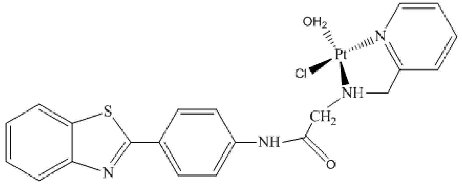
2.3. The 195Pt Chemical Shift for Monoaqua Platinum Complex

Since 1965, when carcinogenic properties of cisplatin were discovered, platinum (II) complexes have been used as important drugs in cancer treatment [15,16,17]. The platinum drugs present activity against cancer due their ability to bind to the N7 atom of guanine from the DNA. An important step in the interaction between platinum and DNA is the aquation of platinum complexes, which is crucial for the following binding step [18,22]. Several studies have shown that the monoaqua cisplatin complex (cis-(Pt[NH3]2[H2O]Cl)+1) is the active hydrolyzed specie at 310 K that binds to DNA, as the diaquo complex (cis-(Pt[NH3]2[H2O]2)+2) is the less likely specie in physiological pH [18].
In this context, it is also important to analyze the behavior of the monoaqua complex as it may be found in this way inside the cell. Thus, to propose platinum complexes as a potential probe, this aspect needs to be considered. In this sense, for monoaqua complex (Figure 2) calculations were performed including thermal and dynamic effects with an implicit solvent (δ310K (PBEPBEPCM[H2O[//ADMP/PCM[H2O])). Therefore, for the study of the monoaqua compound, this methodology was employed for the evaluation of the thermal, dynamic, and solvent effects, and 195Pt chemical shift calculations were also performed inside the active site of the enzyme. Both results were compared to analyze the behavior of spectroscopic parameters (chemical shifts) in these different environments.
The corresponding δ(195Pt) value for the monoaqua platinum complex was −2813.79 ppm. Comparing this result to result for the complex before hydrolyses (Figure 1) (Table 1), a variation in the obtained 195Pt chemical shift values was observed. Therefore, when a water molecule was added in place of a chlorine atom, the respective value increased by 1255.79 ppm. This was expected, as it is known that platinum chemical shifts are sensitive to the coordination sphere.
In this sense, the docking study was performed to better understand the interaction of the monoaqua complex inside the active site of the enzyme. The most important moiety of the complexes for this step is the benzothiazole. The ligand inside the enzyme is derived from benzothiazole, as previously mentioned in the docking topic. From the docking studies, it was observed that the monoaquated complex established hydrogen bonds similarly to what was observed for Complex 1. The associated intermolecular interaction energy was around −140 kcal mol−1.
The 195Pt chemical shift of the most stable complex conformation in the active site of PI3K enzyme was performed for Complex 2, which resulted in a δ (195Pt) of −1783.36 ppm. The result was in accordance to the expected value and presented a big difference when compared with results for the (δ310K (PBEPBEPCM[H2O]//ADMP/PCM[H2O])) methodology, which presented a value of 2813.79 ppm. Accordingly, the results for the monoaqua platinum complex (a possible specie for the platinum complex in biological system) indicated a similar behavior between this specie and the first complex. Both presented a large difference in their chemical shifts in the different environments proposed.

3. Methodology

3.1. Optimization and Molecular Dynamics (MD) Procedure

Geometries were fully optimized at the B3LYP theory level [30], with the LanL2dz [31] basis set with effective core potential (ECP) for the Pt atom and Def2-TZVPP [32] for the ligand molecule in the Gaussian 09 program [33]. These calculations were performed either in vacuum or in the presence of polarizable continuum model, using the dielectric constants of water (denoted (PCM)(H2O)) [14,26,27,28,29,30].
The MD simulations were performed using the atom centered density matrix propagation (ADMP) [34,35,36,37], which is a molecular dynamics model. Furthermore, the same level of theory, i.e., B3LYP/Lanl2dz, was chosen for MD. It is worth mentioning that the temperature of 310 K was included in all MD simulations to reproduce the regular biological temperature.
From the obtained MD conformations, the uncorrelated structures were selected employing the statistical inefficiency method, available in the SciLab 2.7 software [38]. This procedure was performed in order to select conformations for further NMR analysis.

3.2. Nuclear Magnetic Resonance (NMR) Calculations

The magnetic shielding parameters (σ) were obtained for optimized geometries, as well as for selected conformations from molecular dynamics. These calculations were performed with the gauge-including atomic orbitals (GIAO)–DFT method involving the PBEPBE functional [27,39,40,41,42,43]. These calculations used the NMRDKH basis set [26], which presented a doubly polarized triple-zeta characteristic.
The 195Pt NMR chemical shifts δ were calculated relative to cisplatin, which presented an experimental chemical shift (δ (195Pt) = −2097 ppm) [27]. Cisplatin compound was employed using the same theory level described above.

3.3. Molecular Docking Studies

The complexes were docked inside of the PI3K enzyme, which presented a crystallographic structure complexed with N-(6-)2-[methylsulfanyl]pyrimidin-4-yl)-1,3-benzothiazol-2-yl)acetamide (active ligand) (Protein Data Bank (PDB) code 3QJZ [44]; resolution = 2,90 Å) using the Molegro Virtual Docker (MVD®) software according to the same procedure adopted previously [14,29].
The binding site was limited to a spherical cavity with variation in the radius of 10–12 Å, and the residues was considered flexible inside a radius of 11 Å around the ligand. For these analyses, around 50 poses were obtained for each compound. Then, the analysis of the ligand–protein interactions was performed, and the overlaps were found with the active ligand inside PI3K. The best conformation of the compound was selected according to its degree of structural similarity between the active ligand and the complexes. The intermolecular interaction energy was also used for choosing the best conformation. The accommodation in the cavity was another important factor that helped evaluate the best energy of interaction with the enzyme [29,45,46].
The appropriate conformation of the studied ligand at the enzyme active site, considering some amino acids residues, was selected for the NMR analysis. Furthermore, the same theory level, i.e., PBEPBE/NMRDKH, was selected for these calculations.

4. Conclusions

In this research, two platinum complexes were investigated in different chemical environments (vacuum, implicit solvent, and inside active site of P13K enzyme) to propose Complex 1 (Figure 1) as a probe in biological systems. This proposal is possible because the 195Pt chemical shifts are very sensitive to the chemical environment. Keeping this in view, the 195Pt chemical shift value is a good spectroscopic parameter to propose platinum complexes as potential spectroscopic probes in biological systems. In this context, we performed chemical shift analysis of platinum-195 complexes in the above-mentioned chemical environments using a NMR-DKH basis set. The difference found in 195Pt chemical shifts when comparing the value in each chemical environment was expected and indicates that platinum complexes may be a potential probe in biological systems. This is possible due to the difference found when comparing the values inside and outside the enzyme. This paper proposed the use of platinum complexes not only in cancer treatments, but also in diagnosis, as these complexes have already been studied as an antitumor agent.

Supplementary Materials

The following are available online.

Author Contributions

T.C.R. and K.K. designed this study; B.T.L.P., M.A.G. and D.T.M. did computational experiments; B.T.L.P., M.A.G. and T.C.R. did data interpretation, B.T.L.P., K.K., T.C.R. and D.T.M. wrote the manuscript.

Funding

This study was financed in part by the Coordenação de Aperfeiçoamento de Pessoal de Nível Superior—Brasil (CAPES)—Finance Code 001.

Acknowledgments

The authors wish to thank the financial support the Conselho Nacional de Desenvolvimento Científico e Tecnológico—Brasil (CNPq) and the Fundação de Amparo à Pesquisa do Estado de Minas Gerais—Brasil (FAPEMIG). This work was also supported by Long-term development plan UHK.

Conflicts of Interest

Authors declare no conflict of interest.

References

  1. Siegel, R.L.; Miller, K.D.; Jemal, A. Cancer Statistics, 2018. CA Cancer J. Clin. 2018, 68, 7–30. [Google Scholar] [CrossRef]
  2. Bray, F.; Ferlay, J.; Soerjomataram, I.; Siegel, R.L.; Torre, L.A.; Jemal, A. Global Cancer Statistics 2018: GLOBOCAN Estimates of Incidence and Mortality Worldwide for 36 Cancers in 185 Countries. CA Cancer J. Clin. 2018, 68, 394–424. [Google Scholar] [CrossRef] [PubMed]
  3. Jafari, S.; Saadatpour, Z.; Salmaninejad, A.; Momeni, F.; Mokhtari, M.; Nahand, J.S.; Rahmati, M.; Mirzaei, H.; Kianmehr, M. Breast cancer diagnosis: Imaging techniques and biochemical markers. J. Cell. Physiol. 2018, 233, 5200–5213. [Google Scholar] [CrossRef] [PubMed]
  4. Park, J.V.; Park, S.J.; Yoo, J.S. Finding characteristics of exceptional breast cancer subpopulations using subgroup mining and statistical test. Expert Syst. Appl. 2019, 118, 553–562. [Google Scholar] [CrossRef]
  5. Wang, Y.; Zhang, G.; Hao, X.; Ma, Y.; Ma, M.; Yan, X.; Jiang, X.; Bie, F.; Yuan, N. Potential biomarker for breast cancer screening: A systematic review and meta-analysis. Futur. Gener. Comput. Syst. 2019, 91, 518–526. [Google Scholar] [CrossRef]
  6. Zonouzy, V.T.; Niknami, S.; Ghofranipour, F.; Montazeri, A. An educational intervention based on the extended parallel process model to improve attitude, behavioral intention, and early breast cancer diagnosis: A randomized trial. Int. J. Women’s Health 2019, 11, 1–10. [Google Scholar] [CrossRef]
  7. Heneghan, H.M.; Miller, N.; Kelly, R.; Newell, J.; Kerin, M.J. Systemic miRNA-195 Differentiates Breast Cancer from Other Malignancies and Is a Potential Biomarker for Detecting Noninvasive and Early Stage Disease. Oncologist 2010, 15, 673–682. [Google Scholar] [CrossRef]
  8. Cui, X.; Li, Z.; Zhao, Y.; Song, A.; Shi, Y. Breast cancer identification via modeling of peripherally circulating miRNAs. PeerJ 2018, 6, e4551. [Google Scholar] [CrossRef]
  9. Khalkhali, I.; Mena, I.; Diggles, L. Review of imaging techniques for the diagnosis of breast cancer: A new role of prone scintimammography using technetium-99m sestamibi. Eur. J. Nucl. Med. 1994, 21, 357–362. [Google Scholar] [CrossRef]
  10. Pereira, B.T.L.; Silva, E.F.; Gonçalves, M.A.; Mancini, D.T.; Ramalho, T.C. Exploring EPR Parameters of99Tc Complexes for Designing New MRI Probes: Coordination Environment, Solvent, and Thermal Effects on the Spectroscopic Properties. J. Chem. 2017, 2017, 8102812. [Google Scholar] [CrossRef]
  11. Duffy, M.J.; Harbeck, N.; Nap, M.; Molina, R.; Nicolini, A.; Senkus, E.; Cardoso, F. Clinical use of biomarkers in breast cancer: Updated guidelines from the European Group on Tumor Markers (EGTM). Eur. J. Cancer 2017, 75, 284–298. [Google Scholar] [CrossRef] [PubMed]
  12. Kurozumi, S.; Yamaguchi, Y.; Kurosumi, M.; Ohira, M.; Matsumoto, H.; Horiguchi, J. Recent trends in microRNA research into breast cancer with particular focus on the associations between microRNAs and intrinsic subtypes. J. Hum. Genet. 2017, 62, 15–24. [Google Scholar] [CrossRef]
  13. Mavroidi, B.; Sagnou, M.; Stamatakis, K.; Paravatou-petsotas, M.; Pelecanou, M.; Methenitis, C. Palladium (II) and platinum (II) complexes of derivatives of 2-(40-aminophenyl) benzothiazole as potential anticancer agents. Inorg. Chim. Acta 2016, 444, 63–75. [Google Scholar] [CrossRef]
  14. Mancini, D.T.; Souza, E.F.; Caetano, M.S.; Ramalho, T.C. 99Tc NMR as a promising technique for structural investigation of biomolecules: Theoretical studies on the solvent and thermal effects of phenylbenzothiazole complex. Magn. Reson. Chem. 2014, 53, 129–137. [Google Scholar] [CrossRef] [PubMed]
  15. Rosernberg, B.; Vancamp, L.; Krigas, T. Inhibition of Cell Division in Escherichia coli by Electrolysis Products from a Platinum Electrode. Nature 1965, 205, 698–699. [Google Scholar] [CrossRef]
  16. Weiss, R.B.; Christian, M.C. New Cisplatin Analogues in Development. A review. Drugs 1993, 46, 360–377. [Google Scholar] [CrossRef]
  17. Dasari, S.; Tchounwou, P.B. Cisplatin in cancer therapy: Molecular mechanisms of action. Eur. J. Pharmacol. 2014, 740, 364–378. [Google Scholar] [CrossRef] [Green Version]
  18. Deo, K.M.; Ang, D.L.; Mcghie, B.; Rajamanickam, A.; Dhiman, A.; Khoury, A.; Holland, J.; Bjelosevic, A.; Pages, B.; Gordon, C.; et al. platinum coordination compounds with potent anticancer activity. Coord. Chem. Rev. 2018, 375, 148–163. [Google Scholar] [CrossRef]
  19. Brabec, V.; Hrabina, O.; Kasparkova, J. Cytotoxic platinum coordination compounds. DNA binding agents. Coord. Chem. Rev. J. 2017, 351, 2–31. [Google Scholar] [CrossRef]
  20. Solomon, V.R.; Hu, C.; Lee, H. Hybrid pharmacophore design and synthesis of isatin-benzothiazole analogs for their anti-breast cancer activity. Bioorg. Med. Chem. 2009, 17, 7585–7592. [Google Scholar] [CrossRef]
  21. Abdelgawad, M.A.; Belal, A.; Omar, H.A.; Hegazy, L.; Rateb, M.E. Synthesis, Anti-Breast Cancer Activity, and Molecular Modeling of Some Benzothiazole and Benzoxazole Derivatives. Arch. Pharm. Chem. Life Sci. 2013, 346, 534–541. [Google Scholar] [CrossRef] [PubMed]
  22. Ahmad, S. Kinetic aspects of platinum anticancer agents. Polyhedron 2017, 138, 109–124. [Google Scholar] [CrossRef]
  23. Benedetti, M.; Girelli, C.R.; Antonucci, D.; Fanizzi, F.P. [PtCl(η1-CH2-CH2OR)(NN). and [PtCl(η2-CH2CH2)(NN).+, NN = dinitrogen ligand, complexes. Sterical and electronic effects evidenced by NMR analysis. J. Organomet. Chem. 2014, 771, 40–46. [Google Scholar] [CrossRef]
  24. Benedetti, M.; Antonucci, D.; Fanizzi, F.P.; Papadia, P.; de Castro, F. General cooperative effects of single atom ligands on a metal: A 195 Pt NMR chemical shift as a function of coordinated halido ligands’ ionic radii overall sum. Dalton Trans. 2015, 44, 15377–15381. [Google Scholar] [CrossRef] [PubMed]
  25. Benedetti, M.; Papadia, P.; Girelli, C.R.; De Castro, F.; Capitelli, F.; Fanizzi, F.P. X-ray structures versus NMR signals in pentacoordinate [PtX2(η2-CH2=CH2)(Me2phen). (X = Cl, Br, I) complexes. Inorganica Chim. Acta 2015, 428, 8–13. [Google Scholar] [CrossRef]
  26. Benedetti, M.; Antonucci, D.; Girelli, C.R.; Fanizzi, F.P. Hindrance, Donor Ability of Men_NN Chelates and Overall Stability of Pentacoordinate [PtCl2(η2-CH2=CH2) (Men_NN). Complexes as Observed by η2-Olefin 1JPt,C Modulation: An NMR Study. Eur. J. Inorg. Chem. 2015, 2308–2316. [Google Scholar] [CrossRef]
  27. Paschoal, D.; Guerra, C.F.; de Oliveira, M.A.; Ramalho, T.C.; Dos Santos, H.F. Predicting Pt-195 NMR Chemical Shift Using New Relativistic All-Electron Basis Set. J. Comput. Chem. 2016, 37, 2360–2373. [Google Scholar] [CrossRef]
  28. Still, B.M.; Kumar, P.G.A.; Aldrich-Wright, J.R.; Price, W.S. 195Pt NMR—Theory and application. Chem. Soc. Rev. 2007, 36, 665–686. [Google Scholar] [CrossRef]
  29. Thomsen, R.; Christensen, M.H. MolDock: A New Technique for High-Accuracy Molecular Docking. J. Med. Chem. 2006, 49, 3315–3321. [Google Scholar] [CrossRef]
  30. Lee, C.; Yang, W.; Parr, R.G. Development of the Colle-Salvetti correlation-energy formula into a functional of the electron density. Phys. Rev. B 1988, 37, 785–789. [Google Scholar] [CrossRef] [Green Version]
  31. Hay, P.J.; Wadt, W.R. Ab initio effective core potentials for molecular calculations. Potentials for the transition metal atoms Sc to Hg. J. Chem. Phys. 1985, 82, 270–283. [Google Scholar] [CrossRef]
  32. Weigend, F.; Ahlrichs, R. Balanced Basis Sets of Split Valence, Triple Zeta Valence and Quadruple Zeta Valence. Phys. Chem. Chem. Phys. 2005, 7, 3297–3305. [Google Scholar] [CrossRef] [PubMed]
  33. Frisch, M.J.; Trucks, G.W.; Schlegel, H.B.; Scuseria, G.E.; Robb, M.A.; Cheeseman, J.R.; Scalmani, G.; Barone, V.; Petersson, G.A.; Nakatsuji, H.; et al. Gaussian 09, Gaussian, Inc.: Wallingford, CT, USA, 2009.
  34. Barone, V.; Cossi, M.; Tomasi, J. Geometry optimization of molecular structures in solution by the polarizable continuum model. J. Comput. Chem. 1998, 19, 404–417. [Google Scholar] [CrossRef]
  35. Cancès, E.; Mennucci, B.; Tomasi, J. A new integral equation formalism for the polarizable continuum model: Theoretical background and applications to isotropic and anisotropic dielectrics. J. Chem. Phys. 1997, 107, 3032–3041. [Google Scholar] [CrossRef]
  36. Gonçalves, M.A.; Santos, L.S.; Prata, D.M.; Peixoto, F.C.; da Cunha, E.F.F.; Ramalho, T.C. Optimal wavelet signal compression as an efficient alternative to investigate molecular dynamics simulations: Application to thermal and solvent effects of MRI probes. Theor. Chem. Acc. 2017, 136, 15. [Google Scholar] [CrossRef]
  37. Iyengar, S.S.; Frisch, M.J. Effect of time-dependent basis functions and their superposition error on atom- centered density matrix propagation (ADMP): Connections to wavelet theory of multiresolution analysis. J. Chem. Phys. 2004, 121, 5061–5070. [Google Scholar] [CrossRef]
  38. SciLab v 2.7. 1989–2003 INRIA/ENPC. Available online: www.scilab.org (accessed on 30 October 2019).
  39. Wolinski, K.; Hinton, J.F.; Pulay, P. Efficient Implementation of the Gauge-Independent Atomic Orbital Method for NMR Chemical Shift Calculations. J. Am. Chem. Soc. 1990, 112, 8251–8260. [Google Scholar] [CrossRef]
  40. Cheeseman, J.R.; Trucks, G.W.; Keith, T.A.; Frisch, M.J. A comparison of models for calculating nuclear magnetic resonance shielding tensors. J. Chem. Phys. 1996, 104, 5497–5509. [Google Scholar] [CrossRef]
  41. Perdew, J.P.; Burke, K.; Ernzerhof, M. Generalized gradient approximation made simple. Phys. Rev. Lett. 1996, 77, 3865–3868. [Google Scholar] [CrossRef] [PubMed]
  42. Perdew, J.P.; Burke, K.; Ernzerhof, M. Generalized Gradient Approximation Made Simple. Phys. Rev. Lett. 1997, 78, 1396. [Google Scholar] [CrossRef]
  43. Ramalho, T.C.; Pereira, C.M.P.; Martins, T.L.C.; Figueroa-Villar, J.D.; Flores, A.F.C. Theoretical and experimental 13C and 15N NMR investigation of guanylhydrazones in solution. Magn. Reson. Chem. 2003, 41, 983–988. [Google Scholar] [CrossRef]
  44. D’Angelo, N.D.; Kim, T.S.; Andrews, K.; Booker, S.K.; Caenepeel, S.; Chen, K.; Amico, D.D.; Freeman, D.; Jiang, J.; Liu, L.; et al. Discovery and Optimization of a Series of Benzothiazole Phosphoinositide 3-Kinase (PI3K)/ Mammalian Target of Rapamycin (mTOR) Dual Inhibitors. J. Med. Chem. 2011, 54, 1789–1811. [Google Scholar] [CrossRef] [PubMed]
  45. Ramalho, T.C.; Taft, C. Thermal and solvent effects on the NMR and UV parameters of some bioreductive drugs. J. Chem. Phy. 2005, 123, 54319. [Google Scholar] [CrossRef] [PubMed]
  46. Guimaraes, A.P.; França, T.C.; Oliveira, A.A.; Da Cunha, E.F.F.; Ramalho, T.C. Design of New Chemotherapeutics Against the Deadly Anthrax Disease. Docking and Molecular Dynamics Studies of Inhibitors Containing Pyrrolidine and Riboamidrazone Rings on Nucleoside Hydrolase from Bacillus anthracis. J. Biomol. Struct. Dyn. 2011, 28, 455–470. [Google Scholar] [CrossRef]
Sample Availability: Samples of the compounds are not available from the authors.
Figure 1. Cis-dichloro(2′pyridinyl)methylamineplatinum(II) bonded to 2-(4′aminophenyl)benzothiazole derivative (Complex 1).
Molecules 24 03970 g001
Figure 2. Monoaquated complex: Monoaquamonochloro(2′pyridinyl)methylamineplatinum(II) ion bonded to 2-(4′aminophenyl)benzothiazole derivative (Complex 2).
Molecules 24 03970 g002
Figure 3. Complex 1 (pink) docked in the active site of PI3K. The active ligand is shown in blue.
Molecules 24 03970 g003
Figure 4. Intermolecular interaction between platinum complex, Val 882 and Asp 884.
Molecules 24 03970 g004
Figure 5. Electrostatic interactions for complex inside active site of PI3K.
Molecules 24 03970 g005
Table 1. 195Pt NMR chemical shifts for Complex (Figure 1) computed at the GIAO–PBEPBE/NMR-DKH.
Level of Approximation195Pt (ppm)
δe(PBEPBE//B3LYP)−1960.49
δe(PBEPBE//B3LYP/PCM(H2O)_−2296.13
δe(PBEPBE/PCM(H2O)//B3LYP/PCM(H2O))−1783.80
δ310K(PBEPBE//ADMP)−2256.00
δ310K (PBEPBE/PCM(H2O)//ADMP/PCM(H2O))−4069.56

