
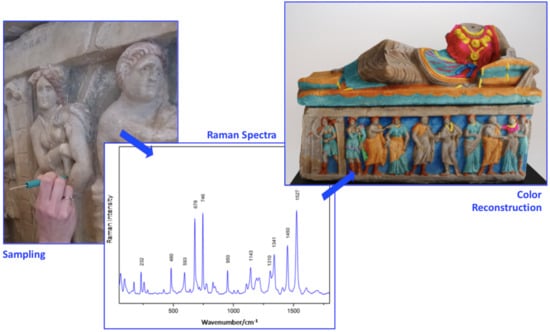
Deepening Inside the Pictorial Layers of Etruscan Sarcophagus of Hasti Afunei: An Innovative Micro-Sampling Technique for Raman/SERS Analyses


 ,
, 

Abstract
1. Introduction
2. Results
2.1. Surface Protection Products
2.2. Pictorial Layers
2.2.1. Standard Raman Spectroscopy
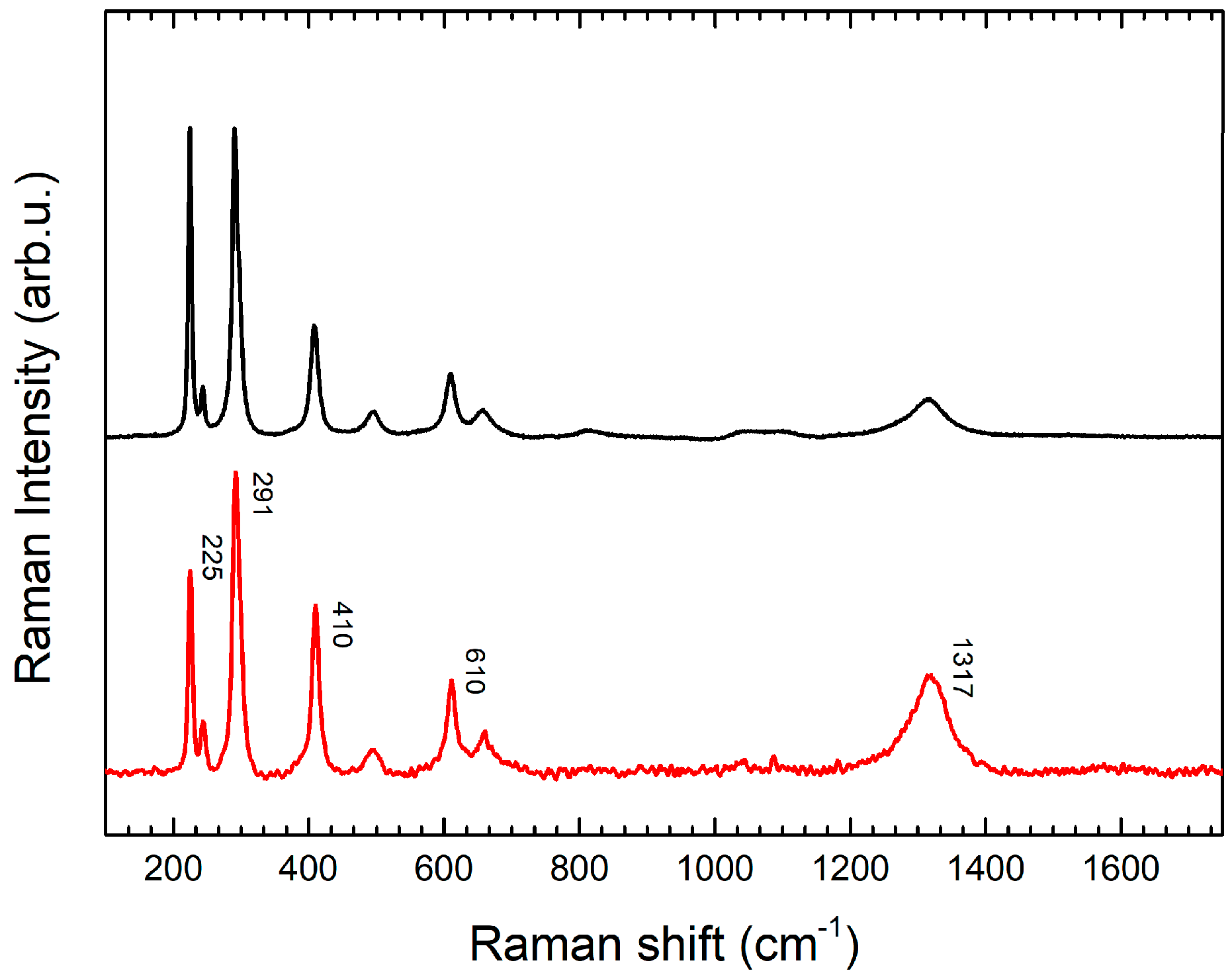
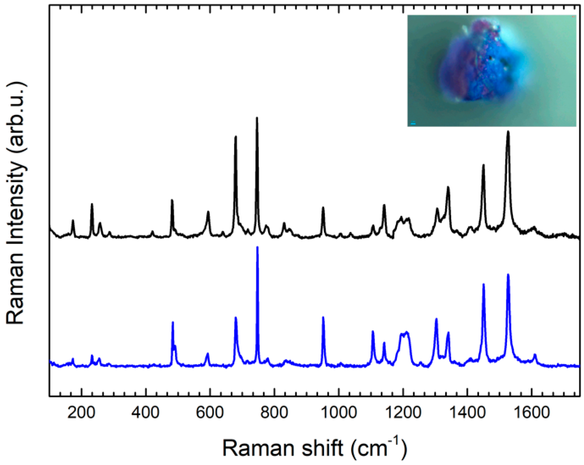
Blue Pigments
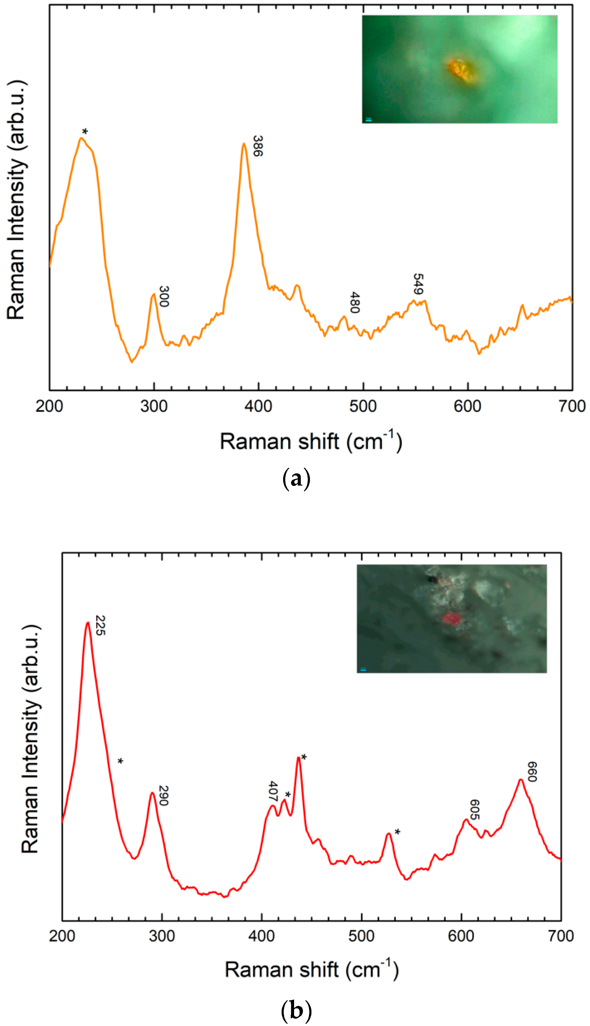
Red Pigments
Purple Pigment
2.2.2. Gel-Transfer Surface Enhanced Raman Spectroscopy (GT-SERS)
Blue Pigments
Brown Pigments
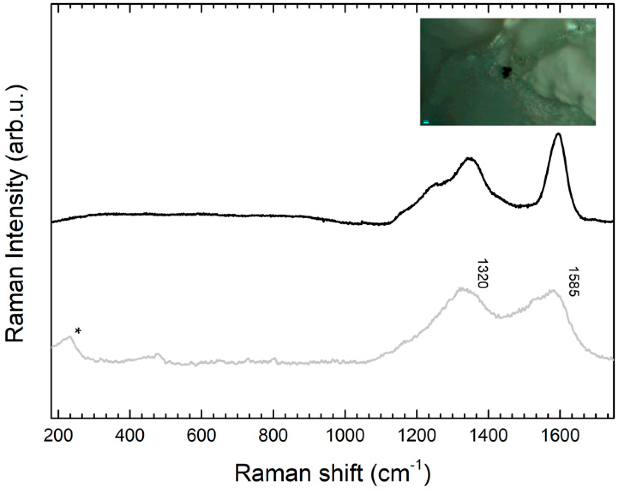
Black Pigment
Green Pigment
Pink Pigment
3. Discussion
4. Experimental
4.1. Microsampling Technique for Gel-Transfer Surface Enhanced Raman Spectroscopy (GT-SERS):
- Place the gel in contact with the surface to be samples; addition of 1–2 µL of organic solvent (ethanol, methanol, acetone, hexane, etc.) or solvent mixtures could be necessary depending the nature of the substance to be sampled.
- Hold in contact for enough time (in the range of 1–30 s). This time range should be sufficient for all the cases, even if some variations could occur depending on several factors.
- After the alloted time, remove the gel matrix from the surface.
- Cut the portion of Ag-gel matrix containing the samples and place it on one of the supports provided in the kit. Sampling effectiveness can be checked through a portable USB optical microscope.
- Leave the Ag-gel matrix portions to dry in the sample holder in horizontal position. After drying (for 2–3 h) they will adhere to a glass slide. Portions are then immobilized and can be moved without any risk.
- Analysis in the laboratory can be performed on the microsample using a Raman micro-spectrometer, even after a long time. After Raman and SER spectroscopy analysis, the sample can be re-extracted from the gel matrix. This allows one to carry out other investigations (e.g.,: HPLC, mass spectrometry) for cross-validated analysis. This is possible because the characteristic insolubility of the gel.
4.2. Raman/SERS Analyses
4.3. NMR Analyses
5. Conclusions
Supplementary Materials
Author Contributions
Funding
Acknowledgments
Conflicts of Interest
References
- Pozzi, F.; Leona, M. Surface-enhanced Raman spectroscopy in art and archaeology. J. Raman Spectrosc. 2016, 47, 67–77. [Google Scholar] [CrossRef]
- Serafini, I.; Ciccola, A. Nanotechnologies and Nanomaterials: An overview for Cultural Heritage. In Nanotechnologies and Nanomaterials for Diagnostic, Conservation and Restoration of Cultural Heritage; Lazzara, G., Fakhrullin, R., Eds.; Elsevier: Amsterdam, The Netherlands, 2018; pp. 325–380. [Google Scholar]
- Leona, M.; Stenger, J.; Ferloni, E. Application of surface-enhanced Raman scattering techniques to the ultrasensitive identification of natural dyes in works of art. J. Raman Spectrosc. 2006, 37, 981–992. [Google Scholar] [CrossRef]
- Lofrumento, C.; Ricci, M.; Platania, E.; Becucci, M.; Castellucci, E. SERS detection of red organic dyes in Ag-agar gel. J. Raman Spectrosc. 2013, 44, 47–54. [Google Scholar] [CrossRef]
- Platania, E.; Lombardi, J.; Leona, M.; Shibayama, N.; Lofrumento, C.; Ricci, M.; Becucci, M.; Castellucci, E. Suitability of Ag-agar gel for the microextraction of organic dyes on different substrates: The case study of wool, silk, printed cotton and a panel painting mock-up. J. Raman Spectrosc. 2014, 45, 1133–1139. [Google Scholar] [CrossRef]
- Platania, E.; Lofrumento, C.; Lottini, E.; Azzaro, E.; Ricci, M.; Becucci, M. Tailored micro-extraction method for Raman/SERS detection of indigoids in ancient textiles. Anal. Bioanal. Chem. 2015, 47, 6505–6514. [Google Scholar] [CrossRef]
- Daher, C.; Drieu, L.; Bellot-Gurlet, L.; Percot, A.; Paris, C.; Le Hô, A. Combined approach of FT-Raman, SERS and IR micro-ATR spectroscopies to enlighten ancient technologies of painted and varnished works of art. J. Raman Spectrosc. 2014, 45, 1207–1214. [Google Scholar] [CrossRef]
- Roldán, M.L.; Centeno, S.A.; Rizzo, A. An improved methodology for the characterization and identification of sepia in works of art by normal Raman and SERS, complemented by FTIR, Py-GC/MS, and XRF. J. Raman Spectrosc. 2014, 4, 1160–1171. [Google Scholar] [CrossRef]
- Lau, D.M.; Livett, M.; Prawer, S. Application of surface-enhanced Raman spectroscopy (SERS) to the analysis of natural resins in artworks. J. Raman Spectrosc. 2008, 39, 545–552. [Google Scholar] [CrossRef]
- Leona, M.; Lombardi, J.R. Identification of berberine in ancient and historical textiles by surface-enhanced Raman scattering. J. Raman Spectrosc. 2007, 38, 853–858. [Google Scholar] [CrossRef]
- Serafini, I.; Lombardi, L.; Fasolato, C.; Sergi, M.; Di Ottavio, F.; Sciubba, F.; Montesano, C.; Guiso, M.; Costanza, R.; Nucci, L.; et al. A new multi analytical approach for the identification of synthetic and natural dyes mixtures. The case of orcein-mauveine mixture in a historical dress of a Sicilian noblewoman of nineteenth century. Nat. Prod. Res. 2017, 33, 1040–1051. [Google Scholar] [CrossRef]
- Bruni, S.; Guglielmi, V.; Pozzi, F. Surface-enhanced Raman spectroscopy (SERS) on silver colloids for the identification of ancient textile dyes: Tyrian purple and madder. J. Raman Spectrosc. 2010, 41, 175–180. [Google Scholar] [CrossRef]
- Bruni, S.; Guglielmi, V.; Pozzi, F.; Mercuri, A.M. Surface-enhanced Raman spectroscopy (SERS) on silver colloids for the identification of ancient textile dyes. Part II: Pomegranate and sumac. J. Raman Spectrosc. 2011, 42, 465–473. [Google Scholar] [CrossRef]
- Chen, K.; Leona, M.; Vo-Dinh, K.; Yan, F.; Wabuyele, M.B.; Vo-Dinh, T. Application of surface-enhanced Raman scattering (SERS) for the identification of anthraquinone dyes used in works of art. J. Raman Spectrosc. 2006, 37, 520–527. [Google Scholar] [CrossRef]
- Pozzi, F.; van den Berg, K.J.; Fiedlera, I.; Casadio, F. A systematic analysis of red lake pigments in French Impressionist and Post-Impressionist paintings by surface-enhanced Raman spectroscopy (SERS). J. Raman Spectrosc. 2014, 45, 1119–1126. [Google Scholar] [CrossRef]
- Lombardi, L.; Bianco, A.; Guiso, M. Metodo e kit per il Campionamento Microinvasivo su Manufatti. Italian Patent n° 102015000042409, 5 August 2015. [Google Scholar]
- Pasquini, G.B. Lettera del Can. Gio. Battista Pasquini Teologo della Cattedrale di Chiusi, e Vicario Generale. Al Direttore dell’Antologia. In Antologia; Gabinetto Scientifico e Letteratio: Firenze, Italy, 1826; p. 113. [Google Scholar]
- David, A.R.; Edwards, H.G.M.; Farwell, D.W.; de Faria, D.L.A. Raman spectroscopic analysis of ancient Egyptian pigments. Archaeometry 2001, 43, 461–473. [Google Scholar] [CrossRef]
- Bell, I.M.; Clark, R.J.H.; Gibbs, P.J. Raman spectroscopic library of natural and synthetic pigments (pre- approximately 1850 AD). Spectrochim. Acta A 1997, 53, 2159–2179. [Google Scholar] [CrossRef]
- Burgio, L.; Clark, R.J.H. Library of FT-Raman spectra of pigments, minerals, pigment media and varnishes, and supplement to existing library of Raman spectra of pigments with visible excitation. Spectrochim. Acta A 2001, 57, 1491–1521. [Google Scholar] [CrossRef]
- Bouherour, S.; Berke, H.; Wiedemann, H.G. Ancient man-made copper silicate pigments studied by Raman spectroscopy. Chimia 2001, 55, 942–951. [Google Scholar]
- Bordignon, F.; Postorino, P.; Dore, P.; Trojsi, G. Raman identification of green and blue pigments in Etruscan polychromes on architectural terracotta panels. J. Raman Spectrosc. 2007, 38, 255–259. [Google Scholar] [CrossRef]
- Hanesch, M. Raman spectroscopy of iron oxides and (oxy)hydroxides at low laser power and possible applications in environmental magnetic studies. Geophys. J. Int. 2009, 177, 941–948. [Google Scholar] [CrossRef]
- de Faria, D.L.A.; Venancio Silva, S.; de Oliveira, M.T. Raman microspectroscopy of some iron oxides and oxyhydroxides. J. Raman Spectrosc. 1997, 28, 873–878. [Google Scholar] [CrossRef]
- Nadim, S.C.; Zumbuehl, S.; Delavy, F.; Fritsch, A.; Kuehnen, R. Synthetic organic pigments of the 20th and 21st century relevant to artist’s paints: Raman spectra reference collection. Spectrochim. Acta A 2009, 73, 505–524. [Google Scholar]
- Basova, T.V.; Kiselev, V.G.; Schuster, B.; Peisert, H.; Chassè, T. Experimental and theoretical investigation of vibrational spectra of copper phthalocyanine: Polarized single crystal Raman spectra. Isotope effect and DFT calculation. J. Raman Spectrosc. 2009, 40, 2080–2087. [Google Scholar] [CrossRef]
- Bruni, S.; Gugliemi, V.; Pozzi, F. Historical organic dyes: A surface-enhanced Raman scattering (SERS) spectral database on Ag Lee–Meisel colloids aggregated by NaClO4. J. Raman Spectrosc. 2011, 42, 1267–1281. [Google Scholar] [CrossRef]
- Ball, P. Colore, una bibliografia tra arte, storia e chimica, la bellezza e i misteri del mondo del colore; Bur Saggi, Rizzoli: Milan, Italy, 2001. [Google Scholar]
- Gettens, R.J.; Stout, G.L.; Forbes, E.W. Paintings Material: A Short Encyclopedia; D. Van Nostrand Company: New York, NY, USA, 1947. [Google Scholar]
- Leona, M.; Decuzzi, P.; Kubic, T.A.; Gates, G.; Lombardi, J.R. Nondestructive identification of natural and synthetic organic colorants in works of art by surface enhanced Raman scattering. Anal. Chem. 2011, 83, 3990–3993. [Google Scholar] [CrossRef]
Sample Availability: Not available. |


© 2019 by the authors. Licensee MDPI, Basel, Switzerland. This article is an open access article distributed under the terms and conditions of the Creative Commons Attribution (CC BY) license (http://creativecommons.org/licenses/by/4.0/).
Share and Cite
Gagliano Candela, R.; Lombardi, L.; Ciccola, A.; Serafini, I.; Bianco, A.; Postorino, P.; Pellegrino, L.; Bruno, M. Deepening Inside the Pictorial Layers of Etruscan Sarcophagus of Hasti Afunei: An Innovative Micro-Sampling Technique for Raman/SERS Analyses. Molecules 2019, 24, 3403. https://doi.org/10.3390/molecules24183403
Gagliano Candela R, Lombardi L, Ciccola A, Serafini I, Bianco A, Postorino P, Pellegrino L, Bruno M. Deepening Inside the Pictorial Layers of Etruscan Sarcophagus of Hasti Afunei: An Innovative Micro-Sampling Technique for Raman/SERS Analyses. Molecules. 2019; 24(18):3403. https://doi.org/10.3390/molecules24183403
Chicago/Turabian StyleGagliano Candela, Rossella, Livia Lombardi, Alessandro Ciccola, Ilaria Serafini, Armandodoriano Bianco, Paolo Postorino, Lorella Pellegrino, and Maurizio Bruno. 2019. "Deepening Inside the Pictorial Layers of Etruscan Sarcophagus of Hasti Afunei: An Innovative Micro-Sampling Technique for Raman/SERS Analyses" Molecules 24, no. 18: 3403. https://doi.org/10.3390/molecules24183403
APA StyleGagliano Candela, R., Lombardi, L., Ciccola, A., Serafini, I., Bianco, A., Postorino, P., Pellegrino, L., & Bruno, M. (2019). Deepening Inside the Pictorial Layers of Etruscan Sarcophagus of Hasti Afunei: An Innovative Micro-Sampling Technique for Raman/SERS Analyses. Molecules, 24(18), 3403. https://doi.org/10.3390/molecules24183403