Article

Terpenoids of the Swamp Cypress Subfamily (Taxodioideae), Cupressaceae, an Overview by GC-MS

1
Department of Chemistry, College of Science, Oregon State University, Corvallis, OR 97331, USA
2
Forschungsinstitut Senckenberg, Sektion Paläobotanik, D-60325 Frankfurt am Main, Germany
3
Consultant, 72 Marina Lakes Drive, Richmond, CA 94804, USA
4
Wood Extractive Laboratory, Forestry and Forest Products Research Institute, Tsukuba, Ibaraki 305-8687, Japan
*
Author to whom correspondence should be addressed.
Molecules 2019, 24(17), 3036; https://doi.org/10.3390/molecules24173036
Submission received: 16 July 2019 / Revised: 28 July 2019 / Accepted: 29 July 2019 / Published: 21 August 2019

Abstract

:
The resins bled from stems and in seed cones and leaves of Cryptomeria japonica, Glyptostrobus pensilis, Taxodium distichum, and T. mucronatum were characterized to provide an overview of their major natural product compositions. The total solvent extract solutions were analyzed as the free and derivatized products by gas chromatography-mass spectrometry to identify the compounds, which comprised minor mono- and sesquiterpenoids, and dominant di- and triterpenoids, plus aliphatic lipids (e.g., n-nonacosan-10-ol). Ferruginol, 7α-p-cymenylferruginol, and chamaecydin were the major characteristic markers for the Taxodioideae conifer subfamily. The mass spectrometric data can aid polar compound elucidation in environmental, geological, archeological, forensic and pharmaceutical studies.

1. Introduction

Natural products, especially terpenoids or their derivatives, are preserved in the ambient environment or geological record. When extracted and characterized by gas chromatography-mass spectrometry (GC-MS) they are used by organic geochemists as tracers for sources, transport and alteration processes of organic matter in any global compartment [1,2,3,4,5,6,7,8,9,10,11,12,13,14,15]. The application of GC-MS in the analysis of natural product mixtures extracted from plants for compound characterization can also be of utility for rapid screening in pharmacological studies [16].
The Coniferae are known as important source plants for resins and are comprised of the Araucariaceae (3 genera), Cupressaceae (27 genera), Pinaceae (11 genera), Podocarpaceae (18 genera), Taxaceae (6 genera), and Sciadopityaceae (1 genus) [17]. Here we focus on the Cupressaceae, specifically the subfamily Taxodioideae with 4 species (Table 1). Fossil remains of Taxodium and Glyptostrobus leaflets, seed cones and wood are often found in the geologic record of the northern hemisphere since the Cretaceous, and are especially abundant in the paleofloras of Tertiary brown coals [18,19,20,21,22] and extant peats of Florida, USA [23].
Structure determinations and pharmacological potential studies of resin terpenoids isolated and purified from species of the Taxodioideae subfamily have been reported by numerous chemists and pharmacologists [24,25,26,27,28,29,30,31,32,33,34,35,36,37,38,39,40,41,42]. However, the connection of the known natural products isolated from these conifers with their application as tracers in other interdisciplinary sciences has been limited [7,8,14,43]. This is due to the lack of reports on the chemical compositions of the total resin mixtures, and the paucity of mass spectra for known compounds, especially the oxygenated natural products. Here we characterize the total compound mixtures in resins from the Taxodioideae subfamily based on correlation and comparison with known standards. We report the GC-MS results and full mass spectra of novel compounds of the resins as free and derivatized products. This total mixture analysis provides an overview of the major compounds present in the resins of the extant Taxodioideae subfamily, and is a guide for their presence in environmental and fossil samples.

2. Results and Discussion
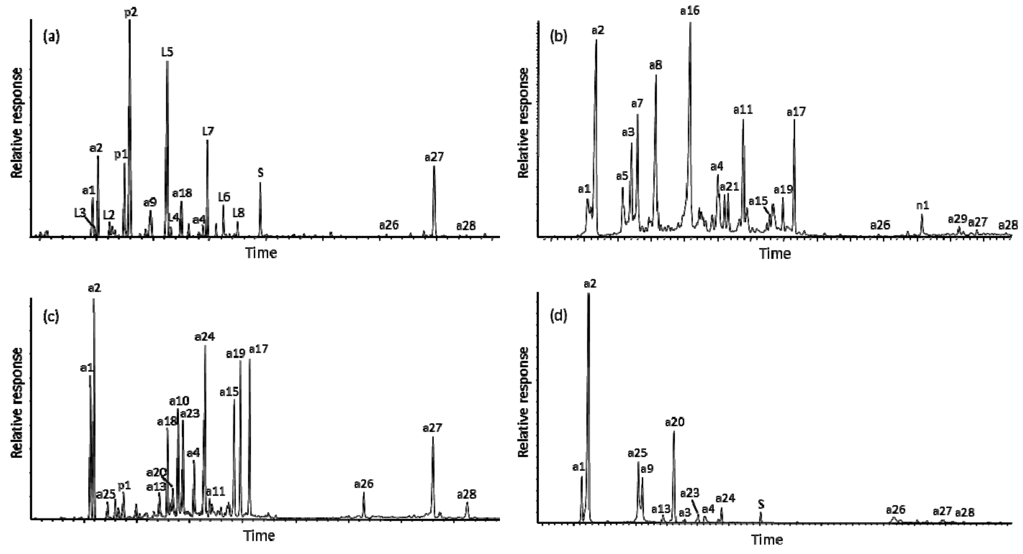
The contents of terpenoid natural products have been documented in many species of the Cupressaceae [17,44,45,46], but the investigation of the former family Taxodiaceae is incomplete. The terpenoid content of Glyptostrobus pensilis (Staunton) Koch (Chinese Water Pine) is limited [17], with only two trace diterpenoids, namely glypensin A and 12-acetoxy-ent-labda-8(17),13E-dien-15-oic acid, identified [37]. According to the merger of Cupressaceae and Taxodiaceae to the Cupressaceae s.l., G. pensilis was placed in the subfamily Taxodioideae [47].
The diterpenoid constituents of Cryptomeria japonica and Taxodium distichum have been studied more extensively [24,25,26,27,28,29,31,32,33,34,35,38,39,40,41,42,48] and references therein]. However, none of these earlier studies reported the natural product compositions of the total resin/plant extracts, but instead the mixtures were separated by liquid or high-pressure liquid chromatography (LC or HPLC), followed by structure determination of each compound by NMR, HRMS for elemental composition, and sometimes MS (underivatized by direct insertion probe). The diterpenoid components of Taxodium mucronatum have had limited examination [7,8,49].
The compounds identified as significant components in the bled resins (as total extracts) of the four conifers in this subfamily (Table 1) are listed here in Table 2. Analyses of extracts of leaves and cones yielded similar results, except the solvent mixture used for extraction also isolated the epicuticular wax and polar cell (e.g., saccharides) components which were superimposed on the terpenoids. Examples of annotated total ion current traces for the GC-MS results are shown in Figure 1. The mass spectra of the major compounds in the extract mixtures are given in the figures of the Supplementary Materials section or can be found in the literature cited in Table 2.

2.1. Cryptomeria

The resin of Cryptomeria japonica (sugi cedar) was comprised of minor diterpenoids as the phenolic abietane-type: Ferruginol (a2, all chemical structures are given in Appendix A), 6,7-dehydroferruginol (a1), sugiol (a4), 7α-p-cymenylferruginol (a26), chamaecydin (a27), iso-chamaecydin (a28), and 6-hydroxychamaecydin (a29); and major diterpenoid resin acids with the pimarane and labdane skeletons: Sandaracopimaric acid (p1), isopimaric acid (p2, 100%), iso-communic acid (L3), 13-epi-cupressic acid (L5), imbricataloic acid (L4), imbricatoloic acid (L7), iso-cupressic acid (L6), 13,14-dihydroagathic acid (L8), and 13E- and 13Z-communic acids (L2). No sesquiterpenoids were detectable, and the lipid compound, n-nonacosan-10-ol (n1), was a carryover from the epicuticular leaf wax.
The essential oil of C. japonica has been analyzed, but only mono- and sesquiterpenoids were reported [50]. Kaurenes and dehydroabietane with the lower terpenoids were reported for sugi leaf oil [51]. The minor resin acids have been characterized in bark and leaf extracts [27,28,29], and the chamaecydins were elucidated in leaf extracts from C. japonica [25,26]. The diterpenoids from bark and fruits of C. fortunei, also known as C. japonica var. sinensis, have been studied [36,52]. Sugiol (a4), 11-hydroxysugiol (a15), and 14-deoxycoleon U (a17), also found in this study, were reported.

2.2. Glyptostrobus

The resin of Glyptostrobus pensilis consisted mainly of diterpenoids as the phenolic abietanes: Ferruginol (a2), 6,7-dehydropisiferol (a3), pisiferol (a7), abeo-pisiferol (a8), abeo-carnosol (a16, 100%), sugiol (a4), salvinolone (a11), and 6-hydroxysalvinolone (14-deoxycoleon U, a17) as major compounds. The minor compounds were 6,7-dehydroferruginol (a1), 7-hydroxytaxodone (a19), sandaracopimaric acid (p1), 7α-p-cymenylferruginol (a26), chamaecydin (a27), iso-chamaecydin (a28), 6-hydroxychamaecydin (a29), and 19(20)-oxoferruginol (a5), also mostly phenolic abietanes. The epicuticular wax extracted from shoots of G. pensilis was comprised of lipids, mainly n-nonacosan-10-ol (n1), n-nonacosan-10-one (n2) and n-alkanes ranging from C27 to C33; with minor diterpenoids: Ferruginol (a2, 100%), 6,7-dehydroferruginol (a1), 6,7-dehydropisiferol (a3), pisiferol (a7), sugiol (a4), salvinolone (a11), 6-hydroxysalvinolone (a17), abeo-carnosol (a16), abeo-pisiferol (a8), and triterpenoids: 24-methylenecycloartan-3-one (T1), and 24-ethylenecycloartan-3-one (T2). No sesquiterpenoids were detected.
The terpenoids of oil steam distilled from wood of G. pensilis from Vietnam were comprised of mono- and sesquiterpenoids with traces of phytol and ferruginol [63]. However, fossil leaves of this conifer back to the Eocene retained mainly the following natural products: ferruginol, 6,7-dehydroferruginol, salvinolone, 5,6-dehydrosugiol, taxodione acetate, abeo-carnosol, sugiol, cymenylferruginol, chamaecydin, and iso-chamaecydin [7,8,54,64,65].

2.3. Taxodium distichum

The resin of Taxodium distichum was comprised mainly of phenolic abietanes: Ferruginol (a2), 6,7-dehydroferruginol (a1), sugiol (a4), taxoquinone (a18), taxodione (a10, R=H), 6α-hydroxytaxoquinone (a23), 6β-hydroxytaxoquinone (a24), taxodone (a14, 100%), 11-hydroxysugiol (a15), 7-hydroxytaxodone (a19), 6-hydroxysalvinolone (a17), salvinolone (a11), and chamaecydin (a27). The other minor compounds that could be identified were: 6-deoxotaxodione (a6), 7α-p-cymenylferruginol (a26), sandaracopimaric acid (p1), iso- and 6-hydroxychamaecydin (a28, a29), royleanone (a13), taxodione acetate (a10, R=Ac), and 7-acetoxy-6,7-dehydroroyleanone (a25).
These compounds, with numerous trace components, have all been reported in the extensive literature [24,31,32,33,34,38,66], also as cited in Table 2. Taxodium peat has been analyzed but only aromatic hydrocarbons derived from diterpenoids were reported [23]. Discrete fossils of Taxodium back to the Eocene contained ferruginol, 6,7-dehydroferruginol, taxodione acetate, sugiol, and the chamaecydins [7,8,65,67,68].

2.4. Taxodium mucronatum

The T. mucronatum resin consisted of significant sesquiterpenoids: Widdrol (s4), widdrene (s2), mannol (L1), cuparene (s1), and mayurone (s3). The diterpenoids were exclusively only phenolics of the abietane-type: Ferruginol (a2, 100%), 6,7-dehydroferruginol (a1), 6-deoxotaxodione (a6), 7-acetoxyroyleanone (a25), taxodone (a14), taxodione acetate (a10, R = Ac), and chamaecydin (a27). The minor components were: Royleanone (a13), sugiol (a4), salvinolone (a11), 7α-p-cymenylferruginol (a26), iso- and 6-hydroxychamaecydin (a28, a29), and the lipid n-nonacosan-10-ol (n1). One study reported the presence of 8β-hydroxypimar-15-en-19-oic acid in this plant [49].

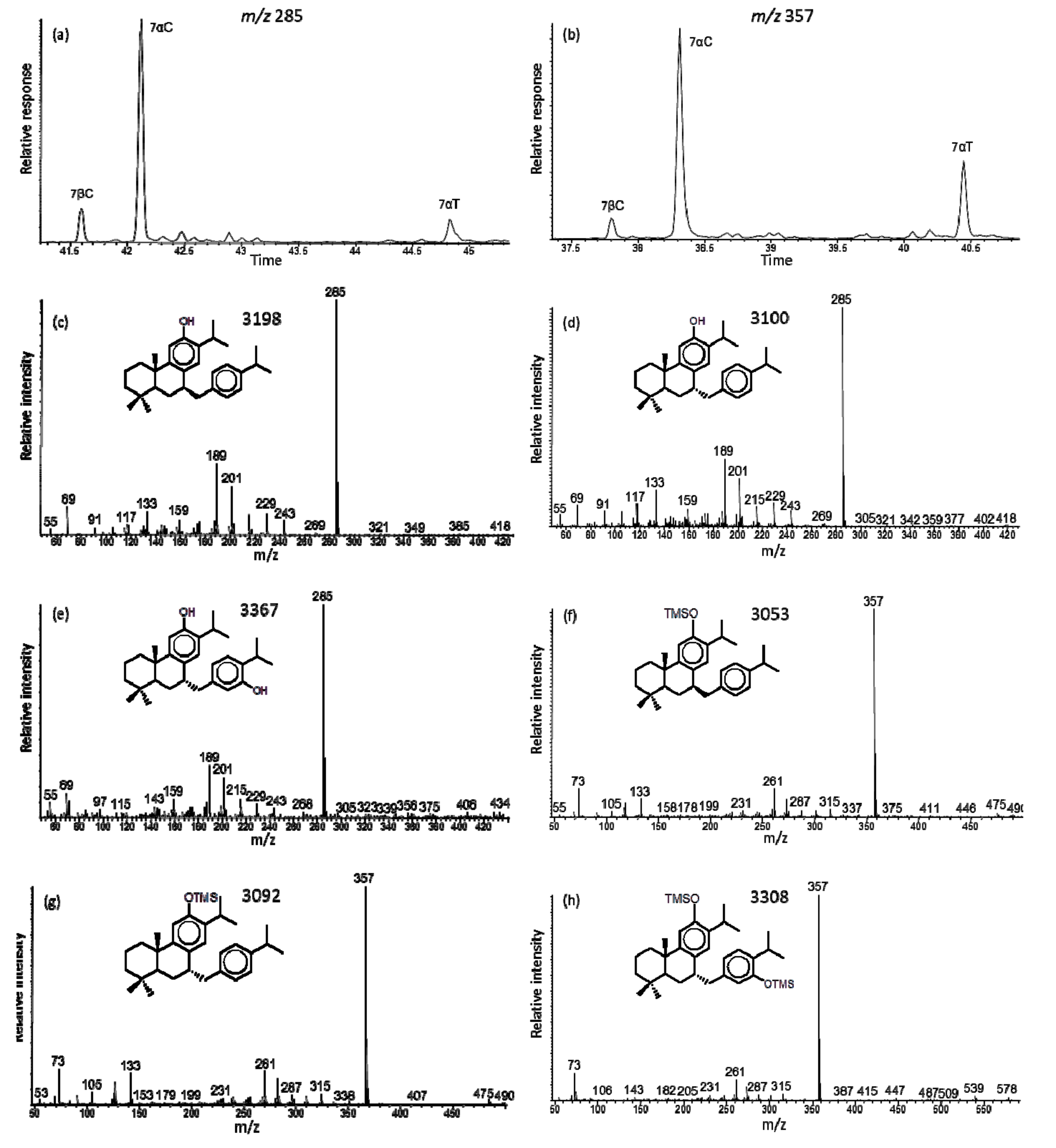
2.5. Mass Spectrometry

The mass spectra of all compounds analyzed by GC-MS are listed in Table 2. Those indicated by S match with the respective reference standards, and those indicated by L match with the literature data cited.
The mass spectra of 6,7-dehydroferruginol (a1), as the free compound and methyl ether [12,65,69,70], as well as the NMR data [27,71] have been published. The interpretation of the mass spectrometric fragmentation, specifically the even mass fragment ion from the retro-Diels–Alder rearrangement loss of ring-A [56] is unusual and should indicate 2,3-dehydroferruginol. The same loss of ring-A by retro-Diels–Alder rearrangement of the C-2 to C-3 double bond is observed in mass spectra of standard cholest-2-ene (loss of C4H6 from M + 370 to m/z 316), friedel-2-ene (loss of C5H8 from M + 410 to m/z 342), or lupa-2,22(29)-diene (loss of C6H10 from M + 408 to m/z 326). This would be consistent for 2,3-dehydroferruginol (loss of C6H10 from M + 284 to m/z 202, Figure 2a) and its silyl derivative (loss of C6H10 from M + 356 to m/z 274, Figure 2d). However, the NMR data indicate the double bond is at C-6 to C-7 [71], so that bond can also induce fragmentation by H-transfer and loss of ring-A to m/z 202 (Figure 3a). Therefore, the double bond position could be misinterpreted based solely on mass spectra, but with the NMR data the assignment as 6,7-dehydoferruginol is correct.
In fossil resins of these conifers we also found 5,6-dehydroferruginol (tentative identification [7,8,67]), where the elimination of C4H8 to m/z 228 with secondary loss of C2H4 to m/z 200 (or direct loss of ring-A to m/z 200) are evident for the free compound and the same fragmentation for the silyl derivative to m/z 300 (Figure 2b,e and Figure 3a). Another isomer was tentatively identified as 15,16-dehydroferruginol in the same fossil resins, but not reported before (Figure 2c). Its key fragment ion is m/z 215 from loss of C5H9 in ring-A, as confirmed in the mass spectrum of its silyl derivative where the same loss of C5H9 results in the fragment ion at m/z 287 (Figure 2f and Figure 3a). We find 6,7-dehydroferruginol and ferruginol primarily in extant conifers, and the tentatively assigned 5,6-dehydroferruginol and 15,16-dehydroferruginol with ferruginol and minor 6,7-dehydroferruginol in fossil conifer resins.
The mass spectra of 7α-p-cymenylferruginol (i.e., 7α-p-isopropyl-benzyl-ferruginol, a26) and its trimethylsilyl derivative (Figure 4d,f) were identified based on the structure determination by NMR and HRMS [36]. The compound has a low intensity molecular ion (M + at m/z 418 and loses the p-cymenyl fragment to the base peak at m/z 285, which is also reflected in the C10H13 ion at m/z 133 (Figure 3c). Its TMS derivative also has a low intensity M + at m/z 490, with losses of CH3 to m/z 475 and C10H13 to the base peak at m/z 357, and the corresponding key ion at m/z 133 (Figure 3c). We also find a lesser amount (typically 20% of the 7α-isomer) of 7β-p-cymenylferruginol (Figure 4c) in all samples analyzed. It is reported as the total of both isomers in Table 2. The extant sample of T. mucronatum and the fossil samples of T. dubium and G. oregonensis had mass spectra of a minor compound, interpreted as 7α-thymylferruginol with M + at m/z 434 and base peak at m/z 285, and for its TMS derivative with M + at m/z 578 and base peak at m/z 357 (Figure 4b,e,h, respectively). It has not yet been reported in the natural products literature.
The identification of cymenylferruginol (a26), a triterpenoid like the chamaecydins, in both the extant and fossil resins is of interest because natural product chemists are identifying numerous dimer terpenoids, i.e., mono-to-diterpenoid, sesqui-to-diterpenoid and diterpenoid dimers, in extant plants [25,26,36,41,48]. Therefore, the formation mechanism is for example ferruginol reacting at C-7 with cymene to produce 7-cymenylferruginol, 6-deoxotaxodione reacting at C-7 and C-14 with sabinene or thujene to form chamaecydin, cadinols reacting with ferruginol at C-7 to yield the various sesquaterpenoids, or peroxidation of ferruginol to dimers [26,41,48,72,73]. Some of these natural products, if adequately concentrated in the extant biomass, may become preserved as tracers in the geological record, as for example cymenylferruginol reported here.
The resin of T. distichum has a significant compound identified in the literature as 11-hydroxy-12-oxoabieta-7,9(11),13-triene (a.k.a. 6-deoxotaxodione, a6). Its mass spectrum is shown in Figure S1b and matches that reported [55]. The interpretation of the fragmentation pattern is based on the analogous fragmentation of 6,7-dehydroferruginol discussed above and correlation with the GC retention index. It also eliminates C6H10 from the M + at m/z 300 to m/z 218 (Figure S1b), indicating that the structure may be 11-hydroxy-6,7-dehydroferruginol (a6) or its isomer 11-hydroxy-12-oxoabieta-7,9(11),13-triene (a6). Silylation of the T. distichum resin extract did not produce a compound with M + at m/z 372, but a mass spectrum of a TMS derivative with M + at m/z 444 that eliminates C6H10 to m/z 362, fitting for 11-hydroxy-6,7-dehydroferruginol-diTMS (Figure S4g). The GC-MS results for the standard of 11-hydroxy-12-oxoabieta-7,9(11),13-triene revealed that it had oxidized to primarily taxodione and numerous other polyoxygenated products. However, silylation of this standard did yield a trace derivative with a mass spectrum showing M + at m/z 372 with fragments at m/z 357, 329 and 287, but no ion for elimination of C6H10 at m/z 290 (Figure S3f). Its structure could be that of 11-trimethylsiloxy-12-oxoabieta-7,9(11),13-triene. Therefore, we conclude that 11-hydroxy-12-oxoabieta-7,9(11),13-triene may isomerize to 11-hydroxy-6,7-dehydroferruginol during the silylation derivatization reaction. Furthermore, the hydroxy functionality at C-11 is preferred rather than at C-14 due to the concurrent presence of 11-hydroxyferruginol (a9).
Another compound that occurred as a minor component in extant and fossil Glyptostrobus sp. had mass spectra that were misinterpreted as hinokione [54,65]. Based on prior mass spectrometric data for hinokione [74,75] we now tentatively suggest that compound to be 19(20)-oxoferruginol (a5). Its mass spectrum has M + at m/z 300 for C20H28O2 with the base peak at m/z 189 due to loss of C7H11O, i.e., rearrangement loss of C-20 and ring-A (Figure 2g and Figure 3b). The same loss is observed in the mass spectrum of its TMS derivative from M + at m/z 372 to the base peak at m/z 261 (Figure 2h and Figure 3b). The mass spectrometric data do not match with that of the known formosanoxide (i.e., 7(20)-oxoferruginol [76]). Therefore, 19(20)-oxoferruginol may be a natural product derived from epoxidation to C-19 of pisiferol (a7) also present in extant G. pensilis.

2.6. Key Molecular Tracers for the Taxodioideae

Ferruginol, 6,7-dehydroferruginol, sugiol, 7α-p-cymenylferruginol, chamaecydin, iso-chamaecydin, and 6β-hydroxychamaecydin were present as generally significant components in the resins of all four species (Table 2). These natural products are stable and have been reported in environmental and fossil samples [7,8,11,14,54,64,65,77].
Resins from G. pensilis contained the following unique natural products in addition to those above: pisiferol, abeo-pisiferol, salvinolone, abeo-carnosol, 6-hydroxysalvinolone, 19(20)-oxoferruginol, and 6α- and 6β,11-dihydroxysugiols. Resins from Eocene fossils of G. pensilis consisted of ferruginol, 6,7-dehydroferruginol, 19(20)-oxoferruginol, sugiol, abeo-carnosol, salvinolone, and chamaecydins, but no pisiferol or abeo-pisiferol [54]. Therefore, the geological fate of the abeo-diterpenoids is not fully known. However, a derivative biomarker, i.e., 10α- and 10β-dehydroicetexane, has been elucidated in sediments and petroleum [78], and it could be derived from pisiferol/abeo-pisiferol by reductive dehydroxylation during diagenesis and preservation of organic matter. The resin of extant G. pensilis contained traces of 7α-p-cymenylferruginol [36]. It was reported first as an unknown compound in fossil G. oregonensis from the Miocene of Emerald Creek, ID (labeled U7 in [65]), then in fossil G. nordenskioeldi from the Eocene of Axel Heiberg Island, Nunavut, Canada, but misidentified as roylean-20-oic acid [54]. It is now correctly identified as 7α-p-cymenylferruginol (a26).
The resins of the two Taxodium species had similar compositions comprised of the compounds present in all four species with additional unique quinoid diterpenoids, specifically: taxodione, royleanone, taxodone, taxoquinone, horminone, 6α- and 6β-hydroxytaxoquinones, taxodione acetate, and 7-acetoxy-6,7-dehydroroyleanone [24]. The natural triterpenoid, 7α-p-cymenylferruginol, was a significant component in the resins of both species (e.g., Figure 3c). Resin from a Miocene T. dubium contained ferruginol, 6,7-dehydroferruginol, taxodione acetate, 7-acetoxy-6,7-dehydroroyleanone, sugiol, and 7α-p-cymenylferruginol [65]. Therefore, the phenolic and quinoid diterpenoids are of utility as environmental and geological tracers for source origin.

3. Samples and Experimental Methods

3.1. Plant Material

The bled resins from trunks and branchlets or seed cones with bled resin blebs of Cryptomeria japonica, Glyptostrobus pensilis, Taxodium distichum, and T. mucronatum were sampled from mature trees with botanical labels in various park and garden reserves (Table 1). The resins were placed in glass vials and cones and branchlets in paper envelopes to allow air drying. We are aware that the structure determinations by natural product chemists typically concentrated on internal compounds extracted from bulk trunk wood, bark, leaves or cones to elucidate the different compound compositions and trace components. However, we examined the external bled compounds in order to determine those that enter the environment before or after tree senescence and may become part of the geological fossil record.

3.2. Extraction

Cones and branchlets were cut into pieces and extracted by ultra-sonication for 10 min with dichloromethane:methanol (DCM:MeOH, 1:1 v/v) and soaking in the solvent mixture for 24 h. The resin samples were dissolved (100% soluble) in the same solvent mixture. The total extracts were filtered through glass fiber filters, and concentrated by use of a rotary evaporator and blow-down with dry nitrogen gas.

3.3. Derivatization

Aliquots of total extracts were converted to trimethylsilyl derivatives by reaction with N,O-bis-(trimethylsilyl)trifluoroacetamide (BSTFA, Sigma-Aldrich, St. Louis, MO, USA) and a trace of pyridine for 3 h at 70 °C. The samples were blown down to dryness with nitrogen gas and dissolved in hexane prior to analysis. Other aliquots were treated with trimethylsilyldiazomethane (2M in hexane, Sigma-Aldrich) at room temperature for 30 min to convert carboxylic acids to methyl esters. The excess reagent was reacted with concentrated acetic acid, followed by blow-down with nitrogen gas, and solution in hexane prior to analysis.

3.4. GC-MS Analysis

The GC-MS analyses of the total and derivatized extracts were performed on a Hewlett-Packard model 6890 GC coupled to a Hewlett-Packard model 5973 MSD (Palo Alto, CA, USA). Separation was achieved on a DB5 (Agilent, Santa Clara, CA, USA) capillary column (30 or 60 m × 0.25 mm i.d., 0.25 µm film thickness). The GC operating conditions were as follows: temperature hold at 65 °C for 2 min, increase from 65 to 300 °C at a rate of 6 °C min−1, with a final isothermal hold for 20 min. Helium was used as carrier gas. The samples were injected splitless (typically 1 µL) with the injector temperature at 280 °C. The MS was operated in the electron impact mode at 70 eV and scanned from 50 to 650 da. Data were acquired and processed with the Chemstation software (Hewlett-Packard, Palo Alto, CA, USA). Individual compounds were identified by comparison of mass spectra with literature and library data, comparison with authentic standards, and interpretation of GC retention times and mass spectrometric fragmentation patterns. The GC retention times are expressed as Kovats indices (KI) and cited on each respective mass spectrum [79].

4. Conclusions

Natural products (diterpenoids) in resins are the best molecular tracers for conifers in the environment and fossil record. Not all compounds survive as such because they are altered or removed by oxidation and diagenetic reactions. However, compounds in resin are protected from oxidation and instead undergo disproportionation or polymerization reactions. The major characteristic markers for the Taxodioideae conifer subfamily are ferruginol, 7α-p-cymenylferruginol and chamaecydin, with secondary salvinolone, abeo-carnosol and taxodione acetate. We provided an overview of the natural product precursors of the Taxodioideae to apply in environmental, geological, archeological, forensic and pharmaceutical studies. Furthermore, we summarized the presence of these tracers in the fossil record.

Supplementary Materials

Additional mass spectra of related and derivatized natural products are collected in the Supplementary Materials section available with this article. Figures S1–7: Mass spectra of the terpenoids from the Taxodioideae resins (ordered in ascending molecular weights).

Author Contributions

B.R.T.S., A.O. and N.K. conceived and designed the work; B.R.T.S., A.O., N.K. and M.Z. performed the experiments; B.R.T.S. and D.R.O. analyzed and interpreted the data; B.R.T.S. wrote the paper, and all coauthors reviewed and edited the paper.

Funding

This research received no external funding.

Acknowledgments

We thank Wei-Min Zhao (Shanghai Institute of Materia Medica, Chinese Academy of Sciences) for generous gifts of standards and HRMS data, and Walter Buechler for supplying shoots and cones of G. pensilis.

Conflicts of Interest

The authors declare no conflicts of interest.

Appendix A

Molecules 24 03036 i001
Molecules 24 03036 i002

References

  1. Simoneit, B.R.T. Diterpenoid compounds and other lipids in deep-sea sediments and their geochemical significance. Geochim. Cosmochim. Acta 1977, 41, 463–476. [Google Scholar] [CrossRef]
  2. Simoneit, B.R.T. The organic chemistry of marine sediments. In Chemical Oceanography, 2nd ed.; Riley, J.P., Chester, R., Eds.; Academic Press: New York, NY, USA, 1978; Volume 7, pp. 233–311. [Google Scholar]
  3. Johns, R.B. Biological Markers in the Sedimentary Record; Elsevier: Amsterdam, The Netherlands, 1986; p. 364. [Google Scholar]
  4. Simoneit, B.R.T. Organic matter of the troposphere—V: Application of molecular marker analysis to biogenic emissions into the troposphere for source reconciliations. J. Atmos. Chem. 1989, 8, 251–275. [Google Scholar] [CrossRef]
  5. Simoneit, B.R.T.; Mazurek, M.A. Organic matter of the troposphere—II. Natural background of biogenic lipid matter in aerosols over the rural western United States. Atmos. Environ. 1982, 16, 2139–2159. [Google Scholar] [CrossRef]
  6. Simoneit, B.R.T. A review of biomarker compounds as source indicators and tracers for air pollution. Environ. Sci. Pollut. Res. 1999, 6, 153–163. [Google Scholar] [CrossRef]
  7. Otto, A.; White, J.D.; Simoneit, B.R.T. Natural product terpenoids in Eocene and Miocene conifer fossils. Science 2002, 297, 1543–1545. [Google Scholar] [CrossRef]
  8. Otto, A.; Simoneit, B.R.T.; Wilde, V.; Kunzmann, L.; Püttmann, W. Terpenoid composition of three fossil resins from Cretaceous and Tertiary conifers. Rev. Palaeobot. Palynol. 2002, 120, 203–215. [Google Scholar] [CrossRef]
  9. Otto, A.; Simoneit, B.R.T.; Wilde, V. Terpenoids as chemosystematic markers in selected fossil and extant species of pine (Pinus, Pinaceae). Bot. J. Linn. Soc. 2007, 154, 129–140. [Google Scholar] [CrossRef]
  10. Evershed, R.P. Organic residue analysis in archaeology: The archaeological biomarker revolution. Archaeometry 2008, 50, 895–924. [Google Scholar] [CrossRef]
  11. Medeiros, P.M.; Simoneit, B.R.T. Multi-biomarker characterization of sedimentary organic carbon in small rivers draining the northwestern United States. Org. Geochem. 2008, 39, 52–74. [Google Scholar] [CrossRef]
  12. Menor-Salván, C.; Najarro, M.; Velasco, F.; Rosales, I.; Tornos, F.; Simoneit, B.R.T. Terpenoids in extracts of Lower Cretaceous ambers from the Basque-Cantabrian basin (El Soplao, Cantabria, Spain): Paleochemotaxonomic aspects. Org. Geochem. 2010, 41, 1089–1103. [Google Scholar] [CrossRef]
  13. Schnell, G.; Schaeffer, P.; Tardiron, H.; Motsch, E.; Connan, J.; Ertlen, D.; Schwarz, D.; Schneider, N.; Adam, P. Contrasting diagenetic pathways of higher plant triterpenoids in buried wood as a function of tree species. Org. Geochem. 2014, 66, 107–123. [Google Scholar] [CrossRef]
  14. Stefanova, M.; Simoneit, B.R.T.; Marinov, S.P.; Zdravkov, A.; Kortenski, J. Novel polar biomarkers of the Miocene Maritza-East lignite, Bulgaria. Org. Geochem. 2016, 96, 1–10. [Google Scholar] [CrossRef]
  15. Simoneit, B.R.T.; Cox, R.C.; Oros, D.R.; Otto, A. Terpenoid compositions of resins from Callitris species (Cupressaceae). Molecules 2018, 23, 3384. [Google Scholar] [CrossRef]
  16. González, M.A. Aromatic abietane diterpenoids: Their biological activity and synthesis. Nat. Prod. Rep. 2015, 32, 684–704. [Google Scholar] [CrossRef]
  17. Otto, A.; Wilde, V. Sesqui-, di-, and triterpenoids as chemosystematic markers in extant conifers—A review. Bot. Rev. 2001, 67, 141–238. [Google Scholar]
  18. Hickey, L.J. The paleobotany and stratigraphy of the Golden Valley Formation (Late Paleocene—Early Eocene) of western North Dakota. Geol. Soc. Am. Mem. 1977, 150, 181. [Google Scholar]
  19. Mai, D.H.; Walther, H. Die oligozänen und untermiozänen Floren NW-Sachsens und des Bitterfelder Raumes. Abh. Staatl. Mus. Mineral. Geol. Dresd. 1991, 38, 1–230. [Google Scholar]
  20. LePage, B.A. The taxonomy and biogeographic history of Glyptostrobus Endlicher (Cupressaceae). Bull. Peabody Mus. Nat. Hist. 2007, 48, 359–426. [Google Scholar] [CrossRef]
  21. Eckenwalder, J.E. Re-evaluation of Curessaceae and Taxodiaceae: A proposed merger. Madroño 2009, 23, 237–256. [Google Scholar]
  22. Ding, W.N.; Kunzmann, L.; Su, T.; Huang, J.; Zhou, Z.K. A new fossil species of Cryptomeria (Cupressaceae) from the Rupelian of the Lühe Basin, Yunnan, East Asia: Implications for palaeogeography and palaeoecology. Rev. Palaeobot. Palynol. 2018, 248, 41–51. [Google Scholar] [CrossRef]
  23. Dehmer, J. Petrological and organic geochemical investigation of recent peats with known environments of deposition. Int. J. Coal Geol. 1995, 28, 111–138. [Google Scholar] [CrossRef]
  24. Kupchan, S.M.; Karim, A.; Marcks, C. Tumor inhibitors. XLVIII. Taxodione and taxodone, two novel diterpenoid quinone methide tumor inhibitors from Taxodium distichum. J. Org. Chem. 1969, 34, 3912–3918. [Google Scholar] [CrossRef]
  25. Shibuya, T. Cryptoquinonemethides D and E, C30-terpene quinone methides, from Cryptomeria japonica. Phytochemistry 1992, 31, 4289–4294. [Google Scholar] [CrossRef]
  26. Su, W.C.; Fang, J.M.; Cheng, Y.S. Hexacarbocyclic triterpenes from the leaves of Cryptomeria japonica. Phytochemistry 1993, 34, 779–782. [Google Scholar] [CrossRef]
  27. Su, W.C.; Fang, J.M.; Cheng, Y.S. Abietanes and kauranes from leaves of Cryptomeria japonica. Phytochemistry 1994, 35, 1279–1284. [Google Scholar] [CrossRef]
  28. Su, W.C.; Fang, J.M.; Cheng, Y.S. Labdanes from Cryptomeria japonica. Phytochemistry 1994, 37, 1109–1114. [Google Scholar]
  29. Su, W.C.; Fang, J.M.; Cheng, Y.S. Diterpenoids from leaves of Cryptomeria japonica. Phytochemistry 1996, 41, 255–261. [Google Scholar]
  30. Su, Z.; Yuan, W.; Wang, P.; Li, S. Ethnobotany, phytochemistry, and biological activities of Taxodium Rich. Pharmaceut. Crops 2013, 4, 1–14. [Google Scholar]
  31. Yamamoto, H.; Hirao, T.; Wakayama, K.-I.; Chida, T. Abietane diterpenes from cones of Taxodium distichum Rich. Bull. Fac. Edu. Ibaraki Univ. Nat. Sci. 2003, 52, 31–39. [Google Scholar]
  32. Kusumoto, N.; Murayama, T.; Kawai, Y.; Ashitani, T.; Ogiyama, K.; Takahashi, K. Taxodal, a novel irregular abietane-type diterpene from the cones of Taxodium distichum. Tetrahedron Lett. 2008, 49, 4845–4847. [Google Scholar] [CrossRef]
  33. Kusumoto, N.; Ashitani, T.; Hayasaka, Y.; Murayama, T.; Ogiyama, K.; Takahashi, K. Antitermitic activities of abietane-type diterpenes from Taxodium distichum cones. J. Chem. Ecol. 2009, 35, 635–642. [Google Scholar] [CrossRef]
  34. Kusumoto, N.; Ashitani, T.; Murayama, T.; Ogiyama, K.; Takahashi, K. Antifungal abietane-type diterpenes from the cones of Taxodium distichum Rich. J. Chem. Ecol. 2010, 36, 1381–1386. [Google Scholar] [CrossRef]
  35. Li, W.H.; Chang, S.T.; Chang, S.C.; Chang, H.T. Isolation of antibacterial diterpenoids from Cryptomeria japonica bark. Nat. Prod. Res. 2008, 22, 1085–1093. [Google Scholar] [CrossRef]
  36. Wu, J.; Zhao, W.M. New sesquiterpene and triterpene from the fruits of Cryptomeria fortunei. J Asian Nat. Prod. Res. 2010, 12, 382–387. [Google Scholar] [CrossRef]
  37. Zhang, Y.M.; Yin, R.T.; Jia, R.R.; Yang, E.H.; Xu, H.M.; Tan, N.H. A new diterpene from Glyptostrobus pensilis. Fitoterapia 2010, 81, 1202–1204. [Google Scholar] [CrossRef]
  38. Naman, C.B.; Gromovsky, A.D.; Vela, C.M.; Fletcher, J.N.; Gupta, G.; Varikuti, S.; Zhu, X.; Zywot, E.M.; Chai, H.; Werbovetz, K.A.; et al. Antileishmanial and cytotoxic activity of some highly oxidized abietane diterpenoids from the bald cypress, Taxodium distichum. J. Nat. Prod. 2016, 79, 598–606. [Google Scholar] [CrossRef]
  39. Chang, C.I.; Chen, C.C.; Wang, S.Y.; Chao, C.Y.; Chao, L.K.; Chen, J.J.; Ko, H.H.; Chen, C.C.; Kuo, Y.H. Three new abietane-type diterpenoids from the bark of Cryptomeria japonica. Helv. Chim. Acta 2016, 99, 710–715. [Google Scholar] [CrossRef]
  40. Chang, C.I.; Chen, C.C.; Wang, S.Y.; Chang, H.S.; Chao, L.K.; Chen, J.J.; Chen, C.C.; Kuo, Y.H. Three new abietane-type diterpenes from the bark of Cryptomeria japonica. Phytochem. Lett. 2017, 19, 46–49. [Google Scholar] [CrossRef]
  41. Chang, C.I.; Wang, S.Y.; Wu, M.D.; Cheng, M.J.; Ko, H.H.; Chang, H.S.; Chen, J.J.; Chen, C.C.; Kuo, Y.H. Two new sesquaterpenoids from the bark of Cryptomeria japonica. Phytochem. Lett. 2017, 22, 56–60. [Google Scholar] [CrossRef]
  42. Chang, C.I.; Lee, T.H.; Li, Y.J.; Chao, C.Y.; You, B.J.; Cheng, M.J.; Wang, S.Y.; Chen, C.C.; Kuo, Y.H. New 7-oxoabietane-type diterpenoids from bark of Cryptomeria japonica and their xanthine oxidase inhibitor activity. Phytochem. Lett. 2018, 27, 69–73. [Google Scholar] [CrossRef]
  43. Lu, Y.; Hautevelle, Y.; Michels, R. Determination of the molecular signature of fossil conifers by experimental palaeochemotaxonomy—Part 1: The Araucariaceae family. Biogeosciences 2013, 10, 1943–1962. [Google Scholar] [CrossRef]
  44. Erdtman, H.; Norin, T. The chemistry of the order Cupressales. Prog. Chem. Org. Nat. Prod. 1966, 24, 207–287. [Google Scholar]
  45. Hegnauer, R. Chemotaxonomie der Pflanzen: Eine Übersicht über die Verbreitung und die Systematische Bedeutung der Pflanzenstoffe. In Band, I: Thallophyten, Bryophyten, Pteridophyten, Gymnospermen; Birkhäuser Verlag: Basel, Switzerland, 1962. [Google Scholar]
  46. Hegnauer, R. Chemotaxonomie der Pflanzen: Eine Übersicht über die Verbreitung und die Systematische Bedeutung der Pflanzenstoffe. In Band VII: Addendum; Birkhäuser Verlag: Basel, Switzerland, 1986. [Google Scholar]
  47. Gadek, P.A.; Alpers, D.L.; Heslewood, M.M.; Quinn, C.J. Relationships within Cupressaceae sensu lato: A combined morphological and molecular analysis. Am. J. Bot. 2000, 87, 1044–1057. [Google Scholar] [CrossRef]
  48. Chang, C.I.; Chen, C.C.; Chen, C.R.; Wu, M.D.; Cheng, M.J.; Sung, P.J.; Kuo, Y.H. Bioactive dimeric abietanoid peroxides from the bark of Cryptomeria japonica. Molecules 2019, 24, 2178. [Google Scholar] [CrossRef] [PubMed]
  49. Ramos, A.R.; Escamilla, E.M.; Calderón, J.; Rodríguez, B. 8β-Hydroxypimar-15-en-19-oic acid from Taxodium mucronatum. Phytochemistry 1984, 23, 1329–1330. [Google Scholar] [CrossRef]
  50. Shieh, B.; Iizuka, Y.; Matsubara, Y. Monoterpenoid and sesquiterpenoid constituents of the essential oil of sugi (Cryptomeria japonica D. Don.). Agric. Biol. Chem. 1981, 45, 1493–1495. [Google Scholar] [CrossRef]
  51. Vernin, G.; Metzger, J.; Mondon, J.P.; Pieribattesti, J.C. GC-MS analysis of the leaf oil of Cryptomeria japonica D. Don from Reunion Island. J. Essent. Oil Res. 1991, 3, 197–207. [Google Scholar] [CrossRef]
  52. Yao, S.; Tang, C.P.; Ke, C.Q.; Ye, Y. Abietane diterpenoids from the bark of Cryptomeria fortunei. J. Nat. Prod. 2008, 71, 1242–1246. [Google Scholar] [CrossRef]
  53. Tulloch, A.P.; Bergter, L. Epicuticular wax of Juniperus scopulorum. Phytochemistry 1981, 20, 2711–2716. [Google Scholar] [CrossRef]
  54. Simoneit, B.R.T.; Otto, A.; Kusumoto, N.; Basinger, J.F. Biomarker compositions of Metasequoia and Glyptostrobus (Cupressaceae) fossils from the Eocene Buchanan Lake Formation, Axel Heiberg Island, Nunavut, Canada reflect diagenesis from terpenoids of their related extant species. Rev. Paleobot. Palynol. 2016, 235, 81–93. [Google Scholar] [CrossRef]
  55. Adams, R.P. Identification of Essential Oil Components by Gas Chromatography/Mass Spectrometry, 4.1st ed.; Allured Publishing: Carol Stream, IL, USA, 2017; ISBN 978-1-932633-21-4. [Google Scholar]
  56. Cox, R.E.; Yamamoto, S.; Otto, A.; Simoneit, B.R.T. Oxygenated di- and tricyclic diterpenoids of southern hemisphere conifers. Biochem. Syst. Ecol. 2007, 35, 342–362. [Google Scholar] [CrossRef]
  57. Hasegawa, S.; Kojima, T.; Hirose, Y. Terpenoids from the seed of Chamaecyparis pisifera: The structure of six diterpenoids. Phytochemistry 1985, 24, 1545–1551. [Google Scholar] [CrossRef]
  58. Wang, S.Y.; Wu, J.H.; Shyur, L.F.; Kuo, Y.H.; Chang, S.T. Antioxidant activity of abietane-type diterpenes from heartwood of Taiwania cryptomerioides Hayata. Holzforschung 2002, 56, 487–492. [Google Scholar] [CrossRef]
  59. Fraga, B.M.; Díaz, C.E.; Guadaño, A.; González-Coloma, A. Diterpenes from Salvia broussonetii transformed roots and their insecticidal activity. J. Agric. Food Chem. 2005, 53, 5200–5206. [Google Scholar] [CrossRef]
  60. Barrero, A.F.; Herrador, M.M.; Arteaga, P.; Arteaga, J.F.; Arteaga, A.F. Communic acids: Occurrence, properties and use as chirons for the synthesis of bioactive compounds. Molecules 2012, 17, 1448–1467. [Google Scholar] [CrossRef]
  61. Carman, R.M.; Craig, W.J.; Shaw, I.M. Diterpenoids XXXI. Three new resin acids. Aust. J. Chem. 1973, 26, 209–214. [Google Scholar] [CrossRef]
  62. Fang, J.M.; Lang, C.I.; Chen, W.L.; Cheng, Y.S. Diterpenoid acids from the leaves of Armand pine. Phytochemistry 1991, 30, 2793–2795. [Google Scholar] [CrossRef]
  63. Dai, D.N.; Thai, T.H. Chemical composition of the woods oil of Glyptostrobus pensilis (Staunton ex D. Don) K. Koch from Vietnam. Tap Chi Sinh Hoc 2012, 34, 204–206. [Google Scholar] [CrossRef]
  64. Otto, A.; Simoneit, B.R.T.; Lesiak, M.; Wilde, V.; Worobiec, G. Resin and wax biomarkers preserved in Miocene Cupressaceae s.l. from Bełchatów and Lipnica Wielka, Poland. Acta Palaeobot. 2001, 41, 195–206. [Google Scholar]
  65. Otto, A.; Simoneit, B.R.T.; Rember, W.C. Resin compounds preserved in the seed cones of three fossil conifer species from the Miocene Clarkia flora, Emerald Creek, Idaho, USA and from related extant species. Rev. Palaeobot. Palynol. 2003, 26, 225–241. [Google Scholar] [CrossRef]
  66. Hirao, T.; Nakano, Y.; Yamamoto, H. Four new 6,7-dioxoabietane diterpenes from cones of Taxodium distichum Rich. Bull. Coll. Edu. Ibaraki Univ. Nat. Sci. 2008, 57, 71–76. [Google Scholar]
  67. Otto, A.; Simoneit, B.R.T. Chemosystematics and diagenesis of terpenoids in fossil conifer species and sediment from the Eocene Zeitz formation, Saxony, Germany. Geochim. Cosmochim. Acta 2001, 65, 3505–3527. [Google Scholar] [CrossRef]
  68. Otto, A.; Walther, H.; Püttmann, W. Sesqui- and diterpenoid biomarkers preserved in Taxodium-rich Oligocene oxbow lake clays, Weisselster basin, Germany. Org. Geochem. 1997, 26, 105–115. [Google Scholar] [CrossRef]
  69. Otto, A.; Simoneit, B.R.T.; Rember, W.C. Conifer and angiosperm biomarkers in clay sediments and fossil plants from the Miocene Clarkia formation, Idaho, USA. Org. Geochem. 2005, 36, 907–922. [Google Scholar] [CrossRef]
  70. Marynowski, L.; Otto, A.; Zatón, M.; Philippe, M.; Simoneit, B.R.T. Biomolecules preserved in ca. 160 million year old fossil conifer wood. Naturwissenschaften 2007, 94, 228–236. [Google Scholar] [CrossRef]
  71. Tsujimura, M.; Goto, M.; Tsuji, M.; Yamaji, Y.; Ashitani, T.; Kimura, K.I.; Ohira, T.; Kofujita, H. Isolation of diterpenoids from sugi wood-drying byproducts and their bioactivities. J. Wood Sci. 2019, 65, 19. [Google Scholar] [CrossRef] [Green Version]
  72. Huang, X.H.; Tao, L.X.; Ke, C.Q.; Tang, C.; Zhang, H.Y.; Ye, Y.; Lin, L.G.; Yao, S. Taxodikaloids A and B, two dimeric abietane-type diterpenoids from Taxodium ascendens possessing an oxazoline ring linkage. Org. Lett. 2017, 19, 556–559. [Google Scholar] [CrossRef]
  73. Shibuya, T. Structures and natural synthesis of terpene quinone methides from Cryptomeria japonica. Symp. Chem. Terps Essen. Oils Arom. (TEAC) Utsunomiya 1995, 39, 1–4. [Google Scholar]
  74. Enzell, C.R.; Ryhage, R. Mass spectrometric studies of diterpenes. 5. Aromatic diterpenes. Arkiv Kemi 1967, 27, 213–229. [Google Scholar]
  75. Asano, N.; Yamamoto, H. Isolation and analysis using gas chromatography-mass spectrometry of terpenes from leaf and cones of Chamaecyparis obtusa. Bull. Fac. Edu. Ibaraki Univ. Nat. Sci. 1994, 43, 61–73. [Google Scholar]
  76. Hsu, K.C.; Fang, J.M.; Cheng, Y.S. Diterpenes from pericarps of Chamaecyparis formosensis. J. Nat. Prod. 1995, 58, 1592–1595. [Google Scholar] [CrossRef]
  77. Rybicki, M.; Marynowski, L.; Misz-Kennan, M.; Simoneit, B.R.T. Molecular tracers preserved in Lower Jurassic “Blanowice brown coals” from southern Poland at the onset of coalification: Organic geochemical and petrological characteristics. Org. Geochem. 2016, 102, 77–92. [Google Scholar] [CrossRef]
  78. Nytoft, H.P.; Kildahl-Andersen, G.; Lindström, S.; Rise, F.; Bechtel, A.; Mitrović, D.; Đoković, N.; Životić, D.; Stojanović, K.A. Dihydroicetexanes in sediments and crude oils: Possible markers for Cupressoideae. Org. Geochem. 2019, 129, 14–23. [Google Scholar] [CrossRef]
  79. Kovats, E. Gas-chromatogaphische Charakterisierung organischer Verbindungen. Teil 1: Retentionsindices aliphatischer Halogenide, Alkohole, Aldehyde und Ketone. Helv. Chim. Acta 1958, 41, 1915–1932. [Google Scholar] [CrossRef]
Sample Availability: The samples of the compounds are not available from the authors.
Figure 1. Annotated examples of GC-MS total ion current traces for the total extracts (analyzed as the trimethylsilyl derivatives) of: (a) Cryptomeria japonica resin from trunk, (b) Glyptostrobus pensilis resin from cone, (c) Taxodium distichum resin from trunk, and (d) T. mucronatum resin from cones. Compound identities are given in Table 2 and structures in Appendix A.
Molecules 24 03036 g001
Figure 2. Mass spectra for: (a) 6,7-dehydroferruginol (a1, extant), (b) 5,6-dehydroferruginol (fossil), (c) 15,16-dehydroferruginol (fossil), (d) 6,7-dehydroferruginol-TMS, (e) 5,6-dehydroferruginol-TMS, (f) 15,16-dehydroferruginol-TMS, (g) 19(20)-oxoferruginol (a5), and (h) 19(20)-oxoferruginol-TMS.
Molecules 24 03036 g002
Figure 3. Interpretation of key fragment ions for the mass spectra of Figure 2 and Figure 4.
Molecules 24 03036 g003
Figure 4. GC-MS data for: (a,b) Key ion plots of m/z 285 and 357 showing the elution of the C30 isomers (a26), (c) mass spectrum of 7β-p-cymenylferruginol (7βC, M + 418), (d) mass spectrum of 7α-p-cymenylferruginol (7αC, M + 418), (e) mass spectrum of 7α-p-thymylferruginol (7αT, M + 434), (f) mass spectrum of 7β-p-cymenylferruginol-TMS (7βC, M + 490), (g) mass spectrum of 7α-p-cymenylferruginol-TMS (7αC, M + 490), and (h) mass spectrum of 7α-p-thymylferruginol-TMS (7αT, M + 578).
Molecules 24 03036 g004
Table 1. Sources of plant materials.
Botanical NameCommon NameSample TypeSampling LocationNumber of Analyses
Cryptomeria japonicaSugi cedarResin on trunk, branchletNarita Temple, Tokyo Metro, Japan7
Glyptostrobus pensilisChinese swamp cypressResin on trunk, cone, shootBotanical Institute, Chinese Academy of Sciences, Guangzhou, China22
Resin on trunk, cone, shootBoise, ID, USA
Taxodium distichumSwamp cypressResin on trunk, shootCypress National Park, FL, USA22
Resin on trunk, shootBotanical Institute, Chinese Academy of Sciences, Guangzhou, China
Resin on trunk, shootLithia Park, Ashland, OR, USA
Taxodium mucronatumMontezuma cypressResin on trunk, coneTule Tree, Oaxaca, Mexico14
Resin on trunk, coneBotanical Garden, Los Angeles, CA, USA
Table 2. Diterpenoids in the resins of Cryptomeria japonica, Glyptostrobus pensilis, Taxodium distichum, and T. mucronatum.
Number (Appendix A)NameFormula 1MWRelative Abundance 2ID 3References
Crytomeria japonicaGlyptostr. pensilisTaxodium distichumTaxodium mucronatum
ALIPHATICS
n1n-Nonacosan-10-olC29H60O424615 8L[53]
n2n-Nonacosan-10-oneC29H58O422 8 L[54]
SESQUITERPENOIDS
s1CupareneC15H22202 3S[55]
s2Widdrene (thujopsene)C15H24204 20S[55]
s3MayuroneC14H20O204 5L[55]
s4Widdrol (thujopsol)C15H24O220 4S[55]
DITERPENOIDS
Abietanes
a16,7-DehydroferruginolC20H28O28418205525L[31,56]
a2FerruginolC20H30O286408080100S[34]
a36,7-DehydropisiferolC20H28O2300 48 I[54]
a4Sugiol (7-ketoferruginol)C20H28O230010303010S[54]
a519(20)-OxoferruginolC20H28O2300 26 I
a611-Hydroxy-12-oxoabieta-7,9(11),13-triene (or 6-deoxotaxodione)C20H28O23008 921S,L,I[55]
a7PisiferolC20H30O2302 60 L, I[7,54]
a8abeo-PisiferolC20H30O2302 75 L[57]
a911-HydroxyferruginolC20H30O230212 23L[58]
a10Taxodione, R=HC20H26O3314 5010S,L[24]
a11SalvinoloneC20H26O3314 5081S[33]
a126,7-DehydroroyleanoneC20H26O3314 64S[34]
a13RoyleanoneC20H28O3316 108S,L[24]
a14TaxodoneC20H28O3316 10012S,L[24]
a1511-HydroxysugiolC20H28O3316 60 L[27,31]
a16abeo-Carnosol (demethyl salvicanol)C20H30O3318 100 L[59]
a176-Hydroxysalvinolone (14-deoxycoleon U)C20H26O4330 6074 S[33,54]
a18TaxoquinoneC20H28O433227 40 L[24]
a197-HydroxytaxodoneC20H28O4332 2470 I[54]
a207-epi-Taxoquinone (horminone)C20H28O4332 12 I[24]
a216α,11-Dihydroxysugiol (5,6-dihydro-6β-hydroxysalvinolone)C20H28O4332 358 L, I[31]
a226β,11-Dihydroxysugiol (5,6-dihydro-6β-hydroxysalvinolone)C20H28O4332 16 I
a236α-Hydroxytaxoquinone C20H28O5348 556S,I[54]
a246β-HydroxytaxoquinoneC20H28O5348 7011S,I[54]
a10Taxodione acetate, R=AcC22H28O4356 1214I[54]
a257-Acetoxy-6,7-dehydroroyleanoneC22H28O5372 520S[54]
a267α-p-CymenylferruginolC30H42O41821155L[36]
a27ChamaecydinC30H40O34484214025S[26,54]
a28Iso-chamaecydinC30H40O34482284L[26,54]
a296β-HydroxychamaecydinC30H40O44642331S[26,54]
Pimaranes
p1Sandaracopimaric acidC20H30O2302351810 S
p2Isopimaric acidC20H30O2302100 S
Labdanes
L1ManoolC20H34O290125 15S
L2E,Z-Communic acidsC20H30O230211 L[55]
L3Iso-communic acidC20H30O23024 L[60]
L4Imbricataloic acidC20H32O33205 S
L513-epi-Cupressic acidC20H32O332075 L[28,61]
L6Iso-cupressic acidC20H32O332020 L[62]
L7Imbricatoloic acidC20H34O332248 S[28,62]
L813,14-Dihydroagathic acidC20H32O43369 S[54]
TRITERPENOIDS
T124-Methylenecycloartan-3-oneC31H50O438 1 I[54]
T224-Ethylenecycloartan-3-oneC32H52O452 0.4 I[54]
1 Analyzed as the natural or trimethysilylated compounds in the extract mixtures. 2 Relative abundance normalized to major peak = 100%. 3 Identification: S = standard; L = published data; I = interpretation of mass spectrometric fragmentation pattern.

