Article

Development of Functional Phthalocyanine-Based Associate towards an Effective Fluorimetric Detection of Hg(II)

Cancer Research Center, Medical College, Xiamen University, Xiamen 361102, China
*
Author to whom correspondence should be addressed.
Molecules 2018, 23(2), 418; https://doi.org/10.3390/molecules23020418
Submission received: 14 January 2018 / Revised: 8 February 2018 / Accepted: 10 February 2018 / Published: 14 February 2018
(This article belongs to the Special Issue Advanced Functional Dyes)

Abstract

:
In acidic media, cationic phthalocyanine Alcian blue 8GX, has an efficient fluorescence quenching effect on anionic phthalocyanine tetrasulphoaluminium phthalocyanines (AlS4Pc), forming an almost non-fluorescent associate. Based on this discovery, a red-emitting fluorescent probe consisted of AlS4PC and Alcian blue 8GX has been developed through molecular assembly. Further studies indicated that the presence of Hg(II) ion has a significant fluorescence recovery effect of the probe. Notably, only Hg(II) can significantly restore the fluorescence of AlS4Pc-Alcian blue 8GX system which was revealed from the screening experiments of common metal ions, which confirmed that the fluorescence recovery by other metal ions is very weak or even unrestored, showing high specificity and sensitivity AlS4Pc-Alcian blue 8GX to Hg(II). Thus, a new fluorimetry for Hg(II) with high specificity and high sensitivity in a wide concentration range has been established using AlS4Pc-Alcian blue 8GX associate as a red-emitting fluorescent probe. It is more noteworthy that this study opens a new way for development and application of functional phthalocyanine based red-emitting fluorescent probes.

Graphical Abstract

1. Introduction

Mercury and its compounds are highly toxic substances that can cause poisoning to the central nervous system, kidneys, mouth, and skin of humans. Mercury can enter the human body directly through the skin, digestive tract, or respiratory tract, and also through the strong enrichment in the food chain accumulated in the atmosphere as well as in soils, oceans, and other environments [1,2]. The inorganic Hg ions in the environment can be converted to more toxic organic Hg under certain conditions. Hg ions can bind to the hydrophobic part of proteins to inhibit the activity of enzymes and block the metabolism of cells, which leads to perception and behavioral disorders and nerve damage, and results in multiple system damages causing mainly neurotoxicity and nephrotoxicity to the body. Mercury not only damages the living environment, but also greatly threatens human health. Therefore, it is quite significant to develop a fast and simple technology to detect mercury ion [3,4,5].
Many methods have been reported for the determination of mercury, including atomic absorption spectrometry (AAS) [6,7], atomic fluorescence spectrometry (AFS) [8,9], atomic emission spectrometry (AES) [10], mass spectrometry [11,12], chromatography [13], and electrochemical methods [14,15]. However, these analytical methods are either require expensive instruments or have complicated operating procedures. Therefore, the development of cheap and simple techniques for the detection of mercury ion has become an important area of research in these days. Fluorescent technology is simple and easy to operate, and has the characteristics of high sensitivity, high selectivity, real-time, online, and in situ detection. It is widely used in the biochemical analysis [16,17], food analysis [18], pharmaceutical analysis [19], environmental analysis [20,21], and more attention is paid in the field of detection of Hg(II). In the last decade, it has been an attractive and impressive topic of development of organic small molecules based fluorescent probes for the analysis or imaging of Hg(II) [3,22,23]. These so-called “on-off” fluorescent probes focus on synthesis based on the parent structure of BOPDY [24], Rhodamine [25], and naphthalimide [26]. Though these newly emerged probes respond to Hg(II) with high specificity and sensitivity, there are still drawbacks as follows: most of the determination should be performed in organic or organic–aqueous media due to the poor solubility of these compounds in water; almost all of these derivatives are laboratory synthesized, and they are commercially unavailable, thus the applications are limited.
In this study, a phthalocyanine associate consisting of oppositely charged phthalocyanines—which are fully water-soluble and commercially available—has been constructed for fluorescent recognition of Hg(II). Tetrasulfoaluminum phthalocyanine (AlS4Pc) is a strong fluorescent compound emitted at the red wavelength range with good water solubility, stability, low photobleaching effect, and high fluorescence quantum yield [27], and thus its application in the field of fluorescence analysis has attracted intensive attention in recent years [28,29,30,31,32]. This study found that Alcian blue, a cationic copper phthalocyanine compound, has an efficient fluorescence quenching effect on the AlS4Pc to form an almost no fluorescence ion-pair associate (AlS4Pc-Alcian blue 8GX) in acidic or weak acidic medium, whereas the fluorescence recovered significantly in the presence of Hg(II). Interestingly, the screening experiments showed that only Hg(II) from the common metal ions can significantly restore the fluorescence of AlS4Pc-Alcian blue 8GX system, while the presence of other metal ions has a slight or no fluorescence recovery, indicating the highly specific responsiveness of Hg(II). Based on this phenomenon, a new method for quantitative analysis of Hg(II) with high specificity and high sensitivity has been established through the red fluorescent probe consisting of AlS4Pc-Alcian blue 8GX.

2. Results and Discussion

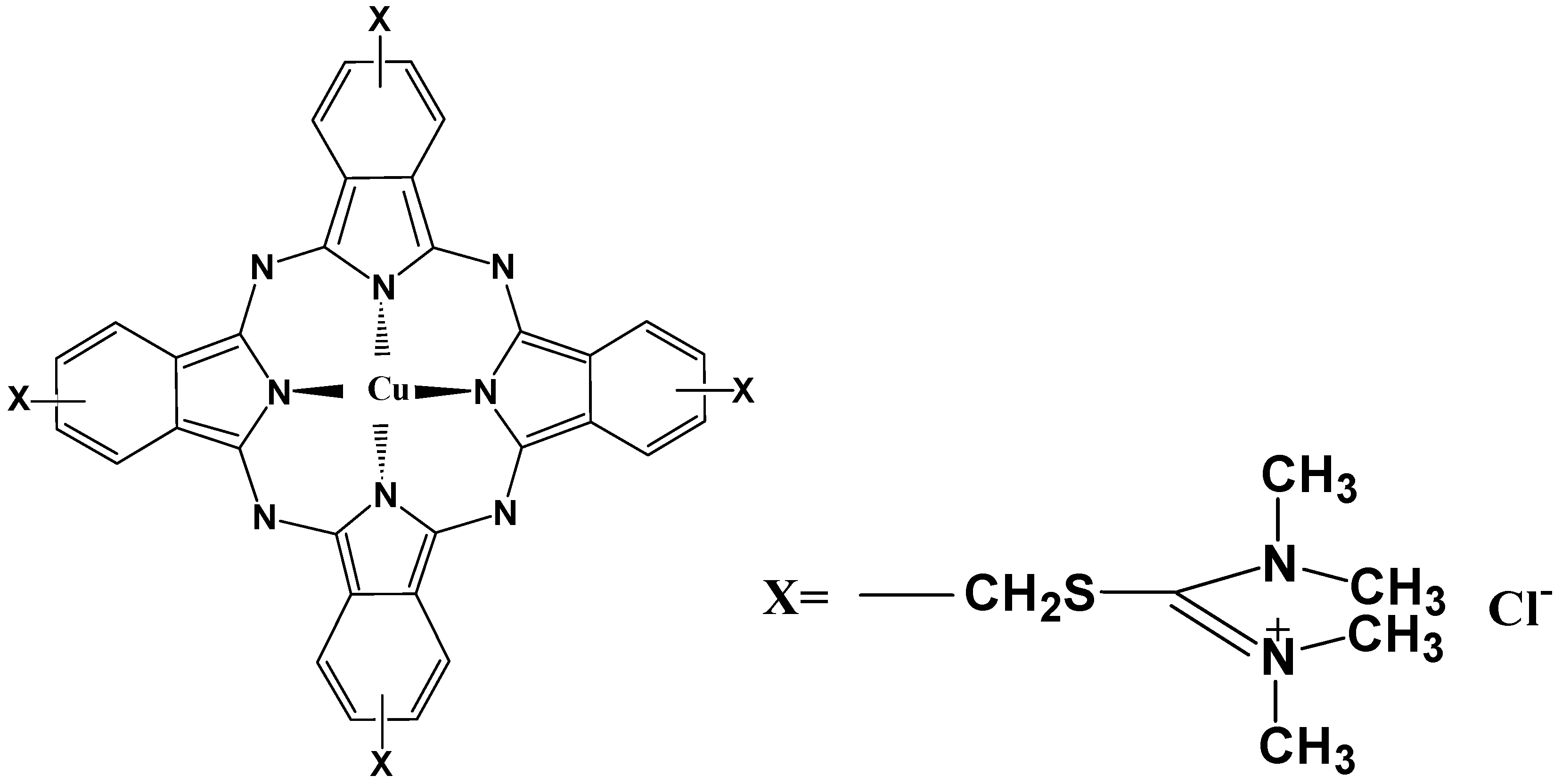
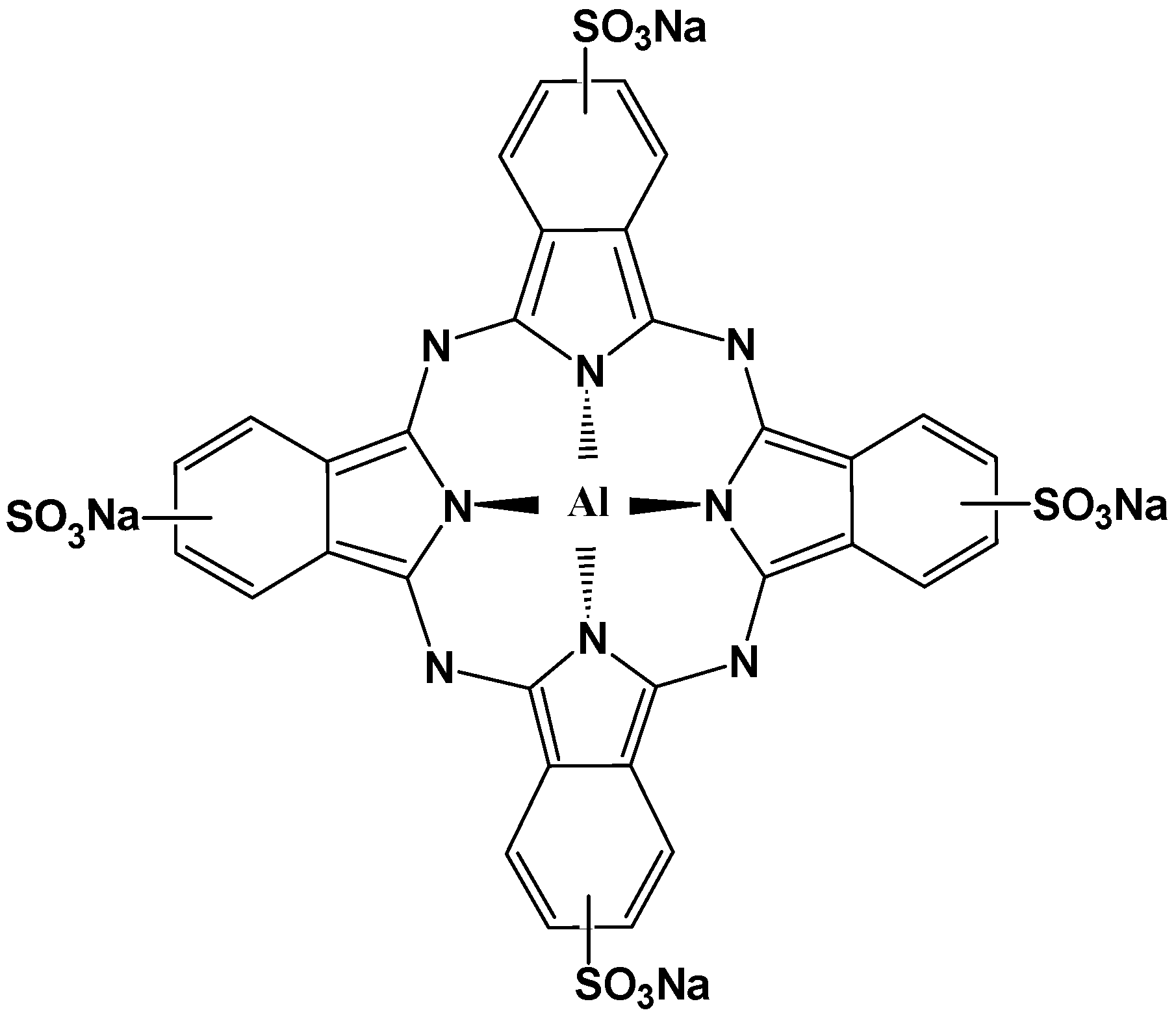
2.1. Molecular Structure of AlS4Pc and Alcian Blue 8GX

Tetrasulfoaluminium phthalocyanine (AlS4Pc) is a strong fluorescent compound with a parent porphyrin-like structure as shown in Figure 1. Its strong polar and negatively-charged sulfonic acid groups on the four peripheral benzene rings greatly enhance its water solubility thereby facilitating its application in aqueous systems. Alcian blue 8GX (Figure 2) has the same parental structure as AlS4Pc, but it does not emit fluorescence because of its central coordinating atom which is a paramagnetic divalent copper ion, and each of the peripheral benzene rings is connected with sulfur-containing positively charged groups through thioether bond.

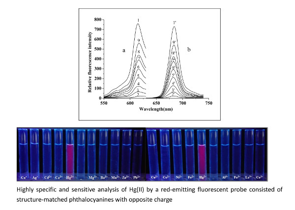
2.2. Fluorescence Properties of AlS4Pc

Tetrasulfoaluminium phthalocyanine (AlS4Pc) was found to be a fluorescent compound with high quantum yield and showed excellent photo-chemical stability [27]. With the attachment of sulphonic acid group on each of the peripheral benzenes, AlS4Pc shows large solubility in water phase. Considering these merits, AlS4Pc exhibits great potential as a novel red-emitting fluorescent probe. It has been used as a fluorescent probe and applied to quantitative analysis based on its distinct characteristics in previous works [28,29,31,32]. The fluorescence emission peak of AlS4Pc locates around 685 nm with two excitation bands; one in the short-wavelength region (called Soret band) and the other in the long-wavelength region (called Q band). As the shortwave excitation will generate secondary scattering strongly around 700 nm (near the fluorescence emission peak), the excitation wavelength is selected in this study at long wavelengths (around 620 nm) to avoid interference from scattering light.

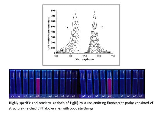
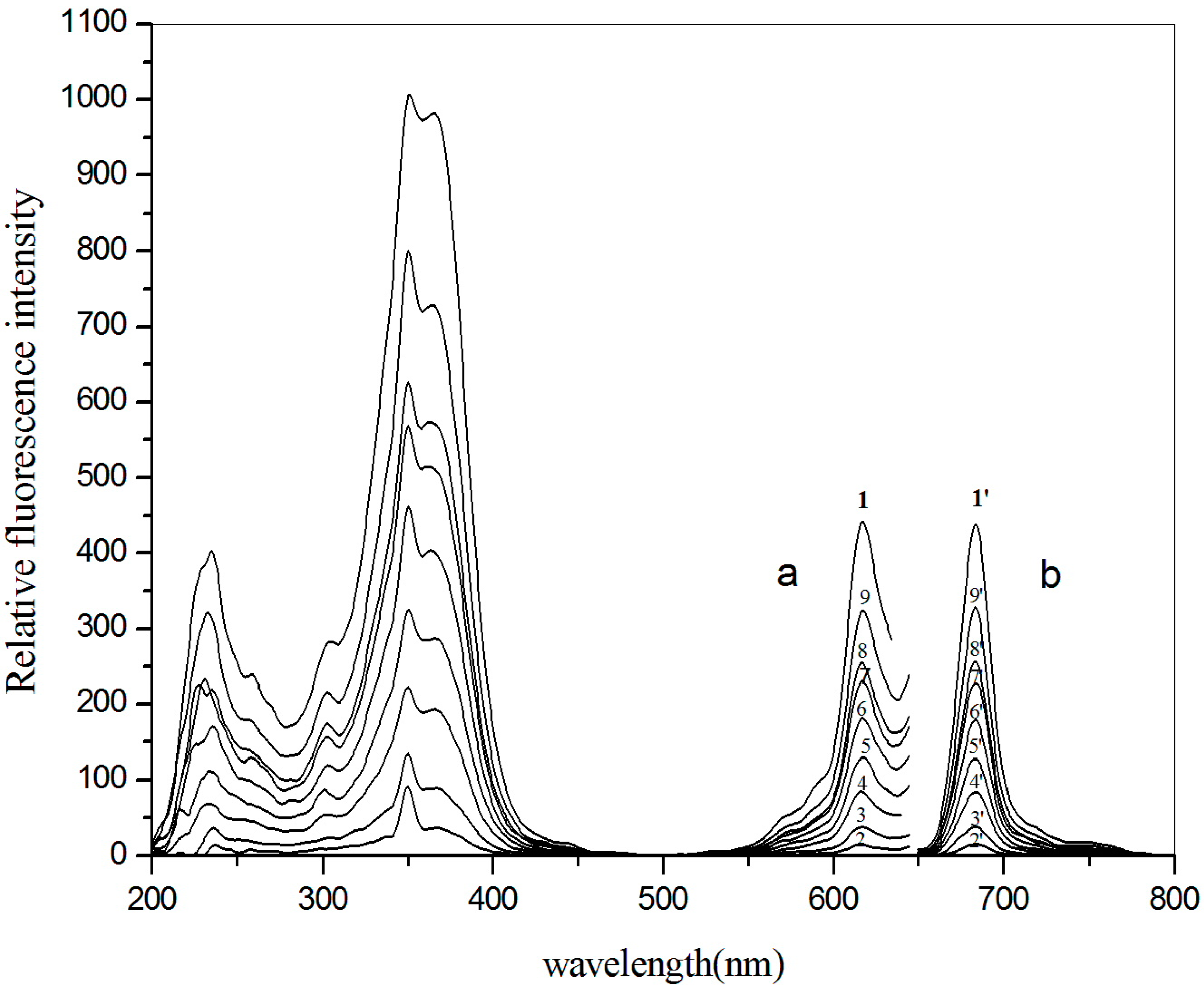
2.3. Spectral Behavior and the Mechanism of the Reaction System

In this study, the fluorescence of AlS4Pc was quenched almost completely after adding Alcian blue 8GX in acidic or weakly acidic media (Figure 3, 2-2’), indicating that Alcian blue 8GX can quench the fluorescence of AlS4Pc with high efficiency. Both AlS4Pc and Alcian blue have similar large-scale planar structure to phthalocyanine, and thus they form a strong association due to their opposite charges and planar hydrophobic interaction, leading to the formation of a non-fluorescence ion-pair associate (AlS4Pc-Alcian blue 8GX) resulting in the quenching of the fluorescence of AlS4Pc. Interestingly, the fluorescence of the above system recovered significantly after adding mercuric chloride, and the recovery degree of fluorescence was linearly and positively correlated over a wide range of Hg(II) concentrations.
It can be seen from the structure of Alcian blue 8GX (Figure 2) that there are four thioether groups which connect the parent structure of phthalocyanine and the cationic groups on periphery, indicating the presence of sulfur atoms in Alcian blue 8GX. It can be deduced that the strong binding nature of Hg(II) to sulfur atoms (Ksp of HgS up to 4 × 10−53) makes it easy to react with sulfur atoms in Alcian blue 8GX. This effect remarkably weakens the binding of Alcian blue 8GX to AlS4Pc and increases fluorescence due to the release of AlS4Pc from the AlS4Pc-Alcian blue 8GX associate. This results in a gradual increase in the concentration of free AlS4Pc in the solution, and thus the fluorescence obviously recovered with an increase in the Hg(II) (Figure 3, 3~9 and 3’~9’). Further investigations revealed that the presence of the majority of common metal ions could not restore the fluorescence of AlS4Pc-Alcian blue 8GX system (Figure 4). It indicates the high specificity for Hg(II) on AlS4Pc-Alcian blue 8GX system, which is an important finding from this study. The visualization of mercury ions can also be achieved using this specificity and the red fluorescence emission characteristics of AlS4Pc (Figure 5). We believe that the highly selective response of AlS4Pc-Alcian blue 8GX to mercury ions lies in the competitive binding effect of Hg(II) with Alcian blue 8GX. Based on the above findings, this study established a new fluorimetric method for the quantitative analysis of Hg(II) with a high selectivity, high sensitivity, and responsiveness in a wide range of concentrations.

2.4. Optimization of Experimental Conditions

2.4.1. pH and the Selection of Buffer Medium

The effects of three buffer systems (phosphate buffer of pH 1.0~12.0, B-R buffer of pH 1.0~12.0, and citric acid–disodium hydrogen phosphate buffer of pH 2.0~8.0) on the fluorescence quenching and recovery were investigated. The results showed that the fluorescence recovery ratio (n = If/If0) of Hg(II) to AlS4Pc-Alcian blue 8GX system was the largest in a buffer solution of pH = 5.0 and the fluorescence quenching ratio [Q%, Q = (IfT − If0)/IfT] was close to 100% at a pH of 5.0, so this buffer was finally chosen as the reaction medium.

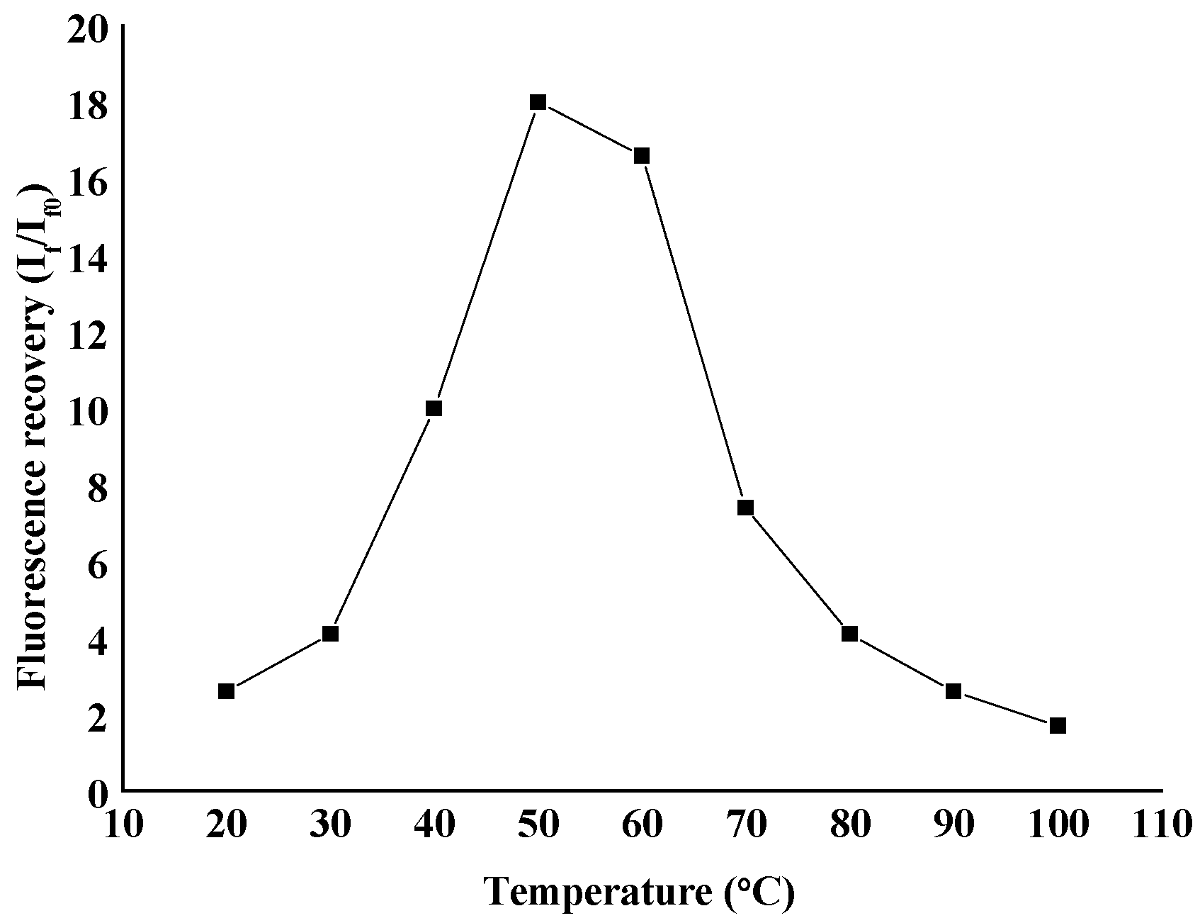
2.4.2. Selection of Temperature

The effect of reaction temperature on the fluorescence quenching and recovery of the reaction system was investigated. It was found that the degree of fluorescence quenching dropped with an increase in the temperature, indicating that this quenching system involves a static quenching process (static quenching constant decreases with an increase in temperature). In the range of 20~100 °C, the fluorescence recovery increases at first and then decreases with an increase in temperature. The fluorescence recovery of the system reached the maximum at 50 °C (Figure 6), due to which the reaction temperature was set at 50 °C.

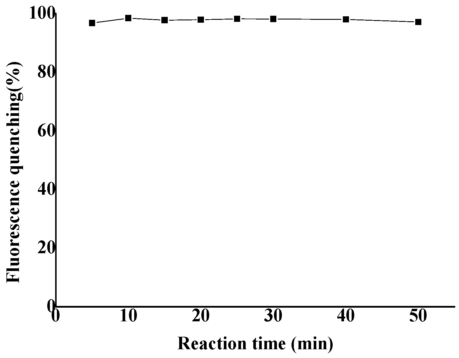
2.4.3. Selection of Reaction Time

The effect of reaction time on the fluorescence quenching and recovery of the system was also investigated. The fluorescence quenching did not show any significant change within 1 h (Figure 7), and the fluorescence recovery increased at first and then decreased with the extending reaction time. The fluorescence recovery of the system reached the maximum after 40 min, which was then chosen as the reaction time. Moreover, after cooling down the reaction for 30 min, both the fluorescence quenching and fluorescence recovery become stable. After 48 h at room temperature, they were basically unchanged, indicating that the reaction system has good stability.

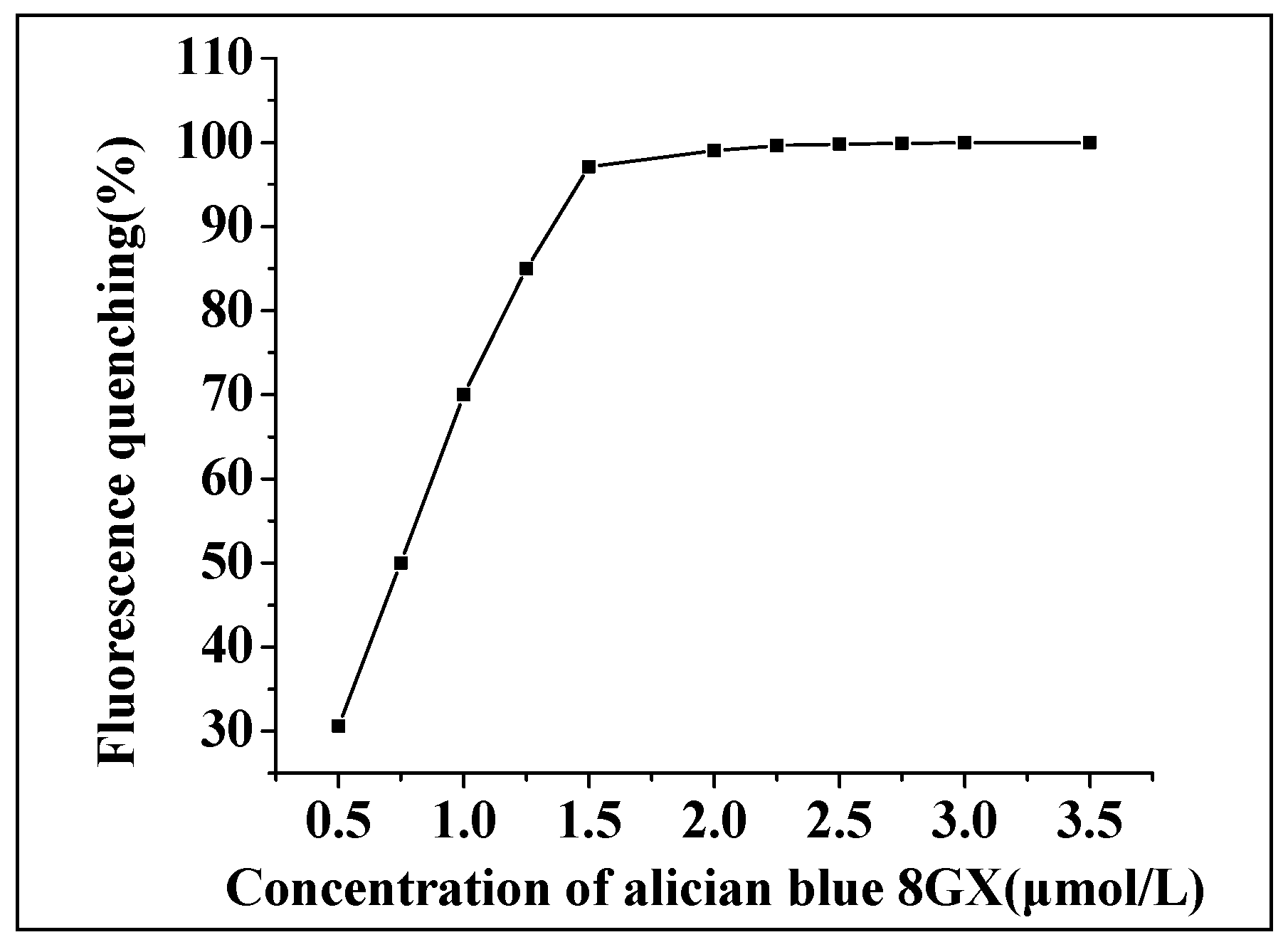
2.4.4. Investigations on the Usage of Alcian Blue 8GX

Under the given concentration (1.0 × 10⁻⁶ mol/L) of AlS4Pc and other optimized conditions, the effects of different concentrations of Alcian blue 8GX on the fluorescence quenching (Figure 8) and recovery of the reaction system were investigated. The other conditions remained the same. It was found that as the concentration of Alcian blue increased, the degree of quenching of fluorescence gradually increased until reaching a platform where the quenching value was close to 100%. The results demonstrated that the relative fluorescence intensity of the system increased at first and then decreased with a gradual addition of Alcian blue, and the relative fluorescence intensity reached the maximum when Alcian blue was at 1.50 μmol/L. Therefore, based on the inflection point of the fluorescence quenching curve and the recovery condition of the system, the dosage of Alcian blue was determined to be 1.50 μmol/L.

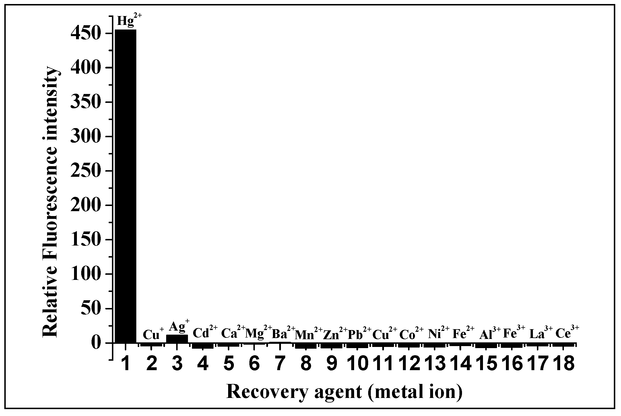
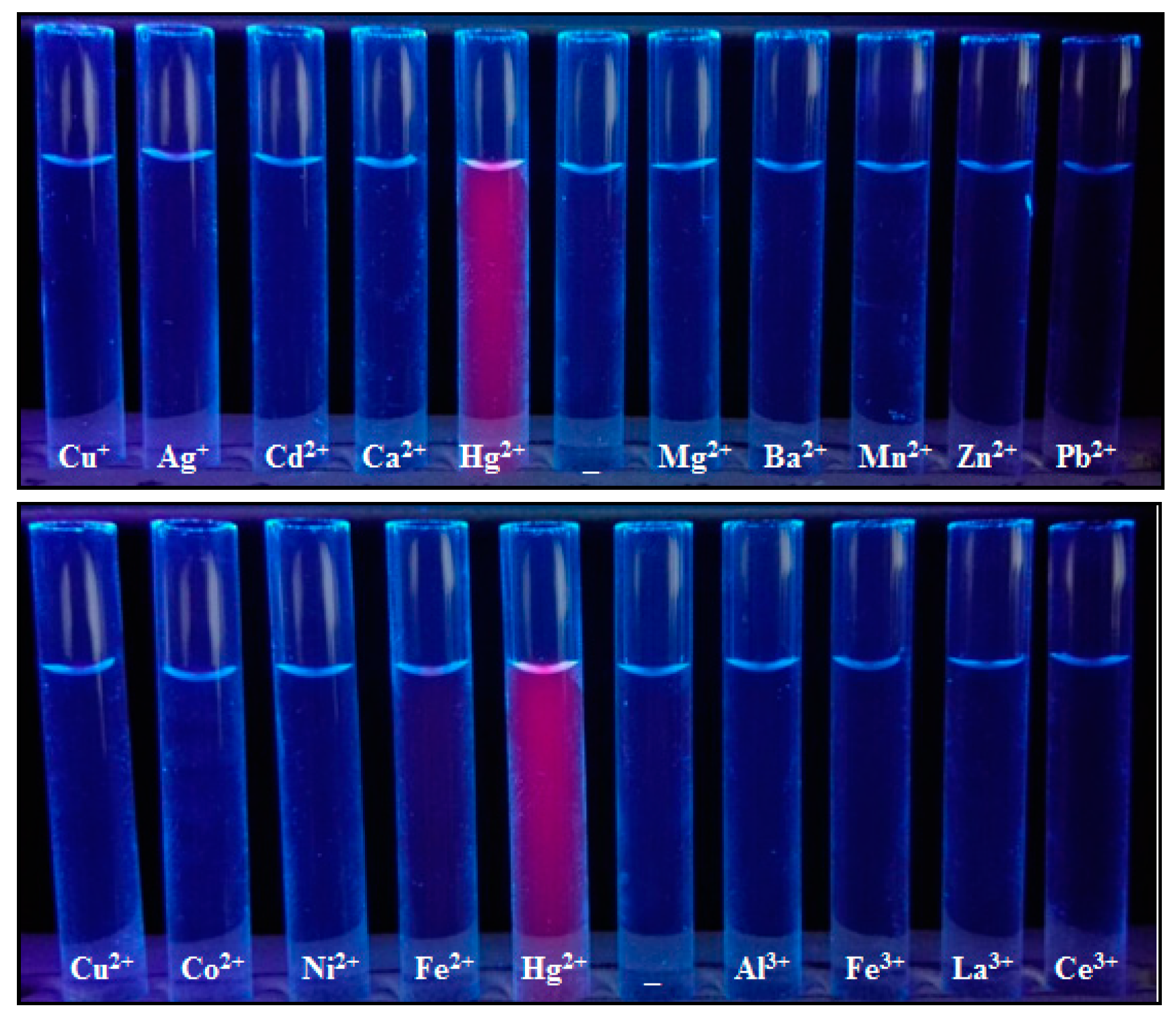
2.5. Effects of Coexisting Substances
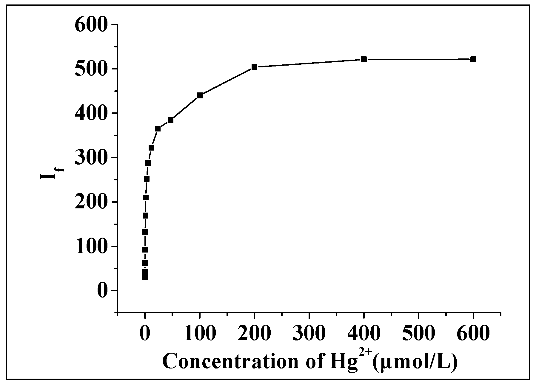
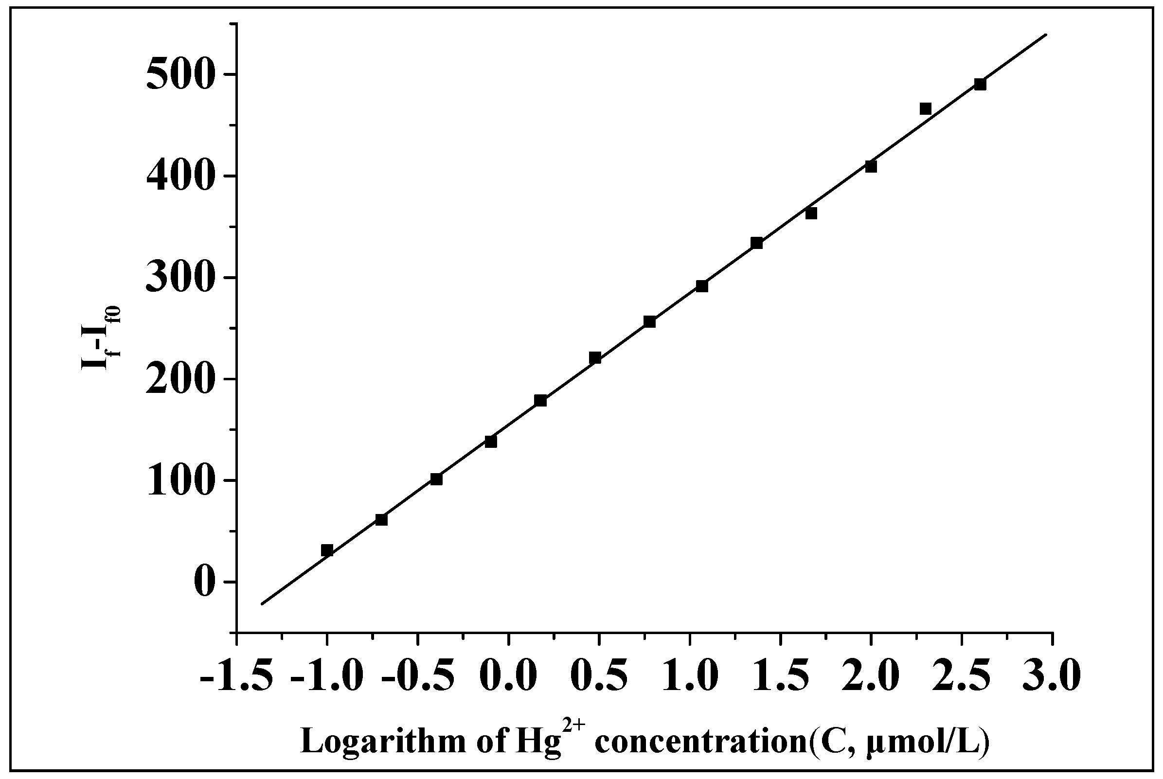
Under optimized conditions, the concentration of Hg(II) was set at 2.0 × 10⁻⁶ mol/L. The interferences of 17 types of common metal ions on the system were investigated and it was found that there were different levels of interference of various metal ions. EDTANa2 was added as a masking agent in order to solve this problem, where the concentration of EDTANa2 was screened to be of 1.0 × 10⁻⁴ mol/L. After screening 17 types of metal ions and 9 types of anions, the relative error of all the interfering metal ions was found to be less than ±10% (Table 1). In addition to the interference from I (I and Hg(II) may form [HgI4]2−, which makes Hg(II)unable to bind to Alcian blue 8GX resulting in no recovery in the fluorescence), the interference by the other eight anions (F, Cl, Br, NO3, CH3COO, SO42−, CO32−, PO43−) was very small, even when the their concentrations were up to 100 times. This indicates that the AlS4Pc-Alcian blue 8GX associate has strong anti-interference ability to most of anions and metal ions after adding the masking agent (EDTANa2).

2.6. Standard Curve

Under the optimal conditions, the fluorescence intensity of the system in the presence of different concentrations of Hg(II) was measured. It was found that the fluorescence intensity at first increased and then remained constant with an increase in the concentration of Hg(II) (Figure 9). A further mathematical analysis showed that the logarithm of the relative fluorescence intensity (ΔIf = If − If0) to Hg(II) concentration showed a good linearity within a certain order of concentration range (Figure 10). The linear regression equation (calibration curve) was obtained by plotting the logarithmic value (lgC) of the concentration of Hg(II) (C, μmol/L) to the restored relative fluorescence intensity (ΔIf = If − If0), which showed ΔIf = 154.82 + 129.71lgC, where r = 0.9994, the linear range was 0.1–400 μmol/L, and the detection limit was 0.0104 μmol/L.

2.7. Analysis of Practical Samples

The prepared fluorescent probe, AlS4Pc-Alcian blue 8GX, was used to determine the content of three types of mineral water, where the mercury ion was not detected. Subsequently, the standard solutions of different concentrations of Hg(II) were added to the above mineral water, followed by the performance of determination described in 4.2. For example, 1 mL of Lungful mineral water and 30 μL of Hg(II) solution (1.0 × 10⁻⁴ moL/L) was added in sequence to a 10 mL glass tube containing reaction reagents, followed by dilution to get a final volume of 3.0 mL. The mixed solution containing Hg(II) with a concentration of 1.0 μmoL/L was measured by a standard procedure, and the recovery was calculated (112.5%). The results obtained from these studies have been shown in Table 2. From these results, it could be noted that the recoveries of Hg(II) in three types of mineral water were satisfactory when the scalar quantity (e.g., the concentration of Hg(II) added is 0.1 µmol/L) was low, so the fluorescent probe could be used for the determination of Hg(II) content in simple real samples at present.

3. Materials and Methods

3.1. Materials

Tetrasulfoaluminium phthalocyanine (AlS4Pc) was purchased from J&K Technology Co., Ltd. (Beijing, China) and stored at 4 °C in the dark at a concentration of 1.0 × 10⁻² mol/L. Alcian blue 8GX was supplied by Bioengineering Co., Ltd. (Shanghai, China) and stored at a concentration of 1.0 × 10⁻² mol/L, and diluted to 1.0 × 10⁻⁴ mol/L before use; Disodium ethylenediaminetetraacetate (Wuhan Chemical Plant); mercuric chloride (Tonghuo Mercury Reagent Factory, Guizhou, China) were utilized as obtained; pH buffer solutions in a wide range (mixed with hydrochloric acid, dipotassium hydrogen phosphate, potassium dihydrogen phosphate, and sodium phosphate); Britton–Robinson (B-R) buffer (mixed preparation with phosphoric acid, acetic acid, boric acid, and sodium hydroxide) and disodium hydrogen phosphate–citrate buffer were used. All other reagents are of analytical grade and the water used was of high purity.

3.2.Experimental Methods

The following solutions were prepared in a beaker: 30 μL of AlS4Pc (1.0 × 10⁻⁴ mol/L), 45.0 μL of Alcian blue 8GX (1.0 × 10−4 mol/L), 30.0 μL of EDTANa2 (1.0 × 10⁻² mol/L), and 300.0 μL of buffer with pH = 5.0. By adding water, 405 μL of the above mixture and Hg(II)solution to a 10.0 mL glass tube, the final volume was made to 3.0 mL, and mixed thoroughly using a vortex mixer. The solution was then heated in a water bath at 50 °C for 40 min and then cooled for 30 min before subjecting it to the measurements of fluorescence. The excitation wavelength was at 613 nm and the excitation slit was 5.0 nm, whereas the emission slit was 4.5 nm. The fluorescence intensity of AlS4PC tube without Alcian blue 8GX and Hg(II) was recorded as IfT. The blank tube with intensity recorded as If0 without Hg(II). The fluorescence intensity of sample tubes with Hg(II) was recorded as If. The fluorescence quenching ratio (Q%) was then calculated by, Q = (IfT − If0)/IfT; whereas the fluorescence recovery factor (n) was calculated by, n = If/If0; and the difference in the fluorescence intensity before and after recovery could be expressed by ΔIf = If − If0.

4. Conclusions

In summary, the AlS4Pc-Alcian blue 8GX ion-pair fluorescence probe with low fluorescence background was constructed based on the high-efficiency fluorescence quenching effect of Alcian Blue 8GX on AlS4Pc. The screening experiments for common metal ions revealed that only Hg(II) could significantly restore the fluorescence of AlS4Pc-Alcian blue 8GX, indicating that AlS4Pc-Alcian blue 8GX probe has high specificity for the recognition of Hg(II). Inspired by these results, a new fluorimetric method has been established with high specificity and high sensitivity for the quantitative analysis of Hg(II), and its response range is found to be more than three orders of magnitude. It confirms that AlS4Pc-Alcian blue 8GX ion associate is promising as an excellent red fluorescent probe which further opens up new applications of red-emitting fluorescent probes based on functional phthalocyanine compounds. What is more worth mentioning is this study has proposed a novel strategy for constructing new fluorescent probes through molecular assembly. By the association of structure-matched molecules, novel fluorescent probes could be achieved with high specificity to a specific species. Compared with ‘turn-on’ fluorophores reported in literature, compounds used in this study for constructing the associate are commercially available, thus opening an easy way to achieve fluorescent probes without cumbersome organic synthesis. Besides, the constructed probe is fully water-soluble. It can be expected that a series of functional phthalocyanine based fluorescent probes would be developed according to this principle.

Acknowledgments

This work was supported by the Major Science and Technology Project of Fujian Province (no. 2012Y0081), the Basic Scientific Research Project of Fujian Provincial Public Research Institute (no. 2016R1101032-5, 2017R1036-2), all the authors wish to express their grateful acknowledgements.

Author Contributions

Dong-Hui Li conceived and designed the experiments; Guang-Xin Du, Tao Zhou, Meng-Lin Guo, Ping Huang, and Ya-Bin Deng performed the experiments or analyzed the data; Guang-Xin Du and Dong-Hui Li wrote the paper; Dong-Hui Li and Ping Huang supervised the study and proofread the manuscript.

Conflicts of Interest

The authors declare no conflict of interests.

References

  1. Harris, H.H.; Pickering, I.J.; George, G.N. The chemical form of mercury in fish. Science 2003, 301, 1203. [Google Scholar] [CrossRef] [PubMed]
  2. Renzoni, A.; Zino, F.; Franchi, E. Mercury levels along the food chain and risk for exposed populations. Environ. Res. 1998, 77, 68–72. [Google Scholar] [CrossRef] [PubMed]
  3. Nolan, E.M.; Lippard, S.J. Tools and tactics for the optical detection of mercuric ion. Chem. Rev. 2008, 108, 3443–3480. [Google Scholar] [CrossRef] [PubMed]
  4. Krishna, M.V.B.; Ranjit, M.; Karunasagar, D.; Arunachalam, J. A rapid ultrasound-assisted thiourea extraction method for the determination of inorganic and methyl mercury in biological and environmental samples by CVAAS. Talanta 2005, 67, 70–80. [Google Scholar] [CrossRef] [PubMed]
  5. Yardim, M.F.; Budinova, T.; Ekinci, E.; Petrov, N.; Razvigorova, M.; Minkova, V. Removal of mercury (II) from aqueous solution by activated carbon obtained from furfural. Chemosphere 2003, 52, 835–841. [Google Scholar] [CrossRef]
  6. Segade, S.R.; Tyson, J.F. Determination of methylmercury and inorganic mercury in water sample by slurry sampling cold vapor atomic absorption spectrometry in a flow injection system after preconcentration on silica C18 modified. Talanta 2007, 71, 1696–1702. [Google Scholar] [CrossRef] [PubMed]
  7. Zhang, Y.L.; Adeloju, S.B. A novel sequential injection-cold vapour atomic absorption spectrometric system for rapid and reliable determination of mercury. Talanta 2008, 74, 951–957. [Google Scholar] [CrossRef] [PubMed]
  8. Zhang, X.H.; Wang, Q.; Zhao, Q. Determination of mercury in sodium hydroxide as food additives by hydride generation-atomic fluorescence spectrometry. Chin. J. Health Lab. Technol. 2008, 18, 1324–1332. [Google Scholar]
  9. Yu, F.; Yu, T.L. Determination of ultra-trace lead and mercury in groundwater by hydride generation—Atomic Fluorescence Spectrometry with sulfhydryl cotton. Spectrosc. Spectr. Anal. 2000, 20, 898–900. [Google Scholar]
  10. Boaventura, G.R.; Barbosa, A.C.; East, G.A. Multivessel system for cold-vapor mercury generation determination of mercury in hair and fish. Ecol. Biol. Trace Elem. Res. 1997, 60, 153–161. [Google Scholar] [CrossRef] [PubMed]
  11. Seibert, E.L.; Dressler, V.L.; Pozebon, D.; Curtius, A.J. Determination of Hg in seawater by inductively coupled plasma mass spectrometry after on-line pre-concentration. Spectrochim. Acta Part B 2001, 56, 1963–1971. [Google Scholar] [CrossRef]
  12. Iwashita, A.; Nakajima, T.; Takanashi, H.; Ohki, A.; Fujita, Y.; Yamashita, T. Determination of trace elements in coal fly ash by joint-use of ICP-AES and atomic absorption spectrometry. Talanta 2007, 71, 251–257. [Google Scholar] [CrossRef] [PubMed]
  13. Jones, P.; Hardy, S. Development of a capillary electrophoretic method for the separation and determination of trace inorganic and organomercury species utilizing the formation of highly absorbing water soluble dithizone sulphonate complexes. J. Chromatogr. A 1997, 765, 345–352. [Google Scholar] [CrossRef]
  14. Young, T.K.; Yun, J.B.; Yoon, B.S. Determination of Hg2+ ions using a modified glassy carbon electrode with 2,2′:6′:2″-Terpyridine. Bull. Korean Chem. Soc. 2002, 23, 346–350. [Google Scholar]
  15. Liu, Z.H.; Huan, S.Y.; Jiang, J.H.; Shen, G.L.; Yu, R.Q. Molecularly imprinted TiO2 thin film using stable ground state complex as template as applied to selective electrochemical determination of mercury. Talanta 2006, 68, 1120–1125. [Google Scholar] [CrossRef] [PubMed]
  16. Tabak, M.; Borisevitch, I.E. Interaction of dipyridamole with micelles of lysophosphatidylcholine and with bovine serum-albumin-fluorescence studies. Biochim. Biophys. Acta 1992, 1116, 241–249. [Google Scholar] [CrossRef]
  17. Husain, N.; Agbaria, R.A.; Warner, I.M. Spectroscopic analysis of the binding of doxorubicin to human alpha-1-acid glycoprotein. J. Phys. Chem. 1993, 97, 10857–10861. [Google Scholar] [CrossRef]
  18. Karoui, R.; Bosset, J.O.; Mazerolles, G.; Kulmyrzaev, A.; Dufour, E. Monitoring the geographic origin of both experimental French Jura hard cheeses and Swiss Gruyere and L’Etivaz PDO cheeses using mid-infrared and fluorescence spectroscopies: A preliminary investigation. Int. Dairy J. 2005, 15, 275–286. [Google Scholar] [CrossRef]
  19. Wen, Z.Q. Raman spectroscopy of protein pharmaceuticals. J. Pharm. Sci. 2007, 96, 2861–2878. [Google Scholar] [CrossRef] [PubMed]
  20. Liu, R.; Wu, P.; Xu, K.L.; Lv, Y.; Hou, X.D. Highly sensitive and interference-free determination of bismuth in environmental samples by electrothermal vaporization atomic fluorescence spectrometry after hydride trapping on iridium-coated tungsten coil. Spectrochim. Acta Part B 2008, 63, 704–709. [Google Scholar] [CrossRef]
  21. De Silva, A.P.; Gunaratne, H.N.; Gunnlaugsson, T.; Huxley, A.J.M.; McCoy, C.P.; Rademacher, J.T.; Rice, T.E. Signaling recognition events with fluorescent sensors and switches. Chem. Rev. 1997, 97, 1515–1566. [Google Scholar] [CrossRef] [PubMed]
  22. Kim, H.N.; Ren, W.X.; Kim, J.S.; Yoon, J. Fluorescence and colorimetric sensors for detection of lead, cadium, and mercury ion. Chem. Soc. Rev. 2012, 41, 3210–3244. [Google Scholar] [CrossRef] [PubMed]
  23. Chen, G.; Guo, Z.; Zeng, G.; Tang, L. Fluorescent and colorimetric sensors for environmental mercury detection. Analyst 2015, 140, 5400–5443. [Google Scholar] [CrossRef] [PubMed]
  24. Vedamalai, M.; Wu, S.P. A BODIPY-based reactive probe for Hg(II) ions and its application to living cell imaging. Org. Biomol. Chem. 2012, 10, 5410–5416. [Google Scholar] [CrossRef] [PubMed]
  25. Venkatesan, P.; Thirumalivasan, N.; Wu, S.P. A Rhodamine-based chemosensor with diphenylselenium for highly selective fluorescence turn-on detection of Hg2+ in vitro and in vivo. RSC Adv. 2017, 7, 21733–21739. [Google Scholar] [CrossRef]
  26. Dong, M.; Wang, Y.W.; Peng, Y. Highly selective ratiometric fluorescent sensing for Hg2+ and Au3+ respectively in aqueous media. Org. Lett. 2010, 12, 5310–5313. [Google Scholar] [CrossRef] [PubMed]
  27. Ambroz, M.; Beeby, A.; MacRobert, A.J.; Simpson, M.S.C.; Svensen, R.K.; Phillips, D. Preparative, analytical and fluorescence spectroscopic studies of sulphonated aluminum phthalocyanine photosensitizers. J. Photochem. Photobiol. B 1991, 9, 87–95. [Google Scholar] [CrossRef]
  28. Yu, F.; Guo, M.L.; Deng, Y.B.; Yin, L.; Chen, L.; Huang, P.; Li, D.H. Structure-matched phthalocyanine ion pair as a red-emitting fluorescent optical probe for the analysis of sodium dodecylbenzenesulfonate with high specificity and sensitivity. Anal. Sci. 2016, 32, 201–205. [Google Scholar] [CrossRef] [PubMed]
  29. Chen, L.; Huang, P.; Yang, H.Q.; Deng, Y.B.; Guo, M.L.; Li, D.H. Sensitive determination of chondroitin sulfate by fluorescence recovery of an anionic aluminum phthalocyanine-cationic surfactant ion-association complex used as a fluorescent probe emitting at red region. Spectrosc. Spectr. Anal. 2015, 35, 2203–2207. [Google Scholar]
  30. Chen, L.; Huang, P.; Yang, H.Q.; Deng, Y.B.; Li, D.H. Novel Method for Determination of Lysozyme Based on Fluorescence Recovery of a Cationic Aluminum Phthalocyanine-Mucopolysaccharides Association Complex Used as a Red Emitting Fluorogenic Substrate. Chin. J. Anal. Chem. 2014, 42, 962–967. [Google Scholar]
  31. Li, D.H.; Chen, X.L.; Fang, Y.; Xu, J.G. Determination of nucleic acids based on shifting the association equilibrium between tetrasulfonated aluminium phthalocyanine and Acridine Orange. Analyst 2001, 126, 518–522. [Google Scholar] [CrossRef] [PubMed]
  32. Li, D.H.; Yang, H.H.; Zheng, H.; Fang, Y.; Zhu, Q.Z.; Xu, J.G. Fluorimetric determination of albumin and globulin in human serum using tetra-substituted sulphonated aluminum phthalocyanine. Anal. Chim. Acta 1999, 401, 185–189. [Google Scholar] [CrossRef]
Figure 1. Molecular structure of AlS4Pc.
Molecules 23 00418 g001
Figure 2. Molecular structure of Alcian blue 8GX.
Molecules 23 00418 g002
Figure 3. Excitation (a) and emission (b) of AlS4PC-Alcian blue 8GX in the presence of Hg(II) in an aqueous medium with a pH of 5.0. 1-1’. Buffer + AlS4Pc, [AlS4Pc] = 1.0 × 10⁻⁶ mol/L, [EDTANa2] = 1.0 × 10⁻⁴ mol/L. 2-2’. Buffer + AlS4Pc + Alcian blue 8GX, [AlS4Pc] = 1.0 × 10⁻⁶ mol/L, [Alcian blue 8GX] = 1.50 × 10⁻⁶ mol/L, [EDTANa2] = 1.0 × 10⁻⁴ mol/L; 3-3’, 4-4’, 5-5’, 6-6’, 7-7’, 8-8’, 9-9’. Buffer + AlS4Pc + Alcian blue 8GX + Hg(II), [AlS4Pc] = 1.0 × 10⁻⁶ mol/L, [Alcian blue 8GX] = 1.50 × 10⁻⁶ mol/L, [EDTANa2] = 1.0 × 10⁻⁴ mol/L, concentrations of Hg(II) for curves 3–9 and 3’–9’: 0.2, 0.4, 2.0, 10.0, 50.0, 100.0 and 200.0 µmol/L, respectively.
Molecules 23 00418 g003
Figure 4. Comparison of recovery effect on the fluorescence of AlS4Pc-Alcian blue 8GX in the presence of different metal ions in an aqueous medium with a pH of 5.0 containing EDTA. The concentration of AlS4Pc was 1.0 × 10⁻⁶ mol/L, Alcian blue 8GX was 1.50 × 10⁻⁶ mol/L, and EDTANa2 was 1.0 × 10⁻⁴ mol/L. The concentration of all of the metal ions added was 1.0 × 10⁻⁴ mol/L.
Molecules 23 00418 g004
Figure 5. Naked-eye observation of the fluorescence recovery for AlS4PC-Alcian blue 8GX in the presence of different metal ions including Hg(II) in an aqueous medium. The concentration of AlS4Pc was 1.0 × 10⁻⁶ mol/L, Alcian blue 8GX was 1.50 × 10⁻⁶ mol/L and EDTANa2 was 1.0 × 10⁻⁴ mol/L. The concentration of all of the metal ions added was 1.0 × 10⁻⁴ mol/L.
Molecules 23 00418 g005
Figure 6. The influence of temperature on the fluorescence recovery of Alcian 8GX—AlS4Pc.
Molecules 23 00418 g006
Figure 7. The influence of reaction time on the fluorescence quenching Alcian 8GX on AlS4Pc. The concentration of AlS4Pc, Alcian blue 8GX, and Hg(II) were 1.0 × 10⁻⁶ mol/L, 1.50 × 10⁻⁶ mol/L, and 1.0 × 10⁻⁵ mol/L, respectively.
Molecules 23 00418 g007
Figure 8. The effect of dosage of Alcian blue 8GX on the degree of fluorescence quenching of the system. The concentrations of AlS4Pc and Hg(II) were 1.0 × 10⁻⁶ mol/L and 1.0 × 10⁻⁵ mol/L, respectively.
Molecules 23 00418 g008
Figure 9. Relationship between fluorescence intensity and the concentration of Hg(II).
Molecules 23 00418 g009
Figure 10. The calibration curve for the determination of Hg(II).
Molecules 23 00418 g010
Table 1. Effect of 17 metal ions on the determination of Hg(II).
Metal IonsConcentration (mol/L)Relative Error/%Metal IonsConcentration (mol/L)Relative Error/%
Ag+2.0 × 10⁻⁵+8.71Fe3+1.0 × 10⁻⁵−7.99
Cu2+4.0 × 10⁻⁵−7.42Al3+1.5 × 10⁻⁵−7.53
Cd2+1.0 × 10⁻⁵−9.33Ce3+1.0 × 10⁻⁵−9.25
Ca2+2.0 × 10⁻⁵−7.11La3+1.0 × 10⁻⁵−8.55
Ba2+8.0 × 10⁻⁵−5.65F1.0 × 10−5−7.25
Mg2+6.0 × 10⁻⁵−8.84Cl2.0 × 10−4−4.64
Co2+1.0 × 10⁻⁵−9.45Br2.0 × 10−5−6.75
Ni2+1.0 × 10⁻⁵−9.01I2.0 × 10−6−40.37
Cu2+2.0 × 10⁻⁵−8.84N032.0 × 10−4−1.71
Zn2+1.0 × 10⁻⁵−9.31SO42−2.0 × 10⁻⁴−2.53
Mn2+1.0 × 10⁻⁵−9.01CO32−2.0 × 10−57.17
Pb2+2.0 × 10⁻⁵−8.52PO43−2.0 × 10−45.06
Fe2+1.5 × 10⁻⁵−5.81CH3COO2.0 × 10−40.84
([Hg(II)] = 2.0 × 10⁻⁶ mol/L).
Table 2. Determination of Hg(II) in three mineral water samples by the present method.
SamplesHg(II) Added (µmol/L)Hg(II) Detected (µmol/L)Recovery (%)
Lungful Shanquan
(pH 6.89)
10Not detected_
20.10.107 a ± 0.12 b107.0
31.01.125 a ± 0.23 b112.5
Baisuishan
(pH 6.86)
10Not detected_
20.10.108 a ± 0.04 b108.0
31.01.201 a ± 0.21 b120.1
Biaozhi
(pH 7.03)
10Not detected_
20.10.104 a ± 0.05 b104.0
31.00.999 a ± 0.13 b99.9
a average of three results, b standard deviation.

