Discovery of 3-Amino-2-Hydroxypropoxyisoflavone Derivatives as Potential Anti-HCV Agents

 ,
,
Abstract
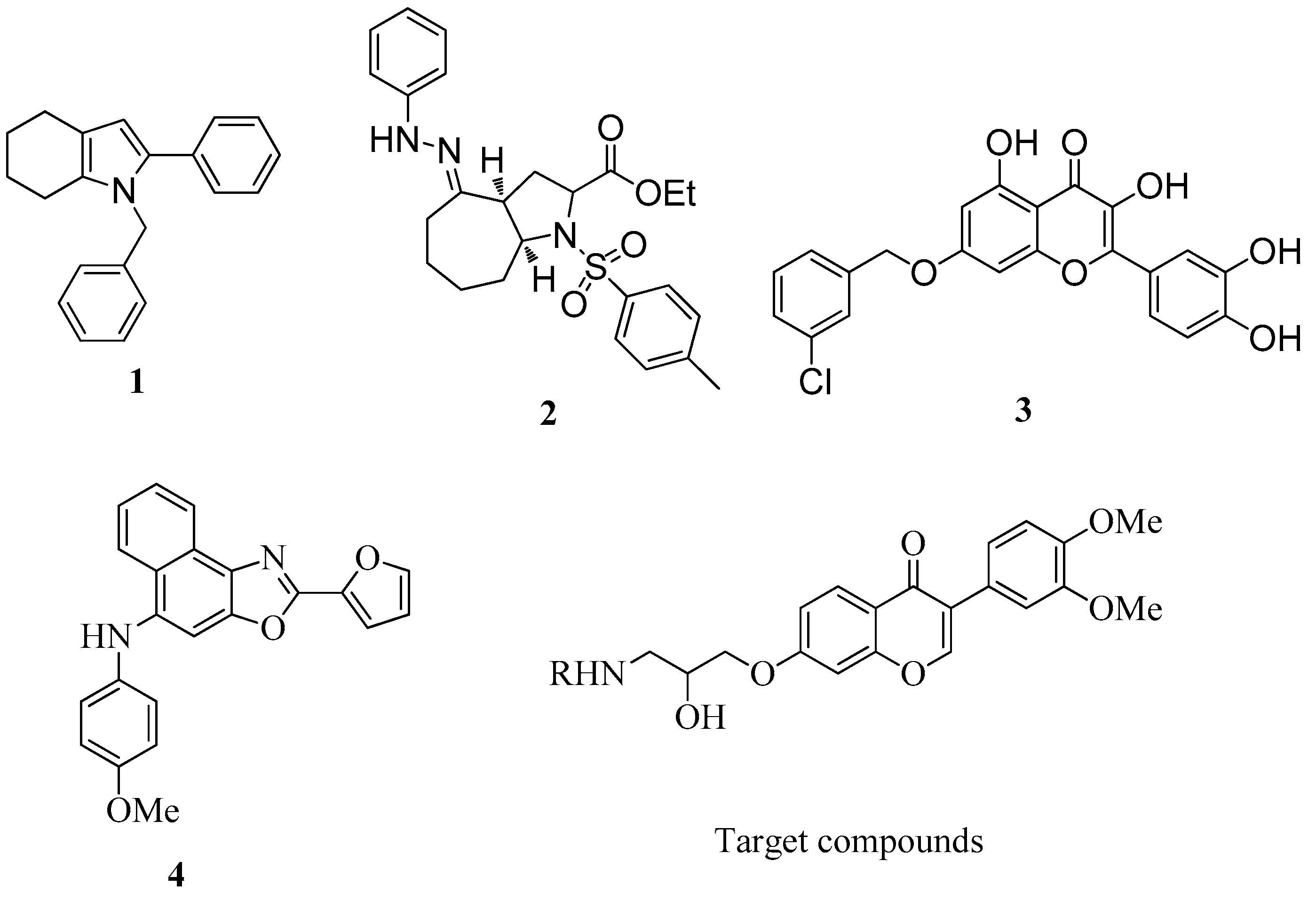
1. Introduction
2. Results and Discussion
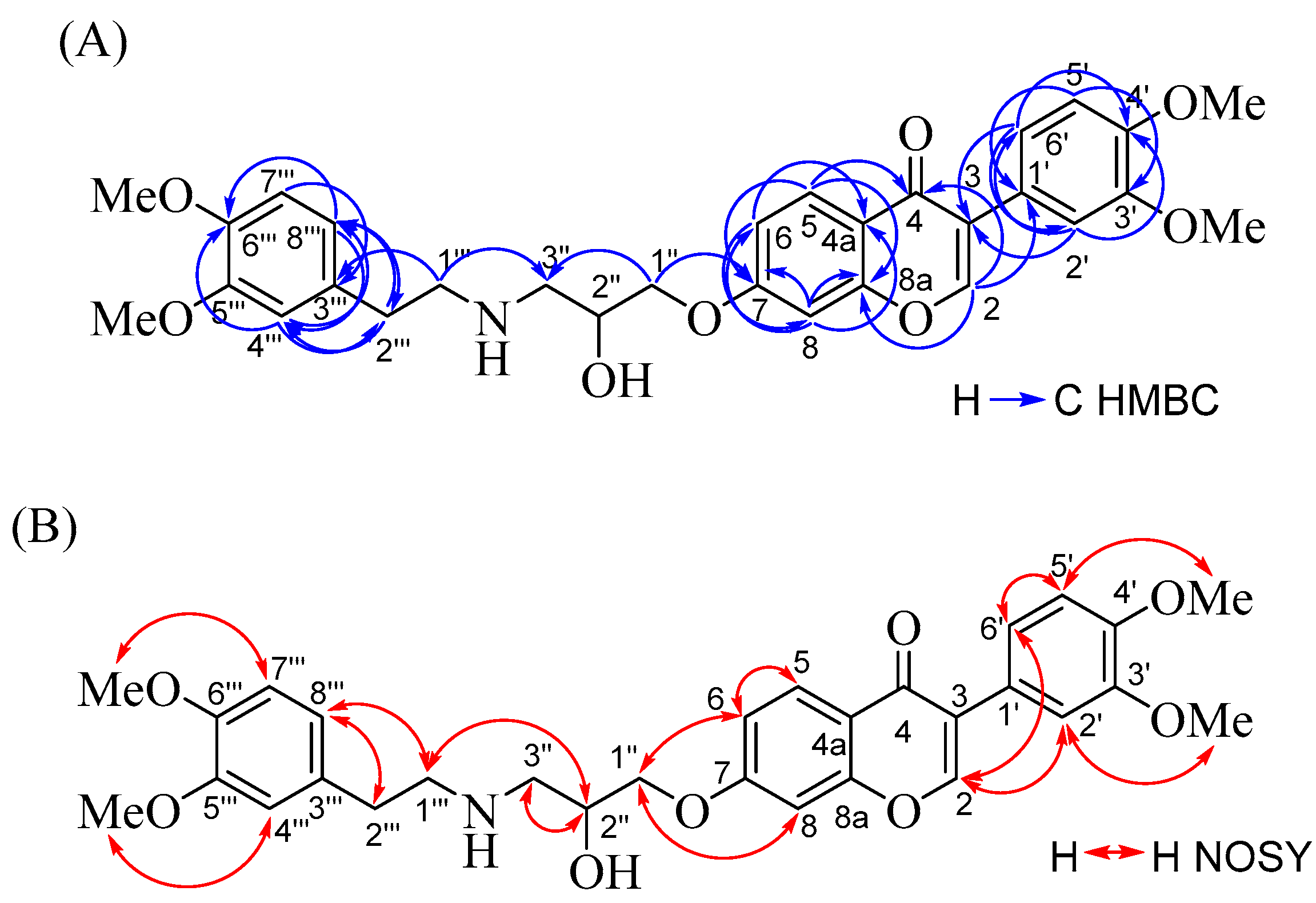
2.1. Chemistry
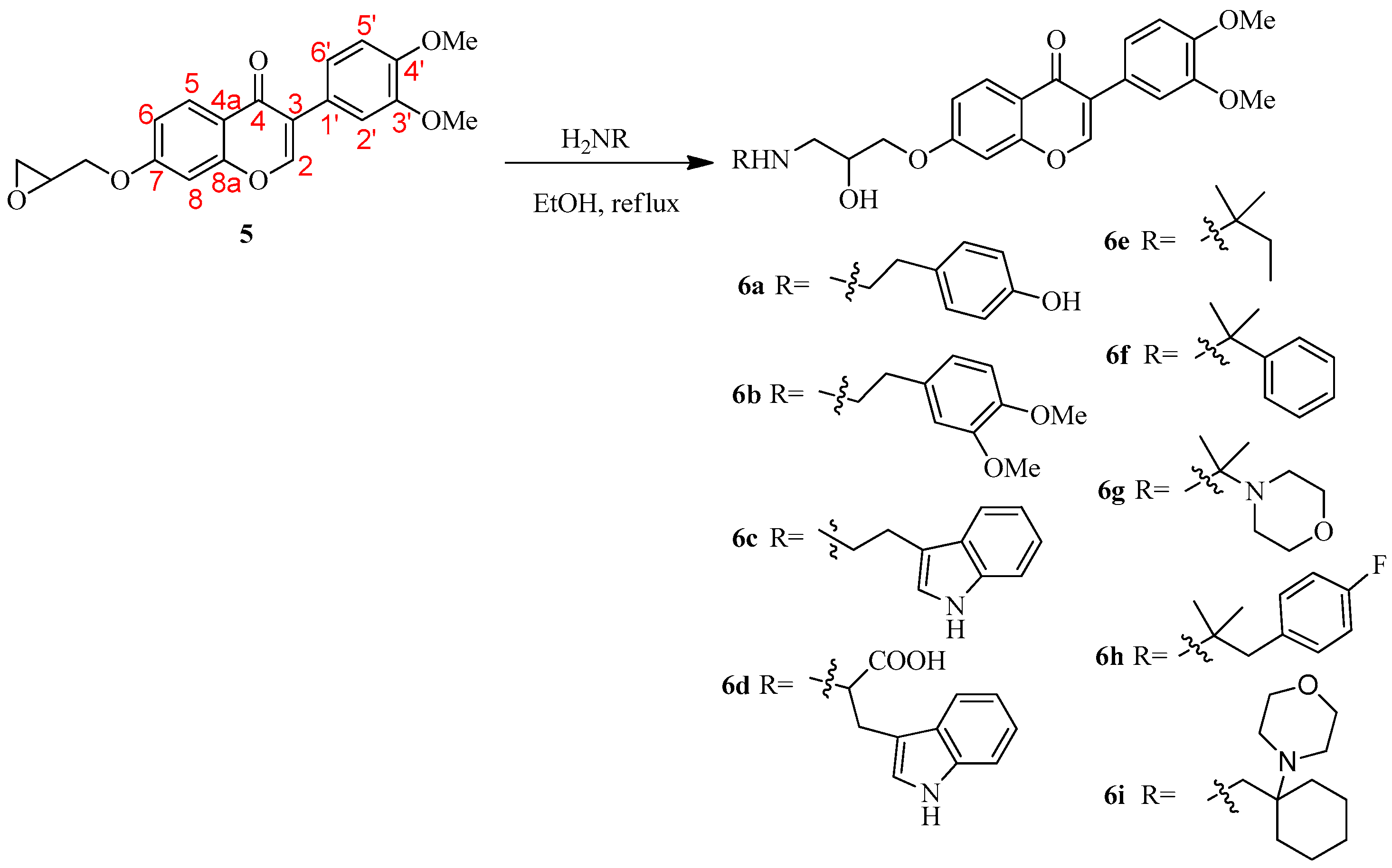
Preparation of 3-Amino-2-Hydroxypropoxyisoflavone Derivatives
2.2. Biological Activities
2.2.1. Anti-HCV Activities and Cytotoxicities
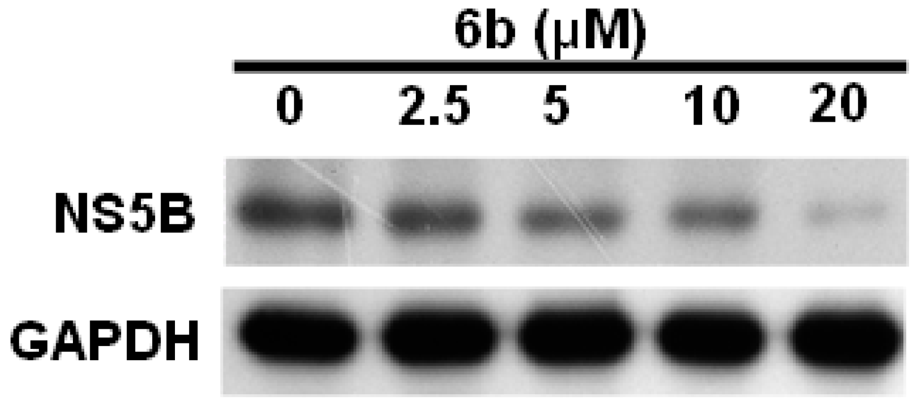
2.2.2. Compound 6b Reduced HCV Replication in HCV-Infected Ava-5 Cells
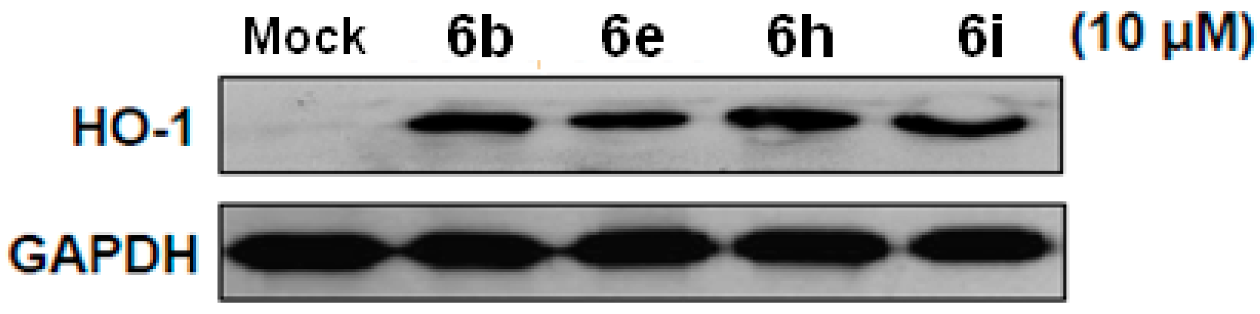
2.2.3. Isoflavones Reduced HCV Replication through Inducing HO-1 Protein Expression
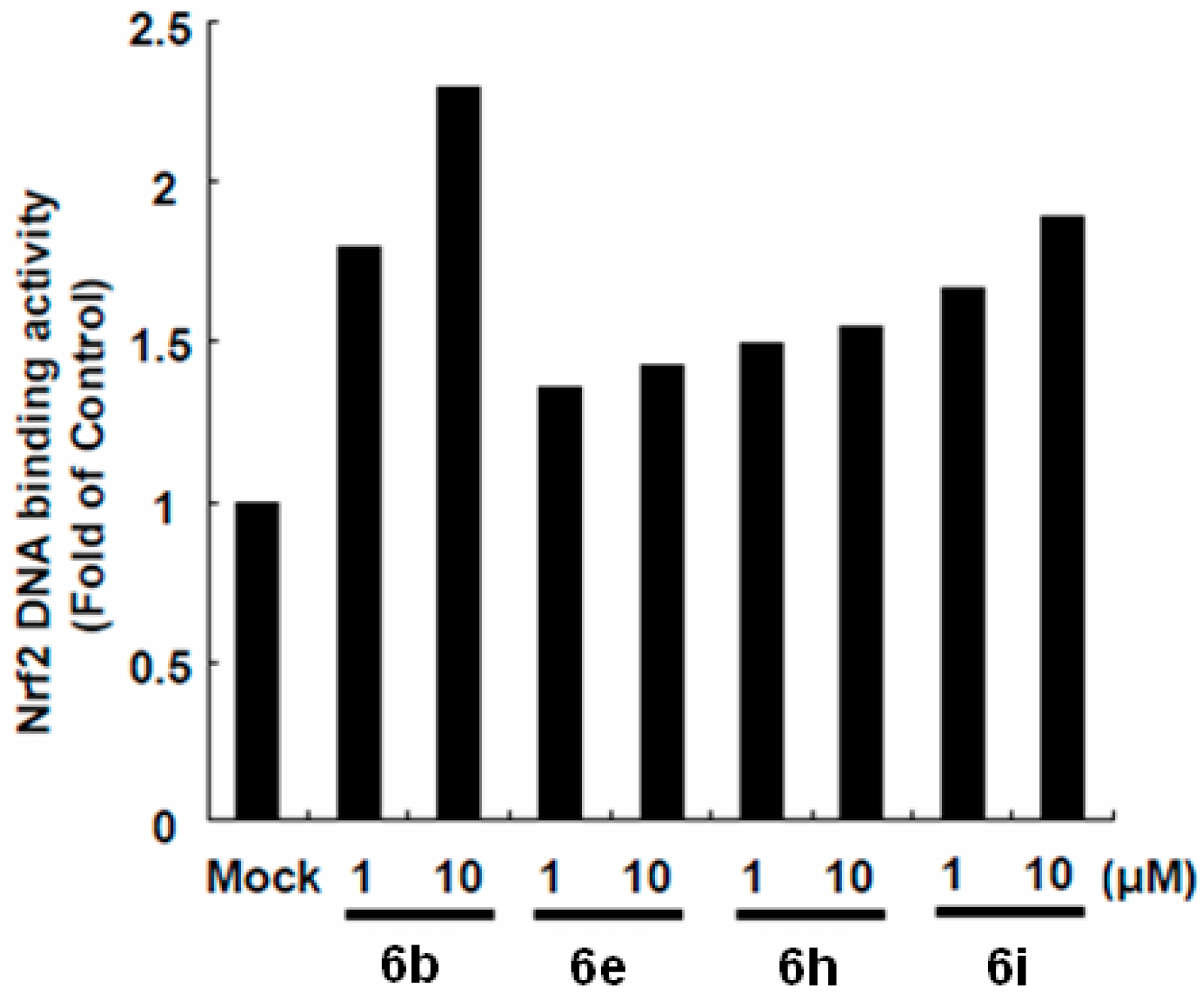
2.2.4. Isoflavones Up-Regulates Nrf2 Transactivating HO-1 Expression to Inhibit HCV Replication
3. Experimental
3.1. Materials and Methods
3.1.1. Chemical Reactions
General
General Procedure for the Preparation of 3-Amino-2-Hydroxypropoxyisoflavone Compounds 6a–i
3.1.2. Cytotoxicity and Antiviral Activity Assays
Compounds
Cell
Cytotoxicity Assays
Transfection and Luciferase Activity Assay
Immunoblot Analysis
4. Conclusions
Supplementary Materials
Author Contributions
Funding
Acknowledgments
Conflicts of Interest
References
- Gravitz, L. Introduction: A smouldering public-health crisis. Nature 2011, 474, 2–4. [Google Scholar] [CrossRef] [PubMed]
- Mohd Hanafiah, K.; Groeger, J.; Flaxman, A.; Wiersma, S. Global epidemiology of hepatitis C virus infection: New estimates of age-specific antibody to HCV seroprevalence. Hepatology 2013, 57, 1333–1342. [Google Scholar] [CrossRef] [PubMed]
- Mohamed, A.A.; Elbedewy, T.A.; El-Serafy, M.; El-Toukhy, N.; Ahmed, W.; Ali El Din, Z. Hepatitis C virus: A global view. World J. Hepatol. 2015, 7, 2676–2680. [Google Scholar] [CrossRef] [PubMed]
- Abdelwahab, K.S.; Ahmed Said, Z.N. Status of hepatitis C virus vaccination: Recent update. World J. Gastroenterol. 2016, 22, 862–873. [Google Scholar] [CrossRef] [PubMed]
- Petta, S.; Craxì, A. Current and future HCV therapy: Do we still need other anti-HCV drugs? Liver Int. 2015, 35 (Suppl. S1), 4–10. [Google Scholar] [CrossRef] [PubMed]
- Gottwein, J.M.; Pham, L.V.; Mikkelsen, L.S.; Ghanem, L.; Ramirez, S.; Scheel, T.K.H.; Carlsen, T.H.R.; Bukh, J. Efficacy of NS5A inhibitors against hepatitis C virus genotypes 1-7 and escape variants. Gastroenterology. 2018, 154, 1435–1448. [Google Scholar] [CrossRef] [PubMed]
- Dousson, C.B. Current and future use of nucleo(s)tide prodrugs in the treatment of hepatitis C virus infection. Antivir. Chem. Chemother. 2018, 26. [Google Scholar] [CrossRef] [PubMed]
- Pinho, P.; Kalayanov, G.; Westerlind, H.; Rosenquist, Å.; Wähling, H.; Sund, C.; Almeida, M.; Ayesa, S.; Tejbrant, J.; Targett-Adams, P.; et al. Discovery of β-d-2′-deoxy-2′-dichlorouridine nucleotide prodrugs as potent inhibitors of hepatitis C virus replication. Bioorg. Med. Chem. Lett. 2017, 27, 3468–3471. [Google Scholar] [CrossRef] [PubMed]
- Liu, B.; Gai, K.; Qin, H.; Liu, X.; Cao, Y.; Lu, Q.; Lu, D.; Chen, D.; Shen, H.; Song, W.; et al. Design, synthesis and identification of silicon-containing HCV NS5A inhibitors with pan-genotype activity. Eur. J. Med. Chem. 2018, 148, 95–105. [Google Scholar] [CrossRef] [PubMed]
- Zhang, X.; Lv, X.Q.; Tang, S.; Mei, L.; Li, Y.H.; Zhang, J.P.; Jiang, J.D.; Peng, Z.G.; Song, D.Q. Discovery and evolution of aloperine derivatives as a new family of HCV inhibitors with novel mechanism. Eur. J. Med. Chem. 2018, 143, 1053–1065. [Google Scholar] [CrossRef] [PubMed]
- Andreev, I.A.; Manvar, D.; Barreca, M.L.; Belov, D.S.; Basu, A.; Sweeney, N.L.; Ratmanova, N.K.; Lukyanenko, E.; Manfroni, G.; Cecchetti, V.; et al. Discovery of the 2-phenyl-4,5,6,7-Tetrahydro-1H-indole as a novel anti-hepatitis C virus targeting scaffold. Eur. J. Med. Chem. 2015, 96, 250–258. [Google Scholar] [CrossRef] [PubMed]
- Kaushik-Basu, N.; Ratmanova, N.K.; Manvar, D.; Belov, D.S.; Cevik, O.; Basu, A.; Yerukhimovich, M.M.; Lukyanenko, E.R.; Andreev, I.A.; Belov, G.M.; et al. Bicyclic octahydrocyclohepta[b]pyrrol-4(1H)one derivatives as novel selective anti-hepatitis C virus agents. Eur. J. Med. Chem. 2016, 122, 319–325. [Google Scholar] [CrossRef] [PubMed]
- Zhong, D.; Liu, M.; Cao, Y.; Zhu, Y.; Bian, S.; Zhou, J.; Wu, F.; Ryu, K.C.; Zhou, L.; Ye, D. Discovery of metal ions chelator quercetin derivatives with potent anti-HCV activities. Molecules 2015, 20, 6978–6999. [Google Scholar] [CrossRef] [PubMed]
- Tseng, C.H.; Lin, C.K.; Chen, Y.L.; Tseng, C.K.; Lee, J.Y.; Lee, J.C. Discovery of naphtho[1,2-d]oxazole derivatives as potential anti-HCV agents through inducing heme oxygenase-1 expression. Eur. J. Med. Chem. 2018, 143, 970–982. [Google Scholar] [CrossRef] [PubMed]
- Su, Q.; Krai, P.; Goetz, M.; Cassera, M.B.; Kingston, D.G. Antiplasmodial isoflavones and pterocarpans from apoplanesia paniculata. Planta Med. 2015, 81, 1128–1132. [Google Scholar] [PubMed]
- Zhang, Y.; Zhong, H.; Lv, Z.; Zhang, M.; Zhang, T.; Li, Q.; Li, K. Anti-hepatitis B virus and anti-cancer activities of novel isoflavone analogs. Eur. J. Med. Chem. 2013, 62, 158–167. [Google Scholar] [CrossRef] [PubMed]
- Jantaratnotai, N.; Utaisincharoen, P.; Sanvarinda, P.; Thampithak, A.; Sanvarinda, Y. Phytoestrogens mediated anti-inflammatory effect through suppression of IRF-1 and pSTAT1 expressions in lipopolysaccharide activated microglia. Int. Immunopharmacol. 2013, 17, 483–488. [Google Scholar] [CrossRef] [PubMed]
- Huang, P.H.; Tseng, C.H.; Lin, C.Y.; Lee, C.W.; Yen, F.L. Preparation, characterizations and anti-pollutant activity of 7,3′,4′-trihydroxyisoflavone nanoparticles in particulate matter-induced HaCaT keratinocytes. Int. J. Nanomed. 2018, 13, 3279–3293. [Google Scholar] [CrossRef] [PubMed]
- Tseng, C.H.; Chen, Y.L.; Lu, C.M.; Wang, C.K.; Tsai, Y.T.; Lin, R.W.; Chen, C.F.; Chang, Y.F.; Wang, G.J.; Ho, M.L.; et al. Synthesis and anti-osteoporotic evaluation of certain 3-amino-2-hydroxypropoxy isoflavone derivatives. Eur. J. Med. Chem. 2009, 44, 3621–3626. [Google Scholar] [CrossRef] [PubMed]
- Chen, W.C.; Wang, S.Y.; Chiu, C.C.; Tseng, C.K.; Lin, C.K.; Wang, H.C.; Lee, J.C. Lucidone suppresses hepatitis C virus replication by Nrf2-mediated heme oxygenase-1 induction. Antimicrob. Agents Chemother. 2013, 57, 1180–1191. [Google Scholar] [CrossRef] [PubMed]
- Reichard, J.F.; Motz, G.T.; Puga, A. Heme oxygenase-1 induction by NRF2 requires inactivation of the transcriptional repressor BACH1. Nucleic Acids Res. 2007, 35, 7074–7086. [Google Scholar] [CrossRef] [PubMed]
- Magesh, S.; Chen, Y.; Hu, L. Small molecule modulators of Keap1-Nrf2-ARE pathway as potential preventive and therapeutic agents. Med. Res. Rev. 2012, 32, 687–726. [Google Scholar] [CrossRef] [PubMed]
- Tkachev, V.O.; Menshchikova, E.B.; Zenkov, N.K. Mechanism of the Nrf2/Keap1/ARE signaling system. Biochemistry 2011, 76, 407–422. [Google Scholar] [CrossRef] [PubMed]
- Roehm, N.W.; Rodgers, G.H.; Hatfield, S.M.; Glasebrook, A.L. An improved colorimetric assay for cell proliferation and viability utilizing the tetrazolium salt XTT. J. Immunol. Methods 1991, 142, 257–265. [Google Scholar] [CrossRef]
Sample Availability: Samples of the compounds reported herein are available from the authors. |


| 13C | 1H b | |||
|---|---|---|---|---|
| Position | δC (mult.) | δH (mult., J, Hz) | HC HMBC | NOESY |
| 2 | 152.26 (CH) | 7.94 (s) | 4, 8a, 1′ | 2′, 6′ |
| 3 | 124.89 (C) | |||
| 4 | 175.81 (C) | |||
| 4a | 118.55 (C) | |||
| 5 | 127.75 (CH) | 8.20 (d, 8.8) | 4, 7, 8a | 6 |
| 6 | 114.74 (CH) | 6.99 (dd, 8.8, 2.4) | 4a, 8 | 5, 1″ |
| 7 | 162.95 (C) | |||
| 8 | 100.84 (CH) | 6.86 (d, 2.4) | 4a, 6, 7, 8a | 1″ |
| 8a | 157.73 (C) | |||
| 1′ | 124.52 (C) | |||
| 2′ | 112.42 (CH) | 7.20 (d, 2.0) | 3, 4′, 6′ | 2, 3′-OMe |
| 3′ | 148.71 (C) | |||
| 3′-OMe | 55.89 (CH3) | 3.84 (s) | 2′ | |
| 4′ | 148.94 (C) | |||
| 4′-OMe | 55.91 (CH3) | 3.88 (s) | 5′ | |
| 5′ | 111.10 (CH) | 6.92 (d, 8.4) | 1′, 3′ | 6′, 4′-OMe |
| 6′ | 120.98 (CH) | 7.04 (dd, 8.4, 2.0) | 3, 2′, 4′ | 2, 5′ |
| 1″ | 67.72 (CH2) | 4.07 (m), overlapped | 7, 3″ | 6, 8 |
| 2″ | 70.95 (CH) | 4.07 (m), overlapped | 3″, 1′″ | |
| 2″-OH | 2.23 (br s), overlapped | |||
| 3″ | 51.32 (CH2) | 2.91 (m), overlapped | 2″ | |
| 3″-NH | 2.23 (br s), overlapped | |||
| 1′″ | 50.98 (CH2) | 2.91 and 2.78 (m), overlapped | 3″, 3′″ | 2″, 8′″ |
| 2′″ | 35.85 (CH2) | 2.78 (m), overlapped | 4′″, 8′″ | 8′″ |
| 3′″ | 132.05 (C) | |||
| 4′″ | 111.89 (CH) | 6.74 (m), overlapped | 2′″, 6′″, 8′″ | 5′″-OMe |
| 5′″ | 147.52 (C) | |||
| 5′″-OMe | 55.82 (CH3) | 3.91 (s) | 4′″ | |
| 6′″ | 149.05 (C) | |||
| 6′″-OMe | 55.87 (CH3) | 3.93 (s) | 7′″ | |
| 7′″ | 111.27 (CH) | 6.80 (d, 8.0) | 3′″ | 6′″-OMe |
| 8′″ | 120.55 (CH) | 6.75 (m), overlapped | 2′″, 4′″, 6′″ | 1′″, 2′″ |
| Compound | EC50 (μM) a | CC50 (μM) b | SI c |
|---|---|---|---|
| 6a | >20 | 71.65 ± 4.44 | <3.58 |
| 6b | 6.53 ± 0.57 | 137.68 ± 6.91 | 21.08 |
| 6c | 16.32 ± 0.95 | 36.93 ± 0.46 | 2.26 |
| 6d | >20 | 98.84 ± 3.67 | <4.94 |
| 6e | 8.14 ± 1.74 | 87.91 ± 2.13 | 10.80 |
| 6f | 14.31 ± 0.84 | 47.19 ± 2.74 | 3.30 |
| 6g | >20 | 143.57 ± 3.82 | <7.17 |
| 6h | 9.35 ± 0.97 | 110.98 ± 4.39 | 11.87 |
| 6i | 10.71 ± 0.87 | 155.87 ± 1.58 | 14.55 |
| ribavirin | 13.16 ± 1.63 | 106.27 ± 3.69 | 8.08 |
© 2018 by the authors. Licensee MDPI, Basel, Switzerland. This article is an open access article distributed under the terms and conditions of the Creative Commons Attribution (CC BY) license (http://creativecommons.org/licenses/by/4.0/).
Share and Cite
Lee, J.-C.; Lin, C.-K.; Tseng, C.-K.; Chen, Y.-L.; Tzeng, C.-C.; Tseng, C.-H. Discovery of 3-Amino-2-Hydroxypropoxyisoflavone Derivatives as Potential Anti-HCV Agents. Molecules 2018, 23, 2863. https://doi.org/10.3390/molecules23112863
Lee J-C, Lin C-K, Tseng C-K, Chen Y-L, Tzeng C-C, Tseng C-H. Discovery of 3-Amino-2-Hydroxypropoxyisoflavone Derivatives as Potential Anti-HCV Agents. Molecules. 2018; 23(11):2863. https://doi.org/10.3390/molecules23112863
Chicago/Turabian StyleLee, Jin-Ching, Chun-Kuang Lin, Chin-Kai Tseng, Yeh-Long Chen, Cherng-Chyi Tzeng, and Chih-Hua Tseng. 2018. "Discovery of 3-Amino-2-Hydroxypropoxyisoflavone Derivatives as Potential Anti-HCV Agents" Molecules 23, no. 11: 2863. https://doi.org/10.3390/molecules23112863
APA StyleLee, J.-C., Lin, C.-K., Tseng, C.-K., Chen, Y.-L., Tzeng, C.-C., & Tseng, C.-H. (2018). Discovery of 3-Amino-2-Hydroxypropoxyisoflavone Derivatives as Potential Anti-HCV Agents. Molecules, 23(11), 2863. https://doi.org/10.3390/molecules23112863