Article

Disubstituted 4-Chloro-3-nitrophenylthiourea Derivatives: Antimicrobial and Cytotoxic Studies

by
Anna Bielenica
1,*,
Giuseppina Sanna
2,
Silvia Madeddu
2,
Gabriele Giliberti
2,
Joanna Stefańska
3,
Anna E. Kozioł
4,
Oleksandra Savchenko
5,
Paulina Strzyga-Łach
1,
Alicja Chrzanowska
1,
Grażyna Kubiak-Tomaszewska
6 and
Marta Struga
1,7
1
Department of Biochemistry, Medical University, 02-097 Warszawa, Poland
2
Department of Biomedical Sciences, Section of Microbiology and Virology, University of Cagliary, Cittadella Universitaria, 09042 Monserrato, Italy
3
Department of Pharmaceutical Microbiology, Medical University, 02-007 Warszawa, Poland
4
Faculty of Chemistry, Maria Curie-Sklodowska University, 20-031 Lublin, Poland
5
Ecotech Complex, Maria Curie-Sklodowska University, 20-031 Lublin, Poland
6
Department of Biochemistry and Pharmacogenomics, Faculty of Pharmacy, Medical University of Warsaw, 02-097 Warszawa, Poland
7
Laboratory of Centre for Preclinical Research, Medical University of Warsaw, 02-097 Warszawa, Poland
*
Author to whom correspondence should be addressed.
Molecules 2018, 23(10), 2428; https://doi.org/10.3390/molecules23102428
Submission received: 5 September 2018 / Revised: 18 September 2018 / Accepted: 19 September 2018 / Published: 21 September 2018

Abstract

:
4-Chloro-3-nitrophenylthioureas 130 were synthesized and tested for their antimicrobial and cytotoxic activities. Compounds exhibited high to moderate antistaphylococcal activity against both standard and clinical strains (MIC values 2–64 μg/mL). Among them derivatives with electron-donating alkyl substituents at the phenyl ring were the most promising. Moreover, compounds 16 and 819 were cytotoxic against MT-4 cells and various other cell lines derived from human hematological tumors (CC50 ≤ 10 μM). The influence of derivatives 11, 13 and 25 on viability, mortality and the growth rate of immortalized human keratinocytes (HaCaT) was observed.

1. Introduction

In the field of medical chemistry, thiourea derivatives are of great interest due to their synthetic utility and biological importance. The thiourea moiety is a structural part of many bioactive compounds, that display antimicrobial [1,2,3,4,5], antifungal [6], antitubercular [2,7,8], antiviral [9,10,11,12], and central nervous system stimulating [13,14,15,16] properties. Derivatives of this group are also viewed as one of the most promising classes of anticancer agents. 1,2,4-Triazole-linked (thio)urea conjugates have been described as cytotoxic and apoptosis inducing substances [17]. The same properties, supported by DNA topoisomerase II inhibition, were proved for podophylltoxin-thiourea congeners [18]. Diaryl thiourea derivatives bearing 1H-indazole-3-amine scaffold displayed anti-angiogenic potential as selective multi-target receptor tyrosine kinase (RTK) inhibitors [19]. Particularly, they have blocked activities of VEGFR-2, Tie-2 and EphB4 kinases, thus as a result can prevent tumorigenesis. On the other hand, bis-thiazoles derived from disubstituted thioureas are supposed to be involved in the regulation of the expression of acetylcholinesterase (AChE) and butyrylcholinesterase (BuChE), overexpressed in pathological conditions; e.g., cancer [20]. Another group, thioureido benzenesulfonamide derivatives, was found to inhibit carbonic anhydrase CA IX, which triggers the migration of tumor cells and results in a raise of the aggressive/invasive phenotype of cancer [6]. Pyrimidinyl acyl thioureas have shown the ability to block Heat Shock Protein 90 (Hsp90) ATP binding site [21]. The role of this protein is stabilization and regulation of oncogenic client proteins, therefore designing its inhibitors is a novel cancer therapy method. What is more, in different types of cancer, overexpression and mutation of the epidermal growth factor receptor (EGFR) have been observed; thus, finding inhibitors of EGFR kinases among (thio)ureido-quinazoline derivatives is also a promising approach to tumor treatment [22,23]. It was proven that the anticancer potency of symmetrical 1,3-phenyl bis-thiourea compounds were combined with their tubulin-binding properties [24]. By halting mitosis of tumor cells in prometaphase, this lead to their death via apoptosis. A series of pyrimidine-thiourea conjugates were found to exhibit an inhibitory effect against histone lysine demethylase 1 (LSD1), overexpressed in gastric, prostate, lung, and breast cancer cells [25,26]. Moreover, miscellaneous thiourea derivatives carrying sulfonamide moieties were proven to possess antiproliferative properties by docking to the active site of the mitogen kinase enzyme (MK-2), controlling the signal transduction pathway in cell proliferation [5,27,28]. The cytotoxicity of acridinylthioureas originates from their ability to intercalate with DNA and as a result inhibit telomerase or topoisomerase action [29]. Thiourea-derived compounds were also employed in the field of coordination chemistry as versatile chelating agents. The Ru(II), Pd(II) and Pt(II)-based complexes of N,N-disubstituted thioureas have exerted cytotoxic properties via DNA binding ability [30,31,32].
A noticeable cytotoxic potential of mentioned thiourea-derived compounds was observed against a wide range of solid tumors, such as breast [4,6,17,21,23,32,33], lung [4,20,22,23,27,29,31,34], liver [5], prostate [4,18,31,32,33], cervical [4,29,30], brain [20,24,29], gastric [25], or colon [27,29,35] carcinoma, as well as human leukemia [29]. Simultaneously some of them, when tested on normal human cells, were found to possess a low cytotoxicity profile [1,17,20]. The main structural element of anticancer active thioureas were the presence of “the butterfly-like” tricyclic ring of acridine [29] or other heterocycles, such as pyrimidine [5,6,21], quinoline [27], quinazoline [22,23], furane [32], benzimidazole [4], chrysene [33], benzodioxole [18], triazole [17] or thiazole [20]. On the other hand, the most promising functionalities of a terminal aromatic ring were mainly electron-withdrawing substituents, such as nitro [27,33,35], cyano [17], trifluoromethyl/methoxy [17,19], halogeno [5,20,22,23], but also electron-withdrawing—methoxy [18,21,25] and methyl [24] groups. Among them, compounds containing the nitro group attached to the aromatic moiety (e.g., nitrofurans, bisnaphthalimides) are typically used as antibiotics and antimicrobials [36,37]. The introduction of an additional deactivating substituent, such as a halogen atom, might increase the bioactivity of a disubtituted derivative, because of a stronger electronegativity effect achieved [1,2].
Prompted by the above facts and as a continuation of our interest in biologically active thioureas [1,2,38], we report the synthesis of some new 4-chloro-3-nitrophenylthiourea derivatives and complete the cytotoxic profile of compounds of this group [2].

2. Results and Discussion

2.1. Chemistry

In this paper new 1,3-disubstituted thioureas 2024 and 2730 were synthesized from 4-chloro-3-nitroaniline and variety of isothiocyanates (Scheme 1, Table 1). Reactions were conducted in anhydrous acetonitrile at room temperature. The structures of new compounds were established by spectroscopic methods (1H NMR, 13C NMR, MS, and IR). The derivatives 119 and 2526 were obtained previously [2].

2.2. Antimicrobial Studies

Newly obtained 4-chloro-3-nitrophenylthioureas 2024 and 2730 were evaluated for their in vitro antimicrobial activity against a set of bacterial and fungal strains. The initial testing by the disc diffusion method [39,40] chose derivatives with meaningful antimicrobial properties. Considering the fact that all compounds exhibited activity against Gram-positive strains (GIZ ≥ 13 mm), in the next step the minimal inhibitory concentrations (MIC) were assessed by the twofold serial microdilution method [41,42]. The results showed that all investigated compounds (except 21) exhibited the potential to moderate antibacterial potency, mainly against standard staphylococcal and M. luteus strains (Table 2).
A close examination of the structures of the active compounds revealed that the nature of the substituent at the thiourea moiety governs their antimicrobial potential. In general, the most prominent activity against standard strains was confined to derivatives comprising the electron-donating alkyl groups at the C2/C4 position of the aromatic ring (20, 22). For these derivatives the MIC values ranged from 2 to 4 μg/mL. The moderate activity (MIC range 8–16 μg/mL) towards staphylococci was denoted for bicyclo[2.2.1]hept-2-yl- (24), phenyl- (23), furan-2-ylmethyl- (30) and (thio)alkylthioureas (27, 29). What is more, compounds possessing an alkyl chain at the thiourea nitrogen (2729) exerted some activity against P. vulgaris, representing Gram-negative strains (MIC 32–128 μg/mL). Only one analog of the series, 4-methoxyphenylthiourea (21), was devoid of activity against tested microorganisms.
The potency of thioureas 22 and 24 towards all hospital methicillin-resistant strains of S. epidermidis was 2–16 stronger than the reference Ciprofloxacin (Table 3). The antistaphylococcal activity of other tested compounds was equal or 2–4 times higher than observed for the standard antibiotic. Selected strains (MRSE 405/11, 406/11, 409/11, 411/11, 422/11) were remarkably less susceptible for the presence of synthesized thiourea than for the reference drug.
It is worth noting that the factor influencing the biological activity could be a molecular flexibility. It is evident from the determined crystal structures of 20 and 24 that molecules of this group of derivatives are able to adopt different conformations (Figure 1). Variable orientations of the thiourea C=S bond versus either 4-chloro-3-nitrophenyl or the second substituent could be described as cis-trans and trans-cis, respectively. Further, the patterns of intermolecular hydrogen bonding are specific for given conformers.

2.3. Cytotoxicity and Anti-HIV Activity

Due to the previously reported anti-HIV activities of thiourea derivatives [11,43], both formerly (119, 25, 26) and newly synthesized compounds (2024, 2730) were tested in cell-based assay against the human immunodeficiency virus type-1 (HIV-1), using Efavirenz as the reference inhibitor. The cytotoxicity against MT-4 cells was evaluated in parallel with the antiviral activity (Table 1).
None of tested compounds showed selective antiviral activity against HIV-1. However, all compounds turned out interestingly cytotoxic for exponentially growing MT-4 cells, with many of them cytotoxic in the low micromolar range (CC50 < 10 µM). The antiproliferative activity against the CD4+ human T cell line derived from a hematological human tumor prompted us to evaluate the antiproliferative activity of a group of cytotoxic compounds (1, 4, 6, 9, 10, 11, 13, 14, 17, 19), also for a panel of other human and solid tumors, as well as for cell lines derived from normal human tissues (Table 4).
Interestingly, the tested compounds showed CC50 values comparable to that obtained with MT-4 cells, but they did not result highly selective, showing cytotoxicity also against the “normal” CRL7065 cell lines, even if with lower values. The highest selectivity versus CCRF-CEM and SK-MEL-28 cells was observed for 4-bromophenyl (6) and 4-iodophenylthiourea (17) derivatives. None of the tested compounds showed CC50 values comparable with Camptotecin, used as the reference compound.

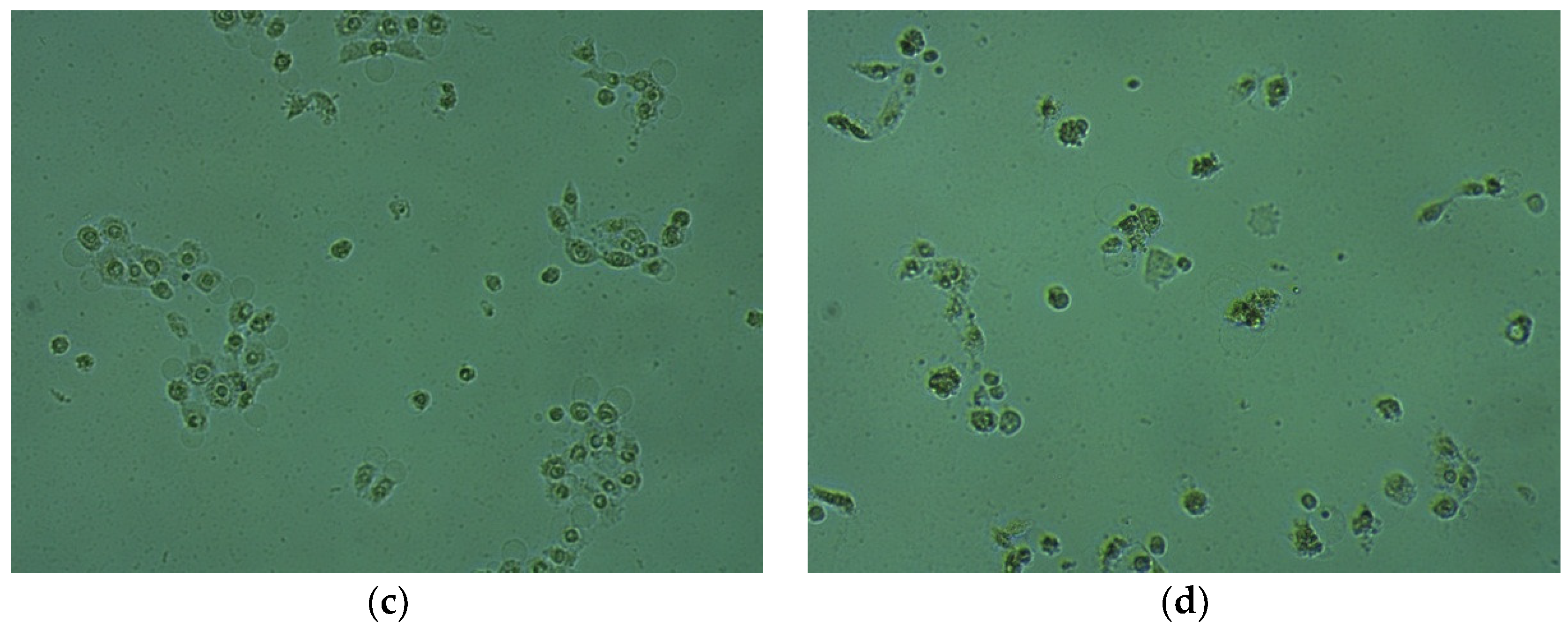
2.4. Cytotoxic Activity in HaCaT Cells

The most promising antistaphylococcal (11, 13) and antimycobacterial (25) agents among derivatives presented in our previous study [2] were now screened with respect to their toxicity against HaCaT cell line (health cell line from adult human skin). The HaCaT cell line was chosen, because it exhibits normal differentiation capacity, and its DNA fingerprint pattern is unaffected by long-term cultivations, transformations, and multiple chromosomal alterations [44].
Human keratinocyte cell lines were exposed to different concentrations of synthesized compounds for 24 h. Derivatives were tested in concentrations equal to their cytotoxic concentrations against MT-4 cells (i.e., 8 µM for 11 and 13, and 37 µM for 25), as well as in concentrations 2 and 3 times higher. At the concentration 8 µM, the thiourea compound 11 caused approximately 20% decrease in cell viability, in comparison with controlled and untreated cells (Figure 2a), that indicated its weak cytotoxic influence against normal cells. On the other hand, cytotoxicity of antibacterial agents 13 and 25 observed at the level of 50%, was found to be moderate according to ISO 10993-5 standards [45].
Results of the LDH (lactate dehydrogenase release) assay indicated that cell mortality in the presence of compounds 11 and 13 increased to 20% and 30%, respectively (Figure 2b). The LD10 value (concentration at which 10% of cells were dead) was determined at the lowest investigated concentration of the compound 11 (8 µM). Substance 13 at concentrations 8 and 16 µM was found to be lowly cytotoxic, resulting in an approximate 10% release of LDH. Compound 25 had the strongest impact on the mortality of tested cells, however, its concentration was the highest among all tested compounds (37 µM). All applied concentrations of that derivative resulted in 7-fold higher cytotoxicity as compared to the LD10 value.
Significant concentration-dependent changes in shape, size, and density of HaCaT cells were observed under a light microscope after 24 h treatment with lowest concentrations of all investigated compounds (Figure 3). At given concentrations, derivatives inhibited viable cell proliferation by about 30–40% (compounds 13 and 25) or 65% (11) (Figure 4). This suggests a direct effect on the HaCaT viability, which was confirmed by the MTT assay. Derivatives 13 and 25 have demonstrated an increased proliferation rate.
The tested substances modified cell viability as well as mortality and growth rate of HaCaT cultures. The presented results demonstrate that derivative 11, one of the strongest antistaphylococcal and antibiofilm agents, at its lowest concentration, possessed the lowest toxic effect on HaCaT cells. Our studies suggest that all tested substances in their lowest concentrations were less toxic in normal immortalized cell lines in comparison to cancer cell lines, however, further studies are needed to describe the underlining mechanism for future application.

3. Materials and Methods

3.1. Chemistry

All reagents and solvents were purchased from Alfa Aesar, Sigma Aldrich or POCh (Polskie Odczynniki Chemiczne, Gliwice, Poland). The infrared (IR) spectra were obtained on Perkin Elmer Spectrum 1000 spectrometer. The nuclear magnetic resonance (NMR) spectra were recorded on Varian VNMRS 300 Oxford NMR spectrometer, using tetramethylsilane (TMS) as the internal reference. Mass spectral electrospray ionization (ESI) measurements were carried out on Waters ZQ Micro-mass instruments with quadruple mass analyzer, at a declustering potential of 40–60 V. Flash chromatography was performed on Merck silica gel 60 (200–400 mesh) using chloroform eluent.
General procedure for the synthesis of (4-chloro-3-nitrophenyl)thiourea derivatives (2024, 2730)
To a solution of 4-chloro-3-nitroaniline (0.0029 mol; 0.5g) in dried acetonitrile (10 mL), a corresponding isothiocyanate (0.0029 mol) was added. The mixture was stirred at room temperature for 12 h. Next the solvent was evaporated and the solid residue was either crystallized from acetonitrile (chloroform) or purified by column chromatography (chloroform).
The synthesis of derivatives 119, 25, 26 was published formerly [2].
1-(4-chloro-3-nitrophenyl)-3-(4-methylphenyl)thiourea (20)
Yield 30%, yellow crystals, m.p. 156–158 °C. FT-IR (KBr, cm−1): 3326.2, 3196.1 (ν N-H); 3113.6, 3024.4 (ν ar C-H); 2977.6 (ν alC-H); 1589.6 (δ N-H); 1533.8 (ν N-O); 1482.0 (ν arC-C); 1407.0 (δ ar(1,4)C-C); 1358.6 (ν N-O); 1299.9 (δ arC-H); 1236.4 (ν alC-C); 1159.7, 1145.4 (δ arC-H); 1043.3 (ν C=S); 864.0 (δ C-Cl); 703.7 (ν C=S). 1H NMR (300 MHz, DMSO) δ: 10.07–10.05 (m, 2H, NH), 8.35 (d, 1H, J = 2.7 Hz, H-2), 7.80 (dd, 1H, J1 = 2.4 Hz, J2 = 2.7 Hz, H-6), 7.69 (d, 1H, J = 8.7 Hz, H-5), 7.34–7.29 (m, 2H, H3′, H-5′), 7.18–7.16 (d, 2H, J = 8.1 Hz, H-2′, H-6′), 2.29 (s, 3H, H-4′a). 13C NMR (75.4 MHz, DMSO) δ: 179.63 (C=S), 146.78 (C-3), 139.86 (C-1), 136.13 (C-1′), 134.40 (C-4′), 131.20 (C-4), 129.13 (C-3′, C-5′), 128.34 (C-5), 124.11 (C-2′, C-6′), 119.69 (C-2), 119.28 (C-6), 20.52 (C-4′a). ESI MS: m/z = 320.0 [M − H] (100%). Rf for TLC (CHCl3:MeOH (1%)): 0.12.
Crystal data: crystal system triclinic, space group P-1, unit cell dimensions at 293 K: a = 7.442(1) Å, b = 8.012(2) Å, c = 12.944(3) Å, α = 98.74(3)°, β = 94.81(3)°, γ = 100.17(3)°, V = 746.0(3) Å3, Z = 2, Dcalc = 1.433 g/cm3, F(000) = 332, μ = 3.647 mm−1, θ range = 3.48 to 75.15°, reflections collected/independent/observed 3147/3042/957, max. and min. transmission 0.7118 and 0.4283; Goodness-of-fit on F3 0.929, final R indices [I > 2σσI)], R1 = 0.0479, wR2 = 0.1266, R indices (all data), R1 = 0.2455, wR2 = 0.1857, residual electron density max./min. 0.38 and −0.36 e Å−3. CCDC No. 1849698.
1-(4-chloro-3-nitrophenyl)-3-(4-methoxyphenyl)thiourea (21)
Yield 28%, yellow powder, m.p. 149–150.5 °C. FT-IR (KBr, cm−1): 3241.8, 3201.5 (ν N-H); 3056.2 (ν arC-H); 1588.4 (δ N-H); 1548.9 (ν N-O); 1484.8 (ν arC-C); 1405.6 (δ ar(1,4)C-C); 1345.5 (ν N-O); 1241.5 (ν C-O-C); 1175.9, 1125.5 (δ arC-H); 1038.3 (ν C=S); 880.2 (δ C-Cl); 697.2 (ν C=S). 1H NMR (300 MHz, DMSO) δ: 9.98 (s, 2H, NH), 8.34 (d, 1H, J = 2.4 Hz, H-2), 7.80 (dd, 1H, J1 = 2.4 Hz, J2 = 2.7 Hz, H-6), 7.68 (d, 1H, J = 8.7 Hz, H-5), 7.34–7.29 (m, 2H, H-3′, H-5′), 6.96–6.91 (m, 2H, H-2′, H-6′), 3.76 (s, 3H, H-4′a). 13C NMR (75.4 MHz, DMSO) δ: 179.54 (C=S), 156.65 (C-4′), 146.46 (C-3), 139.63 (C-1), 131.15 (C-1′), 130.88 (C-4), 128.06 (C-5), 125.88 (C-2′, C-6′), 119.41 (C-2), 118.93 (C-6), 113.61 (C-3′, C-5′), 54.97 (C-4′a). ESI MS: m/z = 336.0 [M − H] (100%). Rf for TLC (CHCl3:MeOH (1%)): 0.17.
1-(4-butyl-2-methylphenyl)-3-(4-chloro-3-nitrophenyl)thiourea (22)
Yield 40%, dark yellow solid, m.p. 67–69 °C. FT-IR (KBr, cm−1): 3341.6, 3187.5 (ν N-H); 3098.1 (ν arC-H); 2954.5, 2859.9 (ν alC-H); 1584.5 (δ N-H); 1533.7 (ν N-O); 1481.5 (ν arC-C); 1407.5 (δ ar(1,4)C-C); 1342.5 (ν N-O); 1126.3 (δ arC-H); 1045.6 (ν C=S); 872.3 (δ C-Cl); 715.2 (ν C=S). 1H NMR (300 MHz, DMSO) δ: 9.95 (s, 1H, NH), 9.66 (s, 1H, NH), 8.36 (d, 1H, J = 2.1 Hz, H-2), 7.82 (dd, 1H, J1 = J2 = 2.7 Hz, H-6), 7.68 (d, 1H, J = 9.0 Hz, H-5), 7.17–6.98 (m, 3H, H-3′, H-5′, H-6′), 2.57–2.55 (m, 2H, H-4a′), 2.20 (s, 3H, H-2′a), 1.58–1.50 (m, 2H, H-4′b), 1.35–1.27 (m, 2H, H-4′c), 0.93–0.87 (m, 3H, H-4′d). 13C NMR (75.4 MHz, DMSO) δ: 180.42 (C=S), 146.69 (C-3), 141.03 (C-2′), 140.57 (C-4′), 139.92 (C-1), 134.53 (C-1′), 131.14 (C-4), 130.356 (C-3′), 128.40 (C-5), 127.65 (C-5′), 126.16 (C-6′), 119.74 (C-2), 119.26 (C-6), 34.35 (C-4′a), 33.04 (C-4′b), 21.77 (C-4′c), 17.75 (C-2′a), 13.76 (C-4′d). ESI MS: m/z = 376.1 [M − H] (100%). Rf for TLC (CHCl3:MeOH (1%)): 0.08.
1-(4-chloro-3-nitrophenyl)-3-phenylthiourea (23)
Yield 56%, pale yellow powder, m.p. 163–165 °C. FT-IR (KBr, cm−1): 3323.5, 3161.3 (ν N-H); 3089.6, 2982.8 (ν arC-H); 1592.0 (δ N-H); 1527.6 (ν N-O); 1482.0 (ν arC-C); 1407.5 (δ ar(1,4)C-C); 1362.7 (ν N-O); 1298.8, 1187.6, 1143.4 (δ arC-H); 1047.1 (ν C=S); 863.7 (δ C-Cl); 695.2 (ν C=S). 1H NMR (300 MHz, DMSO) δ: 10.15 (s, 2H, NH), 8.35 (d, 1H, J = 2.4 Hz, H-2), 7.81 (dd, 1H, J1 = J2 = 2.4 Hz, H-6), 7.70 (d, 1H, J = 9.0 Hz, H-5), 7.47–7.44 (m, 2H, H-2′, H-6′), 7.37 (t, 2H, J = 7.8 Hz, H-3′, H-5′), 7.18 (t, 1H, J = 7.35 Hz, H-4′). 13C NMR (75.4 MHz, DMSO) δ: 179.95 (C=S), 147.09 (C-3), 140.05 (C-1′), 139.08 (C-1), 131.53 (C-4), 128.92 (C-3′, C-5′), 128.62 (C-5), 125.31 (C-4′), 124.20 (C-2′, C-6′), 119.98 (C-2), 119.65 (C-6). ESI MS: m/z = 330.0 [M + Na]+ (100%). Rf for TLC (CHCl3:MeOH (1%)): 0.15.
1-bicyclo[2.2.1]hept-2-yl-3-(4-chloro-3-nitrophenyl)thiourea (24)
Yield 30%, dark yellow solid, m.p. 94–96 °C. FT-IR (KBr, cm−1): 3304.1, 3196.0 (ν N-H); 3033.5 (ν arC-H); 2953.0, 2869.5 (ν alC-H); 1576.1 (δ N-H); 1540.6 (ν N-O); 1478.9 (ν arC-C); 1408.6 (δ ar(1,4)C-C); 1343.4 (ν N-O); 1258.5, 1133.8, 1109.6 (δ arC-H); 1047.2 (ν C=S); 883.3 (δ C-Cl); 714.7 (ν C=S). 1H NMR (300 MHz, DMSO) δ: 9.65 (s, 1H, NH), 8.47 (s, 1H, NH), 8.06 (m, 1H, H-2), 7.74 (dd, 1H, J1 = J2 = 2.4 Hz, H-6), 7.65 (d, 1H, J = 9.0 Hz, H-5), 3.98 (br. s, 1H, H-1′), 2.29–2.26 (m, 2H, H-7′), 1.77–1.70 (m, 1H, H-2′), 1.54–1.30 (m, 4H, H-3′, H-6′), 1.22–1.08 (m, 3H, H-4′, H-5′). 13C NMR (75.4 MHz, DMSO) δ: 178.76 (C=S), 146.20 (C-3), 139.47 (C-1), 130.68 (C-4), 126.10 (C-5), 117.27 (C-2, C-6), 56.34 (C-7′), 40.97 (C-2′, C-6′), 34.70 (C-5′), 34.67 (C-1′), 27.29 (C-4′), 25.48 (C-3′). ESI MS: m/z = 324.1 [M − H] (100%). Rf for TLC (CHCl3:MeOH (1%)): 0.09.
Crystal data: crystal system triclinic, space group P-1, unit cell dimensions at 120 K: a = 7.273(2) Å, b = 9.652(2) Å, c = 11.862(3) Å, α = 105.57(2)°, β = 105.66(3)°, γ = 96.76(2)°, V = 756.0(3) Å3, Z = 2, Dcalc = 1.431 g/cm3, μ = 3.600 mm−1, F(000) = 340, θ range 4.08 to 73.77°, reflections collected/independent/observed 10755/2994/2701; max. and min. transmission 1 and 0.75929; Goodness-of-fit on F2 1.084, final R indices [I > 2σ(I)] R1 = 0.0395, wR2 = 0.0943; R indices (all data) R1 = 0.0456, wR2 = 0.0975, residual electron density max/min 0.45 and−0.54 e Å−3. The cycloalkyl group, viz. 1-bicyclo[2.2.1]hept-2-yl, is disordered over two positions with the site occupancy factors being 0.64:0.36. CCDC No. 1849699.
1-(4-chloro-3-nitrophenyl)-3-(2-methylprop-2-en-1-yl)thiourea (27)
Yield 29%, light brown powder, m.p. 96–98 °C. FT-IR (KBr, cm−1): 3336.7, 3198.9 (ν N-H); 3075.6, 3022.8 (ν arC-H); 1652.3 (ν C=C); 1577.4 (δ N-H); 1535.1 (ν N-O); 1470.4 (ν arC-C, δ alC-H); 1407.5 (δ ar(1,4)C-C); 1347.5 (ν N-O); 1312.2, 1194.6, 1140.4 (δ arC-H); 1040.5 (ν C=S); 882.0 (δ C-Cl); 683.2 (ν C=S). 1H NMR (300 MHz, DMSO) δ: 9.99 (br. s, 1H, NH), 8.42 (br. s, 1H, H-2), 8.23 (br. s, 1H, NH), 7.76 (dd, 1H, J1 = J2 = 2.4 Hz, H-6), 7.68 (d, 1H, J = 9.0 Hz, H-5), 4.84 (s, 2H, H-3′), 4.09 (br. s, 2H, H-1′), 1.72 (s, 3H, H-2′a). 13C NMR (75.4 MHz, DMSO) δ: 181.06 (C=S), 147.07 (C-3), 141.78 (C-1), 140.09 (C-5), 131.60 (C-4), 127.49 (C-3′), 118.79 (C-2′), 110.78 (C-2, C-6), 49.19 (C-1′), 20.65 (C-2′a). ESI MS: m/z = 284.0 [M − H] (100%). Rf for TLC (CHCl3:MeOH (1%)): 0.10.
1-(4-chloro-3-nitrophenyl)-3-prop-2-en-1-ylthiourea (28)
Yield 31%, pale yellow crystals, m.p. 112–114 °C. FT-IR (KBr, cm−1): 3370.1, 3173.8 (ν N-H); 3072.7, 3002.6 (ν arC-H, ν =C-H); 1641.6 (ν C=C); 1600.7 (δ N-H); 1534.5 (ν N-O); 1471.6 (ν arC-C); 1431.1 (δ =C-H); 1409.4 (δ ar(1,4)C-C); 1349.5 (δ alC-H, δ =C-H); 1311.5 (ν N-O); 1195.9, 1127.9 (δ arC-H); 1041.4 (ν C=S); 839.4 (δ C-Cl); 711.5 (ν C=S). 1H NMR (300 MHz, DMSO) δ: 9.97 (s, 1H, NH), 8.40 (d, 1H, J = 2.1 Hz, H-2), 8.24 (s, 1H, NH), 7.76 (dd, 1H, J1 = 2.4 Hz, J2 = 2.7 Hz, H-6), 7.68 (d, 1H, J = 9.0 Hz, H-5), 5.96–5.83 (m, 1H, H-2′), 5.25–5.11 (m, 2H, H-3′), 4.15 (m, 2H, H-1′). 13C NMR (75.4 MHz, DMSO) δ: 180.52 (C=S), 146.82 (C-3), 139.78 (C-1), 134.19 (C-5), 131.32 (C-4), 127.31 (C-3′), 118.64 (C-2′), 116.08 (C-2, C6), 46.02 (C-1′). ESI MS: m/z = 270.0 [M − H] (100%). Rf for TLC (CHCl3:MeOH (1%)): 0.11.
1-(4-chloro-3-nitrophenyl)-3-[3-(methylsulfanyl)propyl]thiourea (29)
Yield 35%, light brown powder, m.p. 102–103.5 °C. FT-IR (KBr, cm−1): 3330.7, 3171.5 (ν N-H); 3070.3, 2999.3 (ν arC-H); 2941.3, 2915.6 (ν alC-H); 1572.8 (δ N-H); 1531.5 (ν N-O); 1475.5 (ν arC-C); 1455.1 (δ alC-H); 1410.5 (δ ar(1,4)C-C); 1387.7 (δ alC-H); 1351.8 (ν N-O); 1298.9, 1154.7, 1134.1 (δ arC-H); 1043.6 (ν C=S); 887.3 (δ C-Cl); 701.7 (ν C=S). 1H NMR (300 MHz, DMSO) δ: 9.88 (br. s, 1H, NH), 8.37 (d, 1H, J = 2.1 Hz, H-2), 8.17 (br. s, 1H, NH), 7.73 (dd, 1H, J1 = J2 = 1.8 Hz, H-6), 7.67 (d, 1H, J = 8.7 Hz, H-5), 3.56–3.54 (m, 2H, H-1′), 2.51–2.49 (m, 2H, H-3′), 2.06–2.05 (m, 3H, H-4′), 1.87–1.78 (m, 2H, H-2′). 13C NMR (75.4 MHz, DMSO) δ: 179.85 (C=S), 146.25 (C-3), 139.24 (C-1), 130.78 (C-4), 126.57 (C-5), 117.86 (C-2, C-6), 42.28 (C-1′), 30.04 (C-2′), 27.03 (C-3′), 14.04 (C-4′). ESI MS: m/z = 318.0 [M − H] (100%). Rf for TLC (CHCl3:MeOH (1%)): 0.09.
1-(4-chloro-3-nitrophenyl)-3-(furan-2-ylmethyl)thiourea (30)
Yield 58%, brown powder, m.p. 144–146 °C. FT-IR (KBr, cm−1): 3299.1, 3180.4 (ν N-H); 3009.7 (ν arC-H); 1578.2 (δ N-H); 1537.3 (ν N-O); 1478.9 (ν arC-C); 1448.8 (δ alC-H); 1407.9 (δ ar(1,4)C-C); 1364.3 (δ alC-H); 1339.2 (ν N-O); 1297.7, 1190.4, 1139.0 (δ arC-H); 1018.2 (ν C=S); 884.6 (δ C-Cl); 714.8 (ν C=S). 1H NMR (300 MHz, DMSO) δ: 9.98 (s, 1H, NH), 8.49 (s, 1H, NH), 8.41 (d, 1H, J = 2.1 Hz, H-2), 7.76 (dd, 1H, J1 = J2 = 2.4 Hz, H-6), 7.68 (d, 1H, J = 8.7 Hz, H-5), 7.62–7.61 (m, 1H, H-5′), 6.42 (dd, 1H, J1 = 2.1 Hz, J2 = 1.8 Hz, H-4′), 6.35 (d, 1H, J1 = 3.3 Hz, H-3′), 4.73 (d, 2H, J = 5.1 Hz, H-1′). 13C NMR (75.4 MHz, DMSO) δ: 180.27 (C=S), 150.75 (C-2′), 146.55 (C-3), 142.09 (C-5′), 139.42 (C-1), 131.07 (C-4), 127.09 (C-5), 118.62 (C-2), 118.37 (C-6), 110.25 (C-4′), 107.44 (C-3′). ESI MS: m/z = 310.0 [M − H] (100%). Rf for TLC (CHCl3:MeOH (1%)):0.15.
The NMR spectra of compounds 2024, 2730 can be found in Supplementary Materials.

3.2. X-ray Crystalography

The X-ray diffraction intensities were measured for 20 and 24 on a SuperNova diffractometer with monochromatized CuKα radiation (λ = 1.54184 Å). Data sets were collected using the ω scan technique. The programs CrysAlisPro [46] were used for data collection, cell refinement and data reduction; empirical absorption correction was applied using spherical harmonics implemented in SCALE3 ABSPACK scaling algorithm. The structure was solved by direct methods using SHELXS-97 and refined by the full-matrix least-squares on F2 using the SHELXL-97 [47]. Non-hydrogen atoms were refined with anisotropic displacement parameters. Hydrogen atoms were positioned geometrically and allowed to ride on their parent atoms, with Uiso(H) = 1.2 Ueq(C).

3.3. In Vitro Evaluation of Antimicrobial Activity

The antimicrobial activity of the compounds was tested on Gram-positive bacteria (Staphylococcus aureus NCTC 4163, Staphylococcus aureus ATCC 25923, Staphylococcus aureus ATCC 6538, Staphylococcus aureus ATCC 29213, Staphylococcus epidermidis ATCC 12228, Staphylococcus epidermidis ATCC 35984, Enterococcus faecalis ATCC 29212, Bacillus subtilis ATCC 6633, Bacillus cereus ATCC 11778, Micrococcus luteus ATCC 9341, Micrococcus luteus ATCC 10240); Gram-negative rods (Escherichia coli ATCC 10538, Escherichia coli ATCC 25922, Escherichia coli NCTC 8196, Proteus vulgaris NCTC 4635, Pseudomonas aeruginosa ATCC 15442, Pseudomonas aeruginosa NCTC 6749, Pseudomonas aeruginosa ATCC 27863, Bordetella bronchiseptica ATCC 4617) and yeasts (Candida albicans ATCC 10231, Candida albicans ATCC 90028, Candida parapsilosis ATCC 22019). Other microorganisms used were obtained from the collection of the Department of Pharmaceutical Microbiology, Medical University of Warsaw, Poland.
Antibacterial activity was examined by the disc-diffusion method under standard conditions using Mueller-Hinton II agar medium (Becton Dickinson) according to CLSI (previously NCCLS) guidelines [39]. Antifungal activities were assessed using Mueller-Hinton agar + 2% glucose and 0.5 µg/mL Methylene Blue Dye Medium [40]. Sterile filter paper discs (9 mm diameter, Whatman No 3 chromatography paper) were dripped with tested compound solutions (in DMSO) to load 400 μg of a given compound per disc. Dry discs were placed on the surface of appropriate agar medium. The results (diameter of the growth inhibition zone) were read after 18 h of incubation at 35 °C. Minimal Inhibitory Concentration (MIC) was tested by the twofold serial microdilution method (in 96-well microtiter plates) using Mueller-Hinton Broth medium (Beckton Dickinson) for bacteria or RPMI-1640 medium for Candida species according to CLSI guidelines [41,42]. The stock solution of tested agent was prepared in DMSO and diluted in sterile water. Concentrations of tested agents ranged from 0.125 to 512 µg/mL. The final inoculum of all studied microorganisms was 105 CFU/ mL−1 (colony forming units per mL). Minimal inhibitory concentrations (the lowest concentration of a tested agent that prevents visible growth of a microorganism) were read after 18 h (bacteria) or 24 h (yeasts) of incubation at 35 °C.

3.4. Cytotoxicity and Anti-HIV Assays

Cell line supporting the multiplication of Human Immunodeficiency Virus type-1 (HIV-1) was the CD4+ human T-cells, derived from human hematological tumors, containing an integrated HTLV-1 genome (MT-4). Cells were purchased from American Type Culture Collection (ATCC). Human Immunodeficiency Virus type-1 (HIV-1) IIIB laboratory strain was obtained from the supernatant of the persistently infected H9/IIIB cells (NIH 1983).
Exponentially growing MT-4 cells were seeded at an initial density of 1 × 10⁵ cells/mL in 96-well plates in RPMI-1640 medium, supplemented with 10% fetal bovine serum (FBS), 100 units/mL penicillin G and 100 μg/mL streptomycin. Cell cultures were then incubated at 37 °C in a humidified, 5% CO2 atmosphere, in the absence or presence of serial dilutions of tested compounds. Cell viability was determined after 96 h at 37 °C by the 3-(4,5-dimethylthiazol-2-yl)-2,5-diphenyl-tetrazolium bromide (MTT) method.
The activity of compounds against HIV-1 was based on inhibition of virus-induced cytopathogenicity in MT-4 cell acutely infected with a multiplicity of infection (m.o.i.) of 0.01. Briefly, 50 μL of RPMI containing 1 × 10⁴ MT-4 cells were added to each well of flat-bottom microtiter trays, containing 50 μL of RPMI without or with serial dilutions of test compounds. Then, 20 μL of a HIV-1 suspension containing 100 CCID50 were added. After a 4-day incubation at 37 °C, cell viability was determined by the MTT method.

3.5. Antiproliferative Assays

Cell lines derived from human hematological tumors were: CD4+ human T-cells containing an integrated HTLV-1 genome (MT-4); CD4+ human acute T-lymphoblastic leukemia (CCRF-CEM); human splenic B-lymphoblastoid cells (WIL-2NS); human acute B-lymphoblastic leukemia (CCRF-SB). Cells were seeded at an initial density of 1 × 10⁵ cells/mL in 96 well plates in RPMI-1640 medium supplemented with 10% fetal calf serum (FCS), 100 units/mL penicillin G and 100 μg/mL streptomycin.
Cell lines derived from human solid tumors were: Skin melanoma (SK-MEL-28); prostate carcinoma (DU-145). Normal tissues foreskin fibroblasts (CRL-7065) were also used. Cells were seeded at 1 × 10⁵ cells/mL in 96 well plates in specific media supplemented with 10% FCS and antibiotics, as above. Cell cultures were then incubated at 37 °C in a humidified, 5% CO2 atmosphere in the absence or presence of serial dilutions of test compounds. Cell viability was determined after 96 h at 37 °C by the MTT method.

3.6. Cytotoxic Activity in HaCaT Cells

3.6.1. Cell Culture: Conditions and Treatments

Human immortal keratinocyte cell line from adult human skin (HaCaT) was purchased from American Type Culture Collection (Rockville, MD, USA), and cultured in Dulbecco’s Modified Eagle’s Medium (DMEM), supplemented with antibiotics (penicillin and streptomycin), 10% heat-inactivated FBS-fetal bovine serum (Gibco Life Technologies, Carlsbad, CA, USA) at 37 °C and 5% CO2 atmosphere. Cells were passaged using trypsin-EDTA (Gibco Life Technologies, Carlsbad, CA, USA) and cultured in 24-well plates (2.5 × 10⁴ cells per well). Experiments were conducted in DMEM with 2% FBS.

3.6.2. Cell Viability Assessment (Mitochondrial Function Assessment)

The cell viability assay was assessed by the determination of MTT salt (3-(4,5-dimethylthiazol-2-yl)-2,5-diphenyltetrazolium bromide, Sigma-Aldrich Chemie, Buchs, Germany) conversion by mitochondrial dehydrogenase. The cells were incubated for 24 h in 24-well plates with tested compounds (11, 13 and 25) at given concentrations, and subsequently for another 2 h with 0.5 mg/mL of MTT solution, which was converted in living cells under the effect of mitochondrial dehydrogenase into insoluble formazan. Then the converted dye was solubilized with the use of 0.04 M HCl in absolute isopropanol. Absorbance of solubilized formazan was measured spectrophotometrically at 570 nm (using Epoch microplate reader, BioTek Inc., Winooski, VT, USA, equipped with Gen5 software (BioTech Instruments, Inc., Biokom, Janki, Polska).
Cell viability was presented as a percent of MTT in the treated cells versus the control (cells incubated in serum-free DMEM without extracts). The relative MTT level (%) was calculated as [A]/[B] × 100, where [A] is the absorbance of the test sample and [B] is the absorbance of the control sample containing the untreated cells. The decreased relative MTT level indicates decreased cell viability.

3.6.3. Lactate Dehydrogenase Release Assay (Cellular Membrane Integrity Assessment)

Release of lactate dehydrogenase (LDH) from the cytosol to culture medium is a marker of cell death. The assay was performed after 24 h incubation of HaCaT cells in 24-well plates with investigated concentrations of each compound. The activity of LDH released from the cytosol of damaged cells to the supernatant was measured using the protocol of the cytotoxicity detection kit LDH test described by the manufacturer (Roche Diagnostics, Berlin, Germany). Absorbance was measured at 490 nm using a microplate reader (using Epoch microplate reader, BioTek Inc., Winooski, VT, USA) equipped with Gen5 software (BioTech Instruments, Inc., Biokom, Janki, Polska)
Compounds mediated cytotoxicity expressed as the LDH release (%) was determined by the following equation: [(A test sample—A low control)/(A high control—A low control)] × 100% (A-absorbance); where “low control” were cells in DMEM with 2% FBS without tested compounds, and “high control” were cells incubated in DMEM with 2% FBS with 10% Triton X-100 (100% LDH release).

3.6.4. Proliferation Rate in Cultures of HaCaT Cells

After 48 h incubation with the lowest concentration of the tested compound the increased number of viable cells were assessed by direct counting of cells using trypan blue with a Countess Automated Cell Counter (Invitrogen, Carlsbad, CA, USA). The results are presented as a % of viable and dead cells versus control.

4. Conclusions

To sum up, this work has revealed the synthesis of new thiourea derivatives and their in vitro antibacterial activity against clinically relevant pathogens. The title compounds showed potent to moderate antimicrobial activity. Representative aryl-thioureas 20, 22, 23 and bicyclic-thiourea derivative 24 were even more active towards selected clinical staphylococci than the reference drug, Ciprofloxacin. To complete the biological profile of the presented thiourea series, their cytotoxic activities in MT-4 cells, as well as human hematological and solid tumors cell lines were assessed. Several compounds (1, 4, 6, 911, 13, 14, 17, 19) turned out cytotoxic in the low micromolar range (CC50 < 10 µM). Performed studies proved that synthesized derivatives weakly to moderately influenced the viability and growth of the human immortal keratinocyte cell line (HaCaT).

Supplementary Materials

Supplementary materials can be found online. The experimental details and final atomic parameters for 20 and 24 have been deposited with the Cambridge Crystallographic Data Centre as supplementary material (CCDC ID: 1849698 and 1849698, respectively). Copies of the data can be obtained free of charge on request via www.ccdc.cam.ac.uk/data_request/cif, or by emailing [email protected], or by contacting The Cambridge Crystallographic Data Centre, 12, Union Road, Cambridge CB2 1EZ, UK; fax: +44 1223 336033.

Author Contributions

Conceptualization, A.B. and G.S.; Methodology, J.S.; Software, A.E.K.; Validation, A.C. and P.S.-Ł.; Formal Analysis, S.M.; Investigation, G.S.; S.M.; G.G.; Resources, J.S.; Data Curation, A.E.K. and O.S.; Writing—Original Draft Preparation, A.B. and G.G.; Writing—Review & Editing, A.B.; Visualization, P.S.-Ł.; Supervision, M.S.; Project Administration, M.S.; Funding Acquisition, G.K.-T.

Funding

This work was supported by the Medical University of Warsaw and carried out with the use of CePT infrastructure financed by the European Union—the European Regional Development Fund within the Operational Programme Innovative Economy for 2007–2013. The research was carried out with the facilities purchased thanks to the financial support of the European Regional Development Fund in the framework of the Operational Program Development of Eastern Poland 2007–2013 (Contract No. POPW.01.03.00-06-009/11-00, Equipping the laboratories of the Faculties of Biology and Biotechnology, Mathematics, Physics and Informatics, and Chemistry for studies of biologically active substances and environmental samples.

Acknowledgments

Authors would like to thank Sylwester Krukowski from the Department of Inorganic and Analytical Chemistry, Faculty of Pharmacy, Medical University of Warsaw, for recording the IR spectra.

Conflicts of Interest

The authors declare no conflict of interest. The funders had no role in the design of the study; in the collection, analyses, or interpretation of data; in the writing of the manuscript, and in the decision to publish the results.

References

  1. Bielenica, A.; Stefańska, J.; Stępień, K.; Napiórkowska, A.; Augustynowicz-Kopeć, E.; Sanna, G.; Madeddu, S.; Boi, S.; Giliberti, G.; Wrzosek, M.; et al. Synthesis, cytotoxicity and antimicrobial activity of thiourea derivatives incorporating 3-(trifluoromethyl)phenyl moiety. Eur. J. Med. Chem. 2015, 101, 111–125. [Google Scholar] [CrossRef] [PubMed]
  2. Bielenica, A.; Stępień, K.; Napiórkowska, A.; Augustynowicz-Kopeć, E.; Krukowski, S.; Włodarczyk, M.; Struga, M. Synthesis and antimicrobial activity of 4-chloro-3-nitrophenylthiourea derivatives targeting bacterial type II topoisomerases. Chem. Biol. Drug Des. 2016, 87, 905–917. [Google Scholar] [CrossRef] [PubMed]
  3. Foley, T.L.; Rai, G.; Yasgar, A.; Daniel, T.; Baker, H.L.; Attene-Ramos, M.; Kosa, N.M.; Leister, W.; Burkart, M.D.; Jadhav, A.; et al. 4-(3-Chloro-5-(trifluoromethyl)pyridin-2-yl)-N-(4-methoxypyridin-2-yl)piperazine-1-carbothioamide (ML267), a potent inhibitor of bacterial phosphopantetheinyl transferase that attenuates secondary metabolism and thwarts bacterial growth. J. Med. Chem. 2014, 57, 1063–1078. [Google Scholar] [CrossRef] [PubMed]
  4. Madabhushi, S.; Mallu, K.K.; Vangipuram, V.S.; Kurva, S.; Poornachandra, Y.; Ganesh Kumar, C. Synthesis of novel benzimidazole functionalized chiral thioureas and evaluation of their antibacterial and anticancer activities. Bioorg. Med. Chem. Lett. 2014, 24, 4822–4825. [Google Scholar] [CrossRef] [PubMed]
  5. Ghorab, M.M.; Alsaid, M.S.; El-Gaby, M.S.A.; Elaasser, M.M.; Nissan, Y.M. Antimicrobial and anticancer activity of some novel fluorinated thiourea derivatives carrying sulfonamide moieties: Synthesis, biological evaluation and molecular docking. Chem. Cent. J. 2017, 11, 1–14. [Google Scholar] [CrossRef] [PubMed]
  6. Ghorab, M.M.; Alsaid, M.S.; El-Gaby, M.S.A.; Safwat, N.A.; Elaasser, M.M.; Soliman, A.M. Biological evaluation of some new N-(2,6-dimethoxypyrimidinyl) thioureido benzenesulfonamide derivatives as potential antimicrobial and anticancer agents. Eur. J. Med. Chem. 2016, 124, 299–310. [Google Scholar] [CrossRef] [PubMed]
  7. Medapi, B.; Renuka, J.; Saxena, S.; Sridevi, J.P.; Medishetti, R.; Kulkarni, P.; Yogeeswari, P.; Sriram, D. Design and synthesis of novel quinoline-aminopiperidine hybrid analogues as Mycobacterium tuberculosis DNA gyraseB inhibitors. Bioorg. Med. Chem. 2015, 23, 2062–2078. [Google Scholar] [CrossRef] [PubMed]
  8. Sriram, D.; Yogeeswari, P.; Dinakaran, M.; Thirumurugan, R. Antimycobacterial activity of novel 1-(5-cyclobutyl-1,3-oxazol-2-yl)-3-(sub)phenyl/pyridylthiourea compounds endowed with high activity toward multidrug-resistant Mycobacterium tuberculosis. J. Antimicrob. Chemother. 2007, 59, 1194–1196. [Google Scholar] [CrossRef] [PubMed]
  9. Bielenica, A.; Sanna, G.; Madeddu, S.; Struga, M.; Jóźwiak, M.; Kozioł, A.E.; Sawczenko, A.; Materek, I.B.; Serra, A.; Giliberti, G. New thiourea and 1,3-thiazolidin-4-one derivatives effective on the HIV-1 virus. Chem. Biol. Drug Des. 2017, 90, 883–891. [Google Scholar] [CrossRef] [PubMed]
  10. D’Cruz, O.J.; Qazi, S.; Yiv, S.; Uckun, F.M. A novel vaginal microbicide containing the rationally designed anti-HIV compound HI-443 (N′-[2-(2-thiophene)ethyl]-N′-[2-(5-bromopyridyl)] thiourea]). Expert Opin. Investig. Drugs 2012, 21, 265–279. [Google Scholar] [CrossRef] [PubMed]
  11. Karakuş, S.; Güniz Küçükgüzel, S.; Küçükgüzel, I.; De Clercq, E.; Pannecouque, C.; Andrei, G.; Snoeck, R.; Sahin, F.; Bayrak, O.F. Synthesis, antiviral and anticancer activity of some novel thioureas derived from N-(4-nitro-2-phenoxyphenyl)-methanesulfonamide. Eur. J. Med. Chem. 2009, 44, 3591–3595. [Google Scholar] [CrossRef] [PubMed]
  12. Bloom, J.D.; Dushin, R.G.; Curran, K.J.; Donahue, F.; Norton, E.B.; Terefenko, E.; Jones, T.R.; Ross, A.A.; Feld, B.; Lang, S.A.; et al. Thiourea inhibitors of herpes viruses. Part 2: N-Benzyl-N′-arylthiourea inhibitors of CMV. Bioorg. Med. Chem. Lett. 2004, 14, 3401–3406. [Google Scholar] [CrossRef] [PubMed]
  13. Bielenica, A.; Kędzierska, E.; Koliński, M.; Kmiecik, S.; Koliński, A.; Fiorino, F.; Severino, B.; Magli, E.; Corvino, A.; Rossi, I.; et al. 5-HT2 receptor affinity, docking studies and pharmacological evaluation of a series of 1,3-disubstituted thiourea derivatives. Eur. J. Med. Chem. 2016, 116, 173–186. [Google Scholar] [CrossRef] [PubMed]
  14. Stefanska, J.; Szulczyk, D.; Koziol, A.E.; Miroslaw, B.; Kedzierska, E.; Fidecka, S.; Busonera, B.; Sanna, G.; Giliberti, G.; La Colla, P.; et al. Disubstituted thiourea derivatives and their activity on CNS: Synthesis and biological evaluation. Eur. J. Med. Chem. 2012, 55, 205–213. [Google Scholar] [CrossRef] [PubMed]
  15. Bielenica, A.; Kedzierska, E.; Fidecka, S.; Maluszynska, H.; Miroslaw, B.; Koziol, A.E.; Stefanska, J.; Madeddu, S.; Giliberti, G.; Sanna, G.; et al. Synthesis, Antimicrobial and Pharmacological Evaluation of Thiourea derivatives of 4H-1,2,4-triazole. Lett. Drug Discov. Dev. 2015, 12, 263–276. [Google Scholar] [CrossRef]
  16. Karakus, S.; Koçyigit-Kaymakcioglu, B.; Toklu, H.Z.; Aricioglu, F.; Rollas, S. Synthesis and anticonvulsant activity of new N-(alkyl/sub-stituted aryl)-N′-[4-(5-cyclohexylamino)-1,3,4-thiadiazole-2-yl)phenyl]thioureas. Arch. Pharm. Weinheim 2009, 342, 48–53. [Google Scholar] [CrossRef] [PubMed]
  17. Tokala, R.; Bale, S.; Janrao, I.P.; Vennela, A.; Kumar, N.P.; Senwar, K.R.; Godugu, C.; Shankaraiah, N. Synthesis of 1,2,4-triazole-linked urea/thiourea conjugates as cytotoxic and apoptosis inducing agents. Bioorg. Med. Chem. Lett. 2018, 28, 1919–1924. [Google Scholar] [CrossRef] [PubMed]
  18. Shankaraiah, N.; Kumar, N.P.; Amula, S.B.; Nekkanti, S.; Jeengar, M.K.; Naidu, V.G.; Reddy, T.S.; Kamal, A. One-pot synthesis of podophyllotoxin-thiourea congeners by employing NH2SO3H/NaI: Anticancer activity, DNA topoisomerase-II inhibition, and apoptosis inducing agents. Bioorg. Med. Chem. Lett. 2015, 25, 4239–4244. [Google Scholar] [CrossRef] [PubMed]
  19. Sun, Y.; Shan, Y.; Li, C.; Si, R.; Pan, X.; Wang, B.; Zhang, J. Discovery of novel anti-angiogenesis agents. Part 8: Diaryl thiourea bearing 1H-indazole-3-amine as multi-target RTKs inhibitors. Eur. J. Med. Chem. 2017, 141, 373–385. [Google Scholar] [CrossRef] [PubMed]
  20. Turan-Zitouni, G.; Altıntop, M.D.; Özdemir, A.; Kaplancıklı, Z.A.; Çiftçi, G.A.; Temel, H.E. Synthesis and evaluation of bis-thiazole derivatives as new anticancer agents. Eur. J. Med. Chem. 2016, 107, 288–294. [Google Scholar] [CrossRef] [PubMed]
  21. Koca, İ.; Özgür, A.; Er, M.; Gümüş, M.; Açikalin Coşkun, K.; Tutar, Y. Design and synthesis of pyrimidinyl acyl thioureas as novel Hsp90 inhibitors in invasive ductal breast cancer and its bone metastasis. Eur. J. Med. Chem. 2016, 122, 280–290. [Google Scholar] [CrossRef] [PubMed]
  22. Mowafy, S.; Galanis, A.; Doctor, Z.M.; Paranal, R.M.; Lasheen, D.S.; Farag, N.A.; Jänne, P.A.; Abouzid, K.A. Toward discovery of mutant EGFR inhibitors; Design, synthesis and in vitro biological evaluation of potent 4-arylamino-6-ureido and thioureido-quinazoline derivatives. Bioorg. Med. Chem. 2016, 24, 3501–3512. [Google Scholar] [CrossRef] [PubMed]
  23. Hamed, M.M.; Darwish, S.S.; Herrmann, J.; Abadi, A.H.; Engel, M. First Bispecific Inhibitors of the Epidermal Growth Factor Receptor Kinase and the NF-κB Activity as Novel Anticancer Agents. J. Med. Chem. 2017, 60, 2853–2868. [Google Scholar] [CrossRef] [PubMed]
  24. Shing, J.C.; Choi, J.W.; Chapman, R.; Schroeder, M.A.; Sarkaria, J.N.; Fauq, A.; Bram, R.J. A novel synthetic 1,3-phenyl bis-thiourea compound targets microtubule polymerization to cause cancer cell death. Cancer Biol. Ther. 2014, 15, 895–905. [Google Scholar] [CrossRef] [PubMed] [Green Version]
  25. Ma, L.Y.; Zheng, Y.C.; Wang, S.Q.; Wang, B.; Wang, Z.R.; Pang, L.P.; Zhang, M.; Wang, J.W.; Ding, L.; Li, J.; et al. Design, synthesis, and structure-activity relationship of novel LSD1 inhibitors based on pyrimidine-thiourea hybrids as potent, orally active antitumor agents. J. Med. Chem. 2015, 58, 1705–1716. [Google Scholar] [CrossRef] [PubMed]
  26. Suzuki, T.; Miyata, N. Lysine demethylases inhibitors. J. Med. Chem. 2011, 54, 8236–8250. [Google Scholar] [CrossRef] [PubMed]
  27. Ghorab, M.M.; Alsaid, M.S.; Al-Dosary, M.S.; Nissan, Y.M.; Attia, S.M. Design, synthesis and anticancer activity of some novel thioureido-benzenesulfonamides incorporated biologically active moieties. Chem. Cent. J. 2016, 10, 1–13. [Google Scholar] [CrossRef] [PubMed]
  28. Lin, S.; Lombardo, M.; Malkani, S.; Hale, J.J.; Mills, S.G.; Chapman, K.; Thompson, J.E.; Zhang, W.X.; Wang, R.; Cubbon, R.M.; et al. Novel 1-(2-aminopyrazin-3-yl)methyl-2-thioureas as potent inhibitors of mitogen-activated protein kinase-activated protein kinase 2 (MK-2). Bioorg. Med. Chem. Lett. 2009, 19, 3238–3242. [Google Scholar] [CrossRef] [PubMed]
  29. Kožurková, M.; Sabolová, D.; Kristian, P. A review on acridinylthioureas and its derivatives: Biological and cytotoxic activity. J. Appl. Toxicol. 2017, 37, 1132–1139. [Google Scholar] [CrossRef] [PubMed]
  30. Repich, H.H.; Orysyk, V.V.; Palchykovska, L.G.; Orysyk, S.I.; Zborovskii, Y.L.; Vasylchenko, O.V.; Storozhuk, O.V.; Biluk, A.A.; Nikulina, V.V.; Garmanchuk, L.V.; et al. Synthesis, spectral characterization of novel Pd(II), Pt(II) π-coordination compounds based on N-allylthioureas. Cytotoxic properties and DNA binding ability. J. Inorg. Biochem. 2017, 168, 98–106. [Google Scholar] [CrossRef] [PubMed]
  31. Correa, R.S.; de Oliveira, K.M.; Delolo, F.G.; Alvarez, A.; Mocelo, R.; Plutin, A.M.; Cominetti, M.R.; Castellano, E.E.; Batista, A.A. Ru(II)-based complexes with N-(acyl)-N′,N′-(disubstituted)thiourea ligands: Synthesis, characterization, BSA- and DNA-binding studies of new cytotoxic agents against lung and prostate tumour cells. J. Inorg. Biochem. 2015, 150, 63–71. [Google Scholar] [CrossRef] [PubMed]
  32. Plutín, A.M.; Mocelo, R.; Alvarez, A.; Ramos, R.; Castellano, E.E.; Cominetti, M.R.; Graminha, A.E.; Ferreira, A.G.; Batista, A.A. On the cytotoxic activity of Pd(II) complexes of N,N-disubstituted-N′-acyl thioureas. J. Inorg. Biochem. 2014, 134, 76–82. [Google Scholar] [CrossRef] [PubMed]
  33. Liu, S.; Louie, M.C.; Rajagopalan, V.; Zhou, G.; Ponce, E.; Nguyen, T.; Green, L. Synthesis and evaluation of the diarylthiourea analogs as novel anti-cancer agents. Bioorg. Med. Chem. Lett. 2015, 25, 1301–1305. [Google Scholar] [CrossRef] [PubMed]
  34. Rauf, M.K.; Zaib, S.; Talib, A.; Ebihara, M.; Badshah, A.; Bolte, M.; Iqbal, J. Solution-phase microwave assisted parallel synthesis, biological evaluation and in silico docking studies of N,N′-disubstituted thioureas derived from 3-chlorobenzoic acid. Bioorg. Med. Chem. 2016, 24, 4452–4463. [Google Scholar] [CrossRef] [PubMed]
  35. Liu, C.J.; Yu, S.L.; Liu, Y.P.; Dai, X.J.; Wu, Y.; Li, R.J.; Tao, J.C. Synthesis, cytotoxic activity evaluation and HQSAR study of novel isosteviol derivatives as potential anticancer agents. Eur. J. Med. Chem. 2016, 115, 26–40. [Google Scholar] [CrossRef] [PubMed]
  36. El-Hossary, E.M.; Förstner, K.U.; François, P.; Baud, D.; Streker, K.; Schrenzel, J.; Ohlsen, K.; Holzgrabe, U. A Novel Mechanism of Inactivating Antibacterial Nitro Compounds in the Human Pathogen Staphylococcus aureus by Overexpression of a NADH-Dependent Flavin Nitroreductase. Antimicrob. Agents Chemother. 2018, 62, e01510–e01517. [Google Scholar] [CrossRef] [PubMed]
  37. Chu, P.S.; Lopez, M.I.; Abraham, A.; El Said, K.R.; Plakas, S.M. Residue depletion of nitrofuran drugs and their tissue-bound metabolites in channel catfish (Ictalurus punctatus) after oral dosing. J. Agric. Food Chem. 2008, 56, 8030–8034. [Google Scholar] [CrossRef] [PubMed]
  38. Bielenica, A.; Drzewiecka-Antonik, A.; Rejmak, P.; Stefańska, J.; Koliński, M.; Kmiecik, S.; Lesyng, B.; Włodarczyk, M.; Pietrzyk, P.; Struga, M. Synthesis, structural and antimicrobial studies of type II topoisomerase-targeted copper(II) complexes of 1,3-disubstituted thiourea ligands. J. Inorg. Biochem. 2018, 182, 61–70. [Google Scholar] [CrossRef] [PubMed]
  39. Clinical and Laboratory Standards Institute. Methods for Dilution Antimicrobial Susceptibility Tests for Bacteria That Grow Aerobically; Approved Standard M7-A9; CLSI: Wayne, PA, USA, 2012. [Google Scholar]
  40. Clinical and Laboratory Standards Institute. Methods for Determining Bactericidal Activity of Antimicrobial Agents; Approved Guideline M26-A; CLSI: Wayne, PA, USA, 1999. [Google Scholar]
  41. Clinical and Laboratory Standards Institute. Methods for Dilution Antimicrobial Susceptibility Tests for Bacteria That Grow Aerobically; Approved Standard M7-A7; CLSI: Wayne, PA, USA, 2006. [Google Scholar]
  42. Clinical and Laboratory Standards Institute. Reference Method for Broth Dilution Antifungal Susceptibility Testing of Yeasts; Approved Standard. CLSI document M27-A3; CLSI: Wayne, PA, USA, 2008. [Google Scholar]
  43. Küçükgüzel, I.; Tatar, E.; Küçükgüzel, S.G.; Rollas, S.; De Clercq, E. Synthesis of some novel thiourea derivatives obtained from 5-[(4-aminophenoxy)methyl]-4-alkyl/aryl-2,4-dihydro-3H-1,2,4-triazole-3-thiones and evaluation as antiviral/anti-HIV and anti-tuberculosis agents. Eur. J. Med. Chem. 2008, 43, 381–392. [Google Scholar] [CrossRef] [PubMed]
  44. Zhang, Y.; Hwang, H.-M.; Ekunwe, S. Comparing cytotoxicity and genotoxicity in HaCaT cells caused by 6-aminochrysene and 5,6-chrysenequinone under ultraviolet A irradiation. Environ. Toxicol. Chem. 2006, 25, 1920–1925. [Google Scholar] [CrossRef] [PubMed]
  45. ISO 10993-5. Part 5: Tests for In Vitro Cytotoxicity. In 2009 Biological Evaluation of Medical Devices; International Organization for Standardization: Geneva, Switzerland, 2009. [Google Scholar]
  46. Agilent Technologies, version 1.171.37.34; CrysAlisPro.: Oxfordshire, UK, 2014.
  47. Sheldrick, G.M. A short history of SHELX. Acta Crystallogr. 2008, A64, 112–122. [Google Scholar] [CrossRef] [PubMed]
Sample Availability: Samples of compound 130 are available from the authors.
Scheme 1. Synthesis of 4-chloro-3-nitrophenylthiourea derivatives 2024, 2730.
Molecules 23 02428 sch001
Figure 1. Perspective view of molecules 20 and 24. Conformers are defined by relative orientation of 4-chloro-3-nitrophenyl and aryl/alkyl groups to thiourea C=S bond.
Molecules 23 02428 g001
Figure 2. Cell viability assessed by MTT mitochondrial reduction (a), and LDH release (b) as a marker of cell death in HaCaT cells treated for 24 h with compounds 11, 13 and 25. Data are expressed as means ± SD from three independent experiments performed in triplicate. Statistical significance: * p < 0.01 and ** p < 0.001 refer to the control (untreated) cells.
Molecules 23 02428 g002
Figure 3. Morphological changes of HaCaT cells after 24 h treatment with compounds 11, 13 and 25, observed under the light microscope (800×): Untreated cells (a). Cells treated for 24 h with compounds 11, 13 and 25 in concentrations 8 µM, 8 µM and 37 µM, respectively (bd).
Molecules 23 02428 g003aMolecules 23 02428 g003b
Figure 4. Proliferation rate assessment in HaCaT cells treated for 48 h with compounds 11, 13 and 25, applied at concentrations 8 µM, 8 µM and 37 μM, respectively. Data are expressed as means ± SD from three independent experiments performed in triplicate. Statistical significance: * p < 0.01 and ** p < 0.001 refer to the control (untreated) cells.
Molecules 23 02428 g004
Table 1. Cytotoxicity and anti-HIV activity of 4-chloro-3-nitrophenylthiourea derivatives 130.
Comp.RMT-4 *
1 CC50 [µM]
HIV-1IIIB *
2 EC50 [µM]
12-chlorophenyl8.8>8.8
23-chlorophenyl7.0>7.0
34-chlorophenyl7.0>7.0
42-bromophenyl9.6>9.7
53-bromophenyl8.0>8.0
64-bromophenyl6.6>6.6
72-fluorophenyl13.0>13.0
83-fluorophenyl9.0>9.0
94-fluorophenyl7.0>7.0
103-chloro-4-fluorophenyl7.3>7.3
113,4-dichlorophenyl7.9>7.9
122,4-dichlorophenyl8.0>8.0
133-chloro-4-methylphenyl8.0>8.0
145-chloro-2-methylphenyl8.2>8.2
153-(trifluoromethyl)phenyl8.0>8.0
164-(trifluoromethyl)phenyl7.0>7.0
174-iodophenyl7.8>7.8
184-nitrophenyl7.0>7.0
194-cyanophenyl8.2>8.2
204-methylphenyl14.6>14.6
214-methoxyphenyl35.0>35.0
224-butyl-2-methylphenyl18.7>18.7
23phenyl23.0>23.0
24bicyclo[2.2.1]hept-2-yl16.0>16.0
25benzyl36.7>36.7
261-phenylethyl16.7>16.7
27(2-methyl)prop-2-en-1-yl15.5>15.5
28prop-2-en-1-yl43.043.0
293-(methylsulfanyl)propyl33.7>33.7
30furan-2-ylmethyl25.6>25.6
1 Compound concentration (µM) required to reduce the viability of mock-infected MT-4 cells by 50%, as determined by the MTT method; 2 Compound concentration (µM) was required to achieve 50% protection of MT-4 cells from the HIV-1 induced cytopathogenicy, as determined by the MTT method; * Reference compound—Efavirenz (CC50 = 46 µM, EC50 = 0.002 µM).
Table 2. Activity of compounds against standard bacterial strains, expressed as minimal inhibitory concentrations (MIC, µg/mL).
Comp.S. aureus NCTC 4163S. aureus ATCC 25923S. aureus ATCC 6538S. aureus ATCC 29213S. epidermidis ATCC 35984S. epidermidis ATCC 12228B. subtilis ATCC 6633B. cereus ATCC 11778E. faecalis ATCC 29212M. luteus ATCC 9341M. luteus ATCC 10240
202242444444
224222444442
23161616161616881648
2488881616883216
27161616161616168641632
286432326432323232643232
29161616161616168641632
30168161632321616321616
* Ref.0.250.50.250.50.250.25<0.120.25112
* Ref.—Ciprofloxacin.
Table 3. Activity of compounds against hospital methicillin-resistant strains of Staphylococcus epidermidis (MRSE)—minimal inhibitory concentrations (MIC, µg/mL).
Comp.S. epidermidis MRSE 403/11S. epidermidis MRSE 404/11S. epidermidis MRSE 405/11S. epidermidis MRSE 406/11S. epidermidis MRSE 407/11S. epidermidis MRSE 409/11S. epidermidis MRSE 411/11S. epidermidis MRSE 412/11S. epidermidis MRSE 413/11S. epidermidis MRSE 422/11
2016161616161616321632
224848444888
2316161616161616321616
244448444444
2732323232323232643264
286464646464646412864128
2932323232323232323232
3032323264323232323232
* Ref.166440.5642464324
* Ref.—Ciprofloxacin.
Table 4. Antiproliferative activity of selected compounds against human leukaemia/lymphoma, solid tumor and normal cell lines.
Comp.MT-4CCRF-CEMWIL-2NSCCRF-SBSK-MEL-28DU145CRL7065
1 CC50
18.81314148.77.618
49.6223022158.020
66.66.07.08.06.06.030
97.09.1119.4208.422
107.35.07.08.86.66.516
117.98.09.98.68.56.016
138.04.07.66.76.07.016
148.29.4109.89.07.818
177.86.07.08.36.01130
198.29.1109.29.08.020
* Ref0.0040.0030.0050.0040.070.080.3
Data represent mean values for three independent determinations; 1 Compound concentration (µM) required to reduce cell proliferation by 50% under conditions allowing untreated controls to undergo at least three consecutive rounds of multiplication, as determined by the MTT method; * Ref.—Camptotecin.

