Article

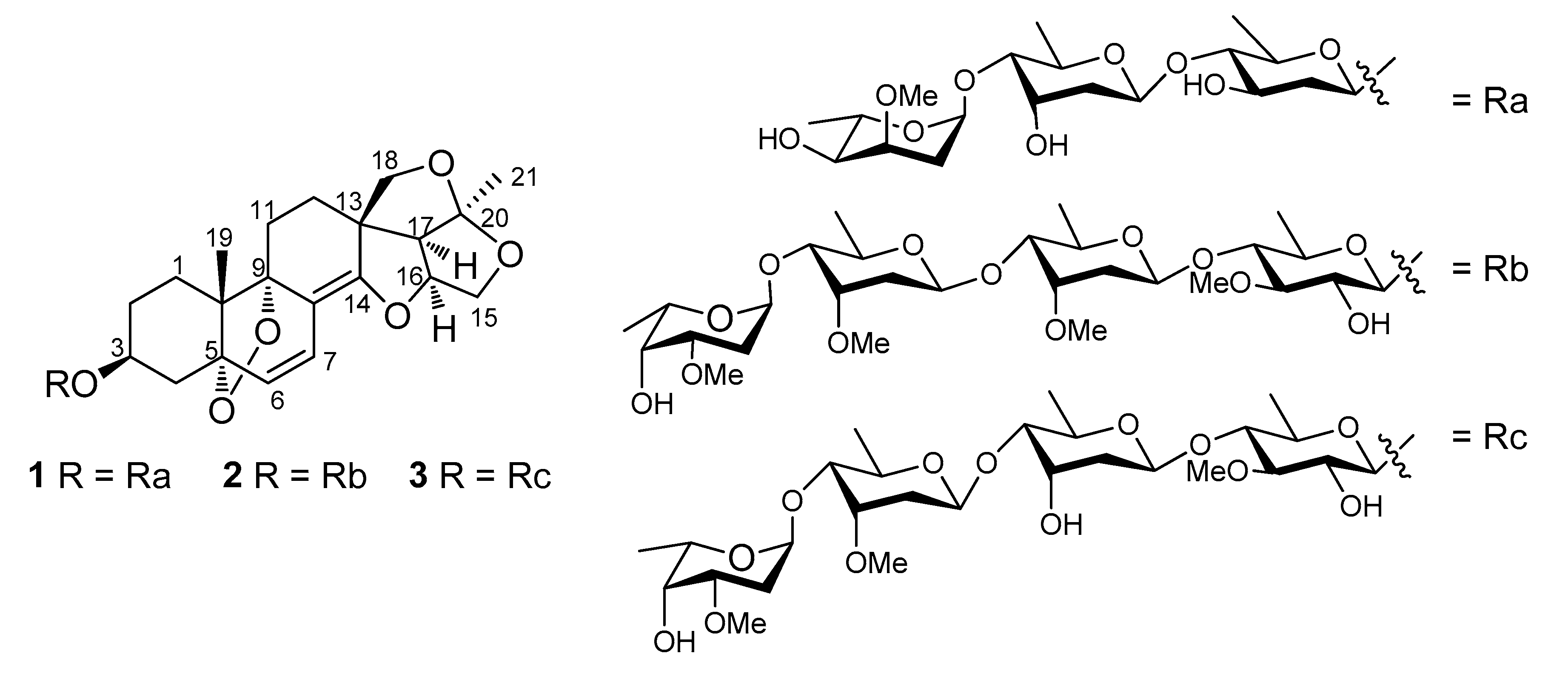
14,15-Secopregnane-Type Glycosides with 5α:9α-Peroxy and Δ6,8(14)-diene Linkages from the Roots of Cynanchum stauntonii

1
State Key Laboratory of Bioactive Substance and Function of Natural Medicines, Institute of Materia Medica, Chinese Academy of Medical Sciences and Peking Union Medical College, Beijing 100050, China
2
Shandong Analysis and Test Center, Shandong Academy of Sciences, Jinan 250014, Shandong, China
*
Author to whom correspondence should be addressed.
Molecules 2017, 22(6), 860; https://doi.org/10.3390/molecules22060860
Submission received: 21 April 2017 / Revised: 18 May 2017 / Accepted: 19 May 2017 / Published: 23 May 2017
(This article belongs to the Section Natural Products Chemistry)

Abstract

:
Three new 14,15-secopregnane-type glycosides, stauntosides UA, UA1, and UA2, were isolated from the roots of Cynanchum stauntonii. The three compounds share the first reported and same basic structural features of 3β-hydroxy-14:16,15:20,18:20-triepoxy-5α:9α-peroxy-14,15-secopregnane-6,8(14)-diene named as stauntogenin G as the aglycones. The structures of the new compounds were characterized on the basis of extensive spectroscopic analyses, mainly 1D and 2D NMR and MS methods and chemical analysis. The isolation and identification of the new compounds graced the structural diversity of pregnane-type steroids from C. stauntonii.

1. Introduction

C21-Pregnane-type natural organic compounds possess the usual skeleton of 17-ethyl-10,13-dimethylhexadecahydro-1H-cyclopenta[a]phenanthrene on the systematic nomenclature. The natural resource of this class of steroids is very affluent in the plant world, with both sugar-free and glycosidated pregnane-type steroids having been isolated by many researchers through phytochemical methods. In addition to the usual four-ring C21-pregnane-type skeleton, there are also several unusual skeletons, such as the 8,14-seco-C21-pregnane-type, 14,15-seco-C21-pregnane-type, and 13,14:14,15-diseco-C21-pregnane-type skeletons, all of these usual and unusual skeletons possessing multiple stereogenic centers and other structural diversities [1,2,3]. It is well known that the Cynanchum species of the Asclepiadaceae family are very rich in C21-steroids, with the unusual 14,15-seco-C21-pregnane-type and 13,14:14,15-diseco-C21-pregnane-type skeletons being most often discovered in previous investigations [4,5,6,7,8,9,10,11]. It is also well known in natural medicinal chemistry that, in addition to the structural diversity, C21-steroids are one class of biologically active compounds, with multiple bioactivities being reported [7,9,10,11,12]. Especially, our group reported that some steroidal glycosides isolated from C. stauntonii (Decne.) Schltr. ex Levl., a perennial medicinal herb naturally growing in the south-central region of China, showed anti-inflammatory activity [3]. This finding provided evidence supporting the application of C. stauntonii in some traditional medicine systems to treat inflammations [3,13,14]. Following the isolation and identification of several steroidal glycosides from the roots of C. stauntonii sharing the first reported aglycones of 8α:14α,14:16,15:20,18:20-tetraepoxy-14,15-secopregn-6-ene-3β,5α,9α-triol or its 5α:9α-peroxy bridge structure [3], our ongoing searches for new steroids in the same subjects lead to the isolation of three new steroidal glycoside, stauntosides UA, UA1, and UA2 (13) (Figure 1). Structural identification affirmed that stauntosides UA, UA1, and UA2 shared an aglycone of 3β-hydroxy-5α:9α-peroxy-14:16,15:20,18:20-triepoxy-14,15-secopregnane-6, 8(14)-diene, similar to, but somewhat different from the aforementioned first reported aglycones. Thus, the three new steroids were regarded as another subcategory of 14,15-secopregnane-type steroids. In order to grace the structural diversity of pregnane-type steroids in C. stauntonii, this paper describes the isolation and structure elucidation of these new compounds.

2. Results and Discussion

The roots of C. stauntonii were extracted with 95% EtOH. The 95% EtOH extract was concentrated and partitioned using petroleum ether and EtOAc. The EtOAc-soluble fraction was separated using multiple column chromatographies and preparative HPLC. As a result, three new compounds reported herein were yielded, all as white amorphous powders. All three compounds showed positive Libermann-Burchard and Keller-Kiliani reactions, suggesting their glycosidated steroids or triterpenoids categories with 2-deoxysugars in their sugar moieties [2,15]. The category of steroidal glycosides was determined according to their shared common features in the NMR spectra. The three or four distinguishable anomeric proton signals of sugars in the 1H-NMR spectra indicated the presence of corresponding sugar moieties (Table 1). When putting off the carbon signals of the three or four hexose units, all three compounds left twenty-one carbons from the C21-pregnane moieties (Table 1 and Table 2). All three compounds showed two singlets of methyl groups in their 1H-NMR spectra. The δ values of the relatively lower-field singlet of methyl groups in each compound, i.e., δH 1.54 for all three compounds, along with the carbon signals at δC 118.4, 119.6, and 118.2 in the 13C-NMR spectra for compounds 13, respectively, suggested that they belonged to the unusual 14,15-seco- (or 13,14:14,15-diseco-) C21-pregnane-type steroids. These singlets of methyl groups in the 1H-NMR spectra were born of Me-19 and 21 of the C21-pregnane skeleton, respectively. These carbon signals are the typical features of a dioxygenated secondary carbon-20 structure and they were confirmed by the correlations from Me-21 to C-17 and 20 in the respective HMBC spectrum of the three compounds. No carbonyl carbon was present in the 13C-NMR spectra of all three compounds, assigning them to be the 14,15-seco-C21-pregnane-type steroids [2,3,7].
Compound 1 possessed a molecular formula of C40H58O15 according to its 13C-NMR spectroscopic data and the HRESIMS (positive ion mode) data for the protonated molecular ion at m/z 779.3861 and sodium adduct molecular ion at m/z 801.3694, indicating a hydrogen deficiency index of twelve. This molecular formula has one fewer oxygen atom than that of stauntoside V3, a 14,15-secopregnane-type glycoside with the aglycone of stauntogenin F (3β-hydroxy-8α:14α,14:16,15:20,18:20-tetraepoxy-5α:9α-peroxy-14,15-secopregn-6-ene) previously isolated by our group from C. stauntonii [3]. Its IR spectrum displayed absorption bands for hydroxy (3440 cm−1) and olefinic (1681 cm−1) functionalities, among others. Acid hydrolysis, along with derivatization and GC analysis, indicated the presence of d-canaropyranose, d-digitoxopyranose, and l-cymaropyranose in a 1:1:1 ratio. The entire 1H and 13C-NMR spectroscopic data for 1 are given in Table 1 and Table 2, respectively. A detailed comparison of NMR data between 1 and stauntoside V3 showed that they were very similar. In the 1H-NMR spectrum, all the signals for 1 were nearly superimposable on their counterparts in stauntoside V3, the coupling constants of the three anomeric protons provided evidence that two monosaccharides shared β-glucosidic bonds and one possessed an α-glucosidic bond. In the 13C-NMR spectrum, the primary difference was in the replacement of the signals for an oxygenated tertiary carbon at δC 70.8 (s, C-8) and an dioxygenated secondary carbon at δC 98.9 (s, C-14) in the known compound by the signals of an olefinic quaternary carbon at δC 110.4 (C-8) and an oxygenated olefinic tertiary carbon at δC 156.8 (s, C-14) in 1. The rest of the carbons of compound 1 showed full accordance with categories of carbon types with their respective counterparts in stauntoside V3. The conjugated highfield shifts of C-6 and C-7 by Δδ –10.9 and −2.7, respectively, in compound 1 compared with stauntoside V3 were evident, which proposed a double bond linkage between C-8 and C-14. A highfield shift of Δδ –6.2 for C-10 compared with stauntoside V3 was also observed, which was mainly due to the impact of the change of magnetic anisotropy from the 8:14-epoxy linkage (oxirane) in stauntoside V3 to the Δ8(14) structure in 1. For the rest of carbon atoms, except for C-12 and C-18 which, because of the same causes as C-10, showed up lowfield shifts of Δδ +1.4 and +1.82, respectively, the numerical range of the absolute values of Δδ compared with the corresponding carbons in stauntoside V3 were less than 1.0, including those of the sugar moieties. These consistencies and differences of functional groups and chemical shifts between 1 and stauntoside V3, especially the molecular formula with a hydrogen deficiency index of twelve, indicated the presence of the peroxo bridge structure between C-5 and C-9, just as in stauntoside V3. Further, this identification was confirmed by the finding that the peroxylated downfield chemical shifts of Δδ +11.8 and +11.1 were observed for C-5 and C-9, respectively, in the 13C-NMR spectrum compared with stauntoside U, another 14,15-secopregnane-type glycoside with the aglycone of stauntogenin E (8α:14α,14:16,15:20,18:20-tetraepoxy-14,15-secopregn-6-ene-3β,5α,9α-triol) previously isolated by our group from C. stauntonii [3]. The HMBC spectrum showed the same picture as those in stauntoside V3, with the following correlations being well-marked: H-6 to C-4, 5, 8, and 10; H-7 to C-5, 9, and 14; H-15a to C-16, 17, and 20; H-17 to C-12, 13, 14, 18, and 20; H-18a to C-12 and 14; H-18b to C-12, 14, 17, and 20; Me-19 to C-1, 5, 9, and 10; Me-21 to C-17 and 20; H-1′′′ of α-l-cymaropyranose to C-4′′ of β-d-digitoxopyranose; H-1′′ of β-d-digitoxopyranose to C-4′ of β-d-canaropyranose; and H-1′ of β-d-canaropyranose to C-3, among others (Figure 2). The typical relative configurations of 14,15-secopregnane-type steroids of compound 1, i.e., both CH2-18 and Me-19 in β-orientation and H-16, H-17, and Me-21 all in α-orientation, which was also the same as that of stauntoside V3, were affirmed in the NOESY spectrum by the same picture as those of stauntoside V3, with NOE correlations of H-3/H-2α, H-3/H-4α, H-6/H-4α, H-6/H-4β, H-6/H-7, H-6/Me-19, H-7/Me-19, H-15α/H-17, H-16/H-17, H-17/Me-21, and Me-19/H-11β being evident and NOE correlations between H-3 and H-1α, H-3 and H-1β, H2-18 and Me-21, H2-18 and H-17, and Me-19 and H-4α not being observed (Figure 3). Especially, the relative configurations of 5α:9α-peroxy linkage and H-3α were elucidated by the same picture of reciprocal NOE correlations of H-3, H-6, H-7, and Me-19 (Figure 3), among others, with relevant protons as in stauntoside V3. Thus, the aglycone was elucidated as 3β-hydroxy-14:16,15:20,18:20-triepoxy-5α:9α-peroxy-14,15-secopregnane-6,8(14)-diene according to the number system of pregnanes and was named as stauntogenin G, and the structure of compound 1 was characterized as 14:16,15:20,18:20-triepoxy-5α:9α-peroxy-14,15-secopregn-6,8(14)-dien-3β-yl-4-o-(4-o-α-l-cymaropyranosyl-β-d-digitoxopyranosyl)-β-d-canaropyranoside and named stauntoside UA.
Compound 2 possessed a molecular formula of C49H74O19 according to its 13C-NMR data and the HRESIMS (positive ion mode) of the sodium adduct molecular ion at m/z 989.4729, indicating a hydrogen deficiency index of thirteen. Its IR spectrum displayed absorption bands for hydroxy (3393 cm−1) and olefinic (1646 cm−1) functionalities. Acid hydrolysis of 2, along with derivatization and GC analysis, indicated the presence of d-thevetopyranose, d-cymaropyranose, and l-diginopyranose in a 1:2:1 ratio. A detailed comparison of the 1H- and 13C-NMR data between 1 and 2 showed that 2 was nearly entirely identical to 1, with respect to their aglycone moieties (Table 1 and Table 2), the numerical range of Δδ for all the 13C-NMR signals of the aglycone of 2 compared with the corresponding carbons in 1 was between +1.1 and +1.6, which were obviously systematic errors, suggesting the same aglycone for 2 and 1. This determination was confirmed with a combined interpretation of the 2D NMR spectra of 2, including the 1H,1H-COSY, HSQC, and HMBC correlations (data not shown). In addition to the resonances of the aglycone moiety, the 1H and 13C-NMR data (Table 1 and Table 2) and the 2D NMR spectroscopic features, including the 1H,1H-COSY and HMBC correlations, among others, of the sugar moiety of 2 were consistent with stauntoside V1, a 14,15-secopregnane-type glycoside previously isolated by our group from C. stauntonii [3]. Thus, the structure of 2 is 14:16,15:20,18:20-triepoxy-5α:9α-peroxy-14,15-secopregn-6,8(14)-dien-3β-yl-4-O-[4-O-(4-O-α-l-diginopyranosyl-β-d-cymaropyranosyl)-β-d-cymaropyranosyl]-β-d-thevetopyranoside and named stauntoside UA1.
Compound 3 possessed a molecular formula of C48H72O19 according to its 13C-NMR data and the HRESIMS (positive ion mode) of the sodium adduct molecular ion at m/z 975.4567, indicating a hydrogen deficiency index of thirteen. Its IR spectrum displayed absorption bands for hydroxy (3369 cm−1) and olefinic (1662 cm−1) functionalities. Acid hydrolysis of 3, along with derivatization and GC analysis, indicated the presence of d-thevetopyranose, d-digitoxopyranose, d-cymaropyranose, and l-diginopyranose in a 1:1:1:1 ratio. A detailed comparison of the 1H- and 13C-NMR data of 13 indicated that they shared the same aglycone, compound 3 being nearly entirely identical to 1 and 2, with respect to their aglycone moieties and the numerical range of Δδ for all the 13C-NMR signals of the aglycone of 3 compared with the corresponding carbons in 2 being between −1.4 and −1.6 (Table 1 and Table 2). This determination was confirmed with a combined interpretation of the 2D NMR spectra of 3, including the 1H,1H-COSY, HSQC, and HMBC correlations (data not shown). In addition to the resonances of the aglycone moiety, the 1H- and 13C-NMR data (Table 1 and Table 2) and the 2D NMR spectroscopic features, including the 1H,1H-COSY and HMBC correlations, among others, of the sugar moiety of 3 were consistent with stauntoside W, a 14,15-secopregnane-type glycoside previously isolated by our group from C. stauntonii [3]. Thus, the structure of 3 is 14:16,15:20,18:20-triepoxy-5α:9α-peroxy-14,15-secopregn-6,8(14)-dien-3β-yl-4-O-[4-O-(4-O-α-l-diginopyranosoyl-β-d-cymaropyranosyl)-β-d-digitoxopyranosyl]-β-d-thevetopyranoside and named stauntoside UA2.

3. Experimental Section

3.1. General Experimental Procedures

All the instruments, solvents, reagents, and experimental conditions for the measurements of IR spectra, 1D and 2D NMR spectra, and both ESIMS and HRESIMS data and for the performing of column chromatography (CC), preparative HPLC procedure, and TLC analysis were previously described [2,3].

3.2. Plant Material

The collecting, species identifying, and depositing of the roots of C. stauntonii were previously described [2,3].

3.3. Extraction and Isolation

The extraction of the dried and pulverized roots (30 kg) of C. stauntonii and the preliminary isolation of the 95% EtOH extract and the consequent EtOAc-soluble portion to afford thirteen subfractions (Fr. 1 to Fr. 13) of silica gel CC fractionation were previously described [2,3]. Fr. 3 (68.0 g; eluted with CHCl3:MeOH, 100:1, v/v) was separated with silica gel CC using a gradient elution of petroleum ether:EtOA (25:1→1:1, v/v) to yield seven subfractions, Fr. 3-1 to Fr. 3-7. Fr. 3-5 (5.0 g; eluted with petroleum ether:EtOAc, 10:1, v/v) was subjected to a flash C18 column eluted with a gradient of MeOH:H2O (40:60→100:0, v/v) to yield six subfractions, Fr. 3-5-1 to Fr. 3-5-6. Fr. 3-5-4 (sampled 150 mg; eluted with MeOH:H2O, 60:40, v/v) was subjected to preparative RP-HPLC (mobile phase of MeCN:H2O (38:62, v/v) at a flow rate of 5 mL min−1 with UV detection at 280 nm) to yield, in addition to the reported known compound [3], compound 1 (15 mg). Fr. 5 (12.0 g; eluted with CHCl3:MeOH, 50:1, v/v) was subjected to a flash C18 column eluted with a gradient of MeOH:H2O (40:60→100:0, v/v) to yield five subfractions, Fr. 5-1 to Fr. 5-5. Fr. 5-4 (6.0 g; eluted with MeOH:H2O, 70:30, v/v) was subjected to a flash C18 column eluted with a gradient of MeOH:H2O (40:60→100:0, v/v) to yield five subfractions, Fr. 5-4-1 to Fr. 5-4-5. Fr. 5-4-4 (sampled 800 mg, eluted with MeOH:H2O, 70:30, v/v) was subjected to preparative RP-HPLC (mobile phase of CH3CN:H2O (38:62, v/v) at a flow rate of 5 mL min-1 with UV detection at 280 nm) to yield compound 2 (17 mg). Fr. 8 (6.0 g, eluted with CHCl3:MeOH, 25:1, v/v) was subjected to a flash C18 column eluted with a gradient of MeOH:H2O (40:60→100:0, v/v) to yield five subfractions, Fr. 8-1 to Fr. 8-5. Fr. 8-2 (sampled 300 mg, eluted with MeOH:H2O, 50:50, v/v) was subjected to preparative RP-HPLC (mobile phase of CH3CN:H2O (35:65, v/v) at a flow rate of 5 mL min−1 with UV detection at 280 nm) to yield compound 3 (10 mg).
Stauntoside UA (1). White amorphous powder; IR (KBr) νmax 3440, 2933, 1681, 1450, 1379, 1163, 1095, 1060, 942, and 835 cm−1; for 1H-NMR (500 MHz), see Table 1 and Table 2; for 13C-NMR (125 MHz), see Table 1 and Table 2; positive-ion mode ESIMS m/z 801.5 [M + Na]+; positive-ion mode HRESIMS m/z 779.3861 [M + H]+ (calculated for C40H59O15, 779.3848), m/z 801.3694 [M + Na]+ (calculated for C40H58O15Na, 801.3668).
Stauntoside UA1 (2). White amorphous powder; IR (KBr) νmax: 3393, 2921, 1646, 1468, 1380, 1160, 1104, 1063, 1007, and 721 cm−1; for 1H-NMR (500 MHz), see Table 1 and Table 2; for 13C-NMR (125 MHz), see Table 1 and Table 2; positive-ion mode ESIMS m/z 989.5 [M + Na]+; positive-ion mode HRESIMS m/z 989.4729 [M + Na]+ (calculated for C49H74O19Na, 989.4717).
Stauntoside UA2 (3). White amorphous powder; IR (KBr) νmax: 3369, 2924, 1662, 1465, 1380, 1165, 1065, 1023, and 879 cm−1; for 1H-NMR (500 MHz), see Table 1 and Table 2; for 13C-NMR (125 MHz), see Table 1 and Table 2; positive-ion mode ESIMS m/z 975.5 [M + Na]+; positive-ion mode HRESIMS m/z 975.4567 [M + Na]+ (calculated for C48H72O19Na, 975.4560).

3.4. Determination of Steroidal Category and 2-Deoxysugars

3.4.1. Libermann-Burchard Reaction

To a solution of each compound (1 mg) in acetic anhydride (5 mL) was added a little of a mixture of 98% sulfuric acid and acetic anhydride (1:20, v/v) dropwise. An obvious color change from somewhat yellowish red to purple to blue to dark reddish brown was observed.

3.4.2. Keller-Kiliani Reaction

To a solution of each compound (1 mg) in acetic acid (5 mL) was added one drop of aqueous 20% FeCl3 solution. The solution was fully mixed and then a spot of 98% sulfuric acid was added along the test tube wall, with a light green color being observed in the acetic acid solution.

3.5. Acid Hydrolysis of New Compounds and Determination of Absolute Configurations of Monosaccharides

The acid hydrolysis of new compounds and determination of absolute configurations of monosaccharides were conducted using the method described in a previous paper from our laboratory [2,16]. In this experiment, the known compounds stauntoside B, glaucogenin C mono-d-thevetoside, stauntoside G, and amplexicoside D were used to determine the retention times of the acetylated thiazolidine derivatives of relevant monosaccharides, with tR d-digitoxose 13.09 min, tR l-cymarose 13.46 min, tR d-cymarose 18.46 min, tR l-diginose 14.31 min, tR d-thevetose 16.07 min, and tR d-canarose 16.51 min being determined. Retention times of the monosaccharides released from the new compounds after derivatization were as follows: tR d-digitoxose 13.05 min, tR l-cymarose 13.41 min, and tR d-canarose 16.45 min for compound 1; tR d-cymarose 18.49 min, tR l-diginose 14.38 min, and tR d-thevetose 16.09 min for compound 2; and tR d-digitoxose 13.06 min, tR d-cymarose 18.42 min, tR l-diginose 14.38 min, and tR d-thevetose 16.03 min for compound 3.

Acknowledgments

This work was financially supported by grant from CAMS Initiation Fund for Innovative Medicine (2016-I2M-3-014).

Author Contributions

H.Q. conceived and designed the experiments; A.D. and J.Y. performed the experiments; A.D. and Z.Z. analyzed the data; Z.L and L.M contributed reagents/materials/analysis tools; A.D., J.Y., and H.Q. wrote the paper.

Conflicts of Interest

The authors declare no conflict of interest.

References

  1. Kanchanapoom, T.; Kasai, R.; Ohtani, K.; Andriantsiferana, M.; Yamasaki, K. Preganane and pregnane glycosides from the Malagasy plant, Cynanchum aphyllum. Chem. Pharm. Bull. 2002, 50, 1031–1034. [Google Scholar] [CrossRef] [PubMed]
  2. Yu, J.Q.; Deng, A.J.; Qin, H.L. Nine new steroidal glycosides from the roots of Cynanchum stauntonii. Steroids 2013, 78, 79–90. [Google Scholar] [CrossRef] [PubMed]
  3. Yu, J.Q.; Lin, M.B.; Deng, A.J.; Hou, Q.; Bai, J.Y.; Li, Z.H.; Ma, L.; Zhang, Z.H.; Yuan, S.P.; Jiang, R.T.; et al. 14,15-Secopregnane-type C21-steriosides from the roots of Cynanchum stauntonii. Phytochemistry 2017, 138, 152–162. [Google Scholar] [CrossRef] [PubMed]
  4. Qiu, S.X.; Zhang, Z.X.; Zhou, J. Steroidal glycosides from the root of Cynanchum versicolor. Phytochemistry 1989, 28, 3175–3178. [Google Scholar]
  5. Shibano, M.; Misaka, A.; Sugiyama, K.; Taniguchi, M.; Baba, K. Two secopregnane-type steroidal glycosides from Cynanchum stauntonii (Decne.) Schltr. ex Levl. Phytochem. Lett. 2012, 5, 304–308. [Google Scholar] [CrossRef]
  6. Sugama, K.; Hayashi, K.; Mitsuhashi, H.; Kaneko, K. Studies on the constituents of Asclepiadaceae plants. LXVI. The structures of three new glycosides, Cynapanosides A, B, and C, from the Chinese drug “Xu-Chang-Qing,” Cynanchum paniculatum KITAGAWA. Chem. Pharm. Bull. 1986, 34, 4500–4507. [Google Scholar] [CrossRef] [PubMed]
  7. Wang, P.; Qin, H.L.; Zhang, L.; Li, Z.H.; Wang, Y.H.; Zhu, H.B. Steroids from the roots of Cynanchum stauntonii. Planta Med. 2004, 70, 1075–1079. [Google Scholar] [CrossRef] [PubMed]
  8. Yu, J.Q.; Zhang, Z.H.; Deng, A.J.; Qin, H.L. Three new steroidal glycosides from the roots of Cynanchum stauntonii. BioMed Res. Int. 2013, 2013, 816145. [Google Scholar] [PubMed]
  9. Lai, C.Z.; Liu, J.X.; Pang, S.W.; Dai, Y.; Zhou, H.; Mu, Z.Q.; Wu, J.; Tang, J.S.; Liu, L.; Yao, X.S. Steroidal glycosides from the roots of Cynanchum stauntonii and their effects on the expression of iNOS and COX-2. Phytochem. Lett. 2016, 16, 38–46. [Google Scholar] [CrossRef]
  10. Day, S.H.; Wang, J.P.; Won, S.J.; Lin, C.N. Bioactive constituents of the roots of Cynanchum atratum. J. Nat. Prod. 2001, 64, 608–611. [Google Scholar] [CrossRef] [PubMed]
  11. Li, S.L.; Tan, H.; Shen, Y.M.; Kawazoe, K.; Hao, X.J. A pair of new C-21 steroidal glycoside epimers from the roots of Cynanchum paniculatum. J. Nat. Prod. 2004, 67, 82–84. [Google Scholar] [CrossRef] [PubMed]
  12. Perrone, A.; Plaza, A.; Ercolino, S.F.; Hamed, A.I.; Parente, L.; Pizza, C.; Piacente, S. 14,15-Secopregnane derivatives from the leaves of Solenostemma argel. J. Nat. Prod. 2006, 69, 50–54. [Google Scholar] [CrossRef] [PubMed]
  13. Liang, A.H.; Xue, B.Y.; Yang, Q.; Fu, M.H.; Wang, G. Studies on antitussive, expectorant, and anti-inflammatory effects of Rhizoma Cynanchi stauntonii. China J. Chin. Mater. Med. 1996, 21, 173–175. [Google Scholar]
  14. Liang, A.H.; Xue, B.Y.; Yang, Q.; Li, Z.L.; Wang, J.; Fu, M.H. A pharmacological comparative study on Baiqian and Baiwei. China J. Chin. Mater. Med. 1996, 21, 622–625. [Google Scholar]
  15. Chen, H.; Xu, N.; Zhou, Y.; Qiao, L.; Cao, J.; Yao, Y.; Hua, H.; Pei, Y. Steroidal glycosides from the roots of Cynanchum amplexicaule Sieb. et Zucc. Steroids 2008, 73, 629–636. [Google Scholar] [CrossRef] [PubMed]
  16. Hara, S; Okabe, H; Mihashi, K. Gas-liquid chromatographic separations of aldose enantiomers as trimethylsilyl ethers of methyl 2-(polyhydroxyalkyl)-thiazolidine-4(R)-carboxylates. Chem. Pharm. Bull. 1987, 35, 501–506. [Google Scholar] [CrossRef]
Sample Availability: Samples of compounds 1 and 2 are available from the authors.
Figure 1. Structures of compounds 13.
Molecules 22 00860 g001
Figure 2. Key HMBC correlations (H→C) of 1.
Molecules 22 00860 g002
Figure 3. Key NOE correlations (H↔H) in the aglycone moiety of 1.
Molecules 22 00860 g003
Table 1. 1H- and 13C-NMR spectroscopic data for the aglycone moieties of 13 (in pyridine-d5, TMS).
Position123
δH (J in Hz)δCδH (J in Hz)δCδH (J in Hz)δC
1.34, ov27.61.32, ov28.91.32, ov27.4
2.10, ov 2.10, ov 1.91, ov
2.23, br dd (14.0, 2.5)30.22.39, ov31.42.23, ov29.9
2.08, ov 2.10, ov 2.07, ov
34.31, m73.34.23, m74.94.31, m73.4
2.51, ov33.62.52, dd (14.0, 4.5)34.72.52, dd (14.0, 5.0)33.2
1.96, ov 1.75, ov 1.76, ov
5 85.6 86.8 85.3
65.59, d (9.5)129.15.48, d (9.5)130.35.48, d (9.5)128.8
76.77, d (9.5)125.16.72, d (9.5)126.46.72, d (9.5)124.8
8 110.4 111.7 110.1
9 87.4 88.6 87.1
10 50.6 51.9 50.4
11α2.01, ov24.72.00, ov25.92.00, ov24.4
11β1.77, ov 1.72, ov 1.75, ov
12a2.10, ov28.42.01, ov29.72.08, ov28.2
12b1.92, ov 1.92, ov 1.91, ov
13 55.0 56.2 54.7
14 156.8 158.1 156.6
15α3.79, dd (11.0, 4.5)72.03.79, dd (11.0, 4.5)73.23.79, dd (11.0, 4.5)71.7
15β4.24, ov 4.24, br d (11.0) 4.09, br d (11.0)
164.81, ov86.94.81, ov88.24.81, ov86.7
172.81, d (7.5)61.92.80, d (8.0)63.12.80, d (8.0)61.6
18a3.99, d (9.0)75.33.98, d (10.0)76.53.98, d (8.5)75.0
18b4.11, d (9.0) 4.10, d (10.0) 4.09, d (8.5)
190.95, s15.80.89, s17.00.89, s15.5
20 118.4 119.6 118.2
211.54, s22.41.54, s23.71.54, s22.2
ov: overlapped signals.
Table 2. 1H- and 13C-NMR spectroscopic data for the sugar moieties of 13 (pyridine-d5).
Position123
δH (J in Hz)δCδH (J in Hz)δCδH (J in Hz)δC
β-d-can β-d-the β-d-the
1′4.76, dd (9.5, 2.0)98.84.74, d (8.0)103.94.73, d (8.0)102.4
2′a2.51, ov40.13.91, ov75.93.91, ov74.4
2′b1.96, ov
3′3.95, ov70.13.66, ov86.13.67, ov85.6
4′3.28, ov88.53.67, ov83.93.69, ov82.6
5'3.47, ov70.93.61, ov72.93.69, ov71.4
6′1.31, d (6.5)18.11.42, d (5.5)19.91.42, d (6.0)18.4
3′-OCH3 3.92, s61.83.93, s60.3
β-d-digt β-d-cym β-d-digt
1′′5.24, dd (9.5, 2.0)99.95.30, dd (10.0, 2.0)100.15.55, dd (10.0, 2.0)98.7
2′′a2.41, ov38.21.85, ov38.41.99, ov38.9
2′′b1.96, ov 2.35, ov 2.42, ov
3′′4.48, ov67.44.07, ov79.44.63, ov67.5
4′′3.43, ov80.73.48, dd (10.0, 3.0)84.53.47, ov83.0
5′′4.19, ov69.44.21, ov70.64.29, ov68.6
6′′1.31, d (6.5)18.11.37, d (6.0)19.91.41, d (6.0)18.3
3′′-OCH3 3.61, s60.1
α-l-cym β-d-cym β-d-cym
1′′′5.04, dd (4.0, 3.0)98.65.08, dd (9.5, 1.5)101.55.13, br d (9.5)99.4
2′′′a2.34, ov32.21.74, ov (2.42)36.41.68, ov34.72
2′′′b1.82, ov 2.42, ov 2.32, ov
3′′′3.79, ov76.53.96, ov78.83.92, ov77.2
4′′′3.61, ov72.63.45, dd (10.0, 1.0)83.53.39, ov82.0
5′′′4.48, ov67.64.21, ov70.64.21, ov69.1
6′′′1.41, d (6.5)18. 41.37, d (6.0)19.71.30, d (6.0)18.4
3′′′-OCH33.37, s56.83.52, s58.63.52, s57.1
α-l-dign α-l-dign
1′′′′ 5.22, br d (3.0)102.45.19, br d (3.5)101.0
2′′′′a 2.09, ov32.22.07, ov30.7
2′′′′b 2.39, ov 2.37, ov
3′′′′ 3.85, ov77.13.84, ov75.6
4′′′′ 4.07, ov68.94.07, ov67.4
5′′′′ 4.32, ov68.84.30, ov67.5
6′′′′ 1.57, d (6.5)19.01.56, d (7.0)17.5
3′′′′-OCH3 3.31, s56.33.31, s54.8
ov: overlapped signals.

