Abstract
Chemical investigation of the 90% acetone extract of the branches and leaves of Sabina gaussenii led to the isolation of two new cinnamyl isovalerate derivatives (1–2) and eighteen known compounds (3–20). Their structures were determined mainly by means of MS, 1D- and 2D-NMR data, and this is the first time these compounds have been reported from this plant. The biological activity test results indicated that the 90% acetone extract showed cytotoxicity against the human lung adenocarcinoma (A549) cell line (IC50 = 0.98 ± 0.1 μg/mL), compound 6 showed cytotoxicities against human cervical carcinoma (HeLa) (IC50 = 0.4 ± 0.1 μM ) and human gastric carcinoma (BGC-823) (IC50 = 0.9 ± 0.2 μM) cancer cell lines, and compound 19 showed cytotoxicities against HeLa (IC50 = 1.5 ± 0.4 μM), BGC-823 (IC50 = 7.0 ± 0.8 μM ), and A549 (IC50 = 10.6 ± 1.5 μM ) cancer cell lines.
1. Introduction
Sabina gaussenii is endemic to China and is usually used as a hedge plant. The genus Sabina, which used to belong to genus Juniperus, has about 50 species and spread widely throughout the northern hemisphere [1]. According to the literature, the Sabina plants have been reported to be a rich source of bioactive terpenoids [2]. Up to now, only one diterpenoid and a few flavones have been reported from S. gaussenii [3]. As part of serial investigations on the Gymnospermae plants and in order to seek more novel bioactive compounds, we carried out an extensive chemical study on S. gaussenii [4,5,6,7]. In this paper, we report the isolation and structure elucidation of two new cinnamyl isovalerate derivatives (1–2) together with eighteen other known compounds (3–20) from the branches and leaves of S. gaussenii, in addition to a screening of their cytotoxicities.
2. Results and Discussion
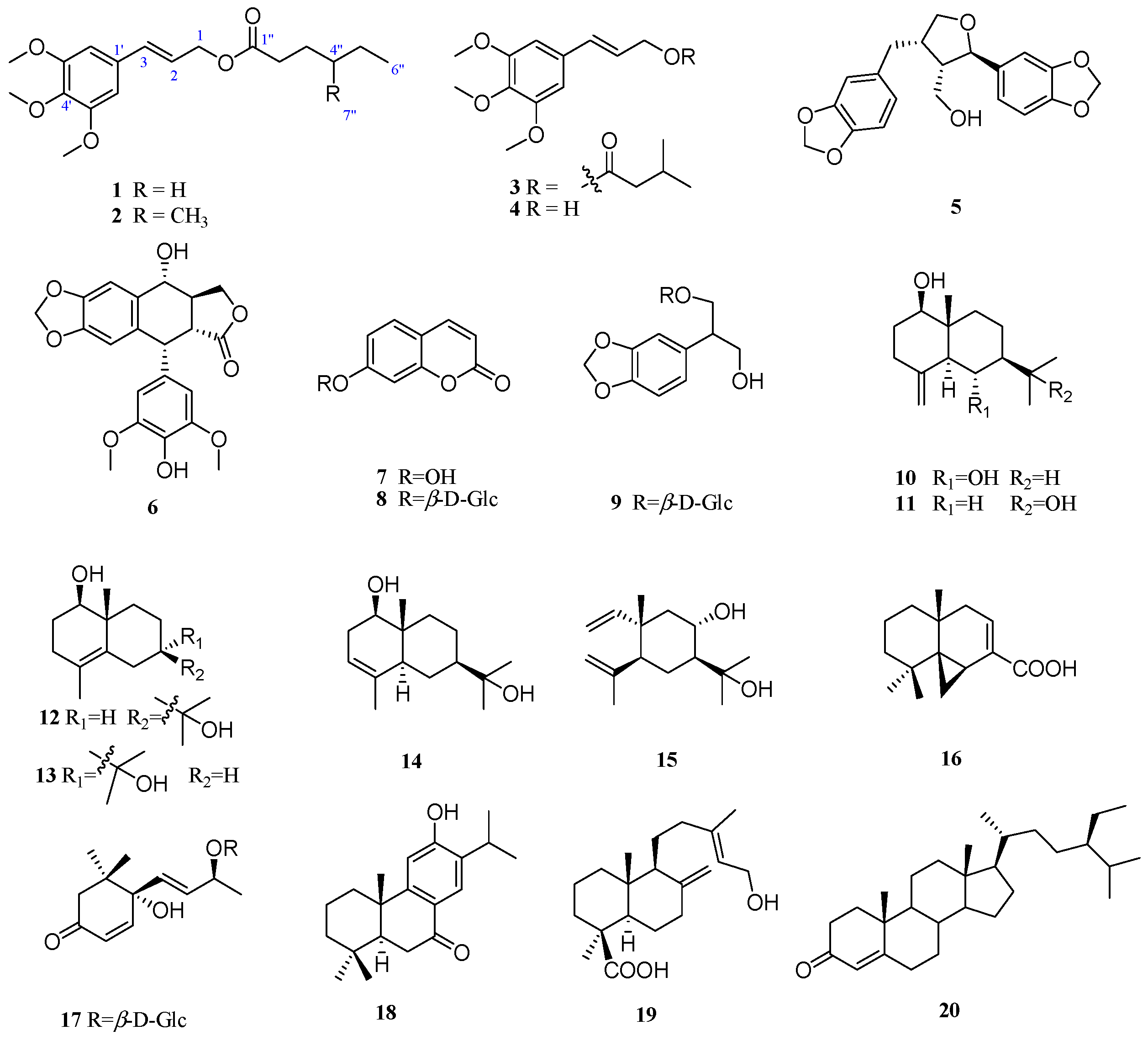
The air-dried powder of the branches and leaves of S. gaussenii was extracted with 90% acetone at room temperature to give a crude extract, which was suspended in H2O and successively partitioned with petroleum ether, ethyl acetate (EtOAc), and n-butyl alcohol (n-BuOH). Column chromatographic separations of these extracts afforded compounds 1–20 (Figure 1). The two new structures (1–2) were identified by spectroscopic analyses and physicochemical properties, while the known compounds were identified as 3’,4’,5’-dimethoxycinnamyl isovalerate (3) [8], 3’,4’,5’-dimethoxycinnamyl alcohol (4) [9], dihydrosesamin (5) [10], 4’-O-demethylepipodophyllotoxin (6) [11], 7-hydroxy coumarin (7) [12], 7-β-d-glucosyloxy coumarin (8) [13], 1-β-d-glucosyloxy-2-(3,4-methylenedioxyphenyl)-propane-l,3-diol (9) [14], lβ,6α-dihydroxy-4(14)-eudesmene (10) [15], selin-4(15)-en-1β, 11-diol (11) [16], 4-eudesmene-1β, 11-diol (12) [17], 7-epi-4-eudesmene-1β, 11-diol (13) [17], 3-eudesmene-1β, 11-diol (14) [17], 8α,11-elemodiol (15) [18], hinokiic acid (16) [19], corchoionoside C (17) [20], hinokiol (18) [21], isocupressic acid (19) [22], and sitostenone (20) [23] by comparison of their spectroscopic data and specific rotations with those obtained in the literature.
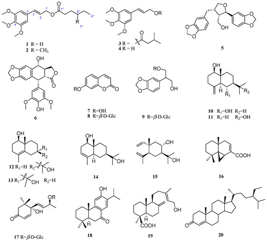
Figure 1.
The chemical structures of compounds 1–20.
2.1. Identification of New Compounds
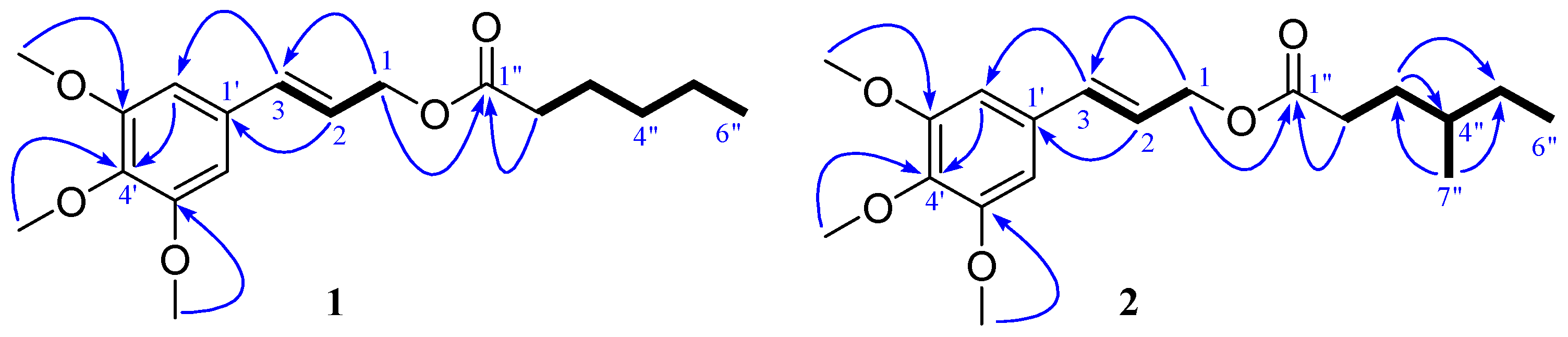
Compound 1 was obtained as a colorless oil. Its molecular was assigned as C18H26O5 on the basis of positive HRESIMS ([M + Na]+ 345.1674, calcd 345.1677) and NMR spectra data (Table 1), which implied six degrees of unsaturation. The IR absorption bands at 1735 cm−1 indicated the presence of carbonyl groups. The 1H-NMR spectrum of 1 showed three methoxy signals (δH 3.87 (s, 6H), 3.84 (s, 3H)). The 13C- and DEPT-NMR spectra of 1 revealed 18 carbon signals: a carbonyl (δC 173.8 (C-1”)), a symmetrical benzene (δC 153.3 (C-3’, 5’), 138.0 (C-4’), 132.0 (C-1’), 103.6 (C-2’, 6’)), a double bond (δC 134.2 (C-3), 122.9 (C-2)), five methylenes (δC 64.9 (C-1), 34.3 (C-2”), 31.4 (C-4”), 24.7 (C-3”), 22.4 (C-5”)), and four methyls (δC 56.1 (C-2*OMe), 61.0 (C-OMe), 14.0 (C-6”)). The NMR data indicated that 1 was a phenylpropanoid, which was very similar with those of 3 [8]. In comparison with 3, the only difference is a hexanoyl (δC 173.8 (C-1”), 34.3 (C-2”), 24.7 (C-3”), 31.4 (C-4”), 22.4 (C-5”), 14.0 (C-6”)) in 1 replaced the isovaleryl (δC 173.0 (C-1”), 43.4 (C-2”), 25.7 (C-3”), 22.4 (C-4”, 5”)) in 3. The 1H-1H COSY correlations (Figure 1) between H-2” and H-3”, H-3” and H-4”, H-4” and H-5”, H-5” and H-6”, and the HMBC cross-peaks of H-2” with C-1” confirmed the presence of the hexanoyl in 1. In the HMBC spectrum, the cross-peak of H-1 with C-1” suggested that the hexanoyl located at C-1 (Figure 2). Hence, the structure of 1 was finally determined as 3’,4’,5’-trimethoxycinnamyl caproate. NMR spectrums show in Supplementary Materials.

Table 1.
1H (600 MHz) and 13C (150 MHz) NMR spectroscopic data of 1–2 in CDCl3. (J in Hz, δ in ppm).
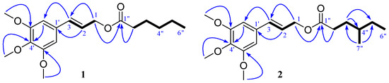
Figure 2.
Key 1H-1H COSY (  ) and HMBC (
  ) correlations of compounds 1–2.
) and HMBC ( ) correlations of compounds 1–2.
Compound 2 was obtained as a colorless oil. The molecular formula of C19H28O5 was determined by HRESIMS ([M + Na]+ 359.1842, calcd 359.1834) and NMR spectra data. The NMR data of 2 was closely similar with those of 1, which suggested that 2 was also a phenylpropanoid. The only difference is that a 4”-methyl-hexanoyl (δC 174.0 (C-1”), 32.1 (C-2”), 31.4 (C-3”), 34.0 (C-4”), 29.1 (C-5”), 18.8 (C-6”)) in 2 replaced the hexanoyl (δC 173.8 (C-1”), 34.3 (C-2”), 24.7 (C-3”), 31.4 (C-4”), 22.4 (C-5”), 14.0 (C-6”), 11.4 (C-7”)) in 1. The 1H-1H COSY correlations between H-2” and H-3”, H-3” and H-4”, H-4” and H-5”, H-4” and H-7”, H-5” and H-6”, and the HMBC cross-peaks of H-2” with C-1”, confirmed the presence of the 4”-methyl-hexanoyl portion in 2. In the HMBC spectrum, the cross-peak of H-1 with C-1” suggested that the 4”-methyl-hexanoyl located at C-1 (Figure 2). Thus, the structure of 2 was assigned as 3’,4’,5’-trimethoxycinnamyl-4”-methyl-caproate.
2.2. Cytotoxicity Assay
The in vitro cytotoxicities of the 90% acetone extract of S. gaussenii and compounds 1–20 were evaluated against three cancer cell lines, including human cervical carcinoma (HeLa), human gastric carcinoma (BGC-823), and human lung adenocarcinoma (A549). The results indicated that the 90% acetone extract showed cytotoxicity against the A549 cell line (IC50 = 0.98 ± 0.1 μg/mL), compound 6 showed cytotoxicities against HeLa (IC50 = 0.4 ± 0.1 μM) and BGC-823 (IC50 = 0.9 ± 0.2 μM) cancer cell lines, and compound 19 showed cytotoxicities against HeLa (IC50 = 1.5 ± 0.4 μM), BGC-823 (IC50 = 7.0 ± 0.8 μM) and A549 (IC50 = 10.6 ± 1.5 μM) cancer cell lines.
3. Materials and Methods
3.1. General Experimental Procedures
Spectra were recorded on a Bio-Rad FTS-135 spectrometer (Bio-Rad, Berkeley, CA, USA) with KBr pellets, ν in cm−1. UV spectra were measured on SHIMADZU UV-2401PC spectrometer (Shimadzu Corporation, Kyoto, Japan). NMR spectra were conducted on Bruker ARX-600 spectrometers (Bruker Corporation, Rheinstetten, Germany) with TMS as internal standard, chemical shift (δ) was expressed in ppm, and coupling constants (J) in Hz. ESI and HR-ESI-MS were taken on an API Qstar-Pulsar-1 mass spectrometer (Thermo Fisher Scientific, Bremen, Germany).
3.2. Plant Material
Branches and leaves of Sabina gaussenii (Cheng) Cheng et W. T. Wang were collected from Kunming Botany Garden, Yunnan Province, People’s Republic of China, in August 2010. It was identified by Prof. Wei-bang Sun at Kunming Institute of Botany, Chinese Academy of Sciences.
3.3. Extraction and Isolation
The powdered air-dried branches and leaves (13 kg) of S. gaussenii were extracted with 90% acetone (3 × 40 L) at room temperature and then concentrated under reduced pressure. The concentrated acetone extract (910 g) was dissolved in 60 °C water and partitioned with petroleum ether, EtOAc, and n-BuOH, respectively, to afford petroleum ether fraction (170 g), EtOAc fraction (130 g), and n-BuOH fraction (250 g).
The petroleum ether fraction (170 g) was separated on an MCI gel column eluted with MeOH–H2O (3:7 to 1:0, v/v) to produce thirteen subfractions A−M. Fraction C (41 g) was separated on a silica gel column and eluted with gradient mixtures of petroleum ether-acetone (from 20:1 to 1:1) and then separated on a column of RP-C18 silica gel (MeOH in H2O, 60%−80%) to yield five major components, with each purified by semipreparative HPLC (SunFire C18 column, 10 mm × 250 mm, 5 μm, CH3CN–H2O, 85:15, 3 mL/min) to afford 1 (2.9 mg), 2 (2.3 mg), 3 (16 mg), 10 (11 mg), and 20 (26 mg), respectively. Fraction E was chromatographed on a RP-C18 silica gel column (MeOH in H2O, 50%−90%) and then purified by semipreparative HPLC with CH3CN–H2O (80:20, 3 mL/min) as the mobile phase to give compounds 4 (29 mg), 11 (21 mg), 12 (17 mg), 13 (35 mg), 14 (11 mg), 16 (13 mg), and 18 (11 mg), respectively. The EtOAc fraction was subjected to silica gel column (CHCl3/MeOH, 9:1 to 7:3) to yield five subfractions N−R. Fraction P was chromatographed on a RP-C18 silica gel column (MeOH in H2O, 50%−90%) to give 5 (44 mg), 6 (28 mg), 7 (25 mg), 15 (27 mg), and 19 (81 mg), respectively. The n-BuOH fraction was subjected to silica gel column (CHCl3–MeOH, 10:1 to 0:1), and then subjected to RP-C18 column and eluted with MeOH–H2O (65:35) to obtain compounds 8 (32 mg), 9 (99 mg), and 17 (28 mg).
3.4. Spectroscopic Data
3’,4’,5’-Trimethoxycinnamyl caproate (1): colorless oil. UV λmax (CH3OH) nm (log ε): 270 (4.32), 221 (4.64). IR (KBr) νmax (cm−1): 2957, 2935, 1735, 1583, 1507, 1462, 1419, 1242, 1128. 1H- and 13C-NMR: Table 1. HRESIMS: m/z 345.1674 (calcd for C18H26O5Na, 345.1677 [M + Na]+).
3’,4’,5’-Trimethoxycinnamyl 4”-methyl-caproate (2): colorless oil. UV λmax (CH3OH) nm (logε): 270 (3.52), 220 (3.86). IR (KBr) νmax (cm−1): 2959, 2928, 1735, 1584, 1508, 1462, 1420, 1242, 1128. 1H- and 13C-NMR: Table 1. HRESIMS: m/z 359.1842 (calcd for C19H28O5Na, 359.1834 [M + Na]+).
3.5. Bioassay
The cytotoxicities of the 90% acetone extract and compounds (1–20) against the HeLa, BGC-823, and A549 cancer cell lines were measured using a sulforhodamine B (SRB, Sigma, Saint Louis, MO, USA) assay as described in the literature [24]. Taxol were used as positive controls. Briefly, cells were plated in 96-well culture plates for 24 h and then treated with serial dilutions of all compounds with a maximum concentration of 20 μg/mL. After being incubated for 48 h under a humidified atmosphere of 5% CO2 at 37 °C, cells were fixed with 25 μL of ice-cold 50% trichloroacetic acid and incubated at 4 °C for 1 h. After washing with distilled water and air-drying, the plate was stained for 15 min with 100 μL of 0.4% SRB in 1% glacial acetic acid. The plates were washed with 1% acetic acid and air-dried. For reading the plate, the protein-bound dye was dissolved in 100 μL of 10 mM Tris base. The absorbance was measured at 560 nm on a microplate spectrophotometer (Molecular Devices SpectraMax 340, MWG-Biotech, Inc., Sunnyvale, CA, USA). All tests were performed in triplicate, and results are expressed as IC50 values.
4. Conclusions
This work was part of a series of investigations on anti-tumor compounds from Gymnospermae plants. Compounds 1–2 were found to be new cinnamyl isovalerate derivatives, and the other eighteen compounds were found for the first time from S. gaussenii. The 90% acetone extract showed significant cytotoxicity against the A549 cell line (IC50 = 0.98 ± 0.1 μg/mL). The next bioassay guided isolation led to the discovery of two cytotoxic compounds, compound 6 showed cytotoxicities against HeLa (IC50 = 0.4 ± 0.1 μM) and BGC-823 (IC50 = 0.9 ± 0.2 μM) cancer cell lines, and compound 19 showed cytotoxicities against HeLa (IC50 = 1.5 ± 0.4 μM), BGC-823 (IC50 = 7.0 ± 0.8 μM), and A549 (IC50 = 10.6 ± 1.5 μM) cancer cell lines. The result indicated that the podophyllotoxin type and the diterpene type compounds were the major cytotoxic constituents in this species, which might be worthy of more extensive investigation so that more novel bioactive compounds can be discovered in the future.
Supplementary Materials
The 1H- and 13C-NMR data of 1–20, HR-ESI-MS, 2D-NMR spectra of compounds 1–2 can be accessed at: http://www.mdpi.com/1420-3049/21/5/571/s1.
Acknowledgments
This work was supported by the grants from the National Natural Science Foundation of China (20972168), the Natural Science Foundation of Yunnan Province (2010CI048 and 2014FA043).
Author Contributions
Z.-H.S. fractionated the extract, isolated the compounds, elucidated the structures and wrote the paper. G.-Z.Z. performed the bioassays. N.-H.T. performed the experiments and analyzed the data. Y.-M.Z. designed and coordinated the study and reviewed the manuscript.
Conflicts of Interest
The authors declare no conflict of interest.
References
- Editorial Committee of Flora Reipublicae Popularis Sinicae. Florae Reipublicae Popularis Sinicae; Science Press: Beijing, China, 1978; Volume 7, pp. 347–348. [Google Scholar]
- Wang, W.S.; Feng, J.C.; Lu, P. The researches on the terpenoids from Juniperus. J. Cent. Univ. Natly. (Nat. Sci. Ed.) 2008, 17, 53–58. [Google Scholar]
- Xu, J.F.; Tan, N.H.; Zhang, Y.M.; Yang, Y.B.; Bai, J.X. The chemical constituents from Sabina gaussenii. Chinese Tradit. Herb. Med. 2006, 37, 838–876. Available online: http://www.tiprpress.com/zcy/ch/reader/create_pdf.aspx?file_no=20060616&year_id=2006&quarter_id=6&falg=1 (accessed on 27 April 2016). [Google Scholar]
- Zhang, Y.M.; Tan, N.H.; Lu, Y.; Chang, Y.; Jia, R.R. Chamobtusin A, a novel skeleton diterpenoid alkaloid from Chamaecyparis obtusa cv. Tetragon. Org. Lett. 2007, 9, 4579–4581. [Google Scholar] [CrossRef] [PubMed]
- Xu, J.; Zeng, G.Z.; Liu, Y.M.; Chen, K.L.; Sun, Z.H.; Zhang, Y.M.; Tan, N.H. Sesquiterpenoids and diterpenes from Chamaecyparis obtusa var. breviramea f. crippsii. Z. Naturforsch. 2014, 69b, 362–368. [Google Scholar]
- He, W.J.; Fu, Z.H.; Zeng, G.Z.; Zhang, Y.M.; Han, H.J.; Yan, H.; Ji, C.J.; Chu, H.B.; Tan, N.H. Terpene and lignan glycosides from the twigs and leaves of an endangered conifer, Cathaya argyrophylla. Phytochemistry 2012, 83, 63–69. [Google Scholar] [CrossRef] [PubMed]
- He, W.J.; Chu, H.B.; Zhang, Y.M.; Han, H.J.; Yan, H.; Zeng, G.Z.; Fu, Z.H.; Olubanke, O.; Tan, N.H. Antimicrobial, cytotoxic lignans and terpenoids from the twigs of Pseudolarix kaempferi. Planta Med. 2011, 77, 1924–1931. [Google Scholar] [CrossRef] [PubMed]
- Feliciano, A.S.; Medarde, M.; Lopez, L.J.; Corral, J.M.M.D.; Barrero, A.F. Two new cinnamyl isovalerate derivatives from Juniperus thurifera leaves. J. Nat. Prod. 1986, 49, 677–679. [Google Scholar] [CrossRef]
- Mohammad, I.; Waterman, P.G.; Thomas, D.W. Chemistry in the Annonaceae, XVII. Phenylpropenes from Uvariodendron connivens seeds. J. Nat. Prod. 1985, 48, 328–329. [Google Scholar] [CrossRef]
- Takaku, N.; Choi, D.H.; Mikame, K.; Okunishi, T.; Suzuki, S.; Ohashi, H.; Umezawa, T.; Shimada, M. Lignans of Chamaecyparis obtusa. J. Wood Sci. 2001, 47, 476–482. [Google Scholar] [CrossRef]
- Fonseca, S.F.; Rúvedaa, E.A.; McChesney, J.D. 13C-NMR analysis of podophyllotoxin and some of its derivatives. Phytochemistry 1980, 19, 1527–1530. [Google Scholar] [CrossRef]
- Ying, C.G.; Peng, W.X.; Yan, D.C.; Jun, Z.; Ri, H.C.; Ping, S.X.; Xin, Z.Q. Chemical constituents in the roots of Calophyllummem branaceum Gardn. Acta Sci. Nat. Univ. Sunyatseni 2009, 48, 52–56. [Google Scholar]
- Shi, J.; Yang, J.Z.; Li, C.J.; Zhang, D.M. Chemical constituents from Hydrangea paniculata. China J. Chin. Mater. Med. 2010, 35, 3007–3009. [Google Scholar]
- Comte, G.; Allais, D.P.; Chulia, A.J.; Vercauteren, J.; Pinaud, N. Three phenylpropanoids from Juniperus phcenicea. Phytochemistry 1997, 44, 1169–1173. [Google Scholar] [CrossRef]
- Moujir, L.M.; Seca, A.M.; Araujo, L.; Silva, A.M.; Barreto, M.C. A new natural spiro heterocyclic compound and the cytotoxic activity of the secondary metabolites from Juniperus brevifolia leaves. Fitoter 2011, 82, 225–229. [Google Scholar] [CrossRef] [PubMed]
- Dama, A.; Venkata, S.K. A new sesquiterpene alcohol from Pterocarpus marsupium. Phytochemistry 1982, 21, 1083–1085. [Google Scholar]
- Su, W.C.; Fang, J.M.; Cheng, Y.S. Sesquiterpenes from leaves of Cryptomeria japonica. Phytochemistry 1995, 39, 603–607. [Google Scholar]
- Mata, R.; Navarrete, A.; Alvarez, L.; Pereda-Miranda, R.; Delgado, G.; Romo de Vivar, A. Chemical studies on Mexican plants used in traditional medicine. Part I. Flavonoids and terpenoids of Chenopodium graveolens. Phytochemistry 1987, 26, 191–193. [Google Scholar] [CrossRef]
- Sisido, K.; Nozaki, H.; Imagawa, T. Structure of thujopsene and hinokiic Acid. J. Org. Chem. 1961, 26, 1964–1967. [Google Scholar] [CrossRef]
- Ozgen, U.; Sevindik, H.; Kazaz, C.; Yigit, D.; Kandemir, A.; Secen, H.; Calis, I. A new sulfated alpha-ionone glycoside from Sonchus erzincanicus Matthews. Molecules 2010, 15, 2593–2599. [Google Scholar] [CrossRef] [PubMed]
- Fang, J.M.; Chen, Y.C.; Wang, B.W.; Cheng, Y.S. Terpenes from heartwood of Juniperus chinensis. Phytochemistry 1996, 41, 1361–1365. [Google Scholar]
- Liu, C.M.; Zhou, H.B.; Zhang, W.D. Terpenoids from stems and leaves of Cupressus gigantea. Chin. J. Nat. Med. 2010, 8, 405–410. [Google Scholar] [CrossRef]
- Greca, M.D.; Monaco, P.; Previtera, L. Stigmasterols from Typha latifolia. J. Nat. Prod. 1990, 53, 1430–1435. [Google Scholar] [CrossRef]
- Fan, J.T.; Kuang, B.; Zeng, G.Z.; Zhao, S.M.; Ji, C.J.; Zhang, Y.M.; Tan, N.H. Biologically active arborinane-type triterpenoids and anthraquinones from Rubia yunnanensis. J. Nat. Prod. 2011, 74, 2069–2080. [Google Scholar] [CrossRef] [PubMed]
- Sample Availability: Samples of the compounds 1–20 are available from the authors.
© 2016 by the authors. Licensee MDPI, Basel, Switzerland. This article is an open access article distributed under the terms and conditions of the Creative Commons Attribution (CC-BY) license ( http://creativecommons.org/licenses/by/4.0/).