Abstract
Mass spectrometry has become a method of choice to characterize bioactive compounds in biological samples because of its sensitivity and selectivity. Hybrid ultra-HPLC hyphenated with Orbitrap mass analyzer is an innovative state of the art technology that allows fast and accurate metabolomic analyses. In this work the metabolites of a Chilean mistletoe endemic to the VIII region of Chile were investigated for the first time using UHPLC mass analysis (UHPLC-PDA-HESI-Orbitrap MSn). The anthocyanins, together with the non-pigmented phenolics were fingerprinted and correlated with the antioxidant capacities measured by the bleaching of the DPPH radical, the ferric reducing antioxidant power (FRAP), the superoxide anion scavenging activity assay (SA), and total content of phenolics, flavonoids and anthocyanins measured by spectroscopic methods. Six anthocyanins were identified, and among them, the 3-O-glycosides of delphinidin and cyanidin were the major ones. In addition, several phenolic acids (including feruloylquinic acid, feruloyl glucose, chlorogenic acid) and several flavonols (luteolin, quercetin, apigenin, isorhamnetin and glycoside derivatives) were also identified. The mistletoe leaves showed the highest antioxidant activity as measured by the DPPH radical bleaching, ferric reducing antioxidant power and superoxide anion scavenging activity tests (13.38 ± 0.47 µg/mL, 125.32 ± 5.96 µmolTE/g DW and 84.06 ± 4.59 at 100 µg/mL, respectively).
Keywords:
muérdago; quintral del álamo; anthocyanins; phenolic acids; flavonoids; antioxidants; ultra HPLC-MS 1. Introduction
The genus Tristerix comprises 11 species growing only in South America in places near the Andes Mountains from Chile-Argentina to Colombia and Ecuador. Tristerix tetrandus Mart. (Loranthaceae, local name quintral or quintral del álamo) is a medicinal mistletoe species native to southern Argentina, and central and southern Chile. It is a parasite plant of aspen (Populus sp.) colliguay (Colliguaya odorifera) Maqui (Aristotelia chilensis) willow (Salix sp.) and other native Chilean species. The mistletoe plants are often gathered by local collectors, dried and sold in local markets. The study of this plant is important because this Chilean mistletoe has traditionally been used in alternative medicine as an anti-inflammatory, digestive [1], hemostatic and hypocholesterolemic [2] remedy and as an anxiolytic agent [3]. The related mistletoe Tristerix corimbosus is used also as an astringent [3]. Anthocyanins, which belong to the flavonoids subclass, are well known pigmented bioactive compounds [4]. They are widely distributed in fruits and vegetables, such as blueberries, blackberries, raspberries, strawberries, blackcurrants, elderberries, grapes, cranberries, red cabbage, red radishes, and spinach [5]. They are very stable in acidic conditions (pH 2) in which they exist as red-colored flavylium (2-phenylbenzopyrilium) cations [5,6]. These compounds, including their associated flavonoids and phenolic acids, have demonstrated ability to protect against a myriad of human diseases, and present several beneficial effects such as antioxidant, anti-allergic, antimicrobial, anti-inflammatory, anti-hyperglycemic and anticancer activities [5,7,8,9,10,11,12], among others. The separation and characterization of phenolics in native plants is important for further research since they can be important for the preparation of nutraceuticals with some of the mentioned activities. The use of liquid chromatography (HPLC, UPLC, UHPLC) coupled to several mass spectrometers such as time of flight (TOF or Q-TOF), quadrupole-Orbitrap (Q or Q-OT), triple quadrupole (TQ) or quadrupole-electrospray ionization (Q-ESI) for metabolic profiling and biological analysis in dietary supplements, plants, fruits and vegetables has increased in the last years [13,14,15,16,17,18]. The hyphenated liquid chromatography-mass spectrometry (LC-MS) methods are superior to gas chromatography-mass spectrometry (GC-MS) methods since no prior derivatization of polar samples (bearing hydroxyl and carboxyl groups) is required [19]. Quality control of herbal drugs and medicinal plants is also performed with LC-MS [13,15]. Indeed, LC-MS was used for the analysis of carotenoids [20], anthocyanins [21], phenolic acids [22] and alkaloids [23] in edible fruits and flowers, among other constituents. Since we were not able to find LC-MS analyses nor reports on phytochemical compounds from this mistletoe species or related ones, and in continuation of our search for interesting polyphenols and other bioactive compounds in native Chilean plants [24,25,26,27,28], in the present work the polyphenolic fingerprints and phenolic content of the leaves and flowers of this species (Figure 1) from the VIII region of Chile were correlated with the antioxidant capacities measured by the bleaching of the DPPH radical, the ferric reducing antioxidant power (FRAP), the superoxide anion scavenging activity assays (SA). The compounds were identified for the first time with the help of PDA analysis and high resolution Orbitrap mass spectrometry (HPLC-ESI-OT-MS) plus comparison with authentic standards.
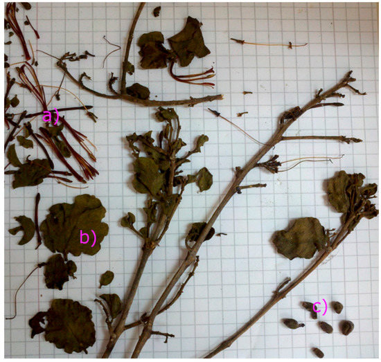
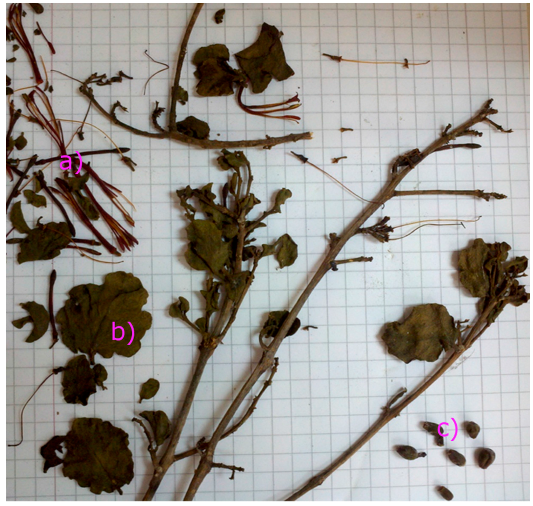
Figure 1.
Pictures of an herborized sample of T. tetrandus collected in the VIII region of Chile in 2012. (a) Flowers; (b) leaves; (c) fruits.
2. Results and Discussion
2.1. Antioxidant Capacity and Total Phenolics, Anthocyanin and Flavonoids Contents
Three antioxidant assays were employed for this study: the DPPH antiradical activity assay, the superoxide anion inhibition assay, and the ferric reducing activity measured as micromoles of the standard Trolox (Table 1). The antioxidant capacities were supported by the measurement of total anthocyanins in the flowers (TAC), as well as the phenolic (TPC) and flavonoid (TFC) contents in flowers and leaves. For the TPC assay, it has to be stressed that the Folin-Ciocalteu reagent employed reacts with all the oxidants present in the plant extract. Consequently, depending on the extract, this method could overestimate the real phenolic level in the extract. Therefore, data for TPC obtained with the Folin-Ciocalteu's method could be artefactual and the interpretation of the results erroneous [29], for this reason we have supported the results of this assay with the other complementary ones. The leaves showed more antiradical DPPH quenching activity than the flowers, possibly due to the quantity of phenolics (Table 1) found in the leaves. The DPPH value of T. tetrandus leaves was close to that of standard cyanidin-3-glucoside (Table 1) and the synthetic antioxidant butylated hydroxytoluene (BHT, 61.13 µM) [30]. The European mistletoe Viscum album has been extensively studied and the antioxidant activity already reported [31,32,33,34], indeed, several studies showed that V. album possess remarkable cholinesterase and tyrosinase inhibitory and antioxidant properties [32]. The TPCs of T. tetrandus leaves were close to that exhibited by V. album L. ssp. album hosting Cerasus vulgaris Miller (Sourcherry), Pinus nigra Arn., (Pine) and Crataegus sp. (around 31, 33 and 37 mg GAE per g extract respectively) [32]. It was also close to that reported from Nolana aplocarioides from Northern Chile (around 30 mg per g) [35]. The TPC of the leaves was also close to that reported for blackberry (Rubus ulmifolium) bud preparations (350 ± 8 mg/100 g fresh weight, considering 90 percent of water loss) [36] and was higher to that reported for the superfruit goji (281.91 mg/100 g fresh weight) [37]. The TFC was also close to the value reported for Nolana aplocarioides (around 22 mg quercetin per g dry weight) [35]. The TPC and FRAP activity of the flowers was similar to that reported for flowers of Helianthus annus [38]. The TAC of the flowers was close to that of standard cyanidin-3-glucoside (Table 1), and was also close to that reported from the Chilean berries Luma apiculata (15.24 ± 1.29 mg cyanidin 3-O-glucoside/g dry weight) [39]. The TAC of the flowers was also double to that reported for the Black Diamond blackberry (Rubus fruticosus) variety (119.3 ± 1.2 mg/100 g fresh weight, considering 90 percent of water loss) [40] and similar to black currant (Ribes nigrus) var. Black Down (170.0 ± 1.7 mg/100 g fresh weight) [41]. The SAA scavenging of the leaves was somehow lower to that reported for leaves of strawberries (67.60% ± 1.01% inhibition) [42].

Table 1.
Scavenging of the 1,1-diphenyl-2-picrylhydrazyl Radical (DPPH), Ferric Reducing Antioxidant Power (FRAP), Superoxide Anion scavenging activity (SAA), Total Phenolic Content (TPC), Total Flavonoid Content (TFC), Total Anthocyanin Content (TAC), and Extraction Yields of a mistletoe from the VIII Region of Chile.
2.2. MS-PDA Identification of Phenolic Acids in Chilean Mistletoe (Lorantaceae) From Southern Chile
The hybrid machine used in this study combines the rapid separation of the ultra-HPLC technique with photodiode (PDA) detection with flow rates up to 2 mL per minute, zero dead volume, the effective ionization of the heated electrospray probe (HESI II), the high resolving power performance of the orbital trap (Orbitrap, OT), and selectivity of a quadrupole, (reaching resolutions of up to 70,000 FWHM at m/z 200), and the outstanding diagnostic power of a high resolution collision (HCD) cell. Qualitative data regarding the phenolic compounds of mistletoe extracts are shown in Table 2. We have identified 28 compounds in the leaves and six in the flowers.The compounds in the flowers and leaves were detected and identified using UHPLC with total ion current (TIC) in positive mode for anthocyanins and negative mode for the other phenolic compounds using OT-HESI-MS (Table 2) and UV-visible data (PDA, Figure 2, Table 2).
Table 2.
Identification of Phenolic Compounds in Tristerix tetrandus leaves and flowers by LC-PDA-HR-OT-ESI-MS Data.
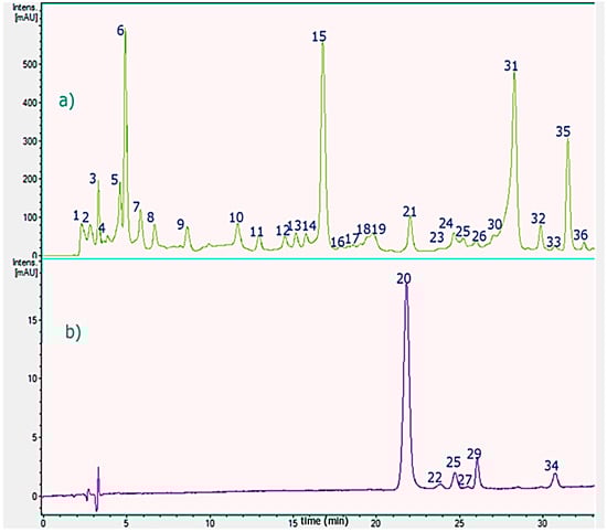
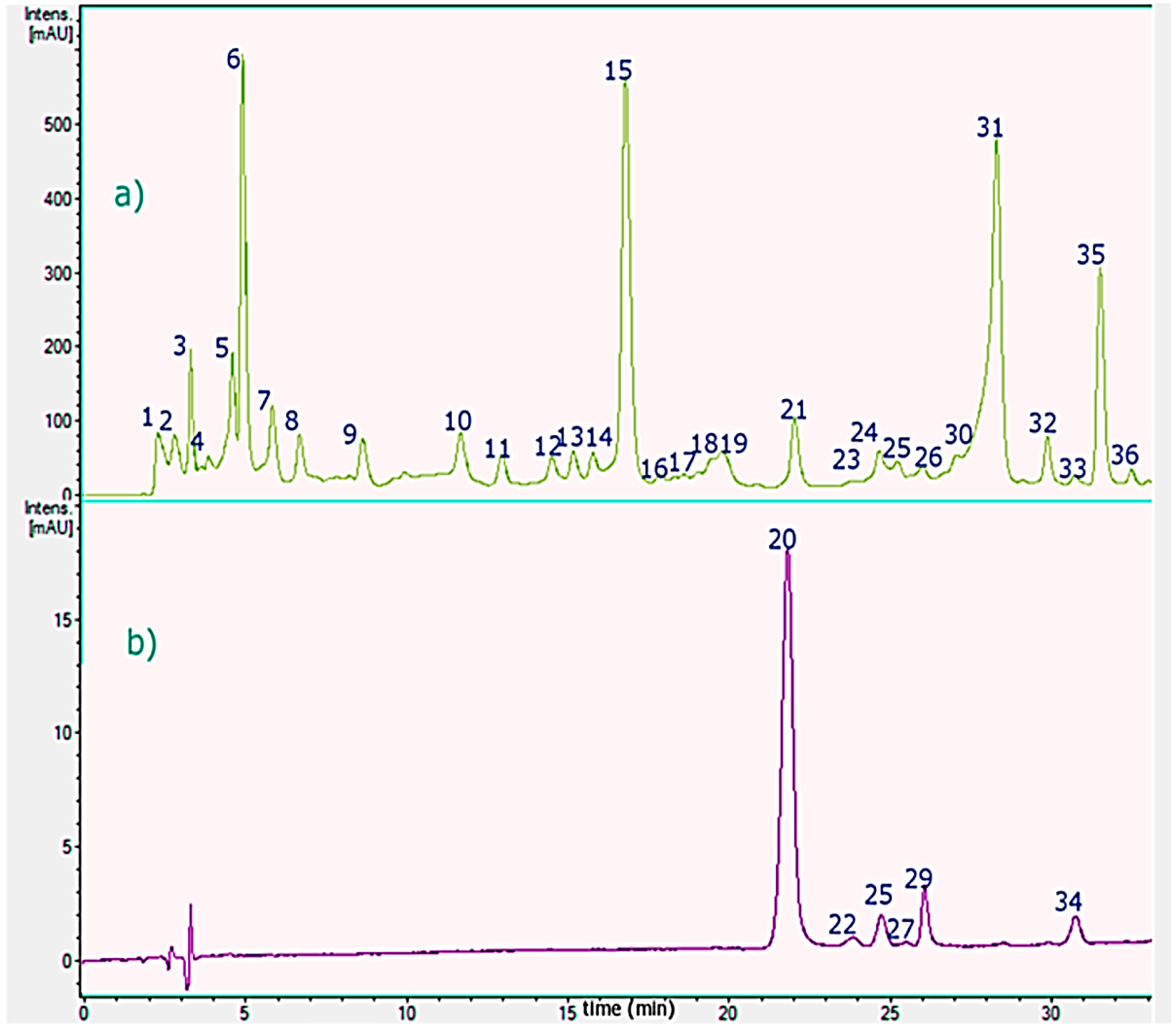
Figure 2.
HPLC-PDA chromatograms of T. tetrandus from the VIII region of Chile. (a) leaves, monitored at 280; and (b) flowers, monitored at 520 nm. Peaks numbers refer to those indicated in Table 2.
The optimal conditions for the separation of the phenolics were obtained with a fast linear gradient solvent system of 0.1% aqueous formic acid (solvent A) and acetonitrile 0.1% formic acid (solvent B) with a flow rate of 1.0 mL/min−1 using an UHPLC C18 column as a stationary phase. Several common compounds were in the present study identified accurately using the HCD cell including proanthocyanidins, phenolic acids and flavonoids. Peaks 20, 22, 25, 27, 29 and 34 were detected in the flowers and the other peaks in the leaves. Below is the detailed explanation of the characterization. Figure 3 shows full MS spectra and structures of several compounds detected.
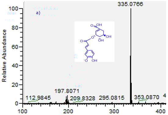
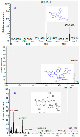
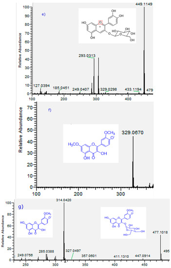
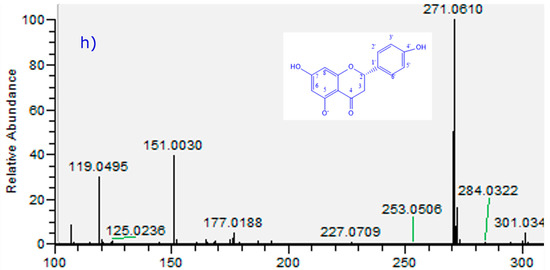
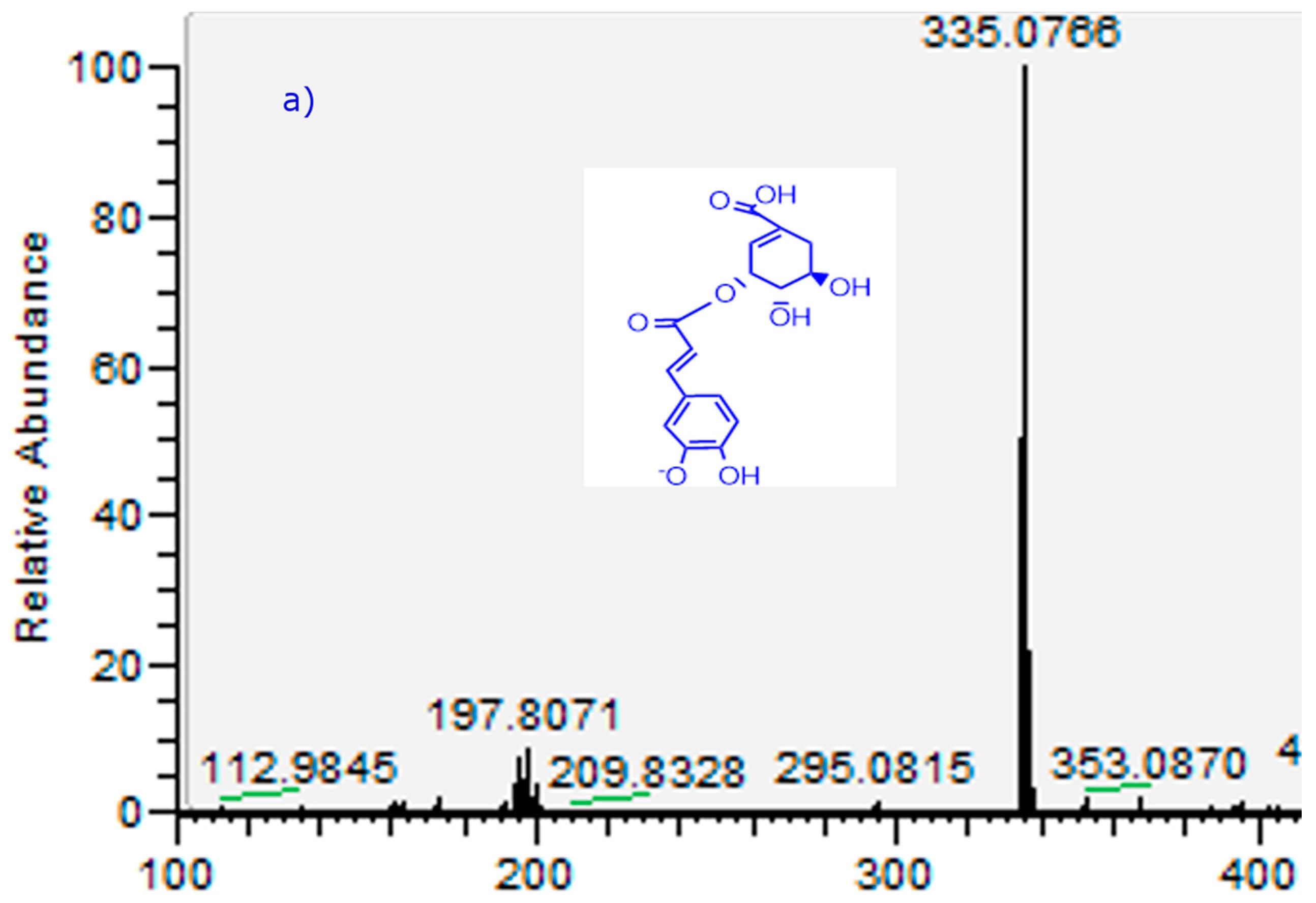
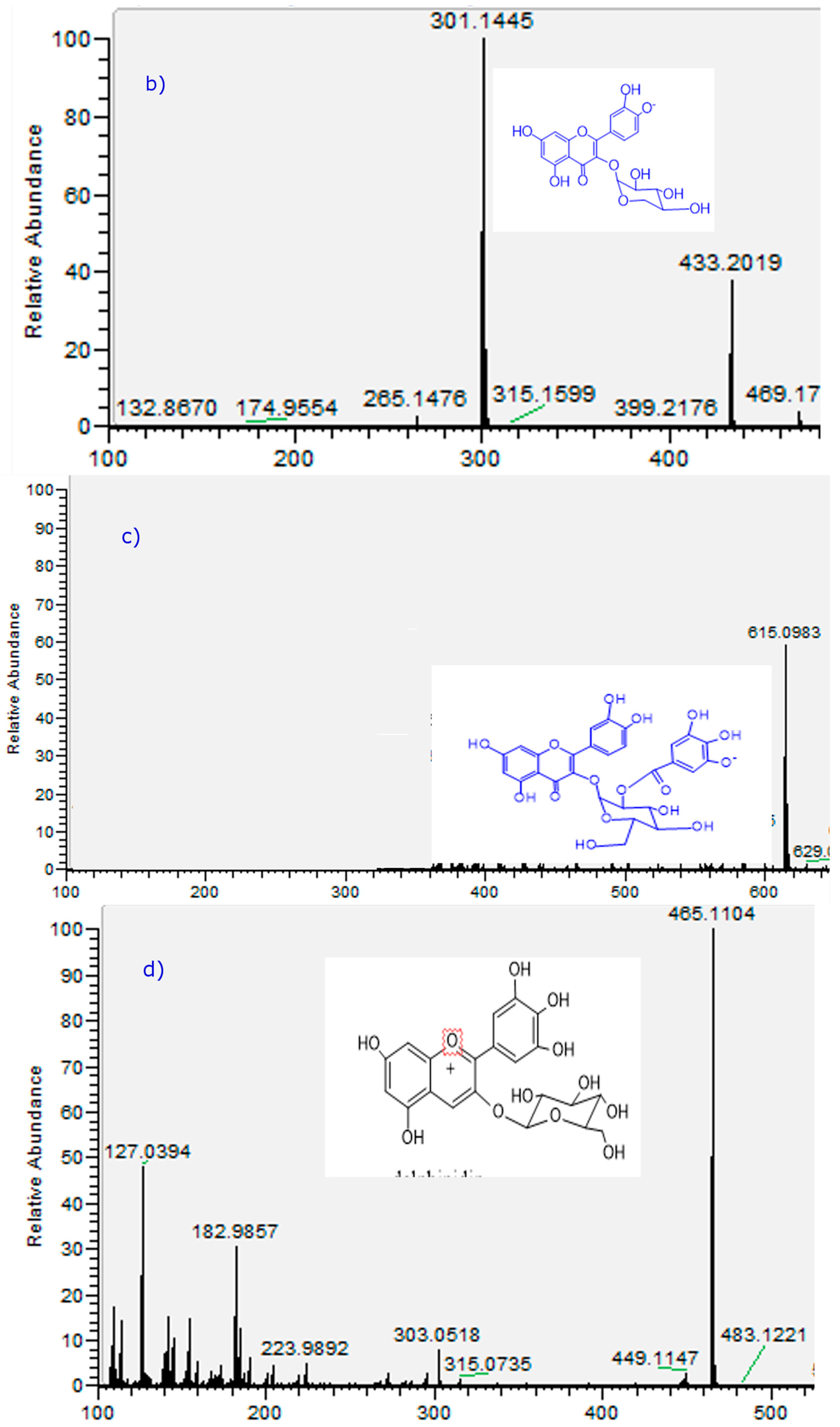
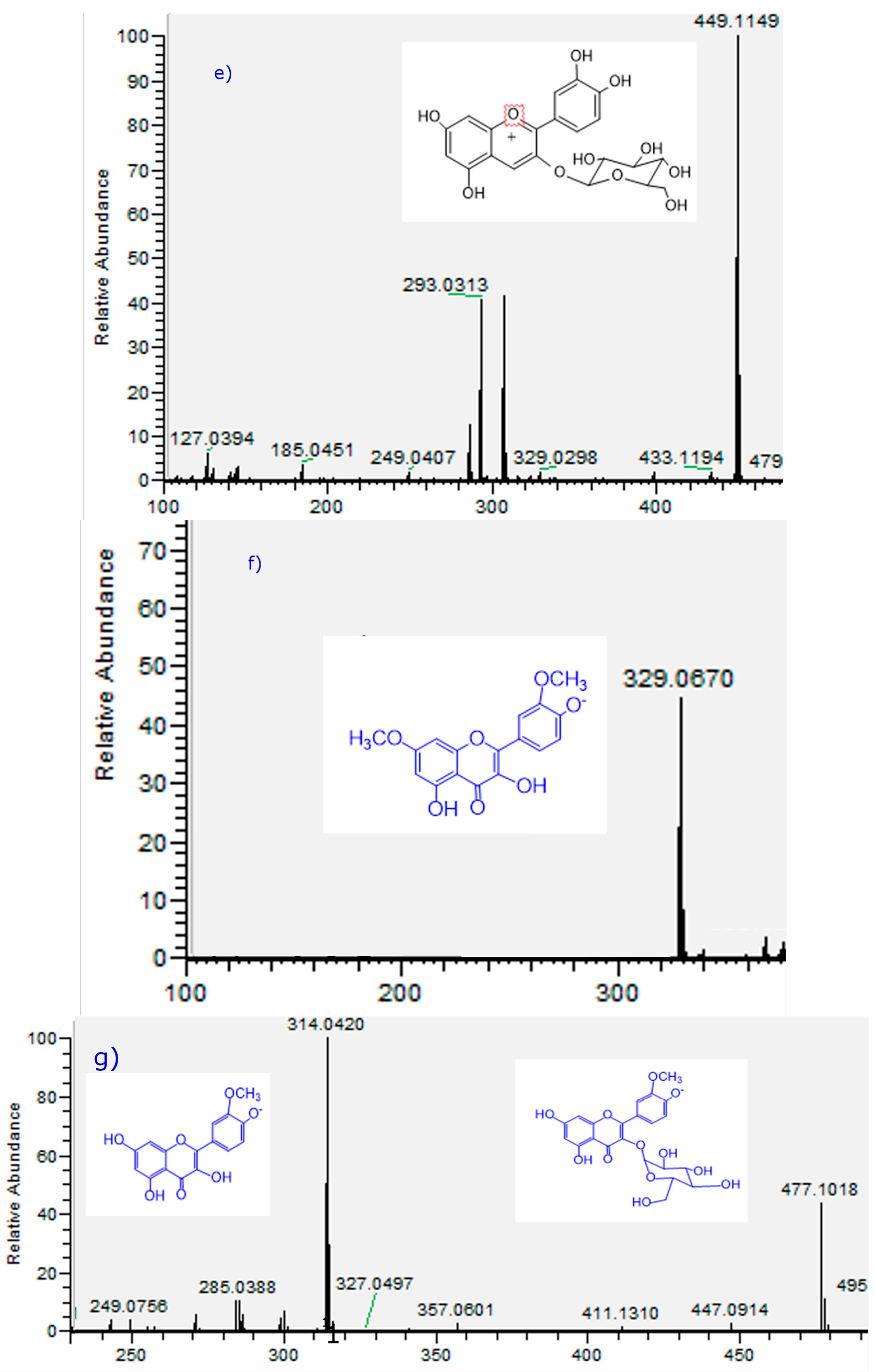
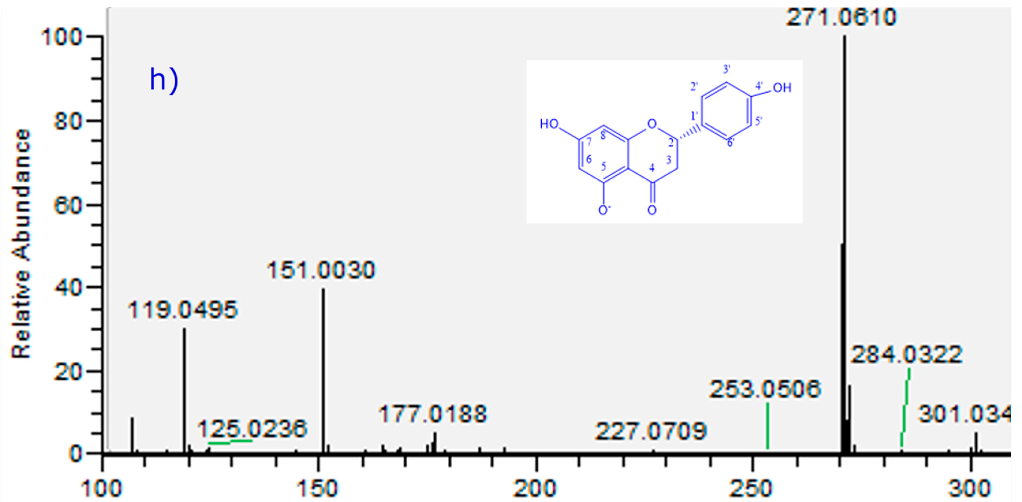
Figure 3.
Full scan OT-MSn spectra of of some representative compounds identified in T. tetrandus from Chile. (a) Peak 4; (b) peak 12; (c) Peak 15; (d) peak 20; (e) peak 22; (f) peak 26; (g) peak 28; and (h) peak 35. Peaks numbers refer to those indicated in Table 2.
2.2.1. Flavonoids
Several flavonols detected were simply aglycones and some were glycosylated. Peak 32 was identified as quercetin, while peaks 33 and 36 (ions at m/z: 285.0392 and 269.0443) were identified as the flavonoids luteolin and apigenin [43]. Peak 31 was identified as isorhamnetin (C16H11O7−, λmax 254–354 nm) [44]. Peaks 26 with a [M − H]− ion at m/z: 329.0670 was identified as 7-O-methyl isorhamnetin, and peak 28 as the glycosylated isorhamnetin-3-O-glucoside [44]. Peak 35 with a [M − H]− ion at m/z: 271.0610 was identified as the flavanone naringenin [35,43,45]. Peak 11 with a [M − H]− ion at m/z: 433.2019 was identified as quercetin 3-O-pentoside. In the same manner peak 12 with a [M − H]− ion at m/z: 463.0876 was identified as quercetin-3-O-glucoside (C21H19O12−) and peak 7 as rutin [44,46] and peak 13 was identified as apiin (apigenin-apioglucoside) [47].
2.2.2. Phenolic Acids
Peak 1 was identified as quinic acid and peak 2 as caffeoyl-glucoside (341.0870, C15H17O9−), while peak 14 as p-coumaroyl malate (ion at m/z: 279.0507, C13H11O7−). Two chlorogenic acids (C16H18O9) were identified. The isomers detected include 3-O-caffeoylquinic acid (peak 3) and 5-O-caffeoylquinic acid (neochlorogenic acid or 5-CQA, peak 17). Another peak could be tentatively identified as 3-O-feruloylquinic acid (or 3-FQA, peak 9), all of them producing a quinic acid MS2 ion at around m/z: 191.0556 (quinic acid C7H11O6−) and the CGA all produced also a 2 M − H adduct ion at around m/z: 707 [9]. They were also identified according to their UV spectra (λmax at 314–330 nm). Peaks 16 and 24 with [M − H]− ions at m/z: 515.1189 and 515.1192 were identified as di-CQA isomers according to the formula C25H23O12− [40]. Peak 24 with pseudomolecular ion at m/z: 337.0356 was identified as 3-p-Coumaroylquinic acid (C18H9O7−) [48]. Peak 30 was identified as phenyl lactic acid hexoside [45].
2.2.3. Oxylipins
Peaks 18 and 19 were tentatively identified as the antioxidant fatty acids known as oxylipins (trihydroxyoctadecadienoic acid and (trihydroxyoctadecaenoic acid, respectively) [24].
2.2.4. Procyanidins
Peak 6, 8 and 10 were identified as catechin, gallocatechin and epigallocatechin respectively (ions at m/z: 289.0715, 305.0304 and 305.0301, respectively) [39,42,44].
2.2.5. Anthocyanins
Six main known anthocyanins were identified in the flowers, peaks 20, 22, 25, 27, 29 and 34, Figure 3) with molecular ions in positive mode at m/z: 465.1033 (delphinidin-3-O-glucoside), 449.1149 (cyanidin-3-O-glucoside), 271.0620 (pelargonidin), 303.0161 (delphinidin), 331.0117 (malvidin), and 287.0571 (cyanidin) respectively. The identity was corroborated by co-elution with standard anthocyanins and literature data.
3. Materials and Methods
3.1. Chemicals and Plant Material
Folin-Ciocalteu phenol reagent (2 N), reagent grade Na2CO3, AlCl3,HCl, FeCl3, NaNO2, NaOH, quercetin, trichloroacetic acid, sodium acetate, HPLC-grade water, HPLC-grade acetonitrile, reagent grade MeOH and formic acid were obtained from Merck (Darmstadt, Germany). Malvidin, delphinidin, pelargonidin, quercetin, luteolin, apigenin, isorhamnetin, naringenin, cyanidin, delphinidin 3-O-galactoside, cyanidin-3-O-galactoside, cyanidin-3-O-glucoside, chlorogenic acid, ferulic and caffeic acids (all standards with purity higher than 95% by HPLC) were purchased either from ChromaDex (Santa Ana, CA, USA), Extrasynthèse (Genay, France) or Wuxi Apptec Co. Ltd. (Shanghai, China). Gallic acid, TPTZ (2, 4, 6-tri(2-pyridyl)-s-triazine), Trolox, tert-butylhydroperoxide, nitroblue tetrazolium, xanthine oxidase and DPPH (1,1-diphenyl-2-picrylhydrazyl radical) were purchased from Sigma-Aldrich Chemical Co. (St. Louis, MO, USA). The plant and flowers (approx. 500 g each, three individuals, plants hosted on a group of aspen species) were collected at Los Ángeles, Región del Bio-Bio, Chile in April 2012. Sampling was performed using sterile disposable gloves and rigid plastic sample containers and the samples (three individuals) were submitted individually by overnight courier to our laboratory in Antofagasta to prevent deterioration. A voucher herbarium specimen was deposited at the Laboratorio de Productos Naturales, Universidad de Antofagasta, Antofagasta, Chile, with the number Tt-121505.
3.2. Sample Preparation
Flowers and leaves (three individuals of each) were separately collected and extracted with acidified methanol and the resulting extracts were processed by solid phase extraction. Fresh flowers were carefully washed, separately homogenized in a blender and freeze-dried (Freezone Freeze dry system plus 2.5 L, Labconco Corporation, Kansas City, MO, USA). Ten grams of the lyophilized flowers and leaves were finally pulverized in a mortar, (separately) defatted thrice with 100 mL of n-hexane and then extracted with 100 mL of 0.1% HCl in MeOH in the dark in an ultrasonic bath for one hour each time. The extracts from each sample were combined, filtered and evaporated in vacuo in the dark (40 °C). The extracts were suspended in 20 mL ultrapure water and loaded onto an XAD-7 (100 g) column. The column was rinsed with water (100 mL) and phenolic compounds were eluted with 100 mL of MeOH acidified with 0.1% HCl. The solutions were combined and evaporated to dryness under reduced pressure (40 °C) to give 567.23 mg and 895.3 mg of extract from T. tetrandus leaves and flowers, respectively.
3.3. Instrumentation
A Thermo Scientific Dionex Ultimate 3000 UHPLC system (Thermo Fisher Scientific, Bremen, Germany) equipped with a quaternary Series RS pump and a Thermo Scientific Dionex Ultimate 3000 Series TCC-3000RS column compartments with a Thermo Fisher Scientific Ultimate 3000 Series WPS-3000RS autosampler (Thermo Fisher Scientific) and a rapid separations PDA detector controlled by Chromeleon 7.2 Software (Thermo Fisher Scientific, Waltham, MA, USA and Dionex Softron GmbH division of Thermo Fisher Scientific) hyphenated with a Thermo high resolution Q Exactive focus mass spectrometer (Thermo Fisher Scientific) were used for analysis. The chromatographic system was coupled to the MS with a Heated Electrospray Ionization Source II (HESI II). Nitrogen (purity > 99.999%) obtained from a Genius NM32LA nitrogen generator (Peak Scientific, Billerica, MA, USA) was employed as both the collision and damping gas. Mass calibration for the Orbitrap was performed once a week, in both negative and positive modes, to ensure a working mass accuracy lowers than or equal to 5 ppm. Caffeine, N-butylamine (Sigma-Aldrich) were the calibration standards for positive ions and buspirone hydrochloride, sodium dodecyl sulfate, and taurocholic acid sodium salt (Sigma-Aldrich) were used to calibrate the mass spectrometer. These compounds were dissolved in a mixture of acetic acid, acetonitrile, water and methanol (Merck) and were infused using a Chemyx Fusion 100 syringe pump (Thermo Fisher Scientific). Q Exactive 2.0 SP 2, XCalibur 2.3 and Trace Finder 3.2 softwares (Thermo Fisher Scientific and Dionex Softron GmbH Part of Thermo Fisher Scientific) were used for UHPLC-mass spectrometer control and data processing, respectively.
3.4. LC Parameters
A portion of each extract (5 mg) obtained as explained above was dissolved in 5 mL 1% formic acid in MeOH, filtered through a 0.45 µm micropore membrane (PTFE, Waters Milford, MA, USA) before use and was injected into the UHPLC-PDA and ESI-orbitrap-MS equipment. Liquid chromatography was performed using an UHPLC C18 column (Acclaim, 150 mm × 4.6 mm ID, 5 µm, Restek Corporation, Bellefonte, PA, USA) operated at 25 °C. The detection wavelengths were 254, 280, 320 and 440 nm, and PDA was recorded from 200 to 800 nm for peak characterization. Mobile phases were 1% formic aqueous solution (A) and acetonitrile (B). The gradient program (time (min), % B) was: (0.00, 5); (5.00, 5); (10.00, 30); (15.00, 30); (20.00, 70); (25.00, 70); (35.00, 5) and 12 min for column equilibration before each injection. The flow rate was 1.00 mL/min−1, and the injection volume was 10 µL. Standards and extracts dissolved in methanol were kept at 10 °C during storage in the autosampler.
3.5. MS Parameters
The HESI parameters were optimized as follows: sheath gas flow rate 75 units; aux. gas unit flow rate 20; capillary temperature 400 °C; aux gas heater temperature 500 °C; spray voltage 2500 V (for ESI−); and S lens RF level 30. Full scan data in both positive and negative was acquired at a resolving power of 70,000 full width half maximum (FWHM) at m/z 200. For the compounds of interest, a scan range of m/z 100–1000 was chosen; the automatic gain control (AGC) was set at 3 × 10⁶ and the injection time was set to 200 ms. Scan-rate was set at 2 scans·s−1. External calibration was performed using a calibration solution in positive and negative modes before each sample series. In addition to the full scan acquisition method, for confirmations purposes, a targeted MS/MS analysis was performed using the mass inclusion list and expected retention times of the target analytes, with a 30 s time window, with the Orbitrap spectrometer operating both in positive and negative mode at 17,500 FWHM (m/z 200). The AGC target was set to 2 × 10⁵, with the maximum injection time of 20 ms. The precursor ions are filtered by the quadrupole which operates at an isolation window of m/z 2. The fore vacuum, high vacuum and ultrahigh vacuum were maintained at approximately 2 mbar, from 105 to below 1010 mbar, respectively. Collision energy (HCD cell) was operated at 30 kv. Detection was based on calculated exact mass and on retention time of target compounds, presented in Table 2. The mass tolerance window was set to 5 ppm for the two analysis modes.
3.6. Antioxidant Assays
3.6.1. Ferric Reducing Antioxidant Power
The determination of ferric reducing antioxidant power or ferric reducing ability (FRAP assay) of the extracts was performed as described by [49] with some modifications. The stock solutions prepared were 300 mM acetate buffer pH 3.6, 10 mM TPTZ (2,4,6-tri (2-pyridyl)-s-triazine) solution in 40 mM HCl, and 20 mM FeCl3·6H2O solution. Plant extracts or standard methanolic Trolox solutions (150 µL) were incubated at 37 °C with 2 mL of the FRAP solution (prepared by mixing 25 mL acetate buffer, 5 mL TPTZ solution, and 10 mL FeCl3.6H2O solution) for 30 min in the dark. Absorbance of the blue ferrous tripyridyltriazine complex formed was then read at 593 nm. Quantification was performed using a standard calibration curve of antioxidant Trolox (from 0.2 to 2.5 µmol/mL, R2: 0.995). Samples were analyzed in triplicate and results are expressed in µmol TE/100 grams fresh mass.
3.6.2. Superoxide Anion Scavenging Activity
The enzyme xanthine oxidase is able to generate superoxide anion radical (O2−) “in vivo” by oxidation of reduced products from intracellular ATP metabolism. The superoxide anion generated in this reaction sequence reduces the nitro blue tetrazolium dye (NBT), leading to a chromophore with a maximum of absorption at 560 nm. Superoxide anion scavengers reduce the speed of generation of the chromophore. The superoxide anion scavenging activities of isolated compounds and fractions were measured spectrophotometrically in a microplate reader as reported previously [24]. All extracts were evaluated at 100 µg/mL. Values are presented as mean ± standard deviation of three determinations.
3.7. Polyphenol and Flavonoids Contents
The total polyphenolic contents (TPC) of mistletoe were determined by the Folin-Ciocalteau method [25,26,50] with some modifications. An aliquot of each processed SPE extract (200 µL, approx. 2 mg/mL) was added to the Folin–Ciocalteau reagent (2 mL, 1:10 v/v in purified water) and after 5 min of reaction at room temperature (25 °C), 2 mL of a 100 g/L solution of Na2CO3 was added. Sixty minutes later the absorbance was measured at 710 nm. The calibration curve was performed with gallic acid (concentrations ranging from 16 to 500 µg/mL, R2= 0.999) and the results were expressed as mg gallic acid equivalents/100 g fresh mass. Determination of total flavonoid content (TFC) of the methanolic extracts was performed as reported previously [51] using the AlCl3 colorimetric method. Quantification was expressed by reporting the absorbance in the calibration graph of quercetin, which was used as a standard (from 0.1 to 65.0 µg/mL, R2 = 0.994). Results are expressed as mg quercetin equivalents/g fresh weight. All spectrometric measurements were performed using a Unico 2800 UV-vis spectrophotometer (Unico Instruments, Co, Ltd., Shanghai, China).
3.8. Statistical Analysis
The statistical analysis was carried out using the originPro 9.0 software packages (Originlab Corporation, Northampton, MA, USA). The determination was repeated at least three times for each sample solution. Analysis of variance was performed using ANOVA. Significant differences between means were determined by Tukey comparison test (p values < 0.05 were regarded as significant).
4. Conclusions
Thirty six compounds including several caffeoyl acids (peaks 2, 3, 4, 9, 16, 17, and 24) three procyanidins (peaks 6, 8 and 10), several flavonols (peaks 11, 12, 15, 21, 23, 26, 28, 31–36) two oxylipins (peaks 18 and 19) were detected in the leaves and six anthocyanins (peaks 22, 25, 26, 27, 29 and 34) were detected in the flowers of a native mistletoe from the VIII region of Chile using PDA and Orbitrap-ESI-MS for the first time. However, significant differences in the total phenolic content and antioxidant activity were found between these two plant parts, probably due to the quantity of phenolic compounds detected. The mistletoe leaves showed the highest antioxidant activity measured as the bleaching of the DPPH radical, the ferric reducing antioxidant power and superoxide anion scavenging activity (13.38 ± 0.47 µg/mL, 125.32 ± 5.96 µmol TE/g DW and 84.06 ± 4.59 at 100 µg/mL, respectively). The mistletoe is thus a good candidate for industrial crop production and has also the potential to produce nutraceuticals.
Acknowledgments
This work was financially supported by FONDECYT (grants No. 1140178 and 1150745) and Universidad Arturo Prat (Iquique, Chile; VRIIP0113-15). We also acknowledge Fondequip (grant EQM140002) for the funding to purchase the UHPLC Orbitrap MS equipment. We also thank to the SAG (Servicio Agrícola y Ganadero) and Corporación Nacional Forestal of Chile (CONAF) for allowing us to collect plants and fruits in the national protected areas of Chile belonging to SNASPE (National System of Protected Areas of Chile).
Author Contributions
Important contributions to the design of the manuscript: B.S. and C.Q. Sample and analysis experiments: B.S., M.S. and C.Q. Analysis of the experimental data: C.A. Critically revising the manuscript for important intellectual content: M.S. and C.A. All authors helped with the preparation of the manuscript and approved the final version.
Conflicts of Interest
The authors declare no conflict of interest.
References
- Burgos, A.N.; Morales, M.A. Qualitative study of use medicinal plants in a complementary or alternative way with the use of among of rural population of the Bulnes City, Bío-Bío Region, Chile. BLCPMA 2010, 9, 377–387. [Google Scholar]
- De Mösbach, E.W. Botánica Indígena de Chile. In Museo Chileno de Arte Precolombino, Fundación Andes y Editorial Andrés Bello; Aldunate, C., Villagrán Santiago, C., Eds.; Andres Bello: Santiago, Chile, 1991; pp. 95–96. [Google Scholar]
- Hoffmann, A.; Arroyo, M.T.K.; Liberona, F.; Muñoz, M.; Watson, J.M. Plantas Altoandinas en la Flora Silvestre de Chile; ediciones Fundación Claudio A. Gay: Santiago de Chile, Chile, 2000. [Google Scholar]
- De Pascual-Teresa, S.; Sanchez-Ballesta, M.T. Anthocyanins: From plant to health. Phytochem. Rev. 2008, 7, 281–299. [Google Scholar] [CrossRef]
- Nile, S.H.; Park, S.W. Edible berries: Bioactive components and their effect on human health. Nutrition 2014, 30, 134–144. [Google Scholar] [CrossRef] [PubMed]
- Woodward, G.; Kroon, P.; Cassidy, A.; Kay, C. Anthocyanin stability and recovery: Implications for the analysis of clinical and experimental samples. J. Sci. Food Agric. 2009, 57, 5271–5278. [Google Scholar] [CrossRef] [PubMed]
- Hou, D.X. Potential mechanisms of cancer chemoprevention by anthocyanins. Curr. Mol. Med. 2003, 3, 149–159. [Google Scholar] [CrossRef] [PubMed]
- Matsui, T.; Ebuchi, S.; Kobayashi, M.; Fukui, K.; Sugita, K.; Terahara, N.; Matsumoto, K. Anti-hyperglycemic effect of diacylated anthocyanin derived from Ipomoea batatas cultivar Ayamurasaki can be achieved through the α-glucosidase inhibitory action. J. Agric. Food Chem. 2002, 50, 7244–7248. [Google Scholar] [CrossRef] [PubMed]
- Simirgiotis, M.J.; Silva, M.; Becerra, J.; Schmeda-Hirschmann, G. Direct characterisation of phenolic antioxidants in infusions from four Mapuche medicinal plants by liquid chromatography with diode array detection (HPLC-DAD) and electrospray ionisation tandem mass spectrometry (HPLC-ESI-MS). Food Chem. 2012, 131, 318–327. [Google Scholar] [CrossRef]
- Brito, A.; Ramirez, J.E.; Areche, C.; Sepúlveda, B.; Simirgiotis, M.J. HPLC-UV-MS Profiles of Phenolic Compounds and Antioxidant Activity of Fruits from Three Citrus Species Consumed in Northern Chile. Molecules 2014, 19, 17400–17421. [Google Scholar] [CrossRef] [PubMed]
- Lea, M.A. Flavonol Regulation in Tumor Cells. J. Cell. Biochem. 2015, 116, 1190–1194. [Google Scholar] [CrossRef] [PubMed]
- Wang, Y.; Han, A.; Chen, E.; Singh, R.K.; Chichester, C.O.; Moore, R.G.; Singh, A.P.; Vorsa, N. The cranberry flavonoids PAC DP-9 and quercetin aglycone induce cytotoxicity and cell cycle arrest and increase cisplatin sensitivity in ovarian cancer cells. Int. J. Oncol. 2015, 46, 1924–1934. [Google Scholar] [PubMed]
- Steinmann, D.; Ganzera, M. Recent advances on HPLC/MS in medicinal plant analysis. J. Pharm. Biomed. Anal. 2011, 55, 744–757. [Google Scholar] [CrossRef] [PubMed]
- Wright, P. Metabolite identification by mass spectrometry: Forty years of evolution. Xenobiotica 2011, 41, 670–686. [Google Scholar] [CrossRef] [PubMed]
- Mattoli, L.; Cangi, F.; Ghiara, C.; Burico, M.; Maidecchi, A.; Bianchi, E.; Ragazzi, E.; Bellotto, L.; Seraglia, R.; Traldi, P. A metabolite fingerprinting for the characterization of commercial botanical dietary supplements. Metabolomics 2011, 7, 437–445. [Google Scholar] [CrossRef]
- Aliferis, K.A.; Chrysayi-Tokousbalides, M. Metabolomics in pesticide research and development: Review and future perspectives. Metabolomics 2011, 7, 35–53. [Google Scholar] [CrossRef]
- Kang, H.J.; Yang, H.J.; Kim, M.J.; Han, E.S.; Kim, H.J.; Kwon, D.Y. Metabolomic analysis of meju during fermentation by ultra performance liquid chromatography-quadrupole-time of flight mass spectrometry (UPLC-Q-TOF MS). Food Chem. 2011, 127, 1056–1064. [Google Scholar] [CrossRef] [PubMed]
- Martínez-Domínguez, G.; Romero-González, R.; Garrido Frenich, A. Multi-class methodology to determine pesticides and mycotoxins in green tea and royal jelly supplements by liquid chromatography coupled to Orbitrap high resolution mass spectrometry. Food Chem. 2016, 197, 907–915. [Google Scholar] [CrossRef] [PubMed]
- Hao, C.; Zhao, X.; Yang, P. GC-MS and HPLC-MS analysis of bioactive pharmaceuticals and personal-care products in environmental matrices. TrAC 2007, 26, 569–580. [Google Scholar] [CrossRef]
- Maoka, T. Recent progress in structural studies of carotenoids in animals and plants. Arch. Biochem. Biophys. 2009, 483, 191–195. [Google Scholar] [CrossRef] [PubMed]
- Barnes, J.S.; Nguyen, H.P.; Shen, S.; Schug, K.A. General method for extraction of blueberry anthocyanins and identification using high performance liquid chromatography-electrospray ionization-ion trap-time of flight-mass spectrometry. J. Chromatogr. A 2009, 1216, 4728–4735. [Google Scholar] [CrossRef] [PubMed]
- Fischer, U.A.; Carle, R.; Kammerer, D.R. Identification and quantification of phenolic compounds from pomegranate (Punica granatum L.) peel, mesocarp, aril and differently produced juices by HPLC-DAD-ESI/MS(n). Food Chem. 2011, 127, 807–821. [Google Scholar] [CrossRef] [PubMed]
- He, D.X.; Shan, Y.; Wu, Y.H.; Liu, G.Z.; Chen, B.; Yao, S.Z. Simultaneous determination of flavanones, hydroxycinnamic acids and alkaloids in citrus fruits by HPLC-DAD-ESI/MS. Food Chem. 2011, 127, 880–885. [Google Scholar] [CrossRef] [PubMed]
- Simirgiotis, M.J.; Ramirez, J.E.; Schmeda Hirschmann, G.; Kennelly, E.J. Bioactive coumarins and HPLC-PDA-ESI-ToF-MS metabolic profiling of edible queule fruits (Gomortega keule), an endangered endemic Chilean species. Food Res. Int. 2013, 54, 532–543. [Google Scholar] [CrossRef]
- Simirgiotis, M.J.; Borquez, J.; Schmeda-Hirschmann, G. Antioxidant capacity, polyphenolic content and tandem HPLC-DAD-ESI/MS profiling of phenolic compounds from the South American berries Luma apiculata and L. chequen. Food Chem. 2013, 139, 289–299. [Google Scholar] [CrossRef] [PubMed]
- Simirgiotis, M.J. Antioxidant Capacity and HPLC-DAD-MS Profiling of Chilean Peumo (Cryptocarya alba) Fruits and Comparison with German Peumo (Crataegus monogyna) from Southern Chile. Molecules 2013, 18, 2061–2080. [Google Scholar] [CrossRef] [PubMed]
- Bórquez, J.; Kennelly, E.J.; Simirgiotis, M.J. Activity guided isolation of isoflavones and hyphenated HPLC-PDA-ESI-ToF-MS metabolome profiling of Azorella madreporica Clos. from northern Chile. Food Res. Int. 2013, 52, 288–297. [Google Scholar] [CrossRef]
- Simirgiotis, M.J.; Vallejos, J.; Areche, C.; Sepúlveda, B. Concise and Straightforward Asymmetric Synthesis of a Cyclic Natural Hydroxy-Amino Acid. Molecules 2014, 19, 19516–19531. [Google Scholar] [CrossRef] [PubMed]
- Plazas, M.; López-Gresa, M.P.; Vilanova, S.; Torres, C.; Hurtado, M.; Gramazio, P.; Andújar, I.; Herráiz, F.J.; Bellés, J.M.; Prohens, J. Diversity and Relationships in Key Traits for Functional and Apparent Quality in a Collection of Eggplant: Fruit Phenolics Content, Antioxidant Activity, Polyphenol Oxidase Activity, and Browning. J. Agric. Food Chem. 2013, 61, 8871–8879. [Google Scholar] [CrossRef] [PubMed]
- López-Gresa, M.P.; Torres, C.; Campos, L.; Lisón, P.; Rodrigo, I.; Bellés, J.M.; Conejero, V. Identification of defence metabolites in tomato plants infected by the bacterial pathogen Pseudomonas syringae. Environ. Exp. Bot. 2011, 74, 216–228. [Google Scholar] [CrossRef]
- Hong, S.M.; Choi, J.-H.; Jo, S.-J.; Song, S.K.; Lee, J.M.; Kusakabe, T. Expression of Recombinant Viscum Album Coloratum lectin B-chain in the Silkworm Expression System and Evaluation of Antioxidant Activity. Biotech. Bioproc. Eng. 2015, 20, 515–522. [Google Scholar] [CrossRef]
- Orhan, D.D.; Senol, F.S.; Hosbas, S.; Orhan, I.E. Assessment of cholinesterase and tyrosinase inhibitory and antioxidant properties of Viscum album L. samples collected from different host plants and its two principal substances. Ind.Crops Prod. 2014, 62, 341–349. [Google Scholar] [CrossRef]
- Pietrzak, W.; Nowak, R.; Olech, M. Effect of extraction method on phenolic content and antioxidant activity of mistletoe extracts from Viscum album subsp abietis. Chem. Pap. 2014, 68, 976–982. [Google Scholar] [CrossRef]
- Raieviawati, S.I.; Ishmaru, K.; Hou, D.-X.; Hayashi, N. Antioxidant Activity and Phenolic Content of Mistletoe Extracts Following High-Temperature Batch Extraction. Food Sci. Technol. Res. 2014, 20, 201–206. [Google Scholar]
- Simirgiotis, M.J.; Benites, J.; Areche, A.; Sepulveda, B. Antioxidant capacities and analisis of phenolic compounds in three endemic Nolana species by HPLC-PDA-ESI-MS. Molecules 2015, 20, 11490–11507. [Google Scholar] [CrossRef] [PubMed]
- Donno, D.; Beccaro, G.L.; Mellano, M.G.; Cerutti, A.K.; Bounous, G. Medicinal plants, chemical composition and quality: May blackcurrant buds and blackberry sprouts be a new polyphenol source for herbal preparations? J. Appl. Bot. Food Qual. 2013, 86, 79–89. [Google Scholar]
- Donno, D.; Beccaro, G.L.; Mellano, M.G.; Cerutti, A.K.; Bounous, G. Goji berry fruit (Lycium spp.): Antioxidant compound fingerprint and bioactivity evaluation. J. Funct. Foods 2015, 18, 1070–1085. [Google Scholar] [CrossRef]
- Ye, F.; Liang, Q.; Li, H.; Zhao, G. Solvent effects on phenolic content, composition, and antioxidant activity of extracts from florets of sunflower (Helianthus annuus L.). Ind. Crops Prod. 2015, 76, 574–581. [Google Scholar] [CrossRef]
- Brito, A.; Areche, C.; Se´púlveda, B.; Kennelly, E.J.; Simirgiotis, M.J. Anthocyanin characterization, total phenolic quantification and antioxidant features of some Chilean berries extracts. Molecules 2014, 19, 10936–10955. [Google Scholar] [CrossRef] [PubMed]
- Abu-Reidah, I.M.; Ali-Shtayeh, M.S.; Jamous, R.M.; Arráez-Román, D.; Segura-Carretero, A. HPLC-DAD-ESI-MS/MS screening of bioactive components from Rhus coriaria L. (Sumac) fruits. Food Chem. 2015, 166, 179–191. [Google Scholar] [CrossRef] [PubMed]
- Benvenuti, S.; Pellati, F.; Melegari, M.; Bertelli, D. Polyphenols, Anthocyanins, Ascorbic Acid, and Radical Scavenging Activity of Rubus, Ribes, and Aronia. J. Food Sci. 2004, 69, FCT164–FCT169. [Google Scholar] [CrossRef]
- Simirgiotis, M.J.; Schmeda-Hirschmann, G. Determination of phenolic composition and antioxidant activity in fruits, rhizomes and leaves of the white strawberry (Fragaria chiloensis spp. chiloensis form chiloensis) using HPLC-DAD-ESI-MS and free radical quenching techniques. J. Food Compos. Anal. 2010, 23, 545–553. [Google Scholar] [CrossRef]
- Ye, M.; Yang, W.-Z.; Liu, K.-D.; Qiao, X.; Li, B.-J.; Cheng, J.; Feng, J.; Guo, D.-A.; Zhao, Y.-Y. Characterization of flavonoids in Millettia nitida var. hirsutissima by HPLC/DAD/ESI-MSn. J. Pharm. Anal. 2012, 2, 35–42. [Google Scholar] [CrossRef]
- Simirgiotis, M.J.; Quispe, C.; Bórquez, J.; Areche, C.; Sepúlveda, B. Fast Detection of Phenolic Compounds in Extracts of Easter Pears (Pyrus communis) from the Atacama Desert by Ultrahigh-Performance Liquid Chromatography and Mass Spectrometry (UHPLC-Q/Orbitrap/MS/MS). Molecules 2016, 21, 92. [Google Scholar] [CrossRef] [PubMed]
- Beelders, T.; de Beer, D.; Stander, M.A.; Joubert, E. Comprehensive phenolic profiling of Cyclopia genistoides (L.) Vent. by LC-DAD-MS and -MS/MS reveals novel xanthone and benzophenone constituents. Molecules 2014, 19, 11760–11790. [Google Scholar] [CrossRef] [PubMed]
- Simirgiotis, M.J.; Theoduloz, C.; Caligari, P.D.S.; Schmeda-Hirschmann, G. Comparison of phenolic composition and antioxidant properties of two native Chilean and one domestic strawberry genotypes. Food Chem. 2009, 113, 377–385. [Google Scholar] [CrossRef]
- Bensouici, C.; Kabouche, A.; Karioti, A.; Ozturk, M.; Duru, M.E.; Bilia, A.R.; Kabouche, Z. Compounds from Sedum caeruleum with antioxidant, anticholinesterase, and antibacterial activities. Pharm. Biol. 2016, 54, 174–179. [Google Scholar] [CrossRef] [PubMed]
- Kolniak-Ostek, J.; Oszmiański, J. Characterization of phenolic compounds in different anatomical pear (Pyrus communis L.) parts by ultra-performance liquid chromatography photodiode detector-quadrupole/time of flight-mass spectrometry (UPLC-PDA-Q/TOF-MS). Int. J. Mass Spect. 2015, 392, 154–163. [Google Scholar] [CrossRef]
- Benzie, I.F.F.; Strain, J.J. The ferric reducing ability of plasma (FRAP) as a measure of ‘‘Antioxidant Power’’: The FRAP assay. Anal. Biochem. 1996, 239, 70–76. [Google Scholar] [CrossRef] [PubMed]
- Simirgiotis, M.J.; Caligari, P.D.S.; Schmeda-Hirschmann, G. Identification of phenolic compounds from the fruits of the mountain papaya Vasconcellea pubescens A. DC. grown in Chile by liquid chromatography-UV detection-mass spectrometry. Food Chem. 2009, 115, 775–784. [Google Scholar] [CrossRef]
- Simirgiotis, M.J.; Adachi, S.; To, S.; Yang, H.; Reynertson, K.A.; Basile, M.J.; Gil, R.R.; Weinstein, I.B.; Kennelly, E.J. Cytotoxic chalcones and antioxidants from the fruits of Syzygium samarangense (Wax Jambu). Food Chem. 2008, 107, 813–819. [Google Scholar] [CrossRef] [PubMed]
- Sample Availability: Samples of the plant material and extracts are available from the authors.
© 2016 by the authors. Licensee MDPI, Basel, Switzerland. This article is an open access article distributed under the terms and conditions of the Creative Commons by Attribution (CC-BY) license ( http://creativecommons.org/licenses/by/4.0/).