
Current Advances of Tubulin Inhibitors in Nanoparticle Drug Delivery and Vascular Disruption/Angiogenesis


Abstract
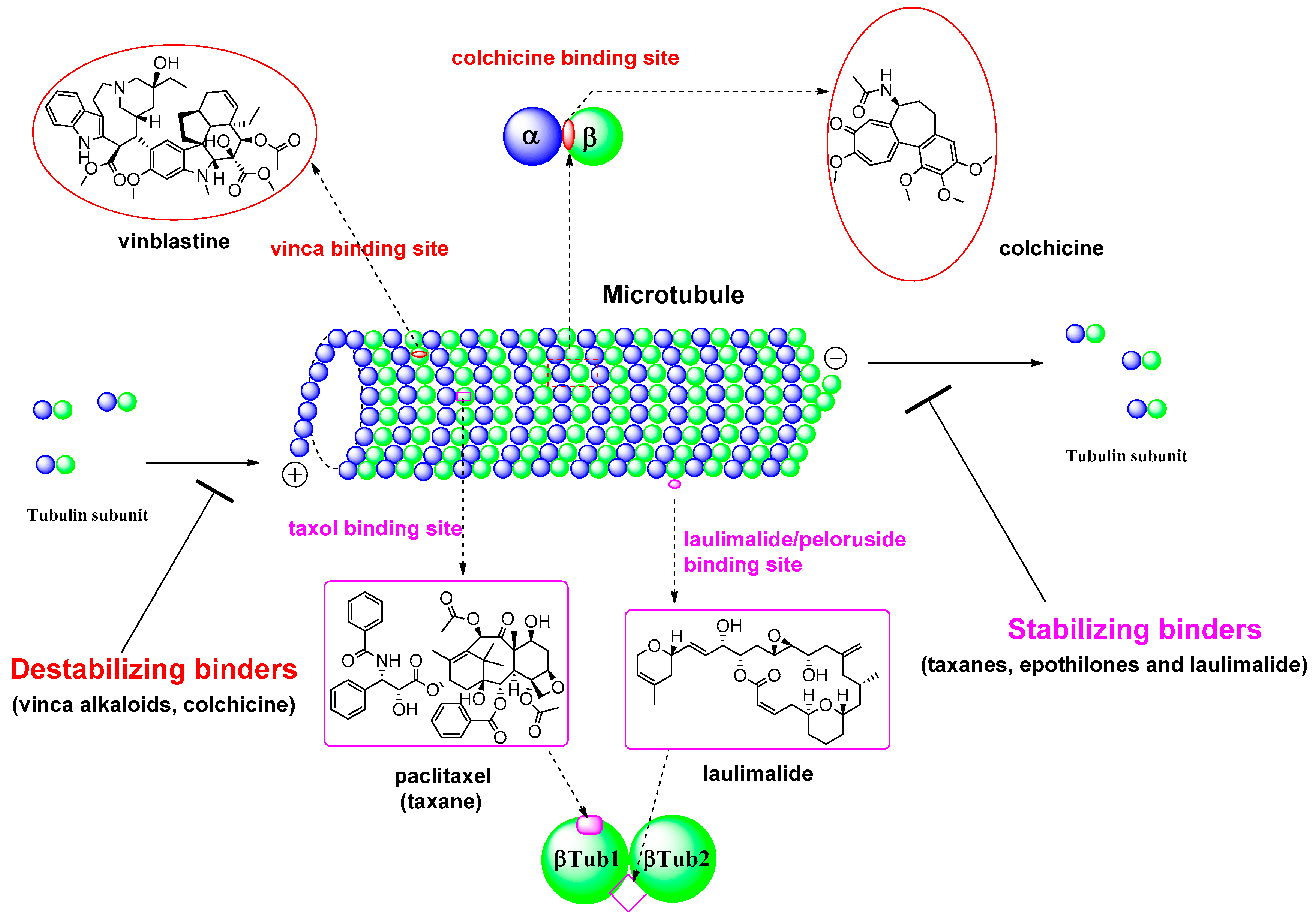
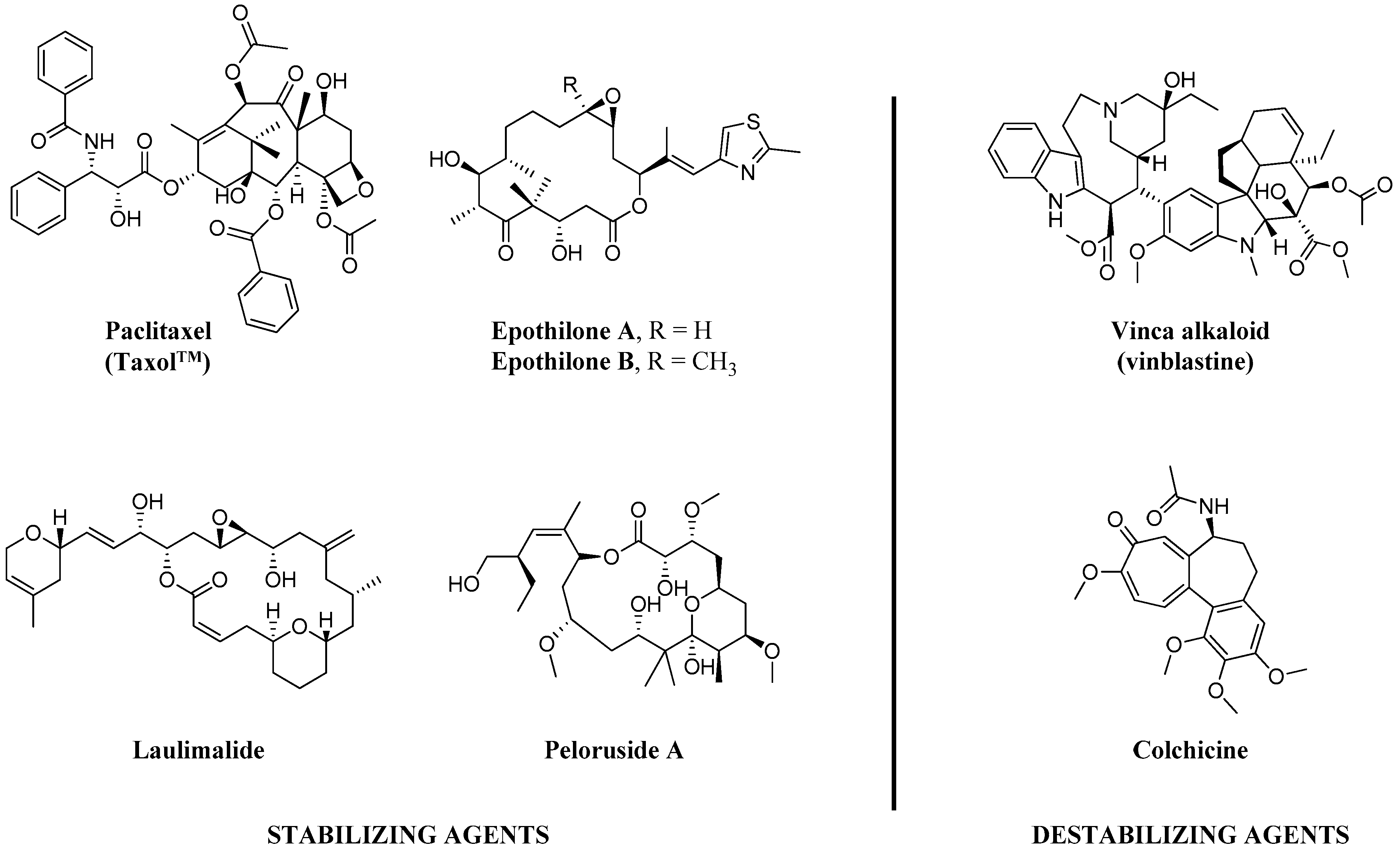
:1. Introduction
2. Nanoparticle Delivery of Tubulin Inhibitors
2.1. Nano Particle Delivery of Tubulin Inhibitors Targeting Vinca Binding Site
2.1.1. Delivery of Tubulysine A (TubA)
2.1.2. Folate Mediated Delivery of Nanoparticle-Loaded Emtansine
2.1.3. α-Cyclodextrin Mediated Delivery of Curcumin to the Cancer Cell
2.2. Nano Particle Delivery of Tubulin Inhibitors Targeting Colchicine Binding Site
2.2.1. Nanoparticle Mediated Delivery of Colchicine Alkaloid
2.2.2. Delivery of LY293
2.2.3. Delivery of Combretastatin A-4
2.2.4. Delivery of Etoposide
2.3. Nano Particle Delivery of Tubulin Inhibitors Targeting Paclitaxel Binding Site
Delivery of Paclitaxel
3. Vascular Disrupting Agents and Antiangiogenic Agents
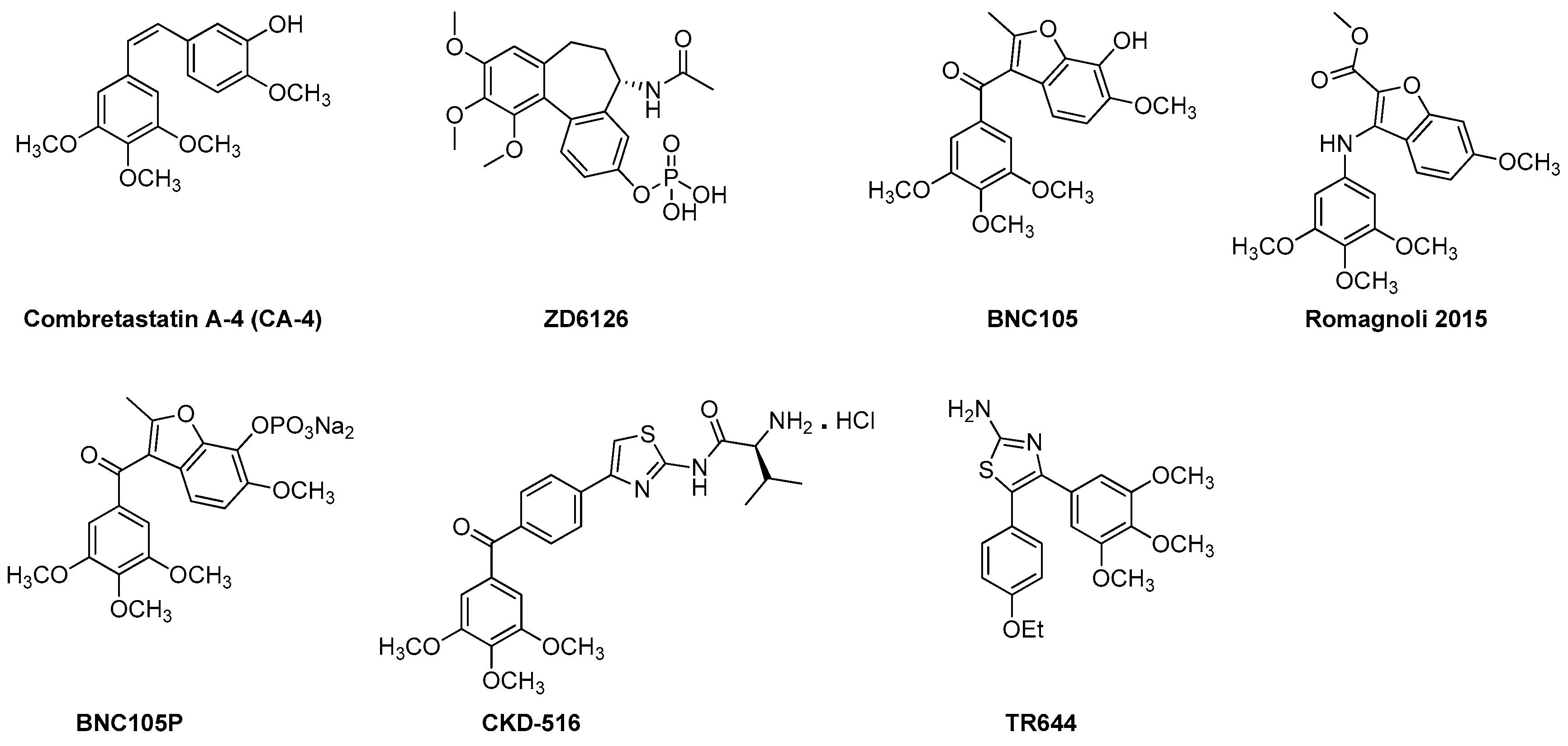
3.1. VDAs and Antiangiogenic Agents from the CA-4 Family
3.1.1. ZD6126
3.1.2. CKD 516
3.1.3. BNC 105 and BNC 105P
3.1.4. Benzofuran CA-4 Derivative
3.1.5. TR644
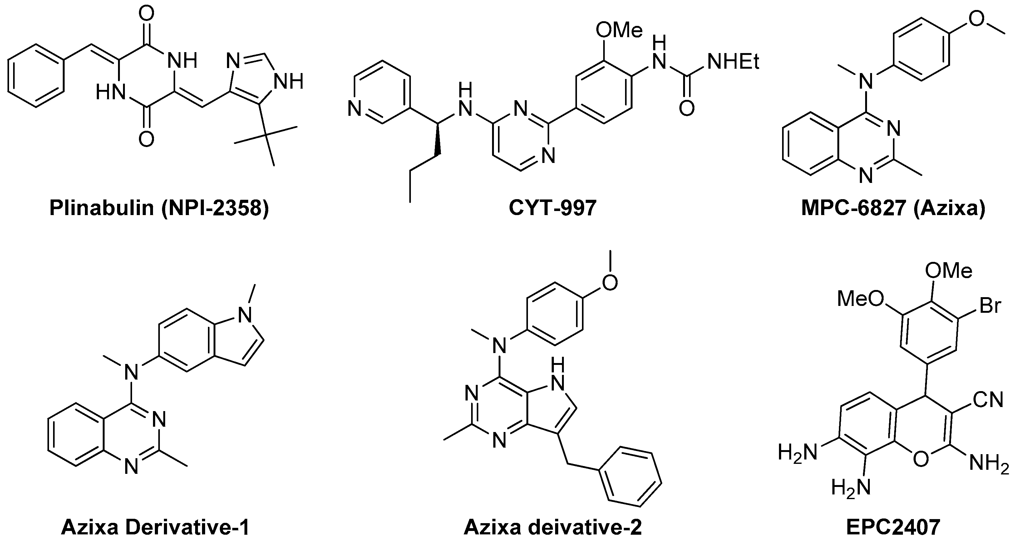
3.2. Miscellaneous Recent VDAs and Antiangiogenic Agents
3.2.1. Plinabulin (NPI-2358)
3.2.2. CYT-997
3.2.3. Azixa and Its Derivatives
3.2.4. EPC2407
4. Conclusions and Future Directions
Acknowledgments
Conflicts of Interest
References
- Borisy, G.G.; Taylor, E.W. The mechanism of action of colchicine. Binding of colchincine-3H to cellular protein. J. Cell Biol. 1967, 34, 525–533. [Google Scholar] [CrossRef] [PubMed]
- Bunker, J.M.; Wilson, L.; Jordan, M.A.; Feinstein, S.C. Modulation of microtubule dynamics by tau in living cells: Implications for development and neurodegeneration. Mol. Biol. Cell 2004, 15, 2720–2728. [Google Scholar] [CrossRef] [PubMed]
- Risinger, A.L.; Dybdal-Hargreaves, N.F.; Mooberry, S.L. Breast cancer cell lines exhibit differential sensitivities to microtubule-targeting drugs independent of doubling time. Anticancer Res. 2015, 35, 5845–5850. [Google Scholar] [PubMed]
- Perez-Elias, M.J.; Morellon, M.L.; Ortega, E.; Hernandez-Quero, J.; Rodriguez-Torres, M.; Clotet, B.; Felizarta, F.; Gutierrez, F.; Pineda, J.A.; Nichols, G.; et al. Pharmacokinetics of fosamprenavir plus ritonavir in human immunodeficiency virus type 1-infected adult subjects with hepatic impairment. Antimicrob. Agents Chemother. 2009, 53, 5185–5196. [Google Scholar] [CrossRef] [PubMed]
- Gan, P.P.; McCarroll, J.A.; Po’uha, S.T.; Kamath, K.; Jordan, M.A.; Kavallaris, M. Microtubule dynamics, mitotic arrest, and apoptosis: Drug-induced differential effects of βIII-tubulin. Mol. Cancer Ther. 2010, 9, 1339–1348. [Google Scholar] [CrossRef] [PubMed]
- Brouhard, G.J.; Rice, L.M. The contribution of αβ-tubulin curvature to microtubule dynamics. J. Cell Biol. 2014, 207, 323–334. [Google Scholar] [CrossRef] [PubMed]
- Zhao, Y.; Mu, X.; Du, G. Microtubule-stabilizing agents: New drug discovery and cancer therapy. Pharmacol. Ther. 2016, 162, 134–143. [Google Scholar] [CrossRef] [PubMed]
- Fanale, D.; Bronte, G.; Passiglia, F.; Calo, V.; Castiglia, M.; Di Piazza, F.; Barraco, N.; Cangemi, A.; Catarella, M.T.; Insalaco, L.; et al. Stabilizing versus destabilizing the microtubules: A double-edge sword for an effective cancer treatment option? Anal. Cell. Pathol. 2015, 2015, 690916. [Google Scholar] [CrossRef] [PubMed]
- Morris, P.G.; Fornier, M.N. Microtubule active agents: Beyond the taxane frontier. Clin. Cancer Res. 2008, 14, 7167–7172. [Google Scholar] [CrossRef] [PubMed]
- Hwang, D.J.; Wang, J.; Li, W.; Miller, D.D. Structural optimization of indole derivatives acting at colchicine binding site as potential anticancer agents. ACS Med. Chem. Lett. 2015, 6, 993–997. [Google Scholar] [CrossRef] [PubMed]
- Prota, A.E.; Bargsten, K.; Northcote, P.T.; Marsh, M.; Altmann, K.H.; Miller, J.H.; Diaz, J.F.; Steinmetz, M.O. Structural basis of microtubule stabilization by laulimalide and peloruside A. Angew. Chem. Int. Ed. Engl. 2014, 53, 1621–1625. [Google Scholar] [CrossRef] [PubMed]
- Perez, E.A. Microtubule inhibitors: Differentiating tubulin-inhibiting agents based on mechanisms of action, clinical activity, and resistance. Mol. Cancer Ther. 2009, 8, 2086–2095. [Google Scholar] [CrossRef] [PubMed]
- Ravelli, R.B.; Gigant, B.; Curmi, P.A.; Jourdain, I.; Lachkar, S.; Sobel, A.; Knossow, M. Insight into tubulin regulation from a complex with colchicine and a stathmin-like domain. Nature 2004, 428, 198–202. [Google Scholar] [CrossRef] [PubMed]
- Spear, M.A.; LoRusso, P.; Mita, A.; Mita, M. Vascular disrupting agents (VDA) in oncology: Advancing towards new therapeutic paradigms in the clinic. Curr. Drug Targets 2011, 12, 2009–2015. [Google Scholar] [CrossRef] [PubMed]
- Siemann, D.W. The unique characteristics of tumor vasculature and preclinical evidence for its selective disruption by tumor-vascular disrupting agents. Cancer Treat. Rev. 2011, 37, 63–74. [Google Scholar] [CrossRef] [PubMed]
- Prokopiou, E.M.; Ryder, S.A.; Walsh, J.J. Tumour vasculature targeting agents in hybrid/conjugate drugs. Angiogenesis 2013, 16, 503–524. [Google Scholar] [CrossRef] [PubMed]
- Lobert, S.G.; Mary, E. Regulation of tubulin expression by micro-RNA: Implications drug resistance. Methods Cell Biol. 2013, 115, 63–74. [Google Scholar] [PubMed]
- Lu, Y.; Chen, J.; Xiao, M.; Li, W.; Miller, D.D. An overview of tubulin inhibitors that interact with the colchicine binding site. Pharm. Res. 2012, 29, 2943–2971. [Google Scholar] [CrossRef] [PubMed]
- Ji, Y.T.; Liu, Y.N.; Liu, Z.P. Tubulin colchicine binding site inhibitors as vascular disrupting agents in clinical developments. Curr. Med. Chem. 2015, 22, 1348–1360. [Google Scholar] [CrossRef] [PubMed]
- Wang, X.-F.; Guan, F.; Ohkoshi, E.; Guo, W.; Wang, L.; Zhu, D.-Q.; Wang, S.-B.; Wang, L.-T.; Hamel, E.; Yang, D.; et al. Optimization of 4-(N-cycloamino)phenylquinazolines as a novel class of tubulin-polymerization inhibitors targeting the colchicine site. J. Med. Chem. 2014, 57, 1390–1402. [Google Scholar] [CrossRef] [PubMed]
- Solum, E.J.; Cheng, J.-J.; Sørvik, I.B.; Paulsen, R.E.; Vik, A.; Hansen, T.V. Synthesis and biological evaluations of new analogs of 2-methoxyestradiol: Inhibitors of tubulin and angiogenesis. Eur. J. Med. Chem. 2014, 85, 391–398. [Google Scholar] [CrossRef] [PubMed]
- O’Boyle, N.M.; Pollock, J.K.; Carr, M.; Knox, A.J.S.; Nathwani, S.M.; Wang, S.; Caboni, L.; Zisterer, D.M.; Meegan, M.J. Β-lactam estrogen receptor antagonists and a dual-targeting estrogen receptor/tubulin ligand. J. Med. Chem. 2014, 57, 9370–9382. [Google Scholar] [CrossRef] [PubMed]
- Gangjee, A.; Pavana, R.K.; Ihnat, M.A.; Thorpe, J.E.; Disch, B.C.; Bastian, A.; Bailey-Downs, L.C.; Hamel, E.; Bai, R. Discovery of antitubulin agents with antiangiogenic activity as single entities with multitarget chemotherapy potential. ACS Med. Chem. Lett. 2014, 5, 480–484. [Google Scholar] [CrossRef] [PubMed]
- Patil, S.A.; Wang, J.; Li, X.S.; Chen, J.; Jones, T.S.; Hosni-Ahmed, A.; Patil, R.; Seibel, W.L.; Li, W.; Miller, D.D. New substituted 4H-chromenes as anticancer agents. Bioorg. Med. Chem. Lett. 2012, 22, 4458–4461. [Google Scholar] [CrossRef] [PubMed]
- Chen, W.; Seefeldt, T.; Young, A.; Zhang, X.; Zhao, Y.; Ruffolo, J.; Kaushik, R.S.; Guan, X. Microtubule S-glutathionylation as a potential approach for antimitotic agents. BMC Cancer 2012, 12. [Google Scholar] [CrossRef] [PubMed]
- Mundra, V.; Peng, Y.; Kumar, V.; Li, W.; Miller, D.D.; Mahato, R.I. Systemic delivery of nanoparticle formulation of novel tubulin inhibitor for treating metastatic melanoma. Drug Deliv. Transl. Res. 2015, 5, 199–208. [Google Scholar] [CrossRef] [PubMed]
- Athawale, R.B.; Jain, D.S.; Singh, K.K.; Gude, R.P. Etoposide loaded solid lipid nanoparticles for curtailing B16F10 melanoma colonization in lung. Biomed. Pharmacother. 2014, 68, 231–240. [Google Scholar] [CrossRef] [PubMed]
- Mundra, V.; Lu, Y.; Danquah, M.; Li, W.; Miller, D.D.; Mahato, R.I. Formulation and characterization of polyester/polycarbonate nanoparticles for delivery of a novel microtubule destabilizing agent. Pharm. Res. 2012, 29, 3064–3074. [Google Scholar] [CrossRef] [PubMed]
- Schluep, T.; Gunawan, P.; Ma, L.; Jensen, G.S.; Duringer, J.; Hinton, S.; Richter, W.; Hwang, J. Polymeric tubulysin-peptide nanoparticles with potent antitumor activity. Clin. Cancer Res. 2009, 15, 181–189. [Google Scholar] [CrossRef] [PubMed]
- Reddy, J.A.; Dorton, R.; Westrick, E.; Dawson, A.; Smith, T.; Xu, L.-C.; Vetzel, M.; Kleindl, P.; Vlahov, I.R.; Leamon, C.P. Preclinical evaluation of EC145, a folate-vinca alkaloid conjugate. Cancer Res. 2007, 67, 4434–4442. [Google Scholar] [CrossRef] [PubMed]
- Carter, P.J.; Senter, P.D. Antibody-drug conjugates for cancer therapy. Cancer J. 2008, 14, 154–169. [Google Scholar] [CrossRef] [PubMed]
- Thomas, A.; Teicher, B.A.; Hassan, R. Antibody-drug conjugates for cancer therapy. Lancet Oncol. 2016, 17, e254–e262. [Google Scholar] [CrossRef]
- Polakis, P. Antibody drug conjugates for cancer therapy. Pharmacol. Rev. 2016, 68, 3–19. [Google Scholar] [CrossRef] [PubMed]
- Vicent, M.J. Polymer-drug conjugates as modulators of cellular apoptosis. AAPS J. 2007, 9, E200–E207. [Google Scholar] [CrossRef] [PubMed]
- Kaur, G.; Hollingshead, M.; Holbeck, S.; Schauer-Vukašinović, V.; Camalier, R.F.; Dömling, A.; Agarwal, S. Biological evaluation of tubulysin A: A potential anticancer and antiangiogenic natural product. Biochem. J. 2006, 396, 235–242. [Google Scholar] [CrossRef] [PubMed]
- Steinmetz, H.; Glaser, N.; Herdtweck, E.; Sasse, F.; Reichenbach, H.; Höfle, G. Isolation, crystal and solution structure determination, and biosynthesis of tubulysins—Powerful inhibitors of tubulin polymerization from myxobacteria. Angew. Chem. Int. Ed. Engl. 2004, 43, 4888–4892. [Google Scholar] [CrossRef] [PubMed]
- Khalil, M.W.; Sasse, F.; Lünsdorf, H.; Elnakady, Y.A.; Reichenbach, H. Mechanism of action of tubulysin, an antimitotic peptide from myxobacteria. ChemBioChem 2006, 7, 678–683. [Google Scholar] [CrossRef] [PubMed]
- Murray, B.C.; Peterson, M.T.; Fecik, R.A. Chemistry and biology of tubulysins: Antimitotic tetrapeptides with activity against drug resistant cancers. Nat. Prod. Rep. 2015, 32, 654–662. [Google Scholar] [CrossRef] [PubMed]
- Nicolaou, K.C.; Yin, J.; Mandal, D.; Erande, R.D.; Klahn, P.; Jin, M.; Aujay, M.; Sandoval, J.; Gavrilyuk, J.; Vourloumis, D. Total synthesis and biological evaluation of natural and designed tubulysins. J. Am. Chem. Soc. 2016, 138, 1698–1708. [Google Scholar] [CrossRef] [PubMed]
- Colombo, R.; Wang, Z.; Han, J.; Balachandran, R.; Daghestani, H.N.; Camarco, D.P.; Vogt, A.; Day, B.W.; Mendel, D.; Wipf, P. Total synthesis and biological evaluation of tubulysin analogues. J. Org. Chem. 2016. [Google Scholar] [CrossRef] [PubMed]
- Xu, X.; Gregory, K.F.; Yao, L. Recent advances in the synthesis of tubulysins. Mini Rev. Med. Chem. 2013, 13, 1572–1578. [Google Scholar]
- Sendur, M.A.; Aksoy, S.; Ozdemir, Y.; Zengin, N.; Altundag, K. Does trastuzumab-emtansine have better cardiac safety profile in contrast to trastuzumab? J. BUON 2013, 18, 801. [Google Scholar] [PubMed]
- Bighin, C.; Pronzato, P.; Del Mastro, L. Trastuzumab emtansine in the treatment of HER-2-positive metastatic breast cancer patients. Future Oncol. 2013, 9, 955–957. [Google Scholar] [CrossRef] [PubMed]
- Poon, K.A.; Flagella, K.; Beyer, J.; Tibbitts, J.; Kaur, S.; Saad, O.; Yi, J.H.; Girish, S.; Dybdal, N.; Reynolds, T. Preclinical safety profile of trastuzumab emtansine (T-DM1): Mechanism of action of its cytotoxic component retained with improved tolerability. Toxicol. Appl. Pharmacol. 2013, 273, 298–313. [Google Scholar] [CrossRef] [PubMed]
- Tang, X.; Liang, Y.; Zhu, Y.; Cai, S.; Sun, L.; Chen, T. Enhanced anticancer activity of DM1-loaded star-shaped folate-core PLA-TPGS nanoparticles. Nanoscale Res. Lett. 2014, 9. [Google Scholar] [CrossRef] [PubMed]
- Haddley, K. Trastuzumab emtansine for the treatment of HER2-positive metastatic breast cancer. Drugs Today 2013, 49, 701–715. [Google Scholar] [CrossRef] [PubMed]
- Thery, J.-C.; Spano, J.-P.; Azria, D.; Raymond, E.; Penault Llorca, F. Resistance to human epidermal growth factor receptor type 2-targeted therapies. Eur. J. Cancer 2014, 50, 892–901. [Google Scholar] [CrossRef] [PubMed]
- Ye, P.; Zhang, W.; Yang, T.; Lu, Y.; Lu, M.; Gai, Y.; Ma, X.; Xiang, G. Folate receptor-targeted liposomes enhanced the antitumor potency of imatinib through the combination of active targeting and molecular targeting. Int. J. Nanomed. 2014, 9, 2167–2178. [Google Scholar] [CrossRef] [PubMed]
- Hu, Y.; Ma, B.; Zhang, Y.; Wang, M. Small molecule-folic acid modification on nanopatterned PDMS and investigation on its surface property. Biomed. Microdevices 2014, 16, 487–497. [Google Scholar] [CrossRef] [PubMed]
- Leamon, C.P.; Vlahov, I.R.; Reddy, J.A.; Vetzel, M.; Santhapuram, H.K.; You, F.; Bloomfield, A.; Dorton, R.; Nelson, M.; Kleindl, P.; et al. Folate-vinca alkaloid conjugates for cancer therapy: A structure-activity relationship. Bioconjug. Chem. 2014, 25, 560–568. [Google Scholar] [CrossRef] [PubMed]
- Shi, J.; Wang, L.; Gao, J.; Liu, Y.; Zhang, J.; Ma, R.; Liu, R.; Zhang, Z. A fullerene-based multi-functional nanoplatform for cancer theranostic applications. Biomaterials 2014, 35, 5771–5784. [Google Scholar] [CrossRef] [PubMed]
- Zheng, M.; Zhao, P.; Luo, Z.; Gong, P.; Zheng, C.; Zhang, P.; Yue, C.; Gao, D.; Ma, Y.; Cai, L. Robust ICG theranostic nanoparticles for folate targeted cancer imaging and highly effective photothermal therapy. ACS Appl. Mater. Interfaces 2014, 6, 6709–6716. [Google Scholar] [CrossRef] [PubMed]
- Torchilin, V. Tumor delivery of macromolecular drugs based on the EPR effect. Adv. Drug Deliv. Rev. 2011, 63, 131–135. [Google Scholar] [CrossRef] [PubMed]
- Jana, B.; Mohapatra, S.; Mondal, P.; Barman, S.; Pradhan, K.; Saha, A.; Ghosh, S. Α-cyclodextrin interacts close to vinblastine site of tubulin and delivers curcumin preferentially to the tubulin surface of cancer cell. ACS Appl. Mater. Interfaces 2016, 8, 13793–13803. [Google Scholar] [CrossRef] [PubMed]
- Pérez-Pérez, M.-J.; Priego, E.-M.; Bueno, O.; Martins, M.S.; Canela, M.-D.; Liekens, S. Blocking blood flow to solid tumors by destabilizing tubulin: An approach to targeting tumor growth. J. Med. Chem. 2016, 59, 8685–8711. [Google Scholar] [CrossRef] [PubMed]
- Tangutoori, S.; Ohta, A.; Gatley, S.; Campbell, B.R. Repurposing an erstwhile cancer drug: A quantitative and therapeutic evaluation of alternative nanosystems for the delivery of colchicine to solid tumors. J. Cancer Sci. Ther. 2014, 6, 236–246. [Google Scholar] [CrossRef]
- Tortorici, M.A.; Skledar, S.; Barnes, B.; Wasko, M.C. Promoting the safe use of intravenous colchicine. Am. J. Health Syst. Pharm. 2004, 61, 2496–2501. [Google Scholar] [PubMed]
- Deveaux, M.; Hubert, N.; Demarly, C. Colchicine poisoning: Case report of two suicides. Forensic. Sci. Int. 2004, 143, 219–222. [Google Scholar] [CrossRef] [PubMed]
- Fernandez, S.; Castro, P.; Nogue, S.; Nicolas, J.M. Refractory shock and severe leukopenia with multisystemic organ failure due to colchicine intentional overdose. Med. Clin. 2014, 143, 140. [Google Scholar]
- Suh, J.; Choy, K.-L.; Lai, S.K.; Suk, J.S.; Tang, B.C.; Prabhu, S.; Hanes, J. Pegylation of nanoparticles improves their cytoplasmic transport. Int. J. Nanomed. 2007, 2, 735–741. [Google Scholar]
- Lu, Y.; Li, C.-M.; Wang, Z.; Ross, C.R.; Chen, J.; Dalton, J.T.; Li, W.; Miller, D.D. Discovery of 4-substituted methoxybenzoyl-aryl-thiazole as novel anticancer agents: Synthesis, biological evaluation, and structure-activity relationships. J. Med. Chem. 2009, 52, 1701–1711. [Google Scholar] [CrossRef] [PubMed]
- Lu, Y.; Li, C.-M.; Wang, Z.; Chen, J.; Mohler, M.L.; Li, W.; Dalton, J.T.; Miller, D.D. Design, synthesis, and sar studies of 4-substituted methoxylbenzoyl-aryl-thiazoles analogues as potent and orally bioavailable anticancer agents. J. Med. Chem. 2011, 54, 4678–4693. [Google Scholar] [CrossRef] [PubMed]
- Sengupta, S.; Eavarone, D.; Capila, I.; Zhao, G.; Watson, N.; Kiziltepe, T.; Sasisekharan, R. Temporal targeting of tumour cells and neovasculature with a nanoscale delivery system. Nature 2005, 436, 568–572. [Google Scholar] [CrossRef] [PubMed]
- Li, X.; Wu, M.; Pan, L.; Shi, J. Tumor vascular-targeted co-delivery of anti-angiogenesis and chemotherapeutic agents by mesoporous silica nanoparticle-based drug delivery system for synergetic therapy of tumor. Int. J. Nanomed. 2016, 11, 93–105. [Google Scholar] [CrossRef] [PubMed]
- Sumer Bolu, B.; Manavoglu Gecici, E.; Sanyal, R. Combretastatin A-4 conjugated antiangiogenic micellar drug delivery systems using dendron-polymer conjugates. Mol. Pharm. 2016, 13, 1482–1490. [Google Scholar] [CrossRef] [PubMed]
- Houghton, P.J.; Kurmasheva, R.T.; Kolb, E.A.; Gorlick, R.; Maris, J.M.; Wu, J.; Tong, Z.; Arnold, M.A.; Chatterjee, M.; Williams, T.M.; et al. Initial testing (stage 1) of the tubulin binding agent nanoparticle albumin-bound (nab) paclitaxel (Abraxane®) by the pediatric Preclinical Testing Program (PPTP). Pediatr. Blood Cancer 2015, 62, 1214–1221. [Google Scholar] [CrossRef] [PubMed]
- Desai, N.; Trieu, V.; Damascelli, B.; Soon-Shiong, P. Sparc expression correlates with tumor response to albumin-bound paclitaxel in head and neck cancer patients. Transl. Oncol. 2009, 2, 59–64. [Google Scholar] [CrossRef] [PubMed]
- De Leon, M.C.B.; Bolla, S.; Greene, B.; Hutchinson, L.; Del Priore, G. Successful treatment with nab-paclitaxel after hypersensitivity reaction to paclitaxel and docetaxel. Gynecol. Oncol. Case Rep. 2013, 5, 70–71. [Google Scholar] [CrossRef] [PubMed]
- Miele, E.; Spinelli, G.P.; Miele, E.; Tomao, F.; Tomao, S. Albumin-bound formulation of paclitaxel (Abraxane® ABI-007) in the treatment of breast cancer. Int. J. Nanomed. 2009, 4, 99–105. [Google Scholar]
- Kretschmar, C.S.; Kletzel, M.; Murray, K.; Thorner, P.; Joshi, V.; Marcus, R.; Smith, E.I.; London, W.B.; Castleberry, R. Response to paclitaxel, topotecan, and topotecan-cyclophosphamide in children with untreated disseminated neuroblastoma treated in an upfront phase II investigational window: A pediatric oncology group study. J. Clin. Oncol. 2004, 22, 4119–4126. [Google Scholar] [CrossRef] [PubMed]
- Doz, F.; Gentet, J.C.; Pein, F.; Frappaz, D.; Chastagner, P.; Moretti, S.; Vassal, G.; Arditti, J.; Tellingen, O.V.; Iliadis, A.; et al. Phase I trial and pharmacological study of a 3-hour paclitaxel infusion in children with refractory solid tumours: A SFOP study. Br. J. Cancer 2001, 84, 604–610. [Google Scholar] [CrossRef] [PubMed]
- Kolb, E.A.; Gorlick, R.; Reynolds, C.P.; Kang, M.H.; Carol, H.; Lock, R.; Keir, S.T.; Maris, J.M.; Billups, C.A.; DesJardins, C.; et al. Initial testing (stage 1) of eribulin, a novel tubulin binding agent, by the pediatric preclinical testing program. Pediatr. Blood Cancer 2013, 60, 1325–1332. [Google Scholar] [CrossRef] [PubMed]
- Wong, H.; Choo, E.F.; Alicke, B.; Ding, X.; La, H.; McNamara, E.; Theil, F.-P.; Tibbitts, J.; Friedman, L.S.; Hop, C.E.C.A.; et al. Antitumor activity of targeted and cytotoxic agents in murine subcutaneous tumor models correlates with clinical response. Clin. Cancer Res. 2012, 18, 3846–3855. [Google Scholar] [CrossRef] [PubMed]
- Desai, N.P.; Trieu, V.; Hwang, L.Y.; Wu, R.; Soon-Shiong, P.; Gradishar, W.J. Improved effectiveness of nanoparticle albumin-bound (nab) paclitaxel versus polysorbate-based docetaxel in multiple xenografts as a function of HER2 and SPARC status. Anti-Cancer Drugs 2008, 19, 899–909. [Google Scholar] [CrossRef] [PubMed]
- Mielke, S.; Sparreboom, A.; Steinberg, S.M.; Gelderblom, H.; Unger, C.; Behringer, D.; Mross, K. Association of paclitaxel pharmacokinetics with the development of peripheral neuropathy in patients with advanced cancer. Clin. Cancer Res. 2005, 11, 4843–4850. [Google Scholar] [CrossRef] [PubMed]
- Mielke, S.; Sparreboom, A.; Mross, K. Peripheral neuropathy: A persisting challenge in paclitaxel-based regimes. Eur. J. Cancer 2006, 42, 24–30. [Google Scholar] [CrossRef] [PubMed]
- Knemeyer, I.; Wientjes, G.M.; Au, L.S.J. Cremophor reduces paclitaxel penetration into bladder wall during intravesical treatment. Cancer Chemother. Pharmacol. 1999, 44, 241–248. [Google Scholar] [CrossRef] [PubMed]
- Gradishar, W.J.; Tjulandin, S.; Davidson, N.; Shaw, H.; Desai, N.; Bhar, P.; Hawkins, M.; O’Shaughnessy, J. Phase III trial of nanoparticle albumin-bound paclitaxel compared with polyethylated castor oil-based paclitaxel in women with breast cancer. J. Clin. Oncol. 2005, 23, 7794–7803. [Google Scholar] [CrossRef] [PubMed]
- Kundranda, M.N.; Niu, J. Albumin-bound paclitaxel in solid tumors: Clinical development and future directions. Drug Des. Dev. Ther. 2015, 9, 3767–3777. [Google Scholar] [CrossRef] [PubMed]
- Gupta, N.; Hatoum, H.; Dy, G.K. First line treatment of advanced non-small-cell lung cancer—Specific focus on albumin bound paclitaxel. Int. J. Nanomed. 2014, 9, 209–221. [Google Scholar]
- Von Hoff, D.D.; Ervin, T.; Arena, F.P.; Chiorean, E.G.; Infante, J.; Moore, M.; Seay, T.; Tjulandin, S.A.; Ma, W.W.; Saleh, M.N.; et al. Increased survival in pancreatic cancer with nab-paclitaxel plus gemcitabine. N. Engl. J. Med. 2013, 369, 1691–1703. [Google Scholar] [CrossRef] [PubMed]
- Bansal, A.; Kapoor, D.N.; Kapil, R.; Chhabra, N.; Dhawan, S. Design and development of paclitaxel-loaded bovine serum albumin nanoparticles for brain targeting. Acta Pharm. 2011, 61, 141–156. [Google Scholar] [CrossRef] [PubMed]
- Wang, B.; Lv, L.; Wang, Z.; Jiang, Y.; Lv, W.; Liu, X.; Wang, Z.; Zhao, Y.; Xin, H.; Xu, Q. Improved anti-glioblastoma efficacy by IL-13Rα2 mediated copolymer nanoparticles loaded with paclitaxel. Sci. Rep. 2015, 5. [Google Scholar] [CrossRef] [PubMed]
- Yi, X.; Lian, X.; Dong, J.; Wan, Z.; Xia, C.; Song, X.; Fu, Y.; Gong, T.; Zhang, Z. Co-delivery of pirarubicin and paclitaxel by human serum albumin nanoparticles to enhance antitumor effect and reduce systemic toxicity in breast cancers. Mol. Pharm. 2015, 12, 4085–4098. [Google Scholar] [CrossRef] [PubMed]
- Tozer, G.M.; Kanthou, C.; Baguley, B.C. Disrupting tumour blood vessels. Nat. Rev. Cancer 2005, 5, 423–435. [Google Scholar] [CrossRef] [PubMed]
- Kanthou, C.; Tozer, G.M. Selective destruction of the tumour vasculature by targeting the endothelial cytoskeleton. Drug Discov. Today. Ther. Strateg. 2007, 4, 237–243. [Google Scholar] [CrossRef]
- Siemann, D.W.; Bibby, M.C.; Dark, G.G.; Dicker, A.P.; Eskens, F.A.L.M.; Horsman, M.R.; Marmé, D.; LoRusso, P.M. Differentiation and definition of vascular-targeted therapies. Clin. Cancer Res. 2005, 11, 416–420. [Google Scholar] [PubMed]
- Carmeliet, P. Angiogenesis in life, disease and medicine. Nature 2005, 438, 932–936. [Google Scholar] [CrossRef] [PubMed]
- El-Kenawi, A.E.; El-Remessy, A.B. Angiogenesis inhibitors in cancer therapy: Mechanistic perspective on classification and treatment rationales. Br. J. Pharmacol. 2013, 170, 712–729. [Google Scholar] [CrossRef] [PubMed]
- Vasudev, N.S.; Reynolds, A.R. Anti-angiogenic therapy for cancer: Current progress, unresolved questions and future directions. Angiogenesis 2014, 17, 471–494. [Google Scholar] [CrossRef] [PubMed]
- Romagnoli, R.; Baraldi, P.G.; Salvador, M.K.; Prencipe, F.; Lopez-Cara, C.; Schiaffino Ortega, S.; Brancale, A.; Hamel, E.; Castagliuolo, I.; Mitola, S.; et al. Design, synthesis, in vitro, and in vivo anticancer and antiangiogenic activity of novel 3-arylaminobenzofuran derivatives targeting the colchicine site on tubulin. J. Med. Chem. 2015, 58, 3209–3222. [Google Scholar] [CrossRef] [PubMed]
- Porcù, E.; Viola, G.; Bortolozzi, R.; Persano, L.; Mitola, S.; Ronca, R.; Presta, M.; Romagnoli, R.; Baraldi, P.G.; Basso, G. TR-644 a novel potent tubulin binding agent induces impairment of endothelial cells function and inhibits angiogenesis. Angiogenesis 2013, 16, 647–662. [Google Scholar] [CrossRef] [PubMed]
- Ganguly, A.; Cabral, F.; Yang, H.; Patel, K.D. Peloruside a is a microtubule-stabilizing agent with exceptional anti-migratory properties in human endothelial cells. Oncoscience 2015, 2, 585–595. [Google Scholar] [CrossRef] [PubMed]
- Broggini-Tenzer, A.; Sharma, A.; Nytko, K.J.; Bender, S.; Vuong, V.; Orlowski, K.; Hug, D.; O’Reilly, T.; Pruschy, M. Combined treatment strategies for microtubule stabilizing agent-resistant tumors. J. Natl. Cancer Inst. 2015, 107. [Google Scholar] [CrossRef] [PubMed]
- Chan, A.; Singh, A.J.; Northcote, P.T.; Miller, J.H. Inhibition of human vascular endothelial cell migration and capillary-like tube formation by the microtubule-stabilizing agent peloruside A. Investig. New Drugs 2015, 33, 564–574. [Google Scholar] [CrossRef] [PubMed]
- Bocci, G.; Di Paolo, A.; Danesi, R. The pharmacological bases of the antiangiogenic activity of paclitaxel. Angiogenesis 2013, 16, 481–492. [Google Scholar] [CrossRef] [PubMed]
- Lu, H.; Murtagh, J.; Schwartz, E.L. The microtubule binding drug laulimalide inhibits vascular endothelial growth factor-induced human endothelial cell migration and is synergistic when combined with docetaxel (taxotere). Mol. Pharmacol. 2006, 69, 1207–1215. [Google Scholar] [CrossRef] [PubMed]
- Zhu, S.; Kisiel, W.; Lu, Y.J.; Petersen, L.C.; Ndungu, J.M.; Moore, T.W.; Parker, E.T.; Sun, A.; Liotta, D.C.; El-Rayes, B.F.; et al. Tumor angiogenesis therapy using targeted delivery of paclitaxel to the vasculature of breast cancer metastases. J. Drug Deliv. 2014, 2014. [Google Scholar] [CrossRef] [PubMed]
- Lin, C.M.; Singh, S.B.; Chu, P.S.; Dempcy, R.O.; Schmidt, J.M.; Pettit, G.R.; Hamel, E. Interactions of tubulin with potent natural and synthetic analogs of the antimitotic agent combretastatin: A structure-activity study. Mol. Pharmacol. 1988, 34, 200–208. [Google Scholar] [PubMed]
- Massarotti, A.; Coluccia, A.; Silvestri, R.; Sorba, G.; Brancale, A. The tubulin colchicine domain: A molecular modeling perspective. ChemMedChem 2012, 7, 33–42. [Google Scholar] [CrossRef] [PubMed]
- Srivastava, V.; Negi, A.S.; Kumar, J.K.; Gupta, M.M.; Khanuja, S.P.S. Plant-based anticancer molecules: A chemical and biological profile of some important leads. Bioorg. Med. Chem. 2005, 13, 5892–5908. [Google Scholar] [CrossRef] [PubMed]
- Horner, S.A.; Gould, S.; Noakes, J.P.; Rattray, N.J.; Allen, S.L.; Zotova, E.; Arezzo, J.C. Lack of neurotoxicity of the vascular targeting agent ZD6126 following repeated i.v. Dosing in the rat. Mol. Cancer Ther. 2004, 3, 783–791. [Google Scholar] [PubMed]
- Pilat, M.J.; LoRusso, P.M. Vascular disrupting agents. J. Cell. Biochem. 2006, 99, 1021–1039. [Google Scholar] [CrossRef] [PubMed]
- Lee, J.; Bae, S.; Lee, S.-H.; Choi, H.; Kim, Y.H.; Kim, S.J.; Park, G.T.; Moon, S.K.; Kim, D.-H.; Lee, S.; et al. Discovery of a potent tubulin polymerization inhibitor: Synthesis and evaluation of water-soluble prodrugs of benzophenone analog. Bioorg. Med. Chem. Lett. 2010, 20, 6327–6330. [Google Scholar] [CrossRef] [PubMed]
- Lee, J.; Kim, S.J.; Choi, H.; Kim, Y.H.; Lim, I.T.; Yang, H.-M.; Lee, C.S.; Kang, H.R.; Ahn, S.K.; Moon, S.K.; et al. Identification of CKD-516: A potent tubulin polymerization inhibitor with marked antitumor activity against murine and human solid tumors. J. Med. Chem. 2010, 53, 6337–6354. [Google Scholar] [CrossRef] [PubMed]
- Kim, K.W.; Lee, J.M.; Jeon, Y.S.; Lee, I.J.; Choi, Y.; Park, J.; Kiefer, B.; Kim, C.; Han, J.K.; Choi, B.I. Vascular disrupting effect of CKD-516: Preclinical study using DCE-MRI. Investig. New Drugs 2013, 31, 1097–1106. [Google Scholar] [CrossRef] [PubMed]
- Oh, D.-Y.; Kim, T.-M.; Han, S.-W.; Shin, D.-Y.; Lee, Y.G.; Lee, K.-W.; Kim, J.H.; Kim, T.-Y.; Jang, I.-J.; Lee, J.-S.; et al. Phase i study of CKD-516, a novel vascular disrupting agent, in patients with advanced solid tumors. Cancer Res. Treat. 2016, 48, 28–36. [Google Scholar] [CrossRef] [PubMed]
- Kremmidiotis, G.; Leske, A.F.; Lavranos, T.C.; Beaumont, D.; Gasic, J.; Hall, A.; O’Callaghan, M.; Matthews, C.A.; Flynn, B. BNC105: A novel tubulin polymerization inhibitor that selectively disrupts tumor vasculature and displays single-agent antitumor efficacy. Mol. Cancer Ther. 2010, 9, 1562–1573. [Google Scholar] [CrossRef] [PubMed]
- Rischin, D.; Bibby, D.C.; Chong, G.; Kremmidiotis, G.; Leske, A.F.; Matthews, C.A.; Wong, S.S.; Rosen, M.A.; Desai, J. Clinical, pharmacodynamic, and pharmacokinetic evaluation of BNC105P: A phase I trial of a novel vascular disrupting agent and inhibitor of cancer cell proliferation. Clin. Cancer Res. 2011, 17, 5152–5160. [Google Scholar] [CrossRef] [PubMed]
- Yakushiji, F.; Tanaka, H.; Muguruma, K.; Iwahashi, T.; Yamazaki, Y.; Hayashi, Y. Water-soluble prodrug of antimicrotubule agent plinabulin: Effective strategy with click chemistry. Chem. Eur. J. 2011, 17, 12587–12590. [Google Scholar] [CrossRef] [PubMed]
- Millward, M.; Mainwaring, P.; Mita, A.; Federico, K.; Lloyd, G.K.; Reddinger, N.; Nawrocki, S.; Mita, M.; Spear, M.A. Phase 1 study of the novel vascular disrupting agent plinabulin (NPI-2358) and docetaxel. Investig. New Drugs 2012, 30, 1065–1073. [Google Scholar] [CrossRef] [PubMed]
- Burns, C.J.; Harte, M.F.; Bu, X.; Fantino, E.; Joffe, M.; Sikanyika, H.; Su, S.; Tranberg, C.E.; Wilson, N.; Charman, S.A.; et al. Discovery of CYT997: A structurally novel orally active microtubule targeting agent. Bioorg. Med. Chem. Lett. 2009, 19, 4639–4642. [Google Scholar] [CrossRef] [PubMed]
- Burns, C.J.; Fantino, E.; Powell, A.K.; Shnyder, S.D.; Cooper, P.A.; Nelson, S.; Christophi, C.; Malcontenti-Wilson, C.; Dubljevic, V.; Harte, M.F.; et al. The microtubule depolymerizing agent CYT997 causes extensive ablation of tumor vasculature in vivo. J. Pharmacol. Exp. Ther. 2011, 339, 799–806. [Google Scholar] [CrossRef] [PubMed]
- Monaghan, K.; Khong, T.; Smith, G.; Spencer, A. CYT997 causes apoptosis in human multiple myeloma. Investig. New Drugs 2011, 29, 232–238. [Google Scholar] [CrossRef] [PubMed]
- Kasibhatla, S.; Baichwal, V.; Cai, S.X.; Roth, B.; Skvortsova, I.; Skvortsov, S.; Lukas, P.; English, N.M.; Sirisoma, N.; Drewe, J.; et al. MPC-6827: A small-molecule inhibitor of microtubule formation that is not a substrate for multidrug resistance pumps. Cancer Res. 2007, 67, 5865–5871. [Google Scholar] [CrossRef] [PubMed]
- Grossmann, K.F.; Colman, H.; Akerley, W.A.; Glantz, M.; Matsuoko, Y.; Beelen, A.P.; Yu, M.; De Groot, J.F.; Aiken, R.D.; Olsen, J.J.; et al. Phase I trial of verubulin (MPC-6827) plus carboplatin in patients with relapsed glioblastoma multiforme. J. Neurooncol. 2012, 110, 257–264. [Google Scholar] [CrossRef] [PubMed]
- Tsimberidou, A.-M.; Akerley, W.; Schabel, M.C.; Hong, D.S.; Uehara, C.; Chhabra, A.; Warren, T.; Mather, G.G.; Evans, B.A.; Woodland, D.P.; et al. Phase I clinical trial of MPC-6827 (Azixa), a microtubule destabilizing agent, in patients with advanced cancer. Mol. Cancer Ther. 2010, 9, 3410–3419. [Google Scholar] [CrossRef] [PubMed]
- Mahal, K.; Resch, M.; Ficner, R.; Schobert, R.; Biersack, B.; Mueller, T. Effects of the tumor-vasculature-disrupting agent verubulin and two heteroaryl analogues on cancer cells, endothelial cells, and blood vessels. ChemMedChem 2014, 9, 847–854. [Google Scholar] [CrossRef] [PubMed]
- Patil, S.A.; Patil, R.; Pfeffer, L.M.; Miller, D.D. Chromenes: Potential new chemotherapeutic agents for cancer. Future Med. Chem. 2013, 5, 1647–1660. [Google Scholar] [CrossRef] [PubMed]
- Banerjee, S.; Wang, J.; Pfeffer, S.; Ma, D.; Pfeffer, L.; Patil, S.; Li, W.; Miller, D. Design, synthesis and biological evaluation of novel 5H-chromenopyridines as potential anti-cancer agents. Molecules 2015, 20, 17152–17165. [Google Scholar] [CrossRef] [PubMed]


| Drugs | Formulation | Active/Passive | Advantage | References |
|---|---|---|---|---|
| Tubulysine A | Thiol derivative of TubA attached to a linear hexacyclodextrin based polymer via a disulfide linker leading to stable nanoparticles (CDP-TubA) | Active | In vivo CDP-TubA showed a potent antitumor effect and significantly prolonged survival compared with TubA alone | Schluep et al. [29] |
| Emtansine (DM1) | DM1 loaded PLA-TPGS copolymer nanoparticles immobilized with folates (FA-DM1-NPs) | Active | FA-DM1-NPs induce rapid apoptosis avoiding toxicities, side effects and nonspecific distributions exerted by DM1 alone | Tang et al. [46] |
| curcumin | α-Cyclodextrin | Active | α-cyclodextrin-curcumin complex selectively enters human lung cancer cell (A549) as compared to the human normal lung fibroblast (WI38) and delivers hydrophobic curcumin | Jana et al. [55] |
| Colchicine alkaloid | PEGylated Cationic Liposomal-colchicine (PCL-colchicine) nanoparticles | Passive | Microtubules are more efficiently disrupted by nanoparticle-loaded colchicine. In vivo longer survival time for the PCL-colchicine treated group | Tangutoori et al. [56] |
| LY293 | Biodegradable co-polymer, mPEG-b-P (CB-co-LA) | Passive | LY293 loaded nanoparticles demonstrated highly promising efficacy against resistance melanoma cells both in vitro and in vivo without noticeable toxicities to the important organs | Mundra et al. [26,28] |
| Combretastatin A-4 (CA4) | Multi drug delivery system (DDS) based on mesoporous silica nanoparticles (MSNs) followed by anchoring the MSNs onto the iRGD peptide | Active | 1. Co-loading of antiangiogenic CA4 and chemotherapeutic Dox in the MSNs. 2. CA4 is released from the DDS rapidly and target specifically at the tumor vasculature. Later the Dox gets released predominantly within the cells of low pH | Li et al. [64] |
| Combretastatin A-4 (CA4) | Dendron-polymer conjugates (DPDs) | Passive | The DPDs-CA4 construct has shown far superior cytotoxicity than the DPSs alone in the in vitro cellular internalization and toxicity studies | Sanyal et al. [65] |
| Etoposide | Solid lipid nanoparticles (SLN) | Passive | Circumvented the issues associated with its low solubility as well as the low bioavailability. in vivo significant reduction in the metastasized tumor colonies as compared to the etoposide alone | Athawale et al. [27] |
| Paclitaxel | Albumin-bound paclitaxel (nab-pac) | Active | Activity of nab-paclitaxel against pediatric models of rhabdomyosarcoma and neuroblastoma revealed noticeable in vivo activity superior to that of paclitaxel | Houghton et al. [67] |
| Paclitaxel | polysorbate 80/Tween 80 (P80) coated BSA-paclitaxel | Passive | In vivo experiments exhibited that P80 coated BSA-paclitaxel reached the mouse brain in significantly high concentrations than either the uncoated BSA-paclitaxel or paclitaxel itself | Bansal et al. [82] |
| Paclitaxel | Pep-1-conjugated PEGylated nanoparticles (Pep-NP-PTX) | Active | Pep-NP-PTX system has been uptaken by the glioma cells at significantly higher amount than the NP-PTX system | Wang et al. [83] |
© 2016 by the authors. Licensee MDPI, Basel, Switzerland. This article is an open access article distributed under the terms and conditions of the Creative Commons Attribution (CC-BY) license ( http://creativecommons.org/licenses/by/4.0/).
Share and Cite
Banerjee, S.; Hwang, D.-J.; Li, W.; Miller, D.D. Current Advances of Tubulin Inhibitors in Nanoparticle Drug Delivery and Vascular Disruption/Angiogenesis. Molecules 2016, 21, 1468. https://doi.org/10.3390/molecules21111468
Banerjee S, Hwang D-J, Li W, Miller DD. Current Advances of Tubulin Inhibitors in Nanoparticle Drug Delivery and Vascular Disruption/Angiogenesis. Molecules. 2016; 21(11):1468. https://doi.org/10.3390/molecules21111468
Chicago/Turabian StyleBanerjee, Souvik, Dong-Jin Hwang, Wei Li, and Duane D. Miller. 2016. "Current Advances of Tubulin Inhibitors in Nanoparticle Drug Delivery and Vascular Disruption/Angiogenesis" Molecules 21, no. 11: 1468. https://doi.org/10.3390/molecules21111468
APA StyleBanerjee, S., Hwang, D.-J., Li, W., & Miller, D. D. (2016). Current Advances of Tubulin Inhibitors in Nanoparticle Drug Delivery and Vascular Disruption/Angiogenesis. Molecules, 21(11), 1468. https://doi.org/10.3390/molecules21111468