Article

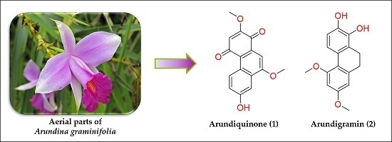
Two New Stilbenoids from the Aerial Parts of Arundina graminifolia (Orchidaceae)

by
Florence Auberon
1,*,
Opeyemi Joshua Olatunji
1,
Stéphanie Krisa
2,
Cyril Antheaume
3,
Gaëtan Herbette
4,
Frédéric Bonté
5,
Jean-Michel Mérillon
2 and
Annelise Lobstein
1
1
Laboratory of Pharmacognosy and Bioactive Natural Products, Faculty of Pharmacy, Strasbourg University, Illkirch-Graffenstaden 67400, France
2
GESVAB Group, Oenology Research Unit, EA 4577, USC 1366 INRA, ISVV, Faculty of Pharmacy, Bordeaux University, Villenave d’Ornon 33140, France
3
Laboratoire Insulaire du Vivant et de l’Environnement, EA 4243, New-Caledonia University, BP R4, Noumea CEDEX 98851, New Caledonia
4
Spectropôle, FR1739, Aix-Marseille University, Campus de St Jerome-Service 511, Marseille 13397, France
5
Louis Vuitton Moët et Hennessy Recherche, 185 avenue de Verdun, St Jean de Braye 45800, France
*
Author to whom correspondence should be addressed.
Molecules 2016, 21(11), 1430; https://doi.org/10.3390/molecules21111430
Submission received: 29 August 2016 / Revised: 21 October 2016 / Accepted: 23 October 2016 / Published: 27 October 2016
(This article belongs to the Section Natural Products Chemistry)

Abstract

:
Two new phenanthrene derivatives, a phenanthrenequinone named arundiquinone (1) and a 9,10-dihydrophenanthrene named arundigramin (2) together with a known lignin dimer (3) and seven known stilbenoids (410) were isolated from the aerial parts of the Asian orchid Arundina graminifolia. The structures of the isolated compounds were elucidated by spectroscopic methods, including extensive 1D, 2D NMR (heteronuclear single quantum coherence (HSQC), heteronuclear multiple-bond correlation spectroscopy (HMBC), and HR-ESI-MS techniques, as well as comparison with respective literature reports. The cytoprotective activity of the isolated compounds were evaluated for their ability to reduce beta amyloid induced toxicity on undifferentiated PC12 cells. Compound 8 showed moderate cytoprotective activity at 0.5 µmol/L (71% of cell viability) while the other compounds showed no significant activity at the highest concentration tested.

Graphical Abstract

1. Introduction

Arundina graminifolia (D. Don) Hochr. (Orchidaceae), known as the bamboo orchid, is an evergreen terrestrial growing orchid and the sole accepted species in the genus Arundina. It is widely distributed in Southeast Asia, from the Himalayas to western Indonesia [1]. The whole plant is mainly used in Chinese traditional Dai medicine as a treatment for blood stasis, food poisoning and as a liver detoxifying agent [2,3,4,5]. It is also used as an antibacterial and emollient in India [6,7] and for the treatment of rheumatism in Bangladesh [8]. Its striking purple flowers are the reason for its harvest for ornamental purposes [9]. Stilbenoids are the major secondary metabolites reported in this orchid based on previous phytochemical studies, revealing an important structural diversity of diphenylethylenes [10,11,12,13,14], bibenzyls [15,16,17,18], phenanthrenes [19], 9,10-dihydrophenanthrene derivatives [20] and other phenolic compounds [21,22,23,24,25,26], and thus could be considered as potential chemotaxonomic markers for the genus. These metabolites are also known to display a wide range of biological activities such as antioxidant, antiviral, cytotoxic and antitumoral properties [27,28,29,30,31,32].
As part of our continuing efforts in contributing to the phytochemical and biological evaluation of tropical orchids [33,34,35], the ethyl acetate extract of the aerial parts of A. graminifolia collected from Chiang Mai Province (Thailand) was screened for its neuroprotective activity against beta amyloid (βA) induced cytotoxicity on PC12 cells and showed promising results. Based on the aforementioned preliminary screening, the extensive investigation on the chemical entities in the plant was pursued. Two new constituents, 1–4 phenanthrenequinone (1) and 9,10-dihydrophenanthrene (2) along with a known lignan dimer (3) and seven known stilbenoids (410) were isolated and characterized. The cytoprotective activity of the compounds was then assessed to establish whether or not it is responsible for the cytoprotective activity of the tested extract.

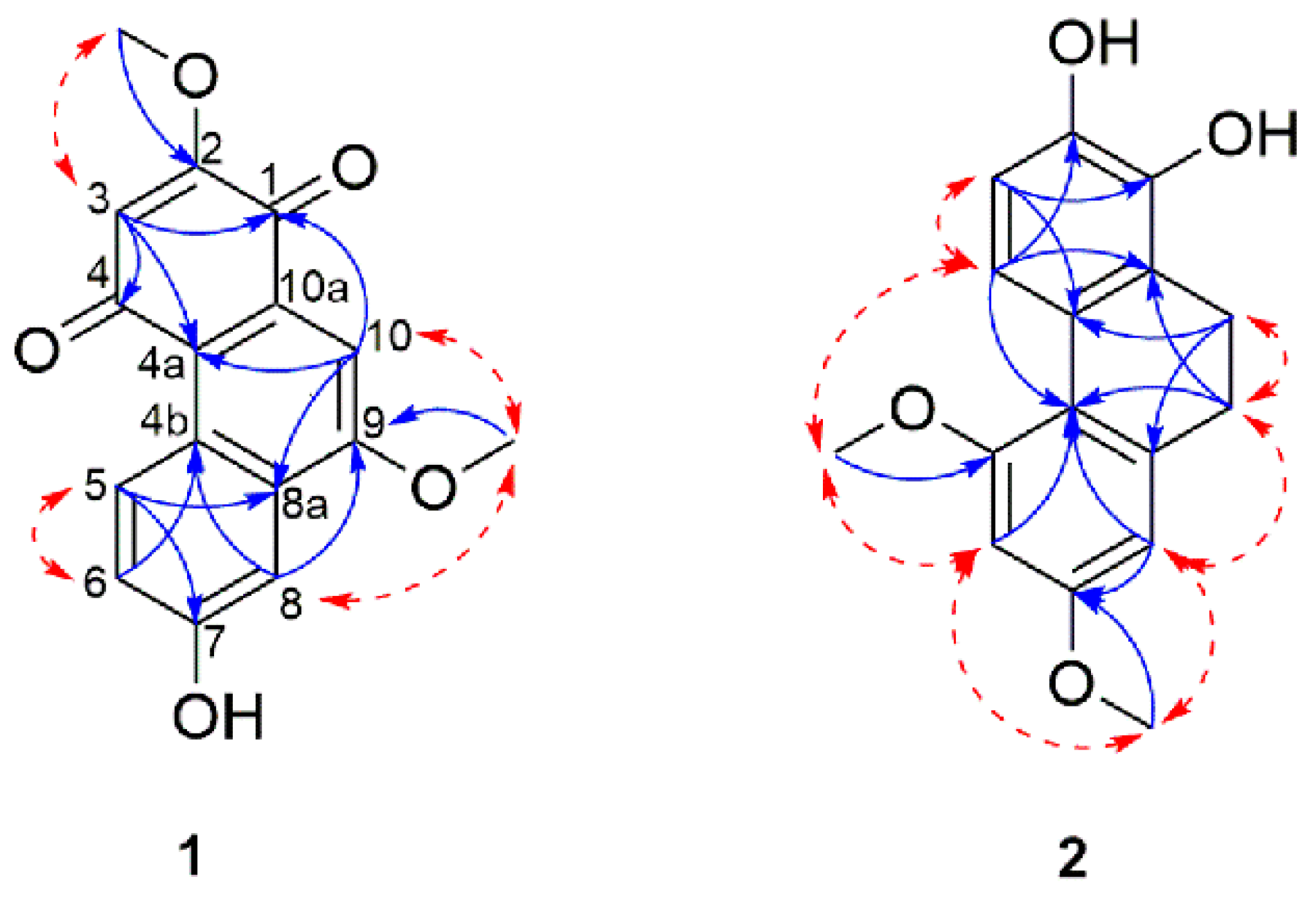
2. Results and Discussion

The ethyl acetate (EtOAc) extract of the aerial parts of A. graminifolia was subjected to a series of chromatographic techniques (silica gel, Sephadex LH-20, Sigma Aldrich, Saint-Louis, MO, USA), and semi-preparative RP-HPLC) to afford two new stilbenoids: arundiquinone and arundigramin (1 and 2), as well as eight known compounds that were identified by comparison of their spectroscopic data to previously published reports as rac-syringaresinol (3) [36,37], orchinol (4) [38], ephemeranthoquinone (5) [39], densiflorol B (6) [40], coelonin (7) [41], lusianthridin (8) [42], batatasin III (9) [43], and flavanthrin (10) [44], (Figure 1). It is noteworthy to add that, besides the two new isolated stilbenoids (1 and 2), compound 3 syringaresinol is herein reported as its first occurrence in A. graminifolia.
Compound 1 was obtained as a red amorphous powder. Its molecular formula was determined to be C16H12O5 based on the molecular ion peak at m/z 285.0760 [M + H]+ (calcd for C16H13O5, 285.0758) as observed in the HR-ESI-MS, which corresponds to eleven degrees of unsaturation. The UV spectrum of 1 showed absorption maxima at 213, 247, 301, 310, 390 and 498 nm, which were similar to those of the phenanthrenequinones [45,46]. The phenolic and quinone moiety in compound 1 was also supported by the IR absorptions at 3292 cm−1 (hydroxyl), 1670 cm−1 (carbonyl) and 1605, 1585, 843, and 797 cm−1 (aromatic ring). The 13C-NMR and HSQC spectra revealed the presence of 16 carbon resonances including nine quaternary carbons, four methines and two methoxy groups. Among the nine quaternary carbons, two were carbonyl carbons according to their chemical shifts at δC 181.4 (C-1) and 188.5 (C-4). The 1H-NMR spectrum of 1 (Table 1) exhibited the presence of an ABX spin system as observed in the aromatic protons at δH 9.50 (1H, d, J = 9.4 Hz, H-5), 7.36 (1H, dd, J = 9.4, 2.3 Hz, H-6) and 7.63 (1H, d, J = 2.3 Hz, H-8), which indicated the presence of a tri-substituted aromatic ring. Additional signals belonging to two isolated aromatic protons at δH 6.10 (1H, s, H-3) and 7.42 (1H, s, H-10), and two methoxy at δH 3.91 (3H, s, 2-OCH3) and 4.15 (3H, s, 9-OCH3) were also noticed. The HMBC cross peaks (T) were observed from H-3 to C-1, C-2, C-4 and C-4a; H-5 to C-4a, C-6, C-7 and C-8a; H-6 to C-4b and C-8; H-8 to C-4b, C-6 and C-9 and H-10 to C-1, C-4a, C-8a and C-9. On the basis of the above evidence, the skeleton of 1 was confirmed as a phenanthrene-1,4-dione. The positions of the methoxy and hydroxyl groups were established by HMBC and NOESY (Nuclear Overhauser Effect Spectroscopy) correlations. HMBC cross peaks were observed between 2-OCH3 to C-2 and 9-OCH3 to C-9 indicating the position of the two methoxy groups on C-2 and C-9. This was further supported by NOESY correlations from 2-OCH3 to H-3, and 9-OCH3 to H-8 and H-10. The remaining substituent, which is the hydroxyl group, was thus substituting on C-7. This was supported by the characteristic chemical shift on an oxygenated carbon (δC 158.7). The structure of compound 1 was established as 7-hydroxy-2,9-dimethoxy-1,4-phenanthrenequinone named arundiquinone. The 1H-NMR, 13C-NMR, HSQC, HMBC, NOESY and HRESIMS spectrum of compounds (1) and (2) are available at Figures S1–S12.
Compound 2 was obtained as a pale rose amorphous solid. The HR-ESI-MS [M + H]+ at m/z 273.1138 supported the molecular formula of C16H16O4 (calcd for C16H17O4, 273.1090), which indicated nine degrees of unsaturation. The UV spectrum exhibited three maxima at 219, 282 and 307 nm, which were similar to 9,10-dihydrophenanthrene derivatives [47,48]. The IR spectrum showed broad absorption bands at 3366 cm−1 for hydroxyl groups and 1588, 163, 1063, 797 cm−1 for aromatic rings. Analysis of the 13C-NMR and HSQC spectra revealed the presence of eight quaternary carbons, four aromatic methines carbons, two methoxy carbons and two methylene carbons. The 1H-NMR data (Table 1) showed signals belonging to a pair of ortho-coupled protons at δH 6.70 (1H, d, J = 7.3 Hz, H-3) and 7.65 (1H, d, J = 7.3 Hz, H-4), two meta-coupled aromatic protons at δH 6.51 (1H, d, J = 2.4 Hz, H-6) and 6.47 (1H, d, J = 2.4 Hz, H-8), two methylenes at δH 2.67 (2H, m, H-9) and 2.74 (2H, m, H-10) and two methoxy groups δH 3.85 (3H, s, 5-OCH3) and 3.81 (3H, s, 7-OCH3). The HMBC correlations (Figure 2) of the methoxy at δH 3.85 to C-5 and δH 3.81 to C-7 confirmed that the methoxy groups were located on C-5 and C-7, respectively. This was also supported by NOESY correlations from 5-OCH3 to H-4 and H-6 as well as from 7-OCH3 with H-6 and H-8. The remaining two hydroxyl groups were thus assigned to C-1 and C-2 with the help of the HMBC cross peaks correlations between H-3 and H-4, respectively, to the last two oxygenated quaternary carbons C-1 and C-2. Thus, the structure of compound 2 was defined as 5,7-dimethoxy-9,10-dihydrophenanthrene-1,2-diol, named arundigramin.
Cell viability, expressed as a percentage relative to the untreated control cells, decreased by more than 60% after exposure to βA25–35 alone. The EtOAc extract of A. graminifolia aerial parts showed promising cytoprotective activity against βA induced cytotoxicity on undifferentiated PC12 cells (86% cell viability at 75 mg/L) (Figure 3). Despite this effect, the isolated compounds (110) were tested for their potential cytoprotective effect. Only compound 8 displayed moderate cytoprotective activity at 0.5 µmol/L, and the other compounds did not exhibit any significant effect at the tested concentrations (Figure 4). Furthermore, compounds 1, 2, 3, 5 and 7 were cytotoxic to the cells at the highest tested concentration (50 µmol/L).

3. Experimental Section

3.1. General Experimental Procedures

Optical rotations were measured with a Perkin Elmer 341 polarimeter (Perkin-Elmer Inc., Waltham, MA, USA). UV spectra were recorded on a Shimadzu UV-2401 PC spectrometer (Shimadzu, Kyoto, Japan). IR spectra were obtained on a 380 FT-IR spectrophotometer (Thermo Fisher Scientific, Waltham, MA, USA). The 1D and 2D NMR spectra were performed on a 500 MHz proton operating system on a Bruker Avance III spectrometer (Bruker BioSpin, Rheinstetten, Germany) Acetone-d6 (Euriso-Top, Saint-Aubin, France) was used as deuterated solvent and its protonated residual signal was used as internal standard at 2.05 ppm relative to TMS. The HR-ESI-MS analyses were performed on an HPLC-DAD/UV-MS Agilent 1200 Series coupled to a 6520 Q-ToF mass spectrometer (Agilent Technologies, Santa Clara, CA, USA). The acquisition of mass spectra was conducted in ESI positive ion mode. A Varian LC-920 HPLC-DAD/UV system (Varian Inc., Palo-Alto, CA, USA) equipped with a Kinetex XB-C18 column (100 mm× 3.0 mm i.d, 2.6 µm) (Phenomenex, Torrance, CA, USA) was used for HPLC-DAD/UV analysis. The prepacked Solid Phase Extraction (SPE) Chromabond cartridge (SiOH, 50 g/150 mL) was purchased from Macherey–Nagel (Macherey–Nagel, Düren, Germany), and SPE fractions were monitored by TLC. The spots were visualized either under UV light (254 nm) and under visible light after heating the plates sprayed with 2% sulfuric vanillin reagent. Sephadex LH-20 (Sigma Aldrich) was used for gel chromatography eluting with methanol. Semi-preparative RP-HPLC experiments were conducted on a Gilson LC system (Gilson Inc., Limburg an der Lahn, Germany) equipped with a semi preparative Kinetex Axia C-18 Column (100 mm × 21.2 mm i.d, 5 µm) (Phenomenex, Torrance, CA, USA). Analytical TLC plates were carried out on pre-coated alumina silica gel 60F254 plates (0.25 mm thickness) (Merck, Darmstadt, Germany). Analytical grade solvents of HPLC quality were purchased from Sigma Aldrich.

3.2. Plant Material

The dried aerial parts (stems and leaves) of A. graminifolia (D. Don) Hochr. (Orchidaceae) of flowering specimens were purchased in September 2010 at Joe’s Orchid Farm in Chiang Mai Province, Thailand and imported to France in compliance with the Convention on International Trade of Endangered Species (CITES). A voucher specimen (No. 05-563) was deposited at the herbarium of the Faculty of Science Chiang Mai University, Chiang Mai, Thailand.

3.3. Extraction and Isolation

The air-dried powder of the aerial parts of A. graminifolia (100 g) was subjected to successive extraction using cyclohexane, EtOAc and CH3OH. Each extraction was performed by maceration for 30 min followed by sonicating in an ultrasonic bath for 10 min at room temperature (1 g raw material per 15 mL of organic solvent) and filtered. Extractions were repeated three times, and the filtrates were combined and evaporated under reduced pressure to afford cyclohexane extract (0.61 g), EtOAc extract (2.49 g) and CH3OH extract (11.01 g).
The EtOAc extract (2.49 g) was subjected to a normal phase SPE cartridge (50 g/150 mL) eluting with CHCl3/EtOAc (100:0 to 0:100) and EtOAc/CH3OH (100:0 to 0:100) to afford 26 fractions (A to Z). Fraction C (67 mg) was subjected to Sephadex LH-20 using CH3OH as eluent to give compound 3 (20 mg). Fraction D (29.5 mg) was purified using a semi-preparative RP-HPLC-DAD system (55% (B) for five min, 55%–70% (B) for 11 min, 70% (B) for two min, 70%–100% (B) for two min, 100% (B) for five min, with solvent B (CH3OH + 0.05% Formic Acid) and solvent A (water + 0.05% Formic Acid), flow rate 28 mL/min, UV monitoring at λ = 280 nm) to obtain compounds 1 (0.8 mg), 2 (1.9 mg), 3 (1.3 mg), 4 (1.3 mg), 5 (1.2 mg) and 6 (1.3 mg). Fraction F (70 mg) was also purified using the same semi-preparative HPLC method stated above to obtain compounds 7 (4.9 mg), 8 (1.7 mg) and 9 (0.8 mg). Fraction G (41 mg) was subjected to Sephadex LH-20 (CH3OH) to afford compound 10 (1.3 mg).

3.4. Compound Characterization

Arundigramin (1). Red amorphous powder (1.2 mg); UV (CH3OH) λmax (log ε): 213 (3.77), 247 (3.53), 301 (3.42), 310 (3.45), 390 (2.56); 498 (2.55); IR (FT-IR) νmax: 3292, 2925, 2852, 1670, 1605, 1465, 1353, 1285, 1243, 1110, 1078, 1019, 843, 797 cm−1; 1H-NMR and 13C-NMR see Table 1; HR-ESI-MS: m/z 285.0759 [M + H]+ (calcd. C16H13O5 for 285.0757).
Arundigramin (2). Rose pale amorphous powder (1.9 mg); UV (CH3OH) λmax (log ε): 219 (4.02), 282 (4.31), 307 (3.93); IR (FT-IR) νmax: 3366, 2923, 1588, 1453, 1258, 1156, 1063 and 797 cm−1; 1H-NMR and 13C-NMR: see Table 1; HR-ESI-MS m/z 273.1138 [M + H]+ (calcd. for C16H17O4 273.1049).

3.5. Cytoprotective Assay

3.5.1. Chemical and Reagents

Dimethyl sulfoxide (DMSO), Dulbecco’s modified eagle’s medium (DMEM-Glutamax), thiazolyl blue tetrazolium bromide (MTT), fetal horse serum and fetal bovine serum were purchased from Sigma-Aldrich (Steinheim, Germany). Rat pheochromocytoma cells (PC12 cells) were supplied from the American Type Culture Collection (ATCC®, Manassas, VA, USA) and Amyloid beta-protein 25–35 (βA25–35) by Synvec (Bordeaux, France).

3.5.2. Cytoprotective Protocol

Undifferentiated PC12 cells were grown in DMEM-Glutamax supplemented with 100 IU/mL of penicillin, 100 µg/mL of streptomycin, 15% fetal horse serum, and 2.5% fetal bovine serum at 37 °C in a humidified atmosphere of 5% CO2. Cells were seeded at a density of 2 × 10⁴ cells/well in 96-well culture plates. After 24 h, cells were incubated with the EtOAc extract (10, 25, 50, 75 and 100 mg/L) and compounds 110 were screened at a concentration of 0.5, 5 and 50 µmol/L in the presence or absence of βA25–35 (5 µM). The extract and compounds were dissolved in DMSO at a final concentration of 0.1%.
The cell viability was determined by the colorimetric MTT reduction assay. After treatment (24 h), PC12 cells were incubated with 0.5 mg/mL (DMEM) of MTT for 3 h at 37 °C. The resulting dark blue formazan crystals were dissolved with 100 µL of DMSO. Absorbance values were read at 540 nm on a microplate reader (MRX Dynex, Dynex Technologies, Denkendorf, Germany) [49]. Cell viability was expressed as a percentage of control cells at 100% viability.
Statistical analysis was performed using GraphPad Prism (version 7.0, GraphPad Software Inc. San Diego, CA, USA). All data are expressed as mean ± SD. Data were analyzed using one-way analysis of variance (ANOVA) followed by post hoc analysis using Dunnett’s multiple test. Differences were considered significant at p < 0.05.

4. Conclusions

Two new phenanthrene derivatives, arundiquinone (1) and arundigramin (2) together with eight known compounds (410) were isolated from the EtOAc extract of A. graminifolia aerial parts, a well-studied Asian orchid. The structures of the new phenanthrenes were elucidated by means of NMR and HR-ESI-MS, as well as comparisons with previous literature reports. Compound 3 was isolated here from A. graminifolia for the first time, whereas the presence of compounds 410 was already signaled in this species. Cytoprotective activity of the isolated compounds was evaluated on their ability to reduce beta amyloid induced toxicity on undifferentiated PC12 cells; however, only compound 8 showed moderate activity while the other isolated compounds did not display any significant activity at the tested concentrations. The EtOAc extract is particularly rich and contains more than 40 compounds based on the HPLC-DAD/UV profiling of this extract and only ten compounds have been isolated and tested. We thus envisage that some minor components that we have yet to isolate may have contributed to the potent bioactivity of the EtOAc extract. Further experiments geared towards identifying these minor components that may be responsible for the displayed cytoprotective activity in the EtOAc extract is in progress.

Supplementary Materials

Supplementary materials can be accessed at: https://www.mdpi.com/1420-3049/21/11/1430/s1.

Acknowledgments

This study was financially supported by the French Ministry of Research and Education (Paris, France) and also by LVMH Recherche and Guerlain (Saint-Jean de Braye, France).

Author Contributions

Florence Auberon performed the extraction, isolation and structural identification of the compounds. Florence Auberon and Opeyemi J. Olatunji wrote this paper. Florence Auberon and Stéphanie Krisa were in charge of the biological evaluation. In addition, Cyril Antheaume and Gaëtan Herbette provided significant advice regarding the NMR structural elucidation. Frédéric Bonté contributed to the plant material selection and its importation. Annelise Lobstein and Jean-Michel Mérillon coordinated the project for the phytochemical and biological parts, respectively.

Conflicts of Interest

The authors declare no conflict of interest.

References

  1. Hong, D.Y.; Lian, Y.S.; Shen, L.D. Orchidaceae. In Flora of China; Chinese Science Press: Beijing, China, 1983; Volume 73, p. 320. [Google Scholar]
  2. Liu, M.; Ding, Y.; Du, L. Chemical components of traditional Dai medicine Arundina graminifolia (D. Don) Hochr. Zhong Cao Yao 2007, 38, 676–677. [Google Scholar]
  3. Hossain, M.M. Therapeutic orchids: Traditional uses and recent advances—An overview. Fitoterapia 2011, 82, 102–140. [Google Scholar] [CrossRef] [PubMed]
  4. Zhang, C.; Chongsuvivatwong, V.; Keawpradub, N.; Lin, Y. Analysis of prescription database extracted from standard textbooks of traditional Dai medicine. J. Ethnobiol. Ethnomed. 2012, 8, 34. [Google Scholar] [CrossRef] [PubMed]
  5. Xiaohua, D.; Jin, Z.; Hui, W.; Haifeng, C.; Chao, Z.; Zepu, Y. Effect of Yajieshaba, a preparation of Dai indigenous medicine, on enhanced liver detoxification. J. Tradit. Chin. Med. 2015, 35, 197–205. [Google Scholar] [CrossRef]
  6. Kumar, S. Arundina. In The Medicinal Plants of North-East India; Scientific Publishers: Judhpur, India, 2002; p. 212. [Google Scholar]
  7. Panda, A.K.; Mandal, D. The folklore medicinal orchids of Sikkim. Anc. Sci. Life 2013, 33, 92–96. [Google Scholar] [CrossRef] [PubMed]
  8. Hossain, M.M. Traditional therapeutic uses of some indigenous orchids of Bangladesh. Med. Aromat. Plant. Sci. Biotechnol. 2009, 42, 101–106. [Google Scholar]
  9. Hegde, S. Orchid wealth of India. Proc. Indian Natl. Sci. Acad. B 1997, 63, 229–244. [Google Scholar]
  10. Li, Y.-K.; Zhou, B.; Ye, Y.-Q.; Du, G.; Niu, D.-Y.; Meng, C.-Y.; Gao, X.-M.; Hu, Q.-F. Two new diphenylethylenes from Arundina graminifolia and their cytotoxicity. Bull. Korean Chem. Soc. 2013, 34, 3257–3260. [Google Scholar] [CrossRef]
  11. Hu, Q.-F.; Zhou, B.; Ye, Y.-Q.; Jiang, Z.-Y.; Huang, X.-Z.; Li, Y.-K.; Du, G.; Yang, G.-Y.; Gao, X.-M. Cytotoxic deoxybenzoins and diphenylethylenes from Arundina graminifolia. J. Nat. Prod. 2013, 76, 1854–1859. [Google Scholar] [CrossRef] [PubMed]
  12. Gao, Y.; Jin, Y.; Yang, S.; Wu, J.; Gao, X.; Hu, Q.; Ma, Y. A new diphenylethylene from Arundina graminifolia and its cytotoxicity. Asian J. Chem. 2014, 26, 3903–3905. [Google Scholar]
  13. Meng, C.-Y.; Niu, D.-Y.; Li, Y.-K.; Zhou, B.; Ye, Y.-Q.; Du, G.; Hu, Q.-F.; Gao, X.-M. A new cytotoxic stilbenoid from Arundina graminifolia. Asian J. Chem. 2014, 26, 2411–2413. [Google Scholar]
  14. Yang, J.X.; Wang, H.; Lou, J.; Li, L.; Liu, G.Y.; Gao, X.; Hu, Q.; Ye, Y. A new cytotoxic diphenylethylene from Arundina graminifolia. Asian J. Chem. 2014, 26, 4517–4518. [Google Scholar]
  15. Majumder, P.L.; Ghosal, S. Two stilbenoids from the orchid Arundina bambusifolia. Phytochemistry 1993, 32, 439–444. [Google Scholar] [CrossRef]
  16. Liu, M.F.; Han, Y.; Xing, D.M.; Shi, Y.; Xu, L.Z.; Du, L.J.; Ding, Y. A new stilbenoid from Arundina graminifolia. J. Asian Nat. Prod. Res. 2004, 6, 229–232. [Google Scholar] [CrossRef] [PubMed]
  17. Liu, M.F.; Han, Y.; Xing, D.M.; Wang, W.; Xu, L.Z.; Du, L.J.; Ding, Y. Chemical constituents from the rhizoma of Arundina graminifolia. Zhongguo Zhong Yao Za Zhi 2004, 29, 147–149. [Google Scholar] [PubMed]
  18. Du, G.; Shen, Y.; Yang, L.; Shu, L.; Wen, M.-L.; Hu, Q.-F. Bibenzyl derivatives of Arundina graminifolia and their cytotoxicity. Chem. Nat. Compd. 2014, 49, 1019–1022. [Google Scholar] [CrossRef]
  19. Liu, M.F.; Ding, Y.; Zhang, D.M. Phenanthrene constituents from rhizome of Arundina graminifolia. Zhongguo Zhong Yao Za Zhi 2005, 30, 353–356. [Google Scholar] [PubMed]
  20. Liu, M.F.; Han, Y.; Xing, D.M.; Wang, W.; Xu, L.Z.; Du, L.J.; Ding, Y. One new benzyldihydrophenanthrene from Arundina graminifolia. J. Asian Nat. Prod. Res. 2005, 7, 767–770. [Google Scholar] [CrossRef] [PubMed]
  21. Gao, X.; Yang, L.; Shen, Y.; Shu, L.; Li, X.; Hu, Q.-F. Phenolic compounds from Arundina graminifolia and their anti-tobacco mosaic virus activity. Bull. Korean Chem. Soc. 2012, 33, 2447–2449. [Google Scholar] [CrossRef]
  22. Niu, D.-Y.; Han, J.-M.; Kong, W.-S.; Cui, Z.-W.; Hu, Q.-F.; Gao, X.-M. Antiviral fluorenone derivatives from Arundina graminifolia. Asian J. Chem. 2013, 25, 9514–9516. [Google Scholar]
  23. Hu, Q.F.; Zhou, B.; Huang, J.M.; Gao, X.M.; Shu, L.D.; Yang, G.Y.; Che, C.T. Antiviral phenolic compounds from Arundina graminifolia. J. Nat. Prod. 2013, 76, 292–296. [Google Scholar] [CrossRef] [PubMed]
  24. Li, Y.; Yang, L.; Shu, L.; Shen, Y.; Hu, Q.; Xia, Z. Flavonoid compounds from Arundina graminifolia. Asian J. Chem. 2013, 25, 4922–4924. [Google Scholar]
  25. Shu, L.; Shen, Y.; Yang, L.; Gao, X.; Hu, Q.-F. Flavonoids derivatives from Arundina graminifolia and their cytotoxicity. Asian J. Chem. 2013, 25, 8358–8360. [Google Scholar]
  26. Li, L.; Xu, W.-X.; Liu, C.-B.; Zhang, C.-M.; Zhao, W.; Shang, S.-Z.; Deng, L.; Guo, Y.-D. A new antiviral phenolic compounds from Arundina graminifolia. Asian J. Chem. 2015, 27, 3525–3526. [Google Scholar] [CrossRef]
  27. Kong, J.-M.; Goh, N.-K.; Chia, L.-S.; Chia, T.-F. Recent advances in traditional plant drugs and orchids. Acta Pharmacol. Sin. 2003, 24, 7–21. [Google Scholar] [PubMed]
  28. Chen, L.; Chen, J.-B. Pharmacological research of stilbenoids. Guangdong Yaoxue 2005, 15, 84–86. [Google Scholar]
  29. Xiao, K.; Zhang, H.-J.; Xuan, L.-J.; Zhang, J.; Xu, Y.-M.; Bai, D.-L. Stilbenoids: Chemistry and bioactivities. Stud. Nat. Prod. Chem. 2008, 34, 453–646. [Google Scholar]
  30. Kovacs, A.; Vasas, A.; Hohmann, J. Natural phenanthrenes and their biological activity. Phytochemistry 2008, 69, 1084–1110. [Google Scholar] [CrossRef] [PubMed]
  31. Tzakou, O. Naturally occurring stilbenoids and their biological activity. Pharmakeutike 2009, 22, 132–141. [Google Scholar]
  32. Williams, R.B.; Martin, S.M.; Hu, J.F.; Garo, E.; Rice, S.M.; Norman, V.L.; Lawrence, J.A.; Hough, G.W.; Goering, M.G.; O’Neil-Johnson, M.; et al. Isolation of apoptosis-inducing stilbenoids from four members of the Orchidaceae family. Planta Med. 2012, 78, 160–165. [Google Scholar] [CrossRef] [PubMed]
  33. Simmler, C.; Antheaume, C.; Lobstein, A. Antioxidant biomarkers from Vanda coerulea stems reduce irradiated HaCaT PGE-2 production as a result of COX-2 inhibition. PLoS ONE 2010, 5, e13713. [Google Scholar] [CrossRef] [PubMed]
  34. Simmler, C.; Antheaume, C.; Andre, P.; Bonte, F.; Lobstein, A. Glucosyloxybenzyl eucomate derivatives from Vanda teres stimulate hacat cytochrome c oxidase. J. Nat. Prod. 2011, 74, 949–955. [Google Scholar] [CrossRef] [PubMed]
  35. Cakova, V.; Urbain, A.; Antheaume, C.; Rimlinger, N.; Wehrung, P.; Bonte, F.; Lobstein, A. Identification of phenanthrene derivatives in Aerides rosea (Orchidaceae) using the combined systems HPLC-ESI-HRMS/MS and HPLC-DAD-MS-SPE-UV-NMR. Phytochem. Anal. 2015, 26, 34–39. [Google Scholar] [CrossRef] [PubMed]
  36. Freudenberg, K.; Dietrich, H. Syringaresinol, a dehydrogenation product of sinapinyl alcohol. Chem. Ber. 1953, 86, 4–10. [Google Scholar] [CrossRef]
  37. Cai, X.F.; Lee, I.S.; Dat, N.T.; Shen, G.; Kang, J.S.; Kim, D.H.; Kim, Y.H. Inhibitory lignans against NFAT transcription factor from Acanthopanax koreanum. Arch. Pharm. Res. 2004, 27, 738–741. [Google Scholar] [CrossRef] [PubMed]
  38. Fisch, M.H.; Flick, B.H.; Arditti, J. Structure and antifungal activity of hircinol, loroglossol and orchinol. Phytochemistry 1973, 12, 437–441. [Google Scholar] [CrossRef]
  39. Tezuka, Y.; Hirano, H.; Kikuchi, T.; Xu, G. Constituents of orchidaceous plants. X. Constituents of Ephemerantha lonchophylla; isolation and structure elucidation of new phenolic compounds, ephemeranthol-A, ephemeranthol-B, and ephemeranthoquinone, and of a new diterpene glucoside, ephemeranthoside. Chem. Pharm. Bull. 1991, 39, 593–598. [Google Scholar] [CrossRef]
  40. Fan, C.Q.; Zhao, W.M.; Qin, G.W. New bibenzyl and phenanthrenedione from Dendrobium densiflorum. Chin. Chem. Lett. 2000, 11, 705–706. [Google Scholar]
  41. Majumder, P.; Laha, S.; Datta, N. Coelonin, a 9,10-dihydrophenanthrene from the orchids Coelogyne ochracea and C. elata. Phytochemistry 1982, 21, 478–480. [Google Scholar] [CrossRef]
  42. Majumder, P.L.; Lahiri, S. Lusianthrin and lusianthridin, two stilbenoids from the orchid Lusia indivisa. Phytochemistry 1990, 29, 621–624. [Google Scholar] [CrossRef]
  43. Hashimoto, T.; Hasegawa, K.; Yamaguchi, H.; Saito, M.; Ishimoto, S. Structure and synthesis of batatasins, dormancy-inducing substances of yam bulbils. Phytochemistry 1974, 13, 2849–2852. [Google Scholar] [CrossRef]
  44. Majumder, P.L.; Banerjee, S. Structure of flavanthrin, the first dimeric 9,10-dihydrophenanthrene derivative from the orchid Eria flava. Tetrahedron 1988, 44, 7303–7308. [Google Scholar] [CrossRef]
  45. Talapatra, B.; Mukhopadhyay, P.; Chaudhury, P.; Talapatra, S.K. Denbinobin, a new phenanthraquinone from Dendrobium Nobile Lindl (Orchidaceae). Indian J. Chem., Sect. B 1982, 21B, 386–387. [Google Scholar]
  46. Wu, T.S.; Jong, T.T.; Tien, H.J.; Kuoh, C.S.; Furukawa, H.; Lee, K.H. Annoquinone a, an antimicrobial and cytotoxic principle from Annona montana. Phytochemistry 1987, 26, 1623–1625. [Google Scholar] [CrossRef]
  47. Harborne, J.B.; Smith, T.A.; Majumder, P.L.; Pal, S. Rotundatin, a new 9,10-didydrophenanthrene derivative from Dendrobium rotundatum. Phytochemistry 1992, 31, 3225–3228. [Google Scholar]
  48. Majumder, P.L.; Sen, S.; Majumder, S. Phenanthrene derivatives from the orchid Coelogyne cristata. Phytochemistry 2001, 58, 581–586. [Google Scholar] [CrossRef]
  49. Papastamoulis, Y.; Richard, T.; Nassra, M.; Badoc, A.; Krisa, S.; Harakat, D.; Monti, J.-P.; Merillon, J.-M.; Waffo-Teguo, P. Viniphenol A, a complex resveratrol hexamer from Vitis vinifera stalks: Structural elucidation and protective effects against amyloid-β-induced toxicity in PC12 cells. J. Nat. Prod. 2014, 77, 213–217. [Google Scholar] [CrossRef] [PubMed]
  • Sample Availability: Not available.
Figure 1. Chemical structures of compounds 110 isolated from A. graminifolia aerial parts.
Molecules 21 01430 g001
Figure 2. NOESY (red dashed arrows) and HMBC (blue arrows) correlations of compounds 1 and 2.
Molecules 21 01430 g002
Figure 3. Effect of the EtOAc extract on PC12 cell viability. PC12 cells were incubated with EtOAc extract (10, 25, 50 and 100 mg/L) for 24 h, and then exposure to 5 μM of βA for 24 h. Results are expressed as mean ± SD (n = 4). * p < 0.05 as compared to the βA treated group.
Molecules 21 01430 g003
Figure 4. Effect of compounds 110 on PC12 cell viability. PC12 cells were incubated with compounds 110 (0, 5 and 50 μM) for 24 h, and then exposure to 5 μM of βA for 24 h. Data are expressed as mean ± SD (n = 4). * p < 0.05 as compared to the βA treated group.
Molecules 21 01430 g004
Table 1. NMR spectral data of compounds 1 and 2 (in acetone-d6, 500 MHz for 1H, 125 MHz for 13C).
No.Compound 1Compound 2
δH (J in Hz)δCHMBCδH (J in Hz)δCHMBC
1 181.4 141.9
2 159.3 143.9
36.10 (s)111.61, 2, 4, 4a6.70 (d, 7.3)112.81, 4a
4 188.5 7.65 (d, 7.3)120.82, 3, 4a, 10a
4a 121.8 126.3
4b 126.6 117.8
59.50 (d, 9.4)131.24a, 6, 7, 8a 158.7
67.36 (dd, 9.4, 2.3)122.74b, 86.51 (d, 2.4)98.44b, 5, 7, 8
7 158.4 159.5
87.63 (d, 2.3)105.14b, 6, 96.47 (d, 2.4)106.04b, 6, 7, 9
8a 131.5 141.3
9 158.9 2.67 (m)31.34b, 8, 8a, 10, 10a
107.42 (s)100.41, 4a, 8a, 92.74 (m)22.48a, 9, 10a
10a 131.5 125.6
2-OCH33.91 (s)56.72
5-OCH3 3.85 (s)55.95
7-OCH3 3.81 (s)55.67
9-OCH34.15 (s)56.69

