
The Structure–Antimicrobial Activity Relationships of a Promising Class of the Compounds Containing the N-Arylpiperazine Scaffold †


 ,
,  ,
,  and
and
Abstract
:
1. Introduction
2. Results
2.1. Electronic, Steric and Lipohydrophilic Properties of the Compounds 5a–l
2.2. Biological Assays in vitro
2.2.1. Antimicrobial Susceptibility Testing
2.2.2. Antiproliferative (Cytotoxicity) Screening
2.3. Structure–Activity Relationships
r = 0.375, R2 = 0.141, F = 2.80, RSS = 1.73, Prob > F = 0.125, n = 12
r = 0.265, R2 = 0.070, F = 1.41, RSS = 1.68, Prob > F = 1.413, n = 12
r = 0.221, R2 = 0.049, F = 1.28, RSS = 1.72, Prob > F = 0.323, n = 12
r = 0.488, R2 = 0.238, F = 2.09, RSS = 0.40, Prob > F = 0.219, n = 8
r = 0.986, R2 = 0.973, F = 109.84, RSS = 0.001, Prob > F = 0.009, n = 4
r = 0.686, R2 = 0.471, F = 10.79, RSS = 0.34, Prob > F = 0.008, n = 12
r = 0.765, R2 = 0.585, F = 5.22, RSS = 0.024, Prob > F = 0.150, n = 4.
r = 0.963, R2 = 0.928, F = 46.32, RSS = 0.01, Prob > F = 6 ×10⁻⁴, n = 8
3. Discussion
3.1. Electronic, Steric and Lipohydrophilic Properties of the Compounds 5a–l
3.2. Biological Assays in vitro
3.2.1. Antimicrobial Susceptibility Testing
3.2.2. Antiproliferative (Cytotoxicity) Screening
3.3. Structure–Activity Relationships
4. Materials and Methods
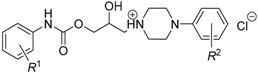
4.1. Tested Compounds
4.2. In silico Investigation
4.2.1. Calculation of Molecular Volume
4.2.2. Prediction of Lipohydrophilic Properties
4.3. Antimicrobial Susceptibility Testing in vitro
4.3.1. Antimycobacterial Evaluation
4.3.2. Antibacterial Evaluation
4.3.3. Candidacidal Evaluation
4.4. Antiproliferative (Cytotoxicity) Screening in vitro
4.4.1. Cell Culture
4.4.2. Analysis of Cell Proliferation and Viability
4.5. Statistical Analysis
5. Conclusions
Supplementary Materials
Acknowledgments
Author Contributions
Conflicts of Interest
Abbreviations
| 5-FC | 5-Flucytosine |
| AARS | Averaged absolute residual sums |
| AMP | Ampicillin |
| Amph. B | Amphotericin B |
| CPX | Ciprofloxacin |
| DMSO | Dimethyl sulfoxide |
| F | Fisher significance ratio |
| INH | Isoniazid |
| log k’ | Capacity (retention) factor |
| log Pexp | Experimentally observed values of a partition coefficient in the octan-1--ol/phosphate buffer (pH = 7.4) system |
| MIC | Minimum inhibitory concentration |
| MV | Molecular volume |
| n | Number of cases |
| PS(s) | Privileged substructure(s) |
| r | Correlation coefficient |
| R2 | Coefficient of determination |
| RIF | Rifampicin |
| RSS | Residual sum of squares |
| SAR | Structure–activity relationship(s) |
References
- Evans, B.E.; Rittle, K.E.; Bock, M.G.; DiPardo, R.M.; Freidinger, R.M.; Whitter, W.L.; Lundell, G.F.; Veber, D.F.; Anderson, P.S.; Chang, R.S.L.; et al. Methods for drug discovery: Development of potent, selective, orally effective cholecystokinin antagonists. J. Med. Chem. 1988, 31, 2235–2246. [Google Scholar] [CrossRef] [PubMed]
- Horton, D.A.; Bourne, G.T.; Smythe, M.L. The combinatorial synthesis of bicyclic privileged structures or privileged substructures. Chem. Rev. 2003, 103, 893–930. [Google Scholar] [CrossRef] [PubMed]
- Panda, G.; Shagufta; Srivastava, A.K.; Sinha, S. Synthesis and antitubercular activity of 2-hydroxy-aminoalkyl derivatives of diaryloxy methano phenanthrenes. Bioorg. Med. Chem. Lett. 2005, 15, 5222–5225. [Google Scholar] [CrossRef] [PubMed]
- Upadhayaya, R.S.; Kulkarni, G.M.; Vasireddy, N.R.; Vandavasi, J.K.; Dixit, S.S.; Sharma, V.; Chattopadhyaya, J. Design, synthesis and biological evaluation of novel triazole, urea and thiourea derivatives of quinoline against Mycobacterium tuberculosis. Bioorg. Med. Chem. 2009, 17, 4681–4692. [Google Scholar] [CrossRef] [PubMed]
- Upadhayaya, R.S.; Vandavasi, J.K.; Kardile, R.A.; Lahore, S.V.; Dixit, S.S.; Deokar, H.S.; Shinde, P.D.; Sarman, M.P.; Chattopadhyaya, J. Novel quinoline and naphthalene derivatives as potent antimycobacterial agents. Eur. J. Med. Chem. 2010, 45, 1854–1867. [Google Scholar] [CrossRef] [PubMed]
- Chai, X.; Zhang, J.; Cao, Y.; Zou, Y.; Wu, Q.; Zhang, D.; Jiang, Y.; Sun, Q. New azoles with antifungal activity: Design, synthesis, and molecular docking. Bioorg. Med. Chem. Lett. 2011, 21, 686–689. [Google Scholar] [CrossRef] [PubMed]
- Che, X.; Sheng, C.; Wang, W.; Cao, Y.; Xu, Y.; Ji, H.; Dong, G.; Miao, Z.; Yao, J.; Zhang, W. New azoles with potent antifungal activity: Design, synthesis and molecular docking. Eur. J. Med. Chem. 2009, 44, 4218–4226. [Google Scholar] [CrossRef] [PubMed]
- Xu, J.; Cao, Y.; Zhang, J.; Yu, S.; Zou, Y.; Chai, X.; Wu, Q.; Zhang, D.; Jiang, Y.; Sun, Q. Design, synthesis and antifungal activities of novel 1,2,4-triazole derivatives. Eur. J. Med. Chem. 2011, 46, 3142–3148. [Google Scholar] [CrossRef] [PubMed]
- Sun, Q.-Y.; Xu, J.-M.; Cao, Y.-B.; Zhang, W.-N.; Wu, Q.-Y.; Zhang, D.-Z.; Zhang, J.; Zhao, H.-Q.; Jiang, Y.-Y. Synthesis of novel triazole derivatives as inhibitors of cytochrome P450 14α-demethylase (CYP51). Eur. J. Med. Chem. 2007, 42, 1226–1233. [Google Scholar] [CrossRef] [PubMed]
- Gan, L.-L.; Fang, B.; Zhou, C.-H. Synthesis of azole-containing piperazine derivatives and evaluation of their antibacterial, antifungal and cytotoxic activities. Bull. Korean Chem. Soc. 2010, 31, 3684–3692. [Google Scholar] [CrossRef]
- Tacon, C.; Guantai, E.M.; Smith, P.J.; Chibale, K. Synthesis, biological evaluation and mechanistic studies of totarol amino alcohol derivatives as potential antimalarial agents. Bioorg. Med. Chem. 2012, 20, 893–902. [Google Scholar] [CrossRef] [PubMed]
- Parai, M.K.; Panda, G.; Chaturvedi, V.; Manju, Y.K.; Sinha, S. Thiophene containing triarylmethanes as antitubercular agents. Bioorg. Med. Chem. Lett. 2008, 18, 289–292. [Google Scholar] [CrossRef] [PubMed]
- Stefańska, J.; Bielenica, A.; Struga, M.; Tyski, S.; Kossakowski, J.; Loddo, R.; Ibba, C.; Collu, D.; Marongiu, E.; La Colla, P. Biological evaluation of 10-(diphenylmethylene)-4-azatricyclo[5.2.1.02,6]dec-8-ene-3,5-dione derivatives. Cent. Eur. J. Biol. 2009, 4, 362–368. [Google Scholar] [CrossRef]
- Bohnert, J.A.; Kern, W.V. Selected arylpiperazines are capable of reversing multidrug resistance in Escherichia coli overexpressing RND efflux pumps. Antimicrob. Agents Chemother. 2005, 49, 849–852. [Google Scholar] [CrossRef] [PubMed]
- Li, L.; Li, Z.; Guo, N.; Jin, J.; Du, R.; Liang, J.; Wu, X.; Wang, X.; Liu, M.; Jin, Q.; et al. Synergistic activity of 1-(1-naphthylmethyl)piperazine with ciprofloxacin against clinically resistant Staphylococcus aureus, as determined by different methods. Lett. Appl. Microbiol. 2011, 52, 372–378. [Google Scholar] [CrossRef] [PubMed]
- Handzlik, J.; Szymańska, E.; Chevalier, J.; Otrębska, E.; Kieć-Kononowicz, K.; Pagès, J.M.; Alibert, S. Amine-alkyl derivatives of hydantoin: New tool to combat resistant bacteria. Eur. J. Med. Chem. 2011, 46, 5807–5816. [Google Scholar] [CrossRef] [PubMed]
- Dymek, A.; Armada, A.; Handzlik, J.; Viveiros, M.; Spengler, G.; Molnar, J.; Kieć-Kononowicz, K.; Amaral, L. The activity of 16 new hydantoin compounds on the intrinsic and overexpressed efflux pump system of Staphylococcus aureus. In Vivo 2012, 26, 223–229. [Google Scholar] [PubMed]
- Colabufo, N.A.; Berardi, F.; Perrone, M.G.; Cantore, M.; Contino, M.; Inglese, C.; Niso, M.; Perrone, R. Multi-drug-resistance-reverting agents: 2-aryloxazole and 2-arylthiazole derivatives as potent BCRP or MRP1 inhibitors. ChemMedChem 2009, 4, 188–195. [Google Scholar] [CrossRef] [PubMed]
- Malík, I. Drug-like properties of some esters of ortho-/meta-/para-alkoxyphenylcarbamic acid containing N-phenylpiperazine fragment. GJMR-B 2013, 13, 23–27. [Google Scholar]
- Kirk, M. Fluorination in medicinal chemistry: Methods, strategies, and recent developments. Org. Process Res. Dev. 2008, 12, 305–321. [Google Scholar] [CrossRef]
- Foley, T.L.; Rai, G.; Yasgar, A.; Daniel, T.; Baker, H.L.; Attene-Ramos, M.; Kosa, N.M.; Leister, W.; Burkart, M.D.; Jadhav, A.; et al. 4-(3-Chloro-5-(trifluoromethyl)pyridin-2-yl)-N-(4-methoxypyridin-2-yl)piperazine-1-carbothioamide (ML267), a potent inhibitor of bacterial phosphopantetheinyl transferase that attenuates secondary metabolism and thwarts bacterial growth. J. Med. Chem. 2014, 57, 1063–1078. [Google Scholar] [CrossRef] [PubMed]
- Patel, R.V.; Kumari, P.; Rajani, D.P.; Chikhalia, K.H. Synthesis, characterization and pharmacological activities of 2-[4-cyano-(3-trifluoromethyl)phenylamino)]-4-(4-quinoline/coumarin-4-yloxy)-6-(fluoro-piperazinyl)-s-triazines. J. Fluor. Chem. 2011, 132, 617–627. [Google Scholar] [CrossRef]
- Kubinyi, H. Methods and Principles in Medicinal Chemistry. In QSAR: Hansch Analysis and Related Approaches; Mannhold, R., Krogsgaard-Larsen, P., Timmerman, H., Eds.; Wiley-VCh Verlag: Weinheim, Germany, 1993; pp. 22–56. [Google Scholar]
- Malík, I.; Sedlárová, E.; Čižmárik, J.; Andriamainty, F.; Csöllei, J. Study of physicochemical properties of 2-, 3-, 4-alkoxyphenylcarbamic acid derivatives with a substituted N-phenylpiperazine moiety in the basic part. Čes. Slov. Farm. 2005, 54, 235–239. [Google Scholar]
- Malík, I.; Sedlárová, E.; Čižmárik, J.; Andriamainty, F.; Csöllei, J. Study of physicochemical properties of 4-alkoxyphenylcarbamic acid derivatives with various substituted N-phenylpiperazin-1-yl moiety in the basic part of the molecule. Farm. Obzor 2005, 74, 211–215. [Google Scholar]
- Domagala, J.M. Structure–activity and structure–side-effect relationships for the quinolone antibacterials. J. Antimicrob. Chemother. 1994, 33, 685–706. [Google Scholar] [CrossRef] [PubMed]
- Nieto, M.J.; Alovero, F.L.; Manzo, R.H.; Mazzieri, M.R. Benzenesulfonamide analogs of fluoroquinolones. Antibacterial activity and QSAR studies. Eur. J. Med. Chem. 2005, 40, 361–369. [Google Scholar] [CrossRef] [PubMed]
- Rodrigues, M.O.; Cantos, J.B.; D’Oca, C.R.; Soares, K.L.; Coelho, T.S.; Piovesan, L.A.; Russowsky, D.; da Silva, P.A.; D’Oca, M.G. Synthesis and antimycobacterial activity of isoniazid derivatives from renewable fatty acids. Bioorg. Med. Chem. 2013, 21, 6910–6914. [Google Scholar] [CrossRef] [PubMed]
- Sztanke, K.; Tuzimski, T.; Rzymowska, J.; Pasternak, K.; Kandefer-Szerszeń, M. Synthesis, determination of the lipophilicity, anticancer and antimicrobial properties of some fused 1,2,4-triazole derivatives. Eur. J. Med. Chem. 2008, 43, 404–419. [Google Scholar] [CrossRef] [PubMed]
- Malík, I.; Sedlárová, E.; Csöllei, J.; Andriamainty, F.; Kurfürst, P.; Vančo, J. Synthesis, spectral description, and lipophilicity parameters determination of phenylcarbamic acid derivatives with integrated N-phenylpiperazine moiety in the structure. Chem. Pap. 2006, 60, 42–47. [Google Scholar] [CrossRef]
- Leo, A.J.; Hoekman, D. Calculating log P(oct) with no missing fragments. The problem of estimating new interaction parameters. Perspect. Drug Discov. Des. 2000, 18, 19–38. [Google Scholar] [CrossRef]
- Ghose, A.K.; Crippen, G.M. Atomic physicochemical parameters for three-dimensional structure-directed quantitative structure–activity relationships. I. Partition coefficients as a measure of hydrophobicity. J. Comput. Chem. 1986, 7, 565–577. [Google Scholar] [CrossRef]
- Ghose, A.K.; Crippen, G.M. Atomic physicochemical parameters for three-dimensional structure-directed quantitative structure–activity relationships. II. Modeling dispersive and hydrophobic interactions. J. Chem. Inform. Comput. Sci. 1987, 27, 21–35. [Google Scholar] [CrossRef]
- Wildman, S.A.; Crippen, G.M. Prediction of physicochemical parameters by atomic contributions. J. Chem. Inform. Comput. Sci. 1999, 39, 868–873. [Google Scholar] [CrossRef]
- Viswanadhan, N.V.; Ghose, K.A.; Reyankar, R.G.; Robins, K.R. Atomic physicochemical parameters for three-dimensional structure-directed quantitative structure–activity relationships 4. Additional parameters for hydrophobic and dispersive interactions and their application for an automated superposition of certain naturally occurring nucleoside antibiotics. J. Chem. Inform. Comput. Sci. 1989, 29, 163–172. [Google Scholar]
- Broto, P.; Moreau, G.; Vandycke, C. Molecular structures: Perception, autocorrelation descriptor and SAR studies. Autocorrelation descriptor. Eur. J. Med. Chem. Chim. Theor. 1984, 19, 66–70. [Google Scholar]
- Tetko, I.V.; Tanchuk, V.Y. Application of associative neural networks for prediction of lipophilicity in ALOGPs 2.1 program. J. Chem. Inf. Comput. Sci. 2002, 42, 1136–1145. [Google Scholar] [CrossRef] [PubMed]
- Mannhold, R.; Petrauskas, A. Substructure versus whole-molecule approaches for calculating log P. QSAR Comb. Sci. 2003, 22, 466–475. [Google Scholar] [CrossRef]
- Zelová, H.; Hanáková, Z.; Čermáková, Z.; Šmejkal, K.; Dalĺ Acqua, S.; Babula, P.; Cvačka, J.; Hošek, J. Evaluation of anti-inflammatory activity of prenylated substances isolated from Morus alba and Morus nigra. J. Nat. Prod. 2014, 77, 1297–1303. [Google Scholar] [CrossRef] [PubMed]
- Nakagawa, S.; Schielzeth, H. General and simple method for obtaining R2 from generalized linear mixed-effects models. Methods Ecol. Evol. 2013, 4, 133–142. [Google Scholar] [CrossRef]
- Hett, E.C.; Rubin, E.J. Bacterial growth and cell division: A mycobacterial perspective. Microbiol. Mol. Biol. Rev. 2008, 72, 126–156. [Google Scholar] [CrossRef] [PubMed]
- Hadda, T.B.; Srivastava, S.; Das, B.; Salgado-Zamora, H.; Shaheen, U.; Bader, A.; Moazzam Naseer, M. POM analyses of antimicrobial activity of some 2,3-armed 4,5,6,7-tetrahydro-1-benzothiophenes: Favourable and unfavourable physico-chemical parameters in design of antibacterial and mycolytic agents. Med. Chem. Res. 2014, 23, 995–1003. [Google Scholar] [CrossRef]
- Hirayama, M. The antimicrobial activity, hydrophobicity and toxicity of sulfonium compounds, and their relationship. Biocontrol Sci. 2011, 16, 23–31. [Google Scholar] [CrossRef] [PubMed]
- Luo, Y.-L.; Baathulaa, K.; Kannekanti, V.K.; Zhou, C.-H.; Cai, G.-X. Novel benzimidazole derived naphthalimide triazoles: Synthesis, antimicrobial activity and interactions with calf thymus DNA. Sci. China Chem. 2015, 58, 483–494. [Google Scholar] [CrossRef]
- Krátký, M.; Vinšová, J.; Rodriguez, N.G.; Stolaříková, J. Antimycobacterial activity of salicylanilide benzenesulfonates. Molecules 2012, 17, 492–503. [Google Scholar] [CrossRef] [PubMed]
- Talath, S.; Gadad, A.K. Synthesis, antibacterial and antitubercular activities of some 7-[4-(5-amino-[1,3,4]-thiadiazole-2-sulfonyl)-piperazin-1-yl] fluoroquinolonic derivatives. Eur. J. Med. Chem. 2006, 41, 918–924. [Google Scholar] [CrossRef] [PubMed]
- Zhang, Y.-Y.; Mi, J.-L.; Zhou, C.-H.; Zhou, X.-D. Synthesis of novel fluconazoliums and their evaluation for antibacterial and antifungal activities. Eur. J. Med. Chem. 2011, 46, 4391–4402. [Google Scholar] [CrossRef] [PubMed]
- Costa, E.C.; Cassamale, T.B.; Carvalho, D.B.; Bosquiroli, L.S.S.; Ojeda, M.; Ximenes, T.V.; Matos, M.F.C.; Kadri, M.C.T.; Baroni, A.C.M.; Arruda, C.C.P. Antileishmanial activity and structure–activity relationship of triazolic compounds derived from the Neolignans Grandisin, Veraguensin, and Machilin G. Molecules 2016, 21. [Google Scholar] [CrossRef] [PubMed]
- Malík, I.; Sedlárová, E.; Csöllei, J.; Andriamainty, F.; Čižmárik, J.; Kečkéšová, S. The physicochemical properties of dibasic alkyl esters of 2- and 3-alkyloxy substituted phenylcarbamic acid. Acta Facult. Pharm. Univ. Comen. 2007, 54, 136–145. [Google Scholar]
- Minnikin, D.E. Lipids: Complex lipids, their chemistry, biosynthesis and roles. In The Biology of the Mycobacteria; Ratledge, C., Stanford, J.L., Eds.; Academic Press: London, UK, 1982; pp. 95–184. [Google Scholar]
- Guérardel, Y.; Maes, E.; Briken, V.; Chirat, F.; Leroy, Y.; Locht, C.; Strecker, G.; Kremer, L. Lipomannan and lipoarabinomannan from a clinical isolate of Mycobacterium kansasii: Novel structural features and apoptosis-inducing properties. J. Biol. Chem. 2003, 278, 36637–36651. [Google Scholar] [CrossRef] [PubMed]
- Burguière, A.; Hitchen, P.G.; Dover, L.G.; Kremer, L.; Ridell, M.; Alexander, D.C.; Liu, J.; Morris, H.R.; Minnikin, D.E.; Dell, A.; et al. LosA, a key glycosyltransferase involved in the biosynthesis of a novel family of glycosylated acyltrehalose lipooligosaccharides from Mycobacterium marinum. J. Biol. Chem. 2005, 23, 42124–42133. [Google Scholar] [CrossRef] [PubMed]
- Van der Woude, A.D.; Sarkar, D.; Bhatt, A.; Sparrius, M.; Raadsen, S.A.; Boon, L.; Geurtsen, J.; van der Sar, A.M.; Luirink, J.; Houben, E.N.; et al. Unexpected link between lipooligosaccharide biosynthesis and surface protein release in Mycobacterium marinum. J. Biol. Chem. 2012, 287, 20417–20429. [Google Scholar] [CrossRef] [PubMed]
- Waisser, K.; Doležal, R.; Čižmárik, J. Graphic demonstration of the structure–antimycobacterial activity relationships in the series of ester phenylcarbamid acid with piperidine or pyrrolidine moiety. Folia Pharm. Univ. Carol. 2008, 37, 65–76. [Google Scholar]
- Balgavý, P.; Devínsky, F. Cut-off effects in biological activities of surfactants. Adv. Colloid Interface Sci. 1996, 12, 23–63. [Google Scholar] [CrossRef]
- Gonec, T.; Zadrazilova, I.; Nevin, E.; Kauerova, T.; Pesko, M.; Kos, J.; Oravec, M.; Kollar, P.; Coffey, A.; O’Mahony, J.; et al. Synthesis and biological evaluation of N-alkoxyphenyl-3-hydroxynaphthalene-2-carboxanilides. Molecules 2015, 20, 9767–9787. [Google Scholar] [CrossRef] [PubMed]
- Kauerova, T.; Kos, J.; Gonec, T.; Jampilek, J.; Kollar, P. Antiproliferative and pro-apoptotic effect of novel nitro-substituted hydroxynaphthanilides on human cancer cell lines. Int. J. Mol. Sci. 2016, 17. [Google Scholar] [CrossRef] [PubMed]
- Tengler, J.; Kapustíková, I.; Peško, M.; Govender, R.; Keltošová, S.; Mokrý, P.; Kollár, P.; O’Mahony, J.; Coffey, A.; Kráľová, K.; et al. Synthesis and biological evaluation of 2-hydroxy-3-[(2-aryloxyethyl)amino]propyl-4-[(alkoxycarbonyl)amino]benzoates. Sci. World J. 2013, 2013. [Google Scholar] [CrossRef] [PubMed]
- Dolezal, M.; Zitko, J.; Jampilek, J. Pyrazinecarboxylic acid derivatives with antimycobacterial activity. In Understanding Tuberculosis—New Approaches to Fighting Against Drug Resistance; Cardona, P.J., Ed.; InTech: Rijeka, Croatia, 2012; pp. 233–262. [Google Scholar]
- Guillemont, J.; Meyer, C.; Poncelet, A.; Bourdrez, X.; Andries, K. Diarylquinolines, synthesis pathways and quantitative structure–activity relationship studies leading to the discovery of TMC207. Future Med. Chem. 2011, 11, 1345–1360. [Google Scholar] [CrossRef] [PubMed]
- Mengelers, M.J.; Hougee, P.E.; Janssen, L.H.; van Miert, A.S. Structure–activity relationships between antibacterial activities and physicochemical properties of sulfonamides. J. Vet. Pharmacol. Ther. 1997, 20, 276–283. [Google Scholar] [CrossRef] [PubMed]
- Richards, C.D.; Martin, K.; Gregory, S.; Keightley, C.A.; Hesketh, T.R.; Smith, G.A.; Warren, G.B.; Metcalfe, J.C. Degenerate perturbations of protein structure as the mechanism of anaesthetic action. Nature 1978, 276, 775–779. [Google Scholar] [CrossRef] [PubMed]
- Mourad, A.E. Charge transfer complexes of N-arylcarbamates with π-acceptors. Z. Naturforschung 1987, 42, 284–288. [Google Scholar] [CrossRef]
- Vaschetto, M.E.; Retamal, B.A. Substituent effect on electronic properties of aniline and oligoanilines. J. Phys. Chem. 1987, 101, 6945–6950. [Google Scholar] [CrossRef]
- Malík, I.; Sedlárová, E.; Csöllei, J.; Račanská, E.; Čižmárik, J.; Kurfürst, P. Synthesis, physico-chemical properties and biological activity of 1-(4-fluorophenyl)-4-[3-(2-, 3- and 4-alkyloxyphenylcarbamoyloxy)-2-hydroxypropyl]piperaziniumchlorides. Sci. Pharm. 2004, 72, 283–291. [Google Scholar]
- Tetko, I.V.; Gasteiger, J.; Todeschini, R.; Mauri, A.; Livingstone, D.; Ertl, P.; Palyulin, V.A.; Radchenko, E.V.; Zefirov, N.S.; Makarenko, A.S.; et al. Virtual computational chemistry laboratory—Design and description. J. Comput. Aided Mol. Des. 2005, 19, 453–463. [Google Scholar] [CrossRef] [PubMed]
- Clinical and Laboratory Standards Institute (CLSI). Methods for Antimicrobial Susceptibility Testing of Anaerobic Bacteria; Approved Standard, 8th ed.; CLSI Document M11-A8; CLSI: Wayne, NJ, USA, 2012; pp. 10–56. [Google Scholar]
- Clinical and Laboratory Standards Institute (CLSI). Performance Standards for Antimicrobial Susceptibility Testing; 24th Informational Supplement M100-S24; CLSI: Wayne, NJ, USA, 2014; pp. 106–211. [Google Scholar]
- Schwalbe, R.; Steele-Moore, L.; Goodwin, A.C. Antimicrobial Susceptibility Testing Protocols; CRC Press: Boca Raton, FL, USA, 2007; pp. 91–274. [Google Scholar]
- Martineau, F.; Picard, F.J.; Roy, P.H.; Ouellette, M.; Bergeron, M.G. Species-specific and ubiquitous-DNA-based assays for rapid identification of Staphylococcus aureus. J. Clin. Microbiol. 1998, 36, 618–623. [Google Scholar] [PubMed]
- Boşgelmez-Tınaz, G.; Ulusoy, S.; Arıdoğan, B.; Coşkun-Arı, F. Evaluation of different methods to detect oxacillin resistance in Staphylococcus aureus and their clinical laboratory utility. Eur. J. Clin. Microbiol. Infect. Dis. 2006, 25, 410–412. [Google Scholar] [CrossRef] [PubMed]
- Oravcova, V.; Zurek, L.; Townsend, A.; Clark, A.B.; Ellis, J.C.; Cizek, A.; Literak, I. American crows as carriers of vancomycin-resistant enterococci with vanA gene. Environ. Microbiol. 2014, 16, 939–949. [Google Scholar] [CrossRef] [PubMed]
- Clinical and Laboratory Standards Institute (CLSI). Methods for Dilution Antimicrobial Susceptibility Tests for Bacteria that Grow Aerobically; Approved Standard, 9th ed.; CLSI Document M07-A9; CLSI: Wayne, NJ, USA, 2012; pp. 10–42. [Google Scholar]
- Clinical Laboratory Standards Institute (CLSI). Performance Standards for Antimicrobial Susceptibility Testing, 12th ed.; Informational Supplement M100-S17; CLSI: Wayne, NJ, USA, 2007; pp. 17–159. [Google Scholar]
- Sato, M.; Tanaka, H.; Yamaguchi, R.; Kato, K.; Etoh, H. Synergistic effects of mupirocin and an isoflavanone isolated from Erythrina variegata on growth and recovery of methicillin-resistant Staphylococcus aureus. Int. J. Antimicrob. Agents 2004, 24, 241–246. [Google Scholar] [CrossRef] [PubMed]
- Sample Availability: Samples of the compounds 5a–l are available from the author Ivan Malík.


 | ||||
|---|---|---|---|---|
| Compound | R1 | R2 | pKa | MV [Å3] 1 |
| 5a | 2-OCH3 | 3′-CF3 | 5.83 | 418.25 |
| 5b | 2-OC2H5 | 3′-CF3 | 6.00 | 436.81 |
| 5c | 3-OCH3 | 3′-CF3 | 5.73 | 417.93 |
| 5d | 3-OC2H5 | 3′-CF3 | 5.35 | 436.49 |
| 5e | 4-OCH3 | 3′-CF3 | 5.66 | 417.85 |
| 5f | 4-OC2H5 | 3′-CF3 | 5.69 | 436.41 |
| 5g | 2-OCH3 | 4′-F | 6.73 | 387.16 |
| 5h | 2-OC2H5 | 4′-F | 6.58 | 405.73 |
| 5i | 3-OCH3 | 4′-F | 6.55 | 386.84 |
| 5j | 3-OC2H5 | 4′-F | 6.31 | 405.40 |
| 5k | 4-OCH3 | 4′-F | 7.24 | 386.77 |
| 5l | 4-OC2H5 | 4′-F | 7.18 | 405.33 |
| Compound | log Pexp | CLOGP 4.0 | log PCf | log PVf | log PBf | ALOGPs | |
|---|---|---|---|---|---|---|---|
| Base 3 | Salt 4 | ||||||
| 5a | 3.57 | 4.21 | 3.62 | 3.71 | 2.52 | 3.12 | 2.36 |
| 5b | 3.60 | 4.74 | 3.95 | 4.06 | 2.86 | 3.64 | 2.41 |
| 5c | 3.61 | 4.21 | 3.62 | 3.71 | 2.52 | 3.14 | 2.35 |
| 5d | 3.72 | 4.74 | 3.95 | 4.06 | 2.86 | 3.66 | 2.45 |
| 5e | 3.60 | 4.21 | 3.62 | 3.71 | 2.52 | 3.17 | 2.35 |
| 5f | 3.71 | 4.74 | 3.95 | 4.06 | 2.86 | 3.69 | 2.46 |
| 5g | 3.61 | 3.34 | 2.85 | 2.97 | 1.67 | 2.24 | 2.52 |
| 5h | 3.90 | 3.87 | 3.19 | 3.31 | 2.02 | 2.76 | 2.88 |
| 5i | 3.25 | 3.34 | 2.85 | 2.97 | 1.67 | 2.31 | 2.52 |
| 5j | 3.59 | 3.87 | 3.19 | 3.31 | 2.02 | 2.78 | 2.87 |
| 5k | 3.42 | 3.34 | 2.85 | 2.97 | 1.67 | 2.37 | 2.54 |
| 5l | 3.28 | 3.87 | 3.19 | 3.31 | 2.02 | 2.76 | 2.89 |
| INH | nd 1 | −0.67 | −0.64 | −0.44 | −0.69 | −0.71 | ng |
| RIF | nd | 4.03 | ng 2 | ng | ng | 2.35 | ng |
| CPX | nd | −0.73 | 1.32 | 1.59 | ng | −0.57 | ng |
| 5-FC | nd | −1.63 | −1.26 | −1.18 | −0.38 | −0.24 | ng |
| AMP | nd | −1.20 | −0.20 | 0.14 | ng | 0.88 | ng |
| Amph. B | nd | −3.65 | ng | ng | ng | −0.66 | ng |
| Compound 1 | CLOGP 4.0 | log PCf | log PVf | log PBf | ALOGPs | |
|---|---|---|---|---|---|---|
| Base 2 | Salt 3 | |||||
| AARS | −0.47 | 0.17 | 0.06 | 1.30 | 0.60 | 1.02 |
| acceptable | 5 | 9 | 10 | 0 | 6 | 1 |
| disputable | 4 | 3 | 2 | 3 | 2 | 2 |
| unacceptable | 3 | 0 | 0 | 9 | 3 | 8 |
| >log Pexp | 9 | 6 | 7 | 12 | 11 | 0 |
| <log Pexp | 3 | 6 | 5 | 0 | 1 | 12 |
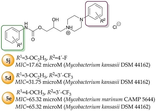
| Compound | MIC (μM) | |||
|---|---|---|---|---|
| MAP 1 | MS 2 | MK 3 | MM 4 | |
| 5a | 510.29 | 522.53 | 130.63 | 130.63 |
| 5b | 1984.32 | 507.99 | 127.00 | 127.00 |
| 5c | 1020.57 | 522.53 | 130.63 | 130.63 |
| 5d | 1984.32 | 127.00 | 31.75 | 127.00 |
| 5e | 1020.57 | 522.53 | 65.32 | 65.32 |
| 5f | 992.16 | 507.99 | 507.99 | 253.99 |
| 5g | 2273.19 | 581.94 | 145.48 | 145.48 |
| 5h | 1101.47 | 563.95 | 281.98 | 281.98 |
| 5i | 1136.60 | 581.94 | 145.48 | 290.97 |
| 5j | 550.73 | 281.98 | 17.62 | 140.99 |
| 5k | 2273.19 | 581.94 | 290.97 | 581.94 |
| 5l | 2202.93 | 563.95 | 563.95 | 281.98 |
| INH | 1822.95 | 117.03 | 29.17 | 466.68 |
| RIF | 109.36 | 19.40 | 0.15 | 2.43 |
| Compound | MIC (μM) | ||||||
|---|---|---|---|---|---|---|---|
| SA 1 | MRSA 2 | EC 3 | EF 4 | CA 5 | CP 6 | CK 7 | |
| 5a | >522.53 | >522.53 | >522.53 | >522.53 | >261.27 | >261.27 | >261.27 |
| 5b | >507.99 | >507.99 | >507.99 | >507.99 | >253.99 | >253.99 | >253.99 |
| 5c | >522.53 | >522.53 | >522.53 | >522.53 | >261.27 | >261.27 | >261.27 |
| 5d | >507.99 | >507.99 | >507.99 | >507.99 | >253.99 | >253.99 | >253.99 |
| 5e | >522.53 | >522.53 | >522.53 | >522.53 | >261.27 | >261.27 | >261.27 |
| 5f | >507.99 | >507.99 | >507.99 | >507.99 | >253.99 | >253.99 | >253.99 |
| 5g | >581.94 | >581.94 | >581.94 | >581.94 | >290.97 | >290.97 | >290.97 |
| 5h | >563.95 | >563.95 | >563.95 | >563.95 | >281.98 | >281.98 | >281.98 |
| 5i | >581.94 | >581.94 | >581.94 | >581.94 | >290.97 | >290.97 | >290.97 |
| 5j | >563.95 | >563.95 | >563.95 | >563.95 | >281.98 | >281.98 | >281.98 |
| 5k | >581.94 | >581.94 | >581.94 | >581.94 | >290.97 | >290.97 | >290.97 |
| 5l | >563.95 | >563.95 | >563.95 | >563.95 | >281.98 | >281.98 | >281.98 |
| CPX | 0.75 | >48.29 | >48.29 | >48.29 | nd | nd | nd |
| 5-FC | nd 8 | nd | nd | nd | 7.69 | 61.50 | 0.96 |
| AMP | 5.72 | >45.79 | >45.79 | >45.79 | nd | nd | nd |
| Amph. B | nd | nd | nd | nd | 0.54 | 1.08 | 0.54 |
© 2016 by the authors. Licensee MDPI, Basel, Switzerland. This article is an open access article distributed under the terms and conditions of the Creative Commons Attribution (CC-BY) license ( http://creativecommons.org/licenses/by/4.0/).
Share and Cite
Malík, I.; Csöllei, J.; Jampílek, J.; Stanzel, L.; Zadražilová, I.; Hošek, J.; Pospíšilová, Š.; Čížek, A.; Coffey, A.; O’Mahony, J. The Structure–Antimicrobial Activity Relationships of a Promising Class of the Compounds Containing the N-Arylpiperazine Scaffold. Molecules 2016, 21, 1274. https://doi.org/10.3390/molecules21101274
Malík I, Csöllei J, Jampílek J, Stanzel L, Zadražilová I, Hošek J, Pospíšilová Š, Čížek A, Coffey A, O’Mahony J. The Structure–Antimicrobial Activity Relationships of a Promising Class of the Compounds Containing the N-Arylpiperazine Scaffold. Molecules. 2016; 21(10):1274. https://doi.org/10.3390/molecules21101274
Chicago/Turabian StyleMalík, Ivan, Jozef Csöllei, Josef Jampílek, Lukáš Stanzel, Iveta Zadražilová, Jan Hošek, Šárka Pospíšilová, Alois Čížek, Aidan Coffey, and Jim O’Mahony. 2016. "The Structure–Antimicrobial Activity Relationships of a Promising Class of the Compounds Containing the N-Arylpiperazine Scaffold" Molecules 21, no. 10: 1274. https://doi.org/10.3390/molecules21101274
APA StyleMalík, I., Csöllei, J., Jampílek, J., Stanzel, L., Zadražilová, I., Hošek, J., Pospíšilová, Š., Čížek, A., Coffey, A., & O’Mahony, J. (2016). The Structure–Antimicrobial Activity Relationships of a Promising Class of the Compounds Containing the N-Arylpiperazine Scaffold. Molecules, 21(10), 1274. https://doi.org/10.3390/molecules21101274