Article

Phytochemicals and Estrogen-Receptor Agonists from the Aerial Parts of Liriope platyphylla

1
Graduate Institute of Natural Products, Kaohsiung Medical University, Kaohsiung 807, Taiwan
2
Department of Laboratory Medicine, Paochien Care Cooperation Paochien Hospital, Pingtung 900, Taiwan
3
Department of Pharmacognosy and Natural Products Chemistry, Faculty of Pharmacy, Ain-Shams University, Organization of African Unity Street, Abassia, Cairo 11566, Egypt
4
Department of Internal Medicine, National Taiwan University Hospital, Taipei 100, Taiwan
5
Chinese Medicine Research and Development Center, China Medical University Hospital, Taichung 404, Taiwan
6
School of Chinese Medicine, College of Chinese Medicine, China Medical University, Taichung 404, Taiwan
7
Center for Molecular Medicine, China Medical University Hospital, Taichung 404, Taiwan
8
Cancer Center, Kaohsiung Medical University Hospital, Kaohsiung 807, Taiwan
9
Research Center for Natural Product and New Drug, Kaohsiung Medical University, Kaohsiung 807, Taiwan
10
Department of Marine Biotechnology and Resources, National Sun Yat-sen University, Kaohsiung 807, Taiwan
*
Authors to whom correspondence should be addressed.
These authors contributed equally to this work.
Molecules 2015, 20(4), 6844-6855; https://doi.org/10.3390/molecules20046844
Submission received: 13 March 2015 / Revised: 12 April 2015 / Accepted: 13 April 2015 / Published: 16 April 2015
(This article belongs to the Section Natural Products Chemistry)

Abstract

:
One new benzofuran, (2R)-(2',4'-dihydroxybenzyl)-6,7-methylenedioxy-2,3-dihydrobenzofuran (1), one new phenylisocoumarin, 3-(2'-hydroxyphenyl)-6,8-dihydroxy-7-methoxy-isocoumarin (2), and one new benzofuroisocoumarin, platyphyllarin C (3), were isolated from the ethanolic extract of Liriope platyphylla aerial parts, along with seventeen known compounds. The structures of the isolates were established by spectroscopic analysis and comparison with the literature data. The results indicated that structures 1–3 are uncommon in Nature. Benzofuroisocoumarin 4, flavonoids 9, 10, and 1315, and homoisoflavonoids 19 and 20 exhibited significant binding activity to estrogen-receptor α and/or β as demonstrated by the SEAP reporter assay system in an MCF-7 cell-line.

Graphical Abstract

1. Introduction

The aerial parts of Liriope platyphylla Wang et Tang (Liliaceae) are used in Asia as nutrients and food supplement [1,2]. They possess reproductive tonic, antitussive, expectorant, and demulcent effects [1]. Phytochemical investigation of Liriope sp. indicated that steroidal compounds are the major class of secondary metabolites in this genus [3]. Other classes of secondary metabolites isolated from this genus included homoisoflavonoids [4], benzofurans [5], anthocyanins [1], amides [4], and sesquiterpenoids [6]. Plants belonging to the genus Liriope were found to possess hepatoprotective [7], antibacterial [8], anti-inflammatory [5], cytotoxic [9], antidiabetic [10], and neuroprotective [11] activities.
Recently, we reported that certain components isolated from L. platyphylla roots showed significant anti-platelet and estrogenic effects [12]. In our continuing investigation to search for novel phytoestrogenic resources, different plants used in Asian folk medicines were assayed for their estrogenic activity. Among the examined plants, the ethanolic extract of L. platyphylla aerial parts was found to be significantly active in the transgenic Arabidopsis pER8:GUS assay [12] (Arabidopsis pER8:GUS possessing a human estrogen ERα receptor activating assay system) at 12.5 μg/mL. Therefore, the extract was selected for further phytochemical and pharmacological investigation. In the current investigation, the isolation and structure elucidation of secondary metabolites isolated from L. platyphylla aerial parts were discussed. The binding activity of the isolates to the estrogen-receptors (ER) α and/or β in a SEAP reporter assay system using the MCF-7 cell-line was also investigated.

2. Results and Discussion

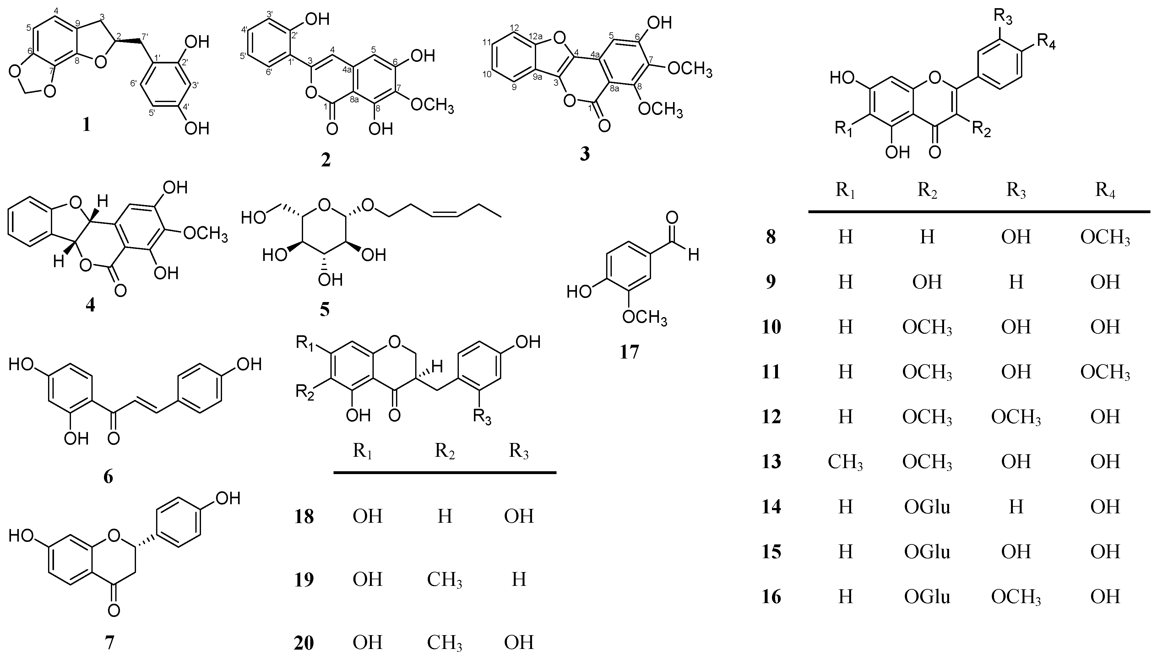
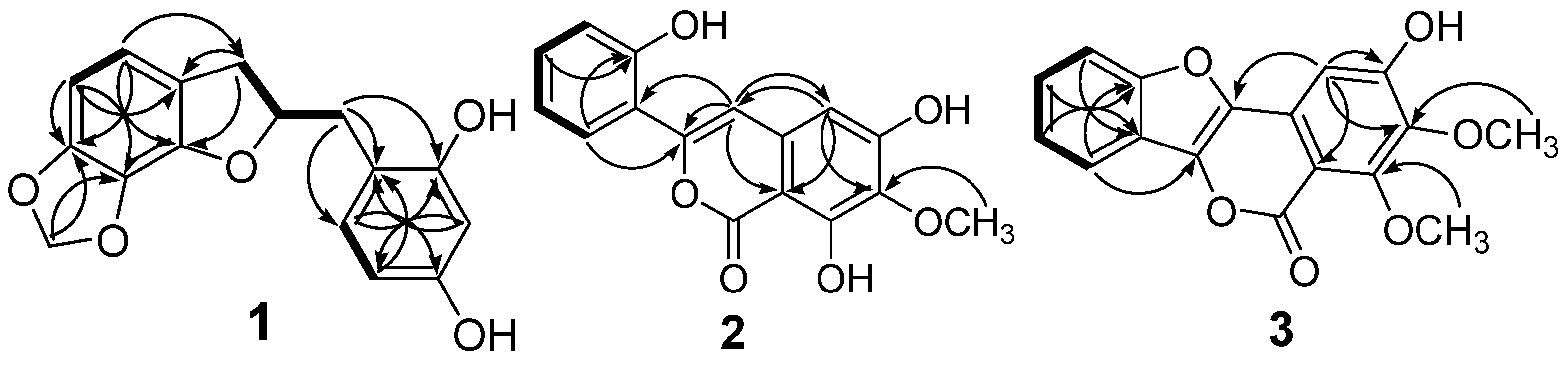
2.1. Structural Elucidation of the Isolated Compounds

The ethanolic extract of L. platyphylla aerial parts was selected for further investigation and subsequently partitioned with n-hexane, ethyl acetate (EtOAc), n-BuOH, and H2O to yield four fractions. The bioassay-guided isolation of the most active (EtOAc) fraction led to the purification of one new 2-benzyl-2,3-dihydrobenzofuran derivative, (2R)-(2',4'-dihydroxybenzyl)-6,7-methylenedioxy-2,3-dihydrobenzofuran (1), one new phenylisocoumarin derivative, 3-(2'-hydroxyphenyl)-6,8-dihydroxy-7-methoxy-isocoumarin (2), and one new benzofuroisocoumarin derivative, platyphyllarin C (3), along with seventeen known compounds (Figure 1). The known compounds were identified as (+)-platyphyllarin A (4) [12], (Z)-3-hexenyl-β-d-glucopyranoside (5) [13], 4,2',4'-trihydroxychalcone (6) [14], liquiritigenin (7) [15], diosmetin (8) [16], kaempferol (9) [17], 3-O-methylquercetin (10) [18], 3,3'-O-dimethylquercetin (11) [19], 3,4'-O-dimethylquercetin (12) [18], 6-C-methylquercetin-3-methyl ether (13) [20], kaempferol-3-O-glucoside (14) [21], quercetin-3-O-glucoside (15) [22], isorhamnetin 3-O-glucoside (16) [23], vanillin (17) [12], disporopsin (18) [24], 3-(2',4'-dihydroxybenzyl)-5,7-dihydroxy-6-methylchroman-4-one (19) [12], and 3-(2',4'-dihydroxy-benzyl)-5,7-dihydroxy-6-methylchroman-4-one (20) [12] by spectroscopic analysis and comparison with the reported physical data.
Figure 1. Chemical structures of compounds 1–21 isolated from the aerial parts of L. platyphylla.
Molecules 20 06844 g001
Compound 1 was isolated as a light-yellow amorphous powder. The molecular formula was calculated as C16H14O5 from the analysis of its HRESIMS (m/z 309.07307 [M + Na]+, calcd. for 309.07334), indicating ten degrees of unsaturation. The IR spectrum indicated the presence of hydroxy (3397 cm−1) and aromatic (1619 and 1600 cm−1) functionalities. The 13C-NMR and DEPT experiments suggested the presence of sixteen carbons, including three methylenes, six methines, five oxygenated aromatic carbons, and two sp2 quaternary carbons (Table 1). All protons and the corresponding carbons in 1 were assigned using coupling constant calculations and 2D NMR spectroscopic analysis. An oxymethine (δH 5.08, δC 86.5, CH-2), two methylenes (δH 2.90, 3.04, δC 35.2, CH2-3; δH 2.81, 3.04, δC 36.6, CH2-7'), a 1,2,3,4-tetrasubstituted aromatic ring (δH 6.59, d, J = 7.6; δC 117.9, CH-4; δH 6.32, d, J = 7.6; δC 101.6, CH-5), a 1,2,4-trisubstituted aromatic ring (δH 6.29, d, J = 2.4; δC 103.4, CH-3'; δH 6.23, dd, J = 8.4, 2.4; δC 107.4, CH-5'; δH 6.92, d, J = 8.4; δC 132.8, CH-6'), and a methylenedioxy (δH 5.86, 2H, δC 102.3) were observed (Table 1). A 2,3-dihydrobenzofuran moiety was suggested by the COSY cross peak between H-2/H-3.
Table 1. 1H and 13C-NMR spectroscopic data of compounds 1–3 (δ in ppm, J in Hz) a.
Position1 b2 b3 c
δHδC, typeδHδC, typeδHδC, type
1 161.3, C 158.1, C
25.08, m86.5, CH
33.04, m d
2.90, dd (15.2, 7.2)
35.2, CH2 150.8, C 138.4, C
46.59, d (7.6)117.9, CH7.49, s108.3, CH 136.9, C
4a 136.5, C 128.9, C
56.32, d (7.6)101.6, CH6.50, s105.3, CH7.16, s102.8, CH
6 150.1, C 156.0, C 159.6. C
7 131.1, C 135.9, C 143.7, C
8 143.2, C 143.5, C 159.4, C
8a 98.7, C 107.2, C
9 124.7, C 7.80, ddd (7.8, 1.8, 0.6)119.9, CH
9a 120.8, C
10 7.43, td (7.2, 1.2)125.5, CH
11 7.51, td (7.2, 1.2)128.3, CH
12 7.68, dd (8.4, 1.2)113.8, CH
12a 155.4, C
1' 115.9, C 119.6, C
2' 157.5, C 156.9, C
3'6.29, d, (2.4)103.4, CH6.93, dd (7.6, 1.2)117.4, CH
4' 158.2, C7.24, ddd (8.0, 7.6, 1.6)131.6, CH
5'6.23, dd (8.4, 2.4)107.4, CH6.95, dd (8.0, 1.2)120.7, CH
6'6.92, d (8.4)132.8, CH7.79, dd (8.0, 1.6)128.6, CH
7'3.04, m d
2.81, dd (14.0, 8.0)
36.6, CH2
7-OCH3 3.90, s (3H)60.9, CH33.94, s (3H)62.3, CH3
8-OCH3 3.97, s (3H)62.6, CH3
OCH2O-5.86, s (2H)102.3, CH2
a The assignments were based on COSY, HSQC, and HMBC experiments; b 1H (400 MHz) and 13C (100 MHz) NMR were measured in methanol-d4; c 1H (600 MHz) and 13C (150 MHz) NMR were measured in acetone-d6; d Signals overlapping.
The HMBC correlations between H-3/C-8 and C-9, H-4/C-3 and C-9, and H-5/C-8 and C-9 (Figure 2) indicated that the 1,2,3,4-tetrasubstituted aromatic ring was connected to the 2,3-dihydrofuran ring. A COSY cross peak between H-2/H-7' and the HMBC correlations between H-7'/C-1', C-2', and C-6' (Figure 2) suggested that the 1,2,4-trisubstituted aromatic ring and 2,3-dihydrobenzofuran was connected at C-7'.
Figure 2. Key COSY (—) and HMBC (→) correlations of 1–3.
Molecules 20 06844 g002
The skeleton of 1 was established as 2-benzyl-2,3-dihydrobenzofuran. Additionally, the position of the methylenedioxy group was confirmed by the HMBC correlations to C-6 and C-7. The configuration of 1 was determined to be 2R by comparing the optical rotation [ α ] D 25 254.9 (c 0.07, MeOH) of 1 to the literature data [25]. Thus, compound 1 was established as (2R)-(2',4'-dihydroxybenzyl)-6,7-methylenedioxy-2,3-dihydrobenzofuran.
Compound 2 was isolated as a yellow amorphous powder. The molecular formula was calculated as C16H12O6 from the analysis of its HRESIMS (m/z 323.05252 [M + Na]+, calcd. for 323.05261), indicating eleven degrees of unsaturation. The IR spectrum indicated the presence of hydroxy (3386 cm−1), carbonyl (1662 cm−1), and aromatic (1609 and 1576 cm−1) functionalities. From the 13C-NMR and DEPT spectra, one methoxy, six aromatic/olefinic methines, five oxygenated aromatic carbons, three aromatic/olefinic quaternary carbons, and one ester carbon were observed (Table 1). Two proton signals at δH 7.49 (s, H-4) and 6.50 (s, H-5) and four proton signals of the 1,2-disubstituted aromatic ring at δH 6.93 (dd, J = 7.6, 1.2, H-3'), 7.24 (ddd, J = 8.0, 7.6, 1.6, H-4'), 6.95 (dd, J = 8.0, 1.2, H-5'), and 7.79 (dd, J = 8.0, 1.6, H-6') were observed in the 1H-NMR spectrum. The 13C-NMR signals along with the HMBC correlations between H-4/C-3 (δC 150.8), C-5 (δC 105.3), C-8a (δC 98.7), and H-5/C-4 (δC 108.3), C-8a (Figure 2) indicated the presence of an isocoumarin moiety formed by connecting the penta-substituted aromatic ring and the 2-pyrone ring systems [26]. The methoxy group (δH 3.90, s, 3H) was connected at C-7 as indicated by the HMBC correlations of H-5/C-7 and OCH3/C-7 as well as by the absence of NOE correlation to H-5. An additional 1,2-disubstituted aromatic ring was confirmed and found to be connected to C-3 of the isocoumarin moiety as suggested by the HMBC correlations between H-4/C-3, C-1' and H-6'/C-3 (Figure 2). Thus, compound 2 was established as 3-(2'-hydroxyphenyl)-6,8-dihydroxy-7-methoxyisocoumarin.
Compound 3 was isolated as a white amorphous powder. The molecular formula was calculated as C17H12O6 from the analysis of its HRESIMS (m/z 335.05254 [M + Na]+, calcd. for 335.05261), indicating 12 degrees of unsaturation. The IR spectrum indicated the presence of hydroxy (3350 cm−1), carbonyl (1708 cm−1), and aromatic (1594 cm−1) functionalities. From the 13C-NMR and DEPT spectra, two methoxy groups, five methines, six oxygenated aromatic carbons, one ester carbon, and three aromatic/olefinic quaternary carbons were observed. The NMR data of 3 suggested a double bond, two aromatic rings, and an ester functionality in the skeleton, similar to the functional groups in 2. Compound 3 showed signals at δH 7.16 (s, H-5), δC 102.8 (CH-5), 159.6 (C-6), 143.7 (C-7), and 159.4 (C-8), three quaternary carbons at δC 138.4 (C-3), 136.9 (C-4), and 128.9 (C-4a), a high field aromatic carbon at δC 107.2 (C-8a), and an unusual ester carbon signal at δC 158.1 (C-1), along with the HMBC correlations between H-5/C-4, C-6, C-7, and C-8a (Figure 2), indicating the presence of the isocoumarin moiety similar to 2, except for an additional substitution at C-4. The 1H-NMR signals at δH 7.80 (ddd, J = 7.8, 1.8, 0.6, H-9), 7.43 (td, J = 7.2, 1.2, H-10), 7.51 (td, J = 7.2, 1.2, H-11), and 7.68 (dd, J = 8.4, 1.2, H-12) indicated the presence of a 1,2-disubstituted aromatic ring. Moreover, the one degree of unsaturation more than 2 and the HMBC correlations between H-9/C-3 and C-12a and H-12/C-9a (Figure 2), suggested the presence of a benzofuran moiety. This proposal was further confirmed by comparing the NMR data of 3 with the reported literature [27]. Through connecting the two moieties, the scaffold of 3 was established as a benzofuroisocoumarin [28]. The position of the methoxy groups (δH 3.94 and 3.97) were confirmed by the HMBC correlations to C-7 and C-8, respectively, as well as by the absence of any NOESY cross peak to H-5. Based on the aforementioned discussion, compound 3 was established as 6-hydroxy-7,8-dimethoxy-benzofuroisocoumarin, and named platyphyllarin C.
2-Benzylbenzofuran [5], phenylisocoumarin [29], and benzofuroisocoumarin [12] structures are uncommon in Nature. 2-Benzyl-2,3-dihydrobenzofuran structures such as compound 1, have never been reported from natural sources. Compound 2 is only the second example of a natural 3-phenyl-isocoumarin and 3 is the third example of a natural benzofuroisocoumarin.

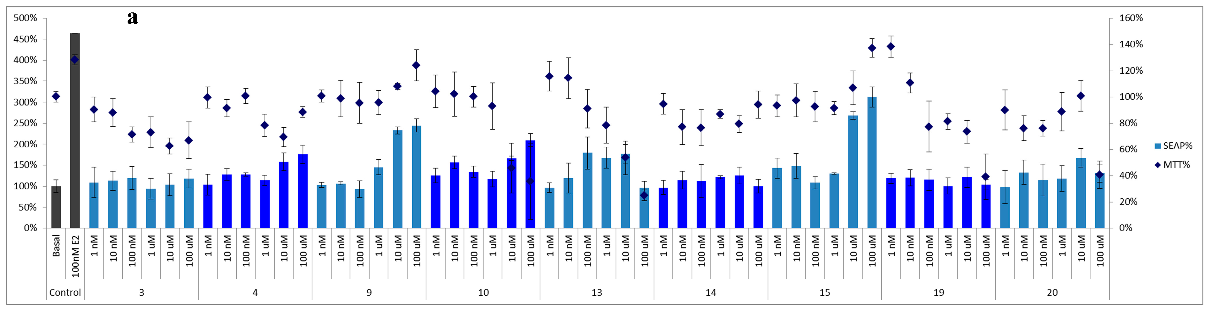
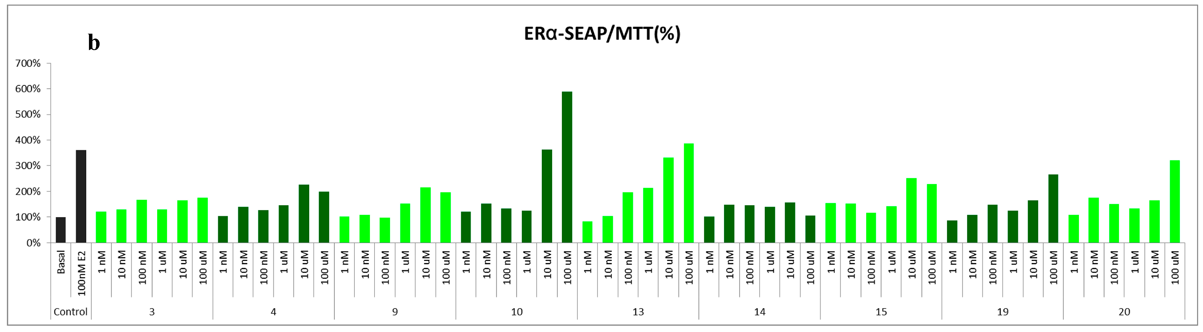
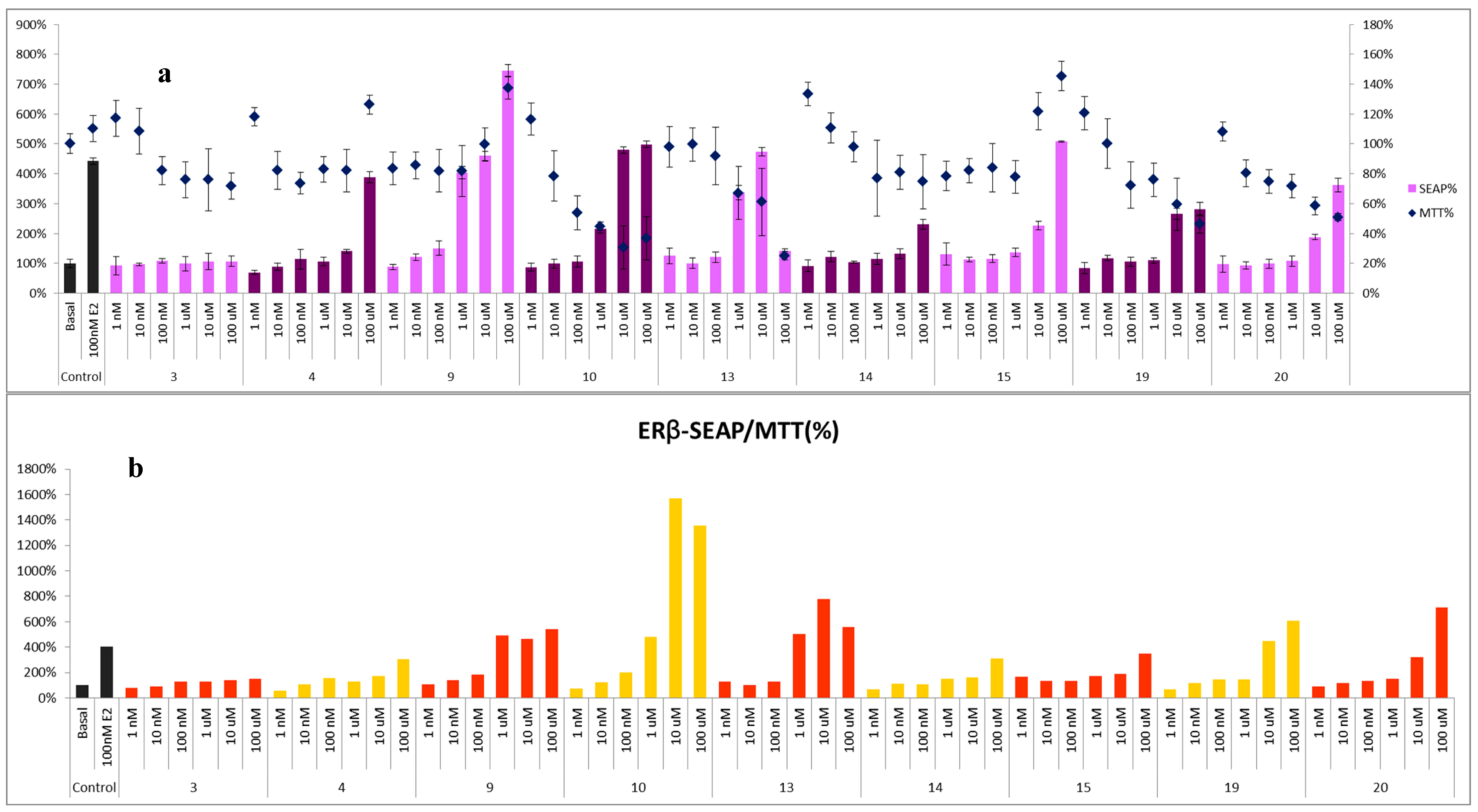
2.2. The Binding Activity of the Isolates to ERα and/or β

Due to the limitated sample availability of some of the isolates, compounds 3, 4, 9, 10, 13–15, 19, and 20 were selected to test their binding potential to the estrogen-receptors α and β at several concentrations (1 nM–100 μM). The basic medium without the addition of the tested compounds (basal) and 17β-estradiol (E2, 100 nM) were used as the blank and positive control, respectively. Cell viability was determined by MTT assay, and the estrogenic activity was evaluated using the SEAP reporter assay system. Both assays were expressed as a percentage based on the basal control, which SEAP% and MTT% of blank were defined as 100% (Figure 3 and Figure 4). For the SEAP% of ERα, compounds 4, 9, 10, 13, and 15 displayed significant binding activity. After excluding the false results from the cell survival ratio, 3-O-methylquercetin (10) and 6-C-methylquercetin-3-methyl ether (13) exhibited the most potent effect in a dose-dependent manner.
Moreover, the new compound, platyphyllarin C (3), showed weak activity to the ERα binding assay, and exhibited weak toxicity to MCF-7 cells at high concentration, 10 and 100 μM (Figure 3). In the ERβ binding assay, all tested isolates exhibited weak to strong activities in a dose-dependent manner. Among the evaluated compounds, flavonoids 9, 10 and, 13, and homoisoflavones 19 and 20 exhibited high binding ability in SEAP/MTT% (Figure 4).
Figure 3. Binding activity to estrogen-receptor alpha (ERα). (a) The SEAP% of ERα activity and the MTT% of MCF-7 cells were presented in percentage, respectively; (b) The SEAP/MTT% was calculated as the final estrogenic data of ERα to avoid false results from cytotoxicity.
Molecules 20 06844 g003aMolecules 20 06844 g003b
Figure 4. Binding activity to estrogen-receptor beta (ERβ). (a) The SEAP% of ERβ activity and the MTT% of MCF-7 cells were presented in percentage, respectively; (b) The SEAP/MTT% was calculated as the final estrogenic data of ERβ to avoid false results from cytotoxicity.
Molecules 20 06844 g004

3. Experimental Section

3.1. General Procedures

Optical rotations were measured with a JASCO P-2000 polarimeter (JASCO Inc., Tokyo, Japan). IR spectra were measured on a PerkinElmer System 2000 FT-IR spectrophotometer (PerkinElmer Inc., Waltham, MA, USA). 1H and 13C-NMR spectra were recorded on Varian VNMRS 600 MHz FT-NMR, Varian Unity-plus 400 MHz FT-NMR (Varian Inc., Palo Alto, CA, USA), or JNM-ECS 400 (JEOL Ltd., Tokyo, Japan) NMR spectrometers. Chemical shifts are reported in parts per million (δ), and coupling constants (J) are expressed in hertz. LRESIMS was measured on a Waters micromass ZQ mass spectrometer (Waters Corporation, Milford, MA, USA). HRESIMS were measured on a Bruker Daltonics APEX II 30e mass spectrometer (Bruker Instruments, Billerica, MA, USA). Silica gel (Kieselgel 60, 70–230 and 230–400 mesh, Merck KGaA, Darmstadt, Germany), C18 reversed phase silica gel (100 Å, 230–400 mesh, Merck KGaA), and Sephadex LH-20 gel (Pharmacia Fine Chemicals AB, Uppsala, Sweden) were used for column chromatography. TLC was carried out using silica gel (Kieselgel 60, F254, Merck KGaA) and RP-18 (F254s, Merck KGaA) precoated plates and compounds were detected with 50% H2SO4 followed by heating on a hot plate. HPLC analyses were performed with a Shimadzu LC-10AT (Shimadzu Inc., Kyoto, Japan) pump interface equipped with a Shimadzu SPD-10A UV-Vis detector using ODS (Thermo Hypersil, 250 × 4 mm; Thermo Hypersil, 250 × 10 mm, Thermo Fisher Scientific Inc., Rockford, IL, USA) and Luna® Phenyl-Hexyl column (5 µm, 250 × 10 mm, Phenomenex Inc., Torrance, CA, USA) columns. ACS grade n-hexane, dichloromethane, ethyl acetate, acetone, methanol, ammonia and 95% sulfuric acid were purchased from ECHO Chemical Co., Ltd. (Miaoli, Taiwan). HPLC grade acetonitrile, methanol, CDCl3, acetone-d6, CD3OD, and C5D5N were obtained from Merck KGaA. Water for chromatographic separation was purified by a Milli-Q water advantage A 10 water system (Merck Millipore, Temecula, CA, USA).

3.2. Plant Material

The plant material was collected in July 2012 from Taichung County, Taiwan, and was identified by a specialist in herbal medicine, Dr. Ming-Hong Yen. A voucher specimen (LP011) was deposited at the Graduate Institute of Natural Products, Kaohsiung Medical University, Kaohsiung, Taiwan. The fresh aerial parts were crushed using a mill (Rong Tsong, Taichung City, Taiwan) before extraction.

3.3. Extraction and Isolation

The fresh aerial parts of L. platyphylla (25 kg) were extracted three times with 95% EtOH (60 L) at room temperature for 24 h. The ethanolic extract (1894.1 g) was concentrated in vacuo, suspended in water and further partitioned into n-hexane, ethyl acetate, and n-BuOH, respectively. The EtOAc layer (76.8 g) was subjected to Sephadex LH-20 column chromatography (CC, 57 × 4.5 cm) and eluted successively with CH2Cl2–EtOAc–MeOH (1:1:6, v/v, repeated 10 times) to afford nine fractions. Fraction 4 was subjected to Sephadex LH-20 CC with CH2Cl2–EtOAc–MeOH (1:1:6, v/v) and further purified using reversed phase (RP) C18 HPLC eluted with MeOH–H2O (40:60, v/v) to obtain 5 (5.1 mg). Fraction 6 was further chromatographed using RP-C18 CC and eluted with gradient MeOH–H2O (10:90 → 60:40 and 100% MeOH for wash, v/v) to yield 14 subfractions, including pure compound 15 (391.9 mg, fr. 6-6). Subfraction 6-7 was isolated using RP-C18 CC with MeOH–H2O (45:55, v/v) to get 14 (25.6 mg) and 16 (0.9 mg). Subfraction 6-9 was subjected to RP-C18 HPLC eluted with MeOH–H2O (50:50, v/v) to yield 18 (2.0 mg). Subfraction 6-10 was separated using RP-C18 CC with MeCN–H2O (25:75 → 40:60, v/v) and was further purified by RP-C18 HPLC with MeCN–H2O (35:65, v/v) to obtain 9 (10.3 mg) and 10 (32.9 mg). Subfraction 6-11 was subjected to silica gel CC and eluted with gradient CH2Cl2-MeOH (35:1 → 25:1, v/v) to yield five subfractions. Subfraction 6-11-1 was further purified by RP-C18 HPLC with MeOH–H2O (60:40, v/v) to yield 3 (3.2 mg) and 4 (4.0 mg). Subfraction 6-11-2 was separated using RP-C18 CC with MeOH–H2O (30:80 → 80:20, v/v) and was further purified by RP-C18 HPLC with MeOH–H2O (60:40, v/v) to obtain 1 (0.8 mg), 6 (1.0 mg), 7 (0.5 mg), 8 (0.6 mg), and a mixture of 11 and 12 (1.2 mg). Subfraction 6-11-4 was subjected to RP-C18 HPLC with MeOH–H2O (60:40, v/v) to yield 13 (25.0 mg) and 20 (4.1 mg). Subfraction 6-12 was isolated using silica gel CC eluted with gradient CH2Cl2–MeOH (20:1 → 15:1, v/v) and was further purified by RP-C18 HPLC eluted with MeCN–H2O (45:55, v/v) to yield 2 (1.5 mg) and 19 (7.3 mg). Fraction 7 was separated by RP-C18 CC eluted with gradient MeOH–H2O (40:60 → 60:40 and 100% MeOH for wash, v/v) to afford 17 (6.4 mg).

3.4. Experimental Data of the New Compounds 1–3

Compound 1: Light-yellow amorphous powder; [ α ] D 25 254.9 (c 0.07, MeOH); UV (MeOH) λmax (log ɛ) 212 (4.75), 280 (3.77) nm; IR ν max neat 3397 (OH), 1619, 1600 (aromatic C=C bond), 1466, 1252, 1057, 1019 cm1; 1H-NMR and 13C-NMR (MeOH-d4): see Table 1; ESIMS found m/z 287 [M + H]+ and 309 [M + Na]+; HR-ESIMS found m/z 309.07307 [M + Na]+ (calcd for C16H14O5Na: 309.07334).
Compound 2: Yellow amorphous powder; UV (MeOH) λmax (log ɛ) 217 (4.10), 260 (4.25), 350 (3.97) nm; IR ν max neat 3386 (OH), 1662 (C=O), 1609, 1576 (aromatic C=C bond), 1452, 1260, 1094 cm1; 1H-NMR and 13C-NMR (MeOH-d4): see Table 1; ESIMS found m/z 301 [M + H]+ and 323 [M + Na]+; HR-ESIMS found m/z 323.05252 [M + Na]+ (calcd for C16H12O6Na: 323.05261).
Compound 3: White amorphous powder; UV (MeOH) λmax (log ɛ) 214 (3.32), 267 (3.60), 314 (3.37), 351 (3.21) nm; IR ν max neat : 3350 (OH), 1708 (C=O), 1594 (aromatic C=C bond), 1369, 1262, 1203, 986 cm−1; 1H-NMR and 13C-NMR (Aceton-d6): see Table 1; ESIMS found m/z 313 [M + H]+; HR-ESIMS found m/z 335.05254 [M + Na]+ (calcd for C17H12O6Na: 335.05261).

3.5. Bioassay Procedure–Estrogenic Activity

The estrogenic activity was determined through adapting the method described by Lai et al. (2011) [30]. The assay was done using SEAP (secreted alkaline phosphatase) reporter gene assay system. Human breast adenocarcinoma cells MCF-7 obtained from Bioresource Collection and Research Center were cultured in phenol-red free minimum essential medium Eagle (MEM) supplemented with dextran-charcoal treated serum, 2 mM l-glutamine, 10% foetal bovine serum (GIBCO BRL, Gaithersburg, MD, USA), penicillin, and streptomycin. The cells were incubated in an atmosphere of 5% CO2 at 37 °C; 0.2 µg of pERE-TA-SEAP plasmid (Clontech, Palo Alto, CA, USA) were transfected into 2 × 10⁴ cells in 100 µL of growth medium per well and incubated for 6 h. Cells were treated with samples of interest in growth medium for 48 h. Aliquots of culture media were analyzed for secreted alkaline phosphatase activity using the Phospha-Light reporter chemiluminescence assay kit (Applied Biosystems, Foster City, CA, USA). The MTT colorimetric assay was performed on the cells for assessing their corresponding cytotoxicity. The estrogenic data were determined by the formula, final SEAP/MTT % = SEAP/cell viability × 100%, to avoid false results from cytotoxicity. Tests were done in triplicate. For the raw data please see supplementary data. The purity of tested compounds was more than 95%.

4. Conclusions

In the current investigation, the estrogenic activity of different fractions obtained from the ethanolic extract of L. platyphylla areal parts was evaluated. Bioassay-guided isolation of the most active fraction (EtOAc) led to the purification of one new 2-benzyl-2,3-dihydrobenzofuran 1, one new phenylisocoumarin 2, and one new benzofuroisocoumarin 3, along with seventeen known compounds. The spectroscopic analyses revealed that 1–3 structures possess unique skeletons which were rarely isolated from nature. 3-O-methylquercetin (10) and 6-C-methylquercetin-3-methyl ether (13) exhibited the most potent estrogenic effect in a dose-dependent manner as demonstrated by the SEAP reporter assay system. Moreover, flavonoids 9, 10 and, 13, and homoisoflavones 19 and 20 expressed high binding ability in the ERβ binding assay. Selective binding to ERβ receptor is an interesting phenomenon from the therapeutic point of view because such binding mimics the beneficial effect of estrogen without the side effects associated with unspecific receptor targeting.

Supplementary Materials

1D and 2D NMR spectra, HR-ESIMS of the new isolates and the raw data of the estrogenic activity can be found in the Supplementary materials at: https://www.mdpi.com/1420-3049/20/04/6844/s1.

Acknowledgments

The authors would like to acknowledge the grants from the Ministry of Science and Technology of Taiwan (MOST 101-2325-B-039-004; 102-2628-B-037-003-MY3; MOST 102-2911-I-002-303; MOST 103-2911-I-002-303; MOST 104-2911-I-002-302; MOST 104-2911-I-037-501). This work was also supported by the Excellence for Cancer Research Center Grant, the Ministry of Health and Welfare, Executive Yuan, Taipei, Taiwan (MOHW104-TDU-B-212-124-003), National Health Research Institutes of Taiwan (NHRI-EX104-10241BI), and the grant for Health and Welfare Surcharge of Tobacco Products and Kaohsiung Medical University “Aim for the Top Universities Grant, grant No. KMU-TP103, and in part from the grant from Chinese Medicine Research Center, China Medical University, Taiwan (the Ministry of Education, the Aim for the Top University Plan). The authors would like to thank the Center for Research Resources and Development (CRRD), Kaohsiung Medical University for the technical supports and services in LC-MS and NMR analyses.

Author Contributions

Y.-C. Tsai and C.-C. Hsu did the major experimental work. Y.-C. Tsai, S.-Y. Chiang, W.-C. Lai, and F.-R. Chang conceived the project and designed the experiments. S.-Y. Chiang and M.-H. Yen collected and identified L. platyphylla. Y.-C. Tsai, M. El-Shazly, Chau-Chung Wu, Chin-Chung Wu, Y.-C. Wu, and F.-R. Chang drafted and revised the manuscript. All authors read and approved the final manuscript.

Conflicts of Interest

The authors declare no conflict of interest.

References

  1. Lee, J.H.; Choung, M.G. Identification and characterisation of anthocyanins in the antioxidant activity-containing fraction of Liriope platyphylla fruits. Food Chem. 2011, 127, 1686–1693. [Google Scholar] [CrossRef]
  2. Yang, S.O.; Ji, G.E. Inhibitory effects of ulmus parvifolia and liriope platyphylla wang et tang on histamine release from rat peritoneal mast cells. Food Sci. Biotechnol. 2006, 15, 363–368. [Google Scholar]
  3. Yu, B.Y.; Qiu, S.X.; Zaw, K.; Xu, G.J.; Hirai, Y.; Shoji, J.; Fong, H.H.; Kinghorn, A.D. Steroidal glycosides from the subterranean parts of Liriope spicata var. Prolifera. Phytochemistry 1996, 43, 201–206. [Google Scholar] [CrossRef]
  4. Li, W.J.; Cheng, X.L.; Liu, J.; Lin, R.C.; Wang, G.L.; Du, S.S.; Liu, Z.L. Phenolic compounds and antioxidant activities of Liriope muscari. Molecules 2012, 17, 1797–1808. [Google Scholar] [CrossRef] [PubMed]
  5. Hu, Z.F.; Chen, L.L.; Qi, J.; Wang, Y.H.; Zhang, H.; Yu, B.Y. Two new benzofuran derivatives with anti-inflammatory activity from Liriope spicata var. Prolifera. Fitoterapia 2011, 82, 190–192. [Google Scholar] [CrossRef]
  6. Cheng, Z.H.; Wu, T.; Bligh, S.A.; Bashall, A.; Yu, B.Y. Cis-eudesmane sesquiterpene glycosides from liriope m uscari and ophiopogon j aponicus. J. Nat. Prod. 2004, 67, 1761–1763. [Google Scholar] [CrossRef] [PubMed]
  7. Wu, F.; Cao, J.; Jiang, J.; Yu, B.; Xu, Q. Ruscogenin glycoside (lm-3) isolated from Liriope muscari improves liver injury by dysfunctioning liver-infiltrating lymphocytes. J. Pharm. Pharmacol. 2001, 53, 681–688. [Google Scholar] [CrossRef] [PubMed]
  8. Kim, S.W.; Chang, I.M.; Oh, K.B. Inhibition of the bacterial surface protein anchoring transpeptidase sortase by medicinal plants. Biosci Biotechnol. Biochem. 2002, 66, 2751–2754. [Google Scholar] [CrossRef] [PubMed]
  9. Wang, H.C.; Wu, C.C.; Cheng, T.S.; Kuo, C.Y.; Tsai, Y.C.; Chiang, S.Y.; Wong, T.S.; Wu, Y.C.; Chang, F.R. Active constituents from liriope platyphylla root against cancer growth in vitro. Evid. Based Complement. Alternat. Med. 2013, 2013. [Google Scholar]
  10. Bai, X.; Chen, X.; Liu, Y.; Tian, L.; Zhou, Q.; Liu, S.; Fang, J.; Chen, J. Effects of water extract and crude polysaccharides from Liriope spicata var. Prolifera on insr/irs-1/pi3k pathway and glucose metabolism in mice. J. Ethnopharmacol. 2009, 125, 482–486. [Google Scholar]
  11. Hur, J.; Lee, P.; Kim, J.; Kim, A.J.; Kim, H.; Kim, S.Y. Induction of nerve growth factor by butanol fraction of Liriope platyphylla in c6 and primary astrocyte cells. Biol. Pharm. Bull. 2004, 27, 1257–1260. [Google Scholar] [CrossRef] [PubMed]
  12. Tsai, Y.C.; Chiang, S.Y.; El-Shazly, M.; Wu, C.C.; Beerhues, L.; Lai, W.C.; Wu, S.F.; Yen, M.H.; Wu, Y.C.; Chang, F.R. The oestrogenic and anti-platelet activities of dihydrobenzofuroisocoumarins and homoisoflavonoids from liriope platyphylla roots. Food Chem. 2013, 140, 305–314. [Google Scholar] [CrossRef] [PubMed]
  13. Lee, S.Y.; Kim, K.H.; Lee, I.K.; Lee, K.H.; Choi, S.U.; Lee, K.R. A new flavonol glycoside from Hylomecon vernalis. Arch. Pharm. Res. 2012, 35, 415–421. [Google Scholar] [CrossRef] [PubMed]
  14. Veitch, N.C.; Sutton, P.S.; Kite, G.C.; Ireland, H.E. Six new isoflavones and a 5-deoxyflavonol glycoside from the leaves of ateleia herbert-smithii. J. Nat. Prod. 2003, 66, 210–216. [Google Scholar] [CrossRef] [PubMed]
  15. Ryu, Y.B.; Kim, J.H.; Park, S.J.; Chang, J.S.; Rho, M.C.; Bae, K.H.; Park, K.H.; Lee, W.S. Inhibition of neuraminidase activity by polyphenol compounds isolated from the roots of Glycyrrhiza uralensis. Bioorg. Med. Chem. Lett. 2010, 20, 971–974. [Google Scholar] [CrossRef] [PubMed]
  16. Ribeiro, D.; Freitas, M.; Tomé, S.M.; Silva, A.M.; Porto, G.; Fernandes, E. Modulation of human neutrophils’ oxidative burst by flavonoids. Eur. J. Med. Chem. 2013, 67, 280–292. [Google Scholar] [CrossRef] [PubMed]
  17. Liao, C.R.; Kuo, Y.H.; Ho, Y.L.; Wang, C.Y.; Yang, C.S.; Lin, C.W.; Chang, Y.S. Studies on cytotoxic constituents from the leaves of elaeagnus oldhamii maxim. In non-small cell lung cancer a549 cells. Molecules 2014, 19, 9515–9534. [Google Scholar]
  18. Yamauchi, K.; Mitsunaga, T.; Inagaki, M.; Suzuki, T. Synthesized quercetin derivatives stimulate melanogenesis in b16 melanoma cells by influencing the expression of melanin biosynthesis proteins mitf and p38 mapk. Bioorg. Med. Chem. 2014, 22, 3331–3340. [Google Scholar] [CrossRef] [PubMed]
  19. Wang, J.; Gao, H.; Zhao, J.; Wang, Q.; Zhou, L.; Han, J.; Yu, Z.; Yang, F. Preparative separation of phenolic compounds from halimodendron halodendron by high-speed counter-current chromatography. Molecules 2010, 15, 5998–6007. [Google Scholar] [CrossRef] [PubMed]
  20. Ibewuike, J.C.; Ogundaini, A.O.; Ogungbamila, F.O.; Martin, M.T.; Gallard, J.F.; Bohlin, L.; Païs, M. Piliostigmin, a 2-phenoxychromone, and c-methylflavonols from Piliostigma thonningii. Phytochemistry 1996, 43, 687–690. [Google Scholar] [CrossRef]
  21. Luyen, B.T.T.; Tai, B.H.; Thao, N.P.; Eun, K.J.; Cha, J.Y.; Xin, M.J.; Lee, Y.M.; Kim, Y.H. Anti-inflammatory components of Euphorbia humifusa willd. Bioorg. Med. Chem. Lett. 2014, 24, 1895–1900. [Google Scholar] [CrossRef] [PubMed]
  22. Stark, T.; Bareuther, S.; Hofmann, T. Sensory-guided decomposition of roasted cocoa nibs (Theobroma cacao) and structure determination of taste-active polyphenols. J. Aagric. Food Chem. 2005, 53, 5407–5418. [Google Scholar] [CrossRef]
  23. Fico, G.; Rodondi, G.; Flamini, G.; Passarella, D.; Tomé, F. Comparative phytochemical and morphological analyses of three italian primula species. Phytochemistry 2007, 68, 1683–1691. [Google Scholar] [CrossRef] [PubMed]
  24. Nguyen, A.T.; Fontaine, J.; Malonne, H.; Duez, P. Homoisoflavanones from Disporopsis aspera. Phytochemistry 2006, 67, 2159–2163. [Google Scholar] [CrossRef] [PubMed]
  25. Ortega, N.; Urban, S.; Beiring, B.; Glorius, F. Ruthenium nhc catalyzed highly asymmetric hydrogenation of benzofurans. Angew. Chem. Int. Ed. 2012, 51, 1710–1713. [Google Scholar] [CrossRef]
  26. Shahzad, S.A.; Venin, C.; Wirth, T. Diselenide-and disulfide-mediated synthesis of isocoumarins. Eur. J. Org. Chem. 2010, 2010, 3465–3472. [Google Scholar] [CrossRef]
  27. Ghosh, S.; Banerjee, I.; Baul, S. Studies on oxygen heterocycles part 2: Synthesis of 2-arylcoumaranones and 2-phenylbenzofuran. Tetrahedron 1999, 55, 11537–11546. [Google Scholar] [CrossRef]
  28. Yamaguchi, S.; Uchiuzoh, Y.; Sanada, K. The synthesis of benzofuroquinolines. IX. A benzofuroisoquinolinone and a benzofuroisocoumarin. J. Heterocycl. Chem. 1995, 32, 419–423. [Google Scholar]
  29. Charubala, R.; Guggisberg, A.; Hesse, M.; Schmid, H. Notiz über das natürliche auftreten von 3-phenylisocumarin. Helv. Chim. Acta 1974, 57, 1096–1097. [Google Scholar] [CrossRef]
  30. Lai, W.C.; Wang, H.C.; Chen, G.Y.; Yang, J.C.; Korinek, M.; Hsieh, C.J.; Nozaki, H.; Hayashi, K.I.; Wu, C.C.; Wu, Y.C. Using the per8: Gus reporter system to screen for phytoestrogens from Caesalpinia sappan. J. Nat. Prod. 2011, 74, 1698–1706. [Google Scholar] [CrossRef] [PubMed]
  • Sample Availability: Samples of the compounds 1–20 are available from the authors.

