Article

A New Phenolic Glycoside from the Barks of Cinnamomum cassia

1
Tongji Hospital Affiliated to Tongji Medical College, Huazhong University of Science and Technology, Wuhan 430030, China
2
Department of Pharmacy, Renmin Hospital of Wuhan University, Wuhan 430060, China
3
Hubei Key Laboratory of Natural Medicinal Chemistry and Resource Evaluation, School of Pharmacy, Tongji Medical College, Huazhong University of Science and Technology, Wuhan 430030, China
*
Author to whom correspondence should be addressed.
These authors contributed equally to this work.
Molecules 2014, 19(11), 17727-17734; https://doi.org/10.3390/molecules191117727
Submission received: 25 September 2014 / Revised: 29 October 2014 / Accepted: 29 October 2014 / Published: 31 October 2014
(This article belongs to the Section Natural Products Chemistry)

Abstract

:
A new phenolic glycoside (1), named methyl 2-phenylpropanoate-2-O-β-d-apiofuranosyl-(1→6)-O-β-d–glucopyranoside, was isolated from the barks of Cinnamomum cassia, along with three known phenolic glycosides and four known lignan glycosides. The structure of 1 was elucidated by extensive interpretation of spectroscopic data and chemical method. Selected compounds were evaluated for their immunosuppressive activities against murine lymphocytes. Compounds 1, 2, 6 and 8 exhibited differential inhibition against ConA-induced T cells proliferation.

Graphical Abstract

1. Introduction

The plant Cinnamomum cassia (Lauraceae), which originates in the south of China, is widely cultivated in tropical or subtropical areas, such as Fujian, Guangdong, Guangxi, Yunnan and Hainan Provinces of China, as well as Taiwan, India, Indonesia, Laos, Malysia, Thailand and Vietnam [1]. The bark of C. cassia, well known as Cinnamomi cortex in China, has long been used as spice and flavoring agents [2]. It also possesses various biological activities, such as insecticidal, anti-allergy, antipyretic, anticancer, anti-Alzheimer′s disease, antidiabetic [3] and immunosuppressive activity [4]. In our previous study [5], three diterpenoids, cinncassiols F and G, and 16-O-β-d-glucopyranosyl-19-deoxycinncassiol G, with unprecedented carbon skeletons, were isolated from the barks of C. cassia. It is notable that two of them showed significant inhibitory effects on the proliferation of murine T cells induced by ConA. Recently, with the aim of discovering more bioactive compounds from C. cassia, the extracts of EtOAc and n-BuOH were further phytochemically studied, which led to the isolation of a new phenolic glycoside (1), together with three known phenolic glycosides (24) and four known lignan glycosides (58). The immunomodulatory activities against murine lymphocytes of the selected compounds were reported in this work.

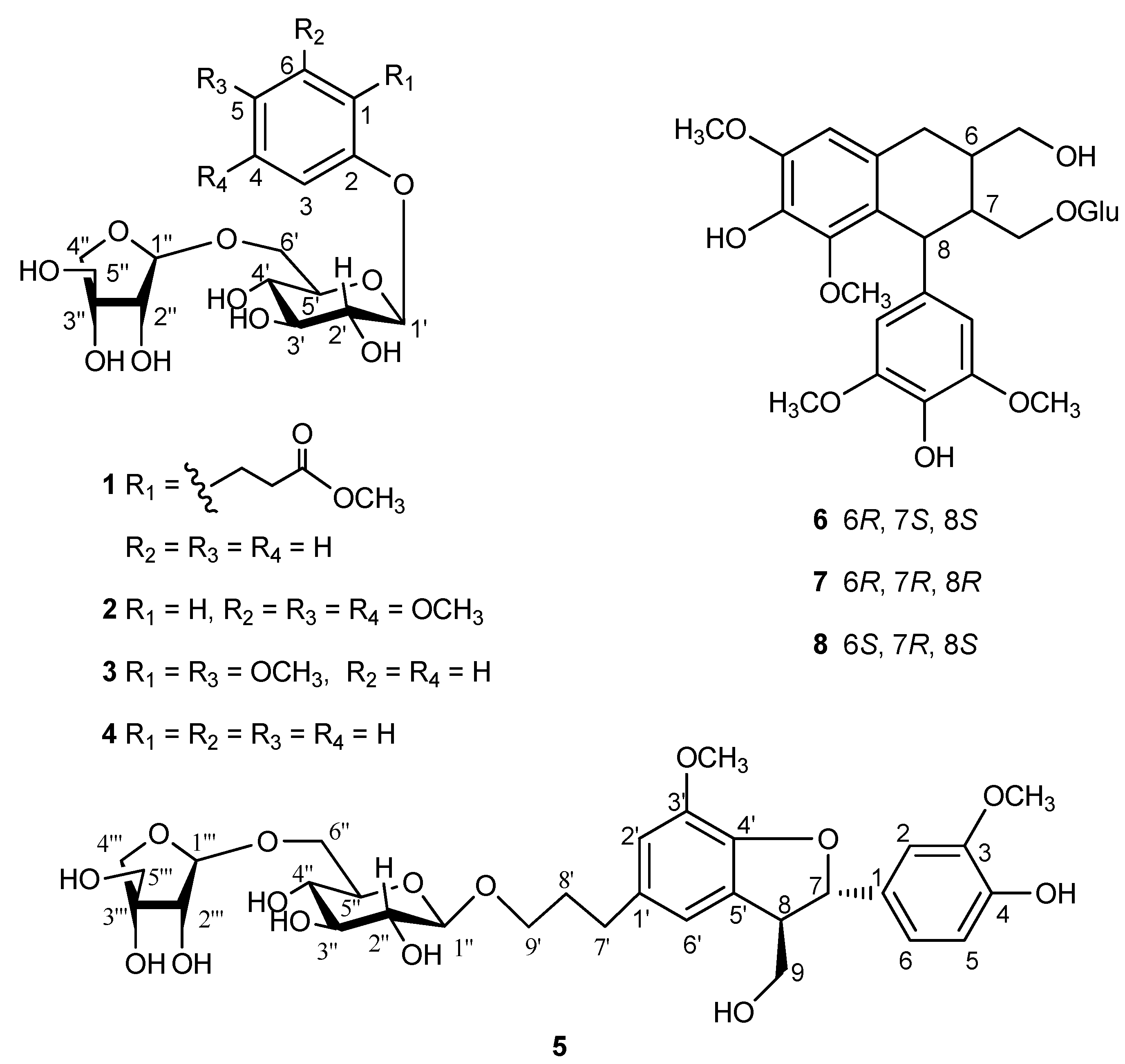
2. Results and Discussion

The EtOAc extract of C. cassia was separated by various chromatographic techniques to yield a new phenolic glycoside (1), along with seven known compounds. The known compounds were identified as 3,4,5-trimethoxyphenol-β-d-apiofuranosyl-(1→6)-O-β-d-glucopyranoside (2) [6], cinnacasolide E (3) [7], phenol-β-d-apiofuranosyl-(1→6)-O-β-d-glucopyranoside (4) [8], samwiside (5) [9], (6R,7R,8R)-7a-[(β-d-glucopyranosyl)oxy]lyoniresinol (6) [10], (6S,7R,8R)-7a-[(β-d-glucopryanosyl)oxy]lyoniresinol (7) [10], and (6R,7S,8S)-7a-[(β-d-glucopyranosyl)oxy]lyoniresinol (8) [11], by comparison of their NMR data with those reported in literatures (Figure 1).
Figure 1. Structures of 18.
Molecules 19 17727 g001
Compound 1 was isolated as amorphous powder, with [ α ] D 25 −65.1 (c = 0.47, MeOH). Its molecular formula, C21H30O12, was deduced from the HRESIMS peak at m/z 497.1616 [M+Na]+ (calcd for C21H30O12Na, 497.1629). The IR spectrum showed the presence of carbonyl (1719 cm−1) and hydroxyl group (3391 cm−1). The 1H NMR spectrum of 1 (Table 1) showed proton signals for a 1,2-disubstituted aromatic ring (δH 7.21, dd, J = 8.1, 1.2 Hz, H-3; 7.18, ddd, J = 8.1, 7.9, 1.9 Hz, H-4; 6.96, ddd, J = 7.9, 7.9, 1.2 Hz, H-5; and 7.17, dd, J = 7.9, 1.9 Hz, H-6), a methoxyl (δH 3.66, s). 13C-NMR and DEPT spectra (Table 1) of 1 showed 21 carbon signals, including one methoxyl, one ester carbonyl, five methylenes, eleven methines, and three quaternary carbons. Comparison of the 1H- and 13C-NMR spectra with those of 4 revealed that 1 differed from 4 by the presence of a methyl propionate at C-1. Thus, compound 1 was determined to be an analogue of 4. The HBMC and 1H-1H COSY spectra of 1 indicated the presence of a methyl propionate moiety, which was connected at C-1.
The HBMC correlations from H-1′ to C-2 and from H-1′′ to C-6′ revealed the linkages of the two sugars to be the same as those of 4. The sugars were finally confirmed by comparing the retention times of the three trimethylsilylthiazolidine derivatives obtained from GC analysis. On the basis of the above evidences, compound 1 was determined and named methyl 2-phenylpropanoate-2-O-β-d-apiofuranosyl-(1→6)-O-β-d-glucopyranoside.
Table 1. 1H-(400 MHz) and 13C-NMR (100 MHz) Data of 1 in CD3OD.
PositionδHδC
1 131.3
2 157.0
37.21 dd (8.1, 1.2)128.9
47.18 ddd (8.1, 7.9, 1.9)116.7
56.96 ddd (7.9, 7.9, 1.2)123.5
67.17 dd (7.9, 1.9)131.0
73.00 m26.9
82.68 t (7.7)35.3
9 175.9
OMe3.66 s52.0
1′4.88 d (7.8)102.8
2′3.50 m75.0
3′3.49 m78.2
4′3.38 m71.6
5′3.60 m77.0
6′a4.04 d (10.6)68.8
6′b3.64 m
1′′5.00 d (2.3)111.0
2′′3.93 d (2.3)78.1
3′′ 80.0
4′′a3.77 d (9.7)75.0
4′′b3.97 d (9.7)
5′′3.61 s65.6
The crude extract of C. cassia has been reported to exhibit potent anti-complement [12] and immunosuppressive activities [4]. Thus, compounds 1, 2, and 58 were evaluated for their immunomodulatory activities against murine lymphocytes. The results (Table 2) showed that compounds 1, 2, 6, and 8 inhibited the proliferation of ConA-induced murine T cells at a concentration of 50 μM, while the positive control at the concentration of 50 μM, resulted in an inhibitory ratio of 106.1% against T cells and an inhibitory ratio of 75.8% against B cells.
Table 2. Effects on Murine Lymphocyte Proliferation Induced by Concanavalin A (ConA) (5 μg/mL) or Lipopolysaccharide (LPS) (10 μg/mL) of the Selected Compounds 1, 2, and 58 a.
CompoundsConcentration (μM)Inhibitory/Enhanced Rate (%) b
ConA−Induced T Cell ProliferationLPS−Induced B Cell Proliferation
1200−56.710.3
100−61.2c25.2
50−35.425.6
25−22.130.1
12.515.27.8
2200−45.2−35.1
100−38.8−5.8
50−8.5−12.5
25−12.510.3
12.5−20.16.2
5200−27.928.4
100−5.422.2
506.510.7
2511.7−1.5
12.52.813.8
6200−47.7−13.2
100−58.020.5
50−23.641.4
25−47.536.1
12.5−11.3−12.5
7200−10.4−22.7
100−13.878.5
502.562.4
2518.758.4
12.535.112.5
8200−80.1 *−46.7
100−58.3−10.3
50−19.525.6
2527.857.8
12.514.552.2
CsA200−109.8 **−106.1 **
100−106.0 **−94.8 **
50−106.1 **−75.8 *
25−97.1 **−60.1
12.5−84.5 **−54.5
a Results are represented as mean ± SD based on three independent experiments. b -Inhibitory effect; +stimulatory effect. (n = 3; * p < 0.05; ** p < 0.01 compared with control). Positive control: ConA or LPS; negative control: DMSO.

3. Experimental

3.1. General Procedures

NMR spectra were recorded on a Bruker AM 400 spectrometer (Bruker, Ettlingcn, Germany), and the 1H- and 13C-NMR chemical shifts were referenced to the solvent peaks for CD3OD at δH 3.31 and δC 49.15. HRESI-MS data were measured on a LC-LTQ-Orbitrap XL spectrometer (Thermo Fisher, Waltham, MA, USA). Optical rotations were determined on a Perkin-Elmer PE-341LC polarimeter (PerkinElmer, Waltham, MA, USA). IR spectra were recorded on a Bruker VERTEX 70 spectrometer (Bruker, Ettlingcn, Germany). UV spectra were recorded on a PerkinElmer Lambda 35 spectrophotometer (PerkinElmer, Waltham, MA, USA). Semi-preparative HPLC was performed on an Agilent 1100 liquid chromatography (Agilent, Santa Clara, CA, USA) with an YMC (10 × 250 mm, 5 μm) column. GC analysis was performed with a capillary column (30 m × 0.32 mm × 0.5 μm) on an Agilent 7890A GC. Silica gel (200–300 mesh, Qingdao Marine Chemical Inc., Qingdao, China), ODS (50 μm, YMC, Kyoto, Japan), and Sephadex LH-20 (Pharmacia Biotech AB, Stockholm, Sweden) were used for column chromatography.

3.2. Plant Material

The barks of C. cassia were collected at Qujing, Yunan Province, China, in July 2010. The plant material was identified by Prof. Changgong Zhang at the School of Pharmacy, Tongji Medical College, Huazhong University of Science and Technology. A voucher specimen (No. 2010-0703) was deposited in the herbarium of Hubei Key Laboratory of Natural Medicinal Chemistry and Resource Evaluation, Tongji Medical College, Huazhong University of Science and Technology.

3.3. Extraction and Isolation

The air-dried stem barks of C. cassia (25 kg) were extracted with 95% ethanol at room temperature three times. The EtOH extract was concentrated in vacuo (2.6 kg), and then suspended in water and partitioned successively with petroleum ether, CHCl3, EtOAc, and n-BuOH. The EtOAc soluble extract was subjected to column chromatography (CC) (100–200 mesh) over silica gel, eluting with a gradient of CHCl3/MeOH to yield fractions 1–6. Fraction 3 (16 g) was chromatographed on YMC gel column (ODS) eluted with MeOH/H2O (10%–70%), and on silica gel with a gradient of CHCl3/MeOH (from 25:1 to 10:1) to afford compounds 2 (25 mg), 3 (18 mg), and 4 (20 mg). Compound 1 (15 mg) was isolated from fraction 4 and purified by semipreparative HPLC (MeOH/H2O, 30:70). Fraction 5 (21 g) was chromatographed on a silica gel CC with a gradient of CHCl3/MeOH (15:1) to yield two subfractions 5.1 and 5.2. Compound 5 (15 mg) was purified from fraction 5.1 by semipreparative HPLC (MeOH/H2O, 20:80), compounds 6 (8 mg), 7 (5 mg), and 8 (10 mg) were isolated from fraction 5.2.

3.4. Acid Hydrolysis

A solution of 1 (2.0 mg), in 2 M aqueous CF3COOH (2.0 mL) was heated at 60 °C for 2 h in a water bath [13]. The reaction mixture was diluted in H2O (2.0 mL) and extracted with EtOAc (2.0 mL × 3), then the aqueous layer was concentrated to remove CF3COOH. The residue was dissolved in pyridine (1.0 mL), to which L-cysteine methyl ester hydrochloride (2.0 mg) in pyridine (1.0 mL) was added. Then, the mixture was kept at 60 °C for 30 min. After the reaction mixture was dried in vacuo, the residue was trimethylsilylated with 1-trimethylsilylimidazole (0.4 mL) at 60 °C for 30 min in a water bath. Finally, the mixture was partitioned between n-hexane and H2O (1 mL each) and the n-hexane extract was analyzed by gas chromatography (GC) under the following conditions: column temperature, 250 °C; injection temperature, 250 °C; carrier N2 gas; flow rate 1.0 mL/min. In the acid hydrolysate of 1, d-glucose and d-apiose were confirmed by comparison of the retention times of their derivatives with those of d-glucose and d-apiose derivatives prepared in a similar way, which showed retention times of 10.03 and 15.62 min.
Methyl 2-phenylpropanoate-2-O-β-d-apiofuranosyl-(1→6)-O-β-d-glucopyranoside (1): amorphous powder; [ α ] D 25 −65.1 (c = 0.47, MeOH); UV (MeOH) λmax (log ε) = 213 (2.9) nm; IR (KBr) νmax 3391, 2926, 2884, 1719, 1493, 1236, 1067, and 756 cm1; 1H-NMR (CD3OD, 400 MHz) and 13C-NMR (CD3OD, 100 MHz) see Table 1; HRESIMS m/z: 497.1616 [M+Na]+ (calcd for C21H30O12Na, 497.1629).

3.5. Lymphocyte Proliferation Test

Splenic lymphocytes were prepared as previously described by Kawaguchi et al. [14]. The lymphocyte proliferation was determined by WST-8 assay using Cell Counting Kit-8 (Dojindo) [15]. The prepared spleen cells (2.5 × 10⁶ cells) were seeded into each well of a 96-well microplate, and various concentrations of compounds and 5 μg/mL of concanavalin A (Con A, from Canavaliaensiformis Type III), for selective stimuli on T cells were added, cyclosporine A (CsA) was used as a positive control. After being cultured at 37 °C with 5% CO2 for 48 h, 20 μL WST-8 was added to each well. The absorbance at 450 nm with a 600 nm reference was detected on a microtiter plate reader (Bio-Rad 680). All optical density (OD) values shown are the mean of triplicate sample ± SD. Controls with and without ConA and LPS were used to establish the baseline proliferation for stimulated and unstimulated cells. The lymphocyte proliferation rate was evaluated according to our reported procedure [5].

4. Conclusions

A new phenolic glycoside, methyl 2-phenylpropanoate-2-O-β-d-apiofuranosyl-(1→6)-O-β-d-glucopyranoside (1), was isolated from the barks of C. cassia, together with seven known compounds (28). The structure and relative configuration of the new isolate was elucidated by careful interpretation of spectroscopic data. This is the first report of samwiside (5) in the genus of Cinnamomum. Compounds 1, 2 and 58 were tested for their in vitro immunomodulatory activities. Compounds 2 and 6 showed weak inhibition against ConA-induced T cell proliferation at the dosage of 12.5–200 μM, and the inhibitory effects are not dose-dependent. Interestingly, at a concentration of 200 μM, compound 8 significantly inhibited ConA-induced T cell proliferation with an inhibition ratio of 80.1%, whilst at low concentrations of 25 and 12.5 μM, stimulated the proliferation of T cell.

Acknowledgments

We are grateful to Changgong Zhang at Huazhong University of Science and Technology for the authentication of this plant material. This project was financially supported by the National Natural Science Foundation of China (31370372, 81202423, and 31200258), Program for New Century Excellent Talents in University, State Education Ministry of China (NCET-2008-0224).

Author Contributions

Jinwen Zhang and Yonghui Zhang designed the research; Junfen Zeng, Yongji Lai, Yongbo Xue, Guangmin Yao, and Zengwei Luo performed research and analyzed the data; Jinwen Zhang and Yonghui Zhang wrote the paper. All authors read and approved the final manuscript.

Conflicts of Interest

The authors declare no conflict of interest.

References

  1. Flora of China Editorial Committee. Flora of China; Science Press: Beijing, China; Missouri Botanical Garden Press: St. Louis, MO, USA, 2008; Volume 7, p. 102. [Google Scholar]
  2. Choi, J.; Lee, K.T.; Ka, H.; Jung, W.T.; Jung, H.J.; Park, H.J. Constituents of the essential oil of the Cinnamomum cassia stem bark and the biological properties. Arch. Pharm. Res. 2001, 24, 418–423. [Google Scholar]
  3. Chinese Pharmacopoeia Commission. Chinese Pharmacopoeia, 2010 ed.; Chemical Industry Press: Beijing, China, 2010; Part I; p. 128. [Google Scholar]
  4. Zeng, X.Y.; Chen, X.F.; Wei, B.W. Study on immunosuppressive activity of Cinnamomum cassia extract. Guangxi Yixue 1984, 6, 62–64. [Google Scholar]
  5. Zeng, J.F.; Xue, Y.B.; Shu, P.H.; Qian, H.Q.; Sa, R.J.; Xiang, M.; Li, X.N.; Luo, Z.W.; Yao, G.M.; Zhang, Y.H. Diterpenoids with Immunosuppressive Activities from Cinnamomum cassia. J. Nat. Prod. 2014, 77, 1948–1954. [Google Scholar]
  6. Tran, M.N.; Nguyen, M.K.; Do, T.H.; Nguyen, X.N.; Bui, H.T.; Dao, V.D.; Hoang, L.; Doan, C.S.; KiHwan, B. Xanthine oxidase inhibitory activity of constituents of Cinnamomum cassia twigs. Bioorg. Med. Chem. Lett. 2012, 22, 4625–4628. [Google Scholar]
  7. Sunghwa, F.; Koketsu, M. Phenolic and bis-iridoid glycosides from Strychnos cocculoides. Nat. Prod. Res. 2009, 23, 1408–1415. [Google Scholar]
  8. Watanabe, S.; Hashimoto, I.; Hayashi, K.; Yagi, K.; Asai, T.; Knapp, H.; Straubinger, M.; Winterhalter, P.; Watanabe, N. Isolation and identification of 2-phenylethyl disaccharide glycosides and mono glycosides from rose flowers, and their potential role in scent formation. Biotechnol. Biochem. 2001, 65, 442–445. [Google Scholar]
  9. Xiao, H.H.; Dai, Y.; Wong, M.S.; Yao, X.S. New lignans from the bioactive fraction of Sambucus williamsii Hance and proliferation activities on osteoblastic-like UMR106 cells. Fitoterapia 2014, 94, 29–35. [Google Scholar]
  10. Yang, Y.L.; Chang, F.R.; Wu, Y.C. Squadinorlignoside: A novel 7, 9′-dinorlignan from the stems of Annona squamosal. Helv. Chim. Acta 2005, 88, 2731–2737. [Google Scholar]
  11. Nie, T.T.; Zhao, H.X.; Bai, H. Chemical constituents of Pittosporum glabratum. Chin. J. Nat. Prod. 2011, 9, 180–184. [Google Scholar]
  12. Yagi, A.; Tokubuchi, N.; Nohara, T.; Nonaka, G.; Nishioka, I.; Koda, A. The constituents of cinnamomi cortex. I. structures of cinncassiol A and its glucoside. Chem. Pharm. Bull. 1980, 28, 1432–1436. [Google Scholar]
  13. Lin, H.C.; Lee, S.S. Dibenzocycloheptanoids from the leaves of Cinnamomum subavenium. J. Nat. Prod. 2012, 75, 1735–1743. [Google Scholar]
  14. Kawaguchi, T.; Matsumoto, I.; Osawa, T.J. Studies on hemagglutinins from Maackia amurensis seeds. Biol. Chem. 1974, 249, 2786–2792. [Google Scholar]
  15. Yamamoto, Y.; Mochida, J.; Sakai, D.; Nakai, T.; Nishimura, K.; Kawada, H.; Hotta, T. Upregulation of the viability of nucleus pulposus cells by bone marrow-derived stromal cells: Significance of direct cell-to-cell contact in coculture system. Spine 2004, 29, 1508–1514. [Google Scholar]
  • Sample Availability: Samples of the compounds 18 are available from the authors.

